Embed Size (px)
Citation preview

1
AWARD NUMBER: W81XWH-15-1-0724
TITLE: Multifunctional PSCA Antibody Fragments for PET and Optical Prostate Cancer Imaging
PRINCIPAL INVESTIGATOR: Robert E. Reiter, MD, MBA
CONTRACTING ORGANIZATION: University of California, Los Angeles Los Angeles, CA 90095-1406
REPORT DATE: October 2016
TYPE OF REPORT: Annual
PREPARED FOR: U.S. Army Medical Research and Materiel Command Fort Detrick, Maryland 21702-5012
DISTRIBUTION STATEMENT: Approved for Public Release; Distribution Unlimited
The views, opinions and/or findings contained in this report are those of the author(s) and should not be construed as an official Department of the Army position, policy or decision unless so designated by other documentation.

2
REPORT DOCUMENTATION PAGE Form Approved
OMB No. 0704-0188 Public reporting burden for this collection of information is estimated to average 1 hour per response, including the time for reviewing instructions, searching existing data sources, gathering and maintaining the data needed, and completing and reviewing this collection of information. Send comments regarding this burden estimate or any other aspect of this collection of information, including suggestions for reducing this burden to Department of Defense, Washington Headquarters Services, Directorate for Information Operations and Reports (0704-0188), 1215 Jefferson Davis Highway, Suite 1204, Arlington, VA 22202-4302. Respondents should be aware that notwithstanding any other provision of law, no person shall be subject to any penalty for failing to comply with a collection of information if it does not display a currently valid OMB control number. PLEASE DO NOT RETURN YOUR FORM TO THE ABOVE ADDRESS. 1. REPORT DATEOctober 2016
2. REPORT TYPEAnnual
3. DATES COVERED30 Sep 2015 - 29 Sep 2016
4. TITLE AND SUBTITLE
Multifunctional PSCA Antibody Fragments for PET and Optical
5a. CONTRACT NUMBER
5b. GRANT NUMBER W81XWH-15-1-0724
Multifunctional PSCA Antibody Fragments for PET and Optical Prostate Cancer Imaging 5c. PROGRAM ELEMENT NUMBER
6. AUTHOR(S)Robert E. Reiter, MD, MBA
5d. PROJECT NUMBER
5e. TASK NUMBER
E-Mail: [email protected]
5f. WORK UNIT NUMBER
7. PERFORMING ORGANIZATION NAME(S) AND ADDRESS(ES)
AND ADDRESS(ES)
8. PERFORMING ORGANIZATIONREPORT NUMBERUniversity of California,
Los Angeles 10889 Wilshire Blvd., Suite 820 Los Angeles, CA 90095-1406 9. SPONSORING / MONITORING AGENCY NAME(S) AND ADDRESS(ES) 10. SPONSOR/MONITOR’S ACRONYM(S)
U.S. Army Medical Research and Materiel Command Fort Detrick, Maryland 21702-5012 11. SPONSOR/MONITOR’S REPORT
NUMBER(S)
12. DISTRIBUTION / AVAILABILITY STATEMENT
Approved for Public Release; Distribution Unlimited
13. SUPPLEMENTARY NOTES
14. ABSTRACT
15. SUBJECT TERMSProstate cancer, imaging, antibody fragment, Positron emission tomography, fluorescence imaging, PSCA 16. SECURITY CLASSIFICATION OF: 17. LIMITATION
OF ABSTRACT 18. NUMBER OF PAGES
19a. NAME OF RESPONSIBLE PERSONUSAMRMC a. REPORT
Unclassified
b. ABSTRACT
Unclassified
c. THIS PAGE
Unclassified Unclassified
19b. TELEPHONE NUMBER (include area code)
Standard Form 298 (Rev. 8-98) Prescribed by ANSI Std. Z39.18
Imaging remains a major unmet need in the management of prostate cancer. We are developing imaging probes based on engineered antibodies that recognize PSCA (prostate stem cell antigen), a cell surface protein highly expressed in prostate cancer. These engineered antibody fragments (cys-minibodies and cys-diabodies) can be labeled with radioisotopes for non-invasive PET imaging for use at multiple points in the prostate cancer treatment continuum, including staging at diagnosis, monitoring treatment, and re-staging at various points during management. Engineered fragments can also be labeled with fluorescent dyes for visual guidance in an intraoperative setting to ensure complete resection with negative margins. In this project, dually-labeled PSCA imaging agents are being developed that can be used for pre- and intra-operative detection of prostate cancer.

3
Table of Contents
Page
1. Introduction…………………………………………………………. 4
2. Keywords……………………………………………………………. 4
3. Accomplishments………..…………………………………………... 4
4. Impact…………………………...…………………………………… 8
5. Changes/Problems...….……………………………………………… 8
6. Products…………………………………….……….….……………. 8
7. Participants & Other Collaborating Organizations…………… 9
8. Special Reporting Requirements…………………………………… 9
9. Appendices…………………………………………………………… 9

4
1. INTRODUCTION
Imaging remains a major unmet need in the management of prostate cancer. We are developing imaging
probes based on engineered antibodies that recognize PSCA (prostate stem cell antigen), a cell surface
protein highly expressed in prostate cancer. These engineered antibody fragments (cys-minibodies and
cys-diabodies) can be labeled with radioisotopes for non-invasive PET imaging for use at multiple points
in the prostate cancer treatment continuum, including staging at diagnosis, monitoring treatment, and re-
staging at various points during management. Engineered fragments can also be labeled with fluorescent
dyes for visual guidance in an intraoperative setting to ensure complete resection with negative margins.
In this project, dually-labeled PSCA imaging agents are being developed that can be used for pre- and
intra-operative detection of prostate cancer.
2. KEYWORDS
Prostate cancer, imaging, antibody fragment, positron emission tomography, fluorescence imaging,
PSCA
3. ACCOMPLISHMENTS
The major goals of the project are:
Specific Aim 1. Develop universal optimized cys-diabody and cys-minibody fragments against PSCA for PET imaging of prostate and pancreatic cancer
1) Major Task 1: Develop and evaluate cys-diabody and cys-minibody fragments (Year 1-2)
2) Major Task 2: Design, optimize and test multifunctional, F-18, and alternatively labeled antibodyfragments (Year 2-3)
3) Major Task 3: New technologies (Year 3)
Specific Aim 2. Evaluate the ability of lead PSCA fragments to imaging prostate cancer in disease progression in xenograft and genetically engineered models of prostate cancer
4) Major Task 4: Image bone and lymph node in xenograft models
5) Major Task 5: Image transgenic mouse models
Specific Aim 3: Evaluate the ability of lead PSCA fragments to visualize prostate cancer “intraoperatively”.
6) Development and evaluation of singly labeled and optimized optical probes for surgery
7) Development of dual labeled probes for PET and optical imaging

5
What was accomplished under these goals: Year 1 has focused on the development, production, and optimization of the PET imaging agents, on
development of metastatic models for imaging, and in vivo fluorescent surgery experiments using singly
labeled fragments.
Two closely-related sequence variants, A2 and A11 have been evaluated. Of note, a novel A11 cys-
minibody has been designed, produced, and purified, and this has been designated the lead candidate for
singly and dually-labeled imaging agents. The protein has been radiolabeled with Zr-89 (site-specific)
(Figure 1) and I-124 (Figure 2) for PET imaging studies.
Importantly, significant progress has been made ahead of schedule on production and evaluation of a
dually-labeled PET/optical probe, as detailed in the abstract below, which was presented at the Antibody
Technology Resource Center Symposium at UC San Francisco, October 2016.
Dual-modality immuno-PET/fluorescence imaging of prostate cancer using anti-PSCA A11 cys-
minibody
Wen-Ting Tsai1, Kirstin Zettlitz1, Richard Tavaré1, Felix Salazar1, Robert Reiter2, and Anna Wu1
1Crump Institute for Molecular Imaging, Department of Molecular and Medical Pharmacology, David
Geffen School of Medicine at UCLA, Los Angeles, CA, USA
2Department of Urology, David Geffen School of Medicine at UCLA, Los Angeles, CA, USA
Prostate cancer can benefit from non-invasive and more accurate diagnosis, as well as improved
visualization during surgery. Immuno-PET can provide information on extent and location of the disease,
while fluorescent image-guided surgery can distinguish cancerous tissue from healthy surrounding
Figure 1. Imaging and biodistribution of A11 cys-minibody site-specifically conjugated using maleimide DFO and radiolabeled with 89Zr. Left, diagram of cys-minibody with C-terminal thiols and radionuclide. Center, immunoPET imaging of PSCA(-) and PSCA (+) tumors at 24 and 44 h post-injection showing antigen-driven localization and renal clearance. Right, biodistribution.

6
tissues for clean resection. Prostate Stem Cell Antigen (PSCA) is upregulated in the majority of prostate
cancers and metastases and is therefore a promising target for imaging (Knowles J Nucl Med 55:429,
2014). Engineered antibody fragments, such as the minibody, exhibit ideal immuno-PET imaging
characteristics due to fast blood clearance for high target-to-background images at short imaging times
post-injection (Wu Methods 65:139, 2014). A dual-labeled minibody probe can reveal the current PSCA-
expressing tumor burden by PET, while also identifying margins of malignancy by near-infrared
fluorescence.
The humanized anti-PSCA A11 minibody (A11 Mb) was previously affinity matured by yeast scFv
display (Lepin Eur. J. Nucl. Med. 37:1529, 2010), then engineered with a C-terminal cys-tag (A11 cMb)
that can be site-specifically labeled by thiol-chemistry. 124I-A11 cMb, 124I-Cy5.5-A11 cMb, 89Zr-
DFO-A11 cMb, and 89Zr-DFO-A11 cMb-Cy5.5 were used to image 22Rv1 tumors expressing PSCA.
For dual-modality imaging, A11 cMb was site-specifically conjugated with Cy5.5-maleimide and
radiolabeled with 124I or 89Zr. PET imaging with 124I-Cy5.5-A11 cMb in nude mice bearing
subcutaneous PSCA-positive and negative tumors resulted in a positive-to-negative tumor ratio of 13:1 at
22 hours post-injection, comparable to the 8:1 ratio when imaged with 124I-A11 cMb. The PSCA-
positive tumors were subsequently visualized by fluorescence in situ and ex vivo (see Figure 2).
In order to radiolabel with 89Zr, a metal chelator desferrioxamine (DFO) was conjugated to A11 cMb by
maleimide chemistry, or SCN-DFO was labeled to random lysines. 89Zr-DFO-A11 cMb demonstrated
specific tumor targeting in subcutaneous PSCA-positive tumors. In an orthotopic model, imaging with
89Zr-DFO-A11 cMb-Cy5.5 resulted in a 3:1 tumor-to-blood ratio, and fluorescence clearly distinguished
prostate tumor from adjacent tissues. In conclusion, the novel A11 cMb has been successfully used for
visualization of tumor burden by immuno-PET and fluorescence imaging, which has the potential for
clinical translation.
Figure 2. Dually-labeled A11 anti-PSCA cys-minibody. A11 cys-minibody was reduced using TCEP and site-specifically conjugated to maleimide Cy5.5 fluorescent dye. Non-conjugated and dye-conjugated cys-minibodies were then radiolabeled with I-124 for PET imaging. Mice bearing 22rv1 (right shoulder) and 22rv1-PSCA (left shoulder) were injected with singly- or dually-labeled A11 PSCA cys-minibodies. Serial PET imaging at 4 and 22 hrs show excellent localization of both probes to PSCA+ tumors by 22 h post injection. Mice were euthanized, and subject to fluorescent imaging following removal of skin (top right); isolated tumors were subsequently removed and also imaged optically (bottom right).
7
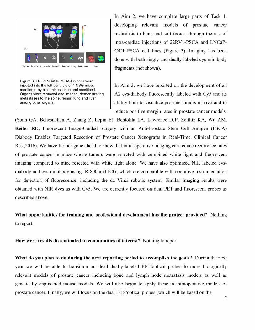
In Aim 2, we have complete large parts of Task 1,
developing relevant models of prostate cancer
metastasis to bone and soft tissues through the use of
intra-cardiac injections of 22RV1-PSCA and LNCaP-
C42b-PSCA cell lines (Figure 3). Imaging has been
done with both singly and dually labeled cys-minibody
fragments (not shown).
In Aim 3, we have reported on the development of an
A2 cys-diabody fluorescently labeled with Cy5 and its
ability both to visualize prostate tumors in vivo and to
reduce positive margin rates in prostate cancer models.
(Sonn GA, Behesnelian A, Zhang Z, Lepin EJ, Bentolila LA, Lawrence DJP, Zettlitz KA, Wu AM,
Reiter RE; Fluorescent Image-Guided Surgery with an Anti-Prostate Stem Cell Antigen (PSCA)
Diabody Enables Targeted Resection of Prostate Cancer Xenografts in Real-Time. Clinical Cancer
Res.,2016). We have further gone ahead to show that intra-operative imaging can reduce recurrence rates
of prostate cancer in mice whose tumors were resected with combined white light and fluorescent
imaging compared to mice resected with white light alone. We have also optimized NIR labeled cys-
diabody and cys-minibody using IR-800 and ICG, which are compatible with operative instrumentation
for detection of fluorescence, including the da Vinci robotic system. Similar imaging results were
obtained with NIR dyes as with Cy5. We are currently focused on dual PET and fluorescent probes as
described above.
What opportunities for training and professional development has the project provided? Nothing
to report.
How were results disseminated to communities of interest? Nothing to report
What do you plan to do during the next reporting period to accomplish the goals? During the next
year we will be able to transition our lead dually-labeled PET/optical probes to more biologically
relevant models of prostate cancer including bone and lymph node metastasis models as well as
genetically engineered mouse models. We will also begin to apply these in intraoperative models of
prostate cancer. Finally, we will focus on the dual F-18/optical probes (which will be based on the
Figure 3. LNCaP-C42b-PSCA-luc cells were injected into the left ventricle of 4 NSG mice, monitored by bioluminescence and sacrificed. Organs were removed and imaged, demonstrating metastases to the spine, femur, lung and liver among other organs.
A
SpineFemurStomachBowelTestesLungProstateLiver
B

8
smaller A2 cys-diabody because its in vivo kinetics are better matched to the physical half-life of F-18),
in collaboration with Dr. Anna Wu’s laboratory.
4. IMPACT What was the impact on the development of the principle discipline of the project?
Nothing to report.
What was the impact on other disciplines? Nothing to report.
What was the impact on technology transfer? Nothing to report.
What was the impact on society beyond science and technology? Nothing to report. After only one
year of funded research it is too early for our findings to have significant impact.
5. CHANGES/PROBLEMS
Nothing to report. There are no significant changes to the objectives, scope, and approaches of the
project.
6. PRODUCTS
Publications, conference papers, and presentations
Abstracts
Tsai, Wen-ting, Tavaré, R., Zettlitz, K.A., Salazar, F.B., Knowles, S., Reiter, R., and Wu, A.M. (2015).
Dual modality immunoPET/fluorescence imaging of prostate cancer. World Molecular Imaging
Congress, Honolulu, HI.
Tsai, W.-T., Tavaré, R., Zettlitz, K.A., Salazar, F.B., Reiter, R.E., and Wu, A.M. (2015) Dual-modality
immunoPET/fluorescence imaging of prostate cancer using anti-PSCA cys-minibody. Antibody
Engineering and Therapeutics 2015, San Diego, CA.
Tsai, W.-T., Zettlitz, K., Tavaré, R., Salazar, F., Reiter, R., and Wu, A. Dual-modality immune-
PET/fluorescence imaging of prostate cancer using anti-PSCA A11 cys-minibody. (2016) Antibody
Technology Resource Center Symposium, San Francisco, CA.

9
7. PARTICIPANTS AND OTHER COLLABORATING ORGANIZATIONS
What individuals have worked on the project?
Name: Robert E Reiter
Project Role: Principal Investigator Researcher Identifier (e.g. ORCID ID): Nearest person month worked: 1 Contribution to Project: Dr. Reiter oversaw all aspects of work
performed and accomplished to-date. Funding Support:
Names: Geoffrey Sonn and Andrew Behesnelian Project Role: Urology Fellow and Residents Researcher Identifier (e.g. ORCID ID): Nearest person month worked: 1 Contribution to Project: Drs. Sonn and Behesneilain performed
the in vivo optical surgical experiments Funding Support:
Name: Evelyn Kono Project Role: Senior Research Assistant Researcher Identifier (e.g. ORCID ID): Nearest person month worked: 1 Contribution to Project: Ms. Kono developed and performed the
metastatic models and imaging of said. Funding Support:
Has there been a change in the active other support of the PD/PI(s) or senior/key personnel since
the last reporting period?
The following funding has been renewed and/or is active:
Renewal funding: R01CA174294 Multifunctional immunoPET tracers for pancreatic and prostate cancer
(Wu, Reiter, Multi-PIs); time commitment of 1.8 calendar months; renewal project period of 8/1/2016 -
7/31/2021
What other organizations were involved as partners? Nothing to report. 8. SPECIAL REPORTING REQUIREMENTS Collaborative awards This report covers the activities of the PI Robert E Reiter and Partnering PI Anna Wu. 9. APPENDICES None

10