Embed Size (px)
Citation preview

Multifunctional protein labeling via enzymatic N-terminal tagging and elaboration by click chemistry.
William P. Heal, Megan H. Wright, Emmanuelle Thinon and Edward W. Tate*
Summary:
A protocol for selective and site-specific enzymatic labeling of proteins is described. The method co-opts the protein co-/post-translational modification known as myristoylation, the transfer of myristic acid (a C14 saturated fatty acid) to an N-terminal glycine catalyzed by the enzyme myristoyl-CoA:protein N-myristoyltransferase (NMT). E. coli, having no endogenous NMT, is used for the co-expression of both the transferase and the target, which participate in the N-terminal attachment of synthetically derived tagged analogues of myristic acid bearing a ‘clickable’ tag (an azide). This tag can be used as a handle for further site-specific labeling; here we provide protocols for in vivo N-terminal tagging of recombinant protein, and the synthesis and application of multifunctional reagents bearing labels for affinity purification and detection by fluorescence.
INTRODUCTION
In the post-genomic era the challenges of mapping, characterizing and quantifying the proteome, the totality of proteins involved in a particular cell/tissue, still presents a colossal challenge in terms of both scale and complexity. Although DNA sequencing and various powerful genetic techniques exist, the degree of fluidity in the proteome caused by mRNA modification and protein co- or post-translational modification (PTM) with small molecules such as lipids, sugars and phosphates or with polypeptides such as in ubiquitinylation or sumoylation, proteolysis, disulfide formation, protein complex assembly, all varying in response to stimuli or cell cycle, means that we are still far from a comprehensive protein atlas. The rapidly growing field of chemical proteomics, denoting the application of chemistry to the study of the proteome, makes use of synthetic chemical tools to modify and probe these biomolecules, resulting in techniques that allow isolation (individually or in classes/families, using affinity tags such as biotin) for characterization or in situ profiling (using techniques ranging from in-gel fluorescence to whole cell/animal imaging) of proteins. One such example of this cross-disciplinary endeavor is the commandeering of cellular machinery to introduce synthetic tagged analogues of small molecule modifiers during co/post-translational modification. Notable examples of this have included glycosylation,1-3 biotin ligation,4-7 prenylation,8-10 cholesterylation,11 phosphorylation12-
15 and lipidation.16-22
Myristoylation
Myristoylation is the attachment of myristic acid (a 14 carbon saturated fatty acid) to an N-terminal glycine in a target protein, catalyzed by the enzyme myristoyl-CoA:protein N-myristoyltransferase (NMT). NMT binds the N-terminal 8-15 residues of a specific set of target proteins for which a canonical motif has been identified, although there is complex species-dependent variation that may also enable or prevent myristoylation outside this motif. The other substrate for NMT is myristoyl-CoA thioester, formed in turn by CoA synthetase in the cell. In eukaryotes, myristate is introduced co-translationally to around 1% of the proteome, and also post-translationally during apoptosis when proteolytic digestion reveals a new N-terminal glycine. Lipidation facilitates protein–protein interactions and protein–membrane associations associated with trafficking and signaling. NMT is ubiquitous in all eukaryotes and has been shown to be essential in several yeasts and, more recently, the protozoan parasites associated with leishmaniasis (L. major) and trypanosomiasis (T. brucei).23 Human NMTs have been reported to have a role in several diseases including cancer and HIV infection, amongst others.24
Protein labeling using enzymatic tagging and bioorthogonal ligation chemistry
It has been shown that NMT and CoA synthetase tolerate small unobtrusive modifications to the terminal region of the myristic acid substrate.25,26 This allows the incorporation of chemical tags that allow further chemical elaboration with a wide variety of features such as fluorescent dyes or affinity handles. It appears that lipidation with such tagged myristate analogues does not interfere significantly with the behavior of the protein targets, and cell or organism growth is generally unaffected.19,20 Bioorthogonal ligation is made possible by the application of aqueous-compatible, selective and efficient covalent bond-forming reactions, commonly termed ‘click’ chemistry.27,28 The most routinely used example is the 3 + 2 copper(I) catalyzed Huisgen-Sharpless cyclisation,29 in which an azide and an alkyne form a triazole ring. The starting materials, azide or alkyne, are well suited to the needs of chemical proteomics as they are small, bear no overall charge, and can be introduced with relative ease to a wide variety of structures, often without disruption to biological behavior.

Advantages over other methods
Site specific protein labeling is widely recognized as a key technique in molecular biology. One common approach involves the use of labeled substrates for PTM. A simple example of this for protein acylation includes tritiated myristic acid, which allows autoradiography after separation of proteins by SDS-PAGE. However, quite aside from safety considerations and the significant cost, autoradiography can take hours to days or more to give sufficient exposure, particularly when considering low copy-number entities. In contrast, in-gel fluorescence provides images in seconds and for a fraction of the cost. Fluorescent proteins can be incorporated into a protein of interest using genetic methods, but these are bulky, typically >10 kDa, implying possible perturbation of protein function. There is no temporal control over label attachment, and the correct tertiary structure is required for fluorescence. A chemical tag (azide or alkyne) is very small by comparison (<50 Da), and in general does not affect protein function adversely. After bioorthogonal ligation the small molecule affinity/fluorescence labels retain their activity throughout down-stream processing that would otherwise denature the subject. Labeling via a PTM, especially with an affinity handle, allows enrichment and study of sub-sets of the proteome, when tagged substrates are introduced to cells/tissues with the intention of identifying novel targets.16 This approach has been adapted as a generic site-specific protein labeling technology for recombinant proteins, where it can provide the advantages of a genetically encoded tag (the consensus site for modification) with the versatility of chemical modification (via bioorthogonal ligation and/or enzyme-substrate engineering). Notable examples include the application of formylglycine generating enzyme,30 biotin ligase,6,7 prenyl transferases,8 and NMT.19,20
Here we provide straightforward and robust protocols for the application of NMT to tag recombinant proteins in live bacteria with an azide and subsequent elaboration to biotin and TAMRA labeled derivatives via bioorthogonal ligation chemistry. This protocol will be of use both to researchers investigating the substrate specificity of NMT, and for the production of proteins site-specifically modified at the N-terminus with small molecule labels.
Figure 1| Illustrative overview of the protocol. In the feeding stage (1), E. coli co-transformed with constructs for the expression of transferase (CaNMT) and target protein (PfARF1) are incubated with tagged myristate (AzC12). E. coli acyl-CoA synthetase catalyzes the formation of AzC12–CoA (the substrate for NMT) in vivo. Tagging (2) is carried out in vivo by the newly synthesized CaNMT, attaching tagged myristate onto the N-terminal glycine of the target PfARF1. After lysis (3), the tagged protein is further elaborated by attachment of biotin (affinity) and TAMRA (fluorescence) labels11 using ‘click’ chemistry (4).
MATERIALS
Reagents

• Biotin-PEG NovaTag™ resin (Merck, cat. no. 855055)
• DMF (N,N-dimethylformamide, National Diagnostics, cat. no. AGMDMF25L)
CRITICAL For peptide bond forming chemistry the DMF must be of peptide synthesis grade, the presence of water or amines typically found in lower grades will greatly diminish the efficiency of the peptide coupling reactions
• Piperidine 20% v/v in N,N-dimethylformamide (DMF80%/PIPERIDINE20%-Premix, AGCT Bioproducts, cat. no. AGMPIPDMF)
• Dichloromethane
• Fmoc-Gly-OH (N-[(9H-fluoren-9-ylmethoxy)carbonyl]glycine, AGTC Biobroducts, cat. no. AGM08)
• HATU (O-(7-azabenzotriazol-1-yl)-N,N,N′,N′-tetramethyluronium hexafluorophosphate, VWR (Novabiochem), cat. no. 01-62-0041-25)
• DIPEA (N,N-diisopropylethylamine, AGCT Bioproducts, cat. no. AGBC7012)
• Fmoc-L-Pra-OH (N-[(9H-fluoren-9-ylmethoxy)carbonyl]propargylglycine, PolyPeptide Laboratories (France), cat. no. FA10902)
• TAMRA (5(6)-carboxytetramethylrhodamine, Merck, cat. no. 8.51030.8500)
• DIC (N,N′-Diisopropylcarbodiimide, Sigma Aldrich, cat. no. D125407)
• HOAt (1-Hydroxy-7-azabenzotriazole, VWR, cat. no. MOLEM45186173)
• Methanol (LC-MS grade, VWR, cat. no. 83638.320)
• Methanol (analytical grade)
• TFA (trifluoroacetic acid, Sigma Aldrich, cat. no. T62200)
• TIPS (triisopropylsilane, Sigma Aldrich, cat. no. 233781)
• TBME (methyl tert-butyl ether, Sigma Aldrich, cat. no. 34875)
• DMSO (dimethylsulfoxide, VWR, cat. no. 23500.297)
• FA (formic acid (mass spectroscopy grade), Sigma Aldrich, cat. no. 94318)
• 12-Bromododecanoic acid (Sigma Aldrich, cat. no. 200999)
• H2SO4 (concentrated sulfuric acid)
• Diethylether
• NaHCO3 (sodium hydrogen carbonate)
• Na2SO4 (sodium sulfate)
• NaN3 (sodium azide, Sigma Aldrich, cat. no. 199931)
• HCl (concentrated hydrochloric acid)
• NaOH (sodium hydroxide pellets)
• Water (Milli-Q water purification system, 18.2 MΩ, UV sterilized or equivalent)
• Ethyl acetate
• Brine (saturated aqueous sodium chloride solution)
• Cyclohexane
• Silica gel (Geduran® Si 60 for column chromatography (60 Å particle size, 40–63 μm), VRW, cat. no. 1.11567.1000)
• Sand
• KMnO4 (potassium permanganate, VWR, cat. no. ALFA36675.22)
• K2CO3 (Potassium carbonate)
• Plasmid cloning sequences
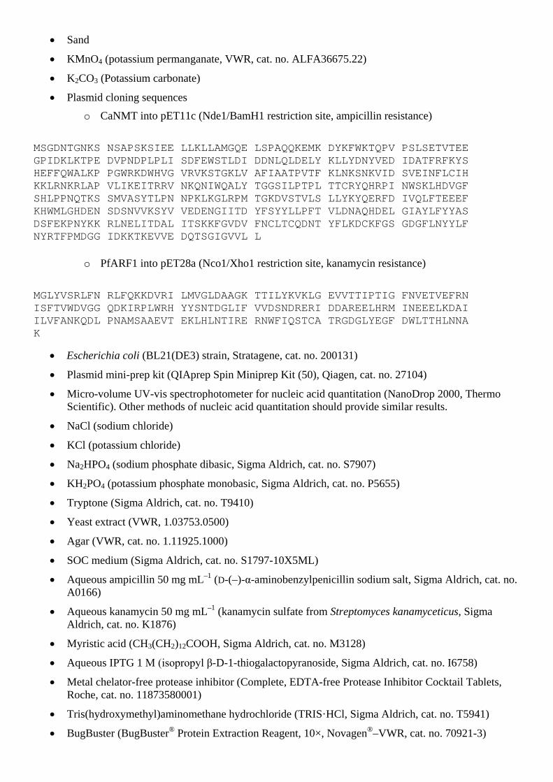
o CaNMT into pET11c (Nde1/BamH1 restriction site, ampicillin resistance)
MSGDNTGNKS NSAPSKSIEE LLKLLAMGQE LSPAQQKEMK DYKFWKTQPV PSLSETVTEE GPIDKLKTPE DVPNDPLPLI SDFEWSTLDI DDNLQLDELY KLLYDNYVED IDATFRFKYS HEFFQWALKP PGWRKDWHVG VRVKSTGKLV AFIAATPVTF KLNKSNKVID SVEINFLCIH KKLRNKRLAP VLIKEITRRV NKQNIWQALY TGGSILPTPL TTCRYQHRPI NWSKLHDVGF SHLPPNQTKS SMVASYTLPN NPKLKGLRPM TGKDVSTVLS LLYKYQERFD IVQLFTEEEF KHWMLGHDEN SDSNVVKSYV VEDENGIITD YFSYYLLPFT VLDNAQHDEL GIAYLFYYAS DSFEKPNYKK RLNELITDAL ITSKKFGVDV FNCLTCQDNT YFLKDCKFGS GDGFLNYYLF NYRTFPMDGG IDKKTKEVVE DQTSGIGVVL L
o PfARF1 into pET28a (Nco1/Xho1 restriction site, kanamycin resistance)
MGLYVSRLFN RLFQKKDVRI LMVGLDAAGK TTILYKVKLG EVVTTIPTIG FNVETVEFRN ISFTVWDVGG QDKIRPLWRH YYSNTDGLIF VVDSNDRERI DDAREELHRM INEEELKDAI ILVFANKQDL PNAMSAAEVT EKLHLNTIRE RNWFIQSTCA TRGDGLYEGF DWLTTHLNNA K
• Escherichia coli (BL21(DE3) strain, Stratagene, cat. no. 200131)
• Plasmid mini-prep kit (QIAprep Spin Miniprep Kit (50), Qiagen, cat. no. 27104)
• Micro-volume UV-vis spectrophotometer for nucleic acid quantitation (NanoDrop 2000, Thermo Scientific). Other methods of nucleic acid quantitation should provide similar results.
• NaCl (sodium chloride)
• KCl (potassium chloride)
• Na2HPO4 (sodium phosphate dibasic, Sigma Aldrich, cat. no. S7907)
• KH2PO4 (potassium phosphate monobasic, Sigma Aldrich, cat. no. P5655)
• Tryptone (Sigma Aldrich, cat. no. T9410)
• Yeast extract (VWR, 1.03753.0500)
• Agar (VWR, cat. no. 1.11925.1000)
• SOC medium (Sigma Aldrich, cat. no. S1797-10X5ML)
• Aqueous ampicillin 50 mg mL–1 (D-(–)-α-aminobenzylpenicillin sodium salt, Sigma Aldrich, cat. no. A0166)
• Aqueous kanamycin 50 mg mL–1 (kanamycin sulfate from Streptomyces kanamyceticus, Sigma Aldrich, cat. no. K1876)
• Myristic acid (CH3(CH2)12COOH, Sigma Aldrich, cat. no. M3128)
• Aqueous IPTG 1 M (isopropyl β-D-1-thiogalactopyranoside, Sigma Aldrich, cat. no. I6758)
• Metal chelator-free protease inhibitor (Complete, EDTA-free Protease Inhibitor Cocktail Tablets, Roche, cat. no. 11873580001)
• Tris(hydroxymethyl)aminomethane hydrochloride (TRIS·HCl, Sigma Aldrich, cat. no. T5941)
• BugBuster (BugBuster® Protein Extraction Reagent, 10×, Novagen®–VWR, cat. no. 70921-3)

• BME (β-mercaptoethanol, Sigma Aldrich, cat. no. M6250)
• Acrylamide (acrylamide-Bis (37.5:1), 30% aqueous solution, VWR, cat. no. 1.00639.1000)
• APS 10% in water (ammonium persulfate for electrophoresis, Sigma Aldrich, cat. no. A3678)
• TEMED (N,N,N',N'-tetramethylethylenediamine, Sigma Aldrich, cat. no. T9281)
• LDS Sample Buffer (NuPAGE® LDS Sample Buffer (4×), Invitrogen, cat. no.NP0007)
• MES monohydrate (4-morpholineethanesulfonic acid monohydrate, Fisher, cat. no. 32776-5000)
• Tris (Trizma® base, Sigma Aldrich, cat. no. T6066)
• EDTA (ethylenediaminetetraacetic acid, Sigma Aldrich, cat. no. 431788)
• Ammonium sulfate (Sigma Aldrich, cat. no. A4418)
• Phosphoric acid 85% in water (Sigma Aldrich, cat. no. 345245)
• G-250 (Coomassie® brilliant blue G 250, Sigma Aldrich, cat. no. 201391)
• Bis-Tris (bis(2-hydroxyethyl)amino-tris(hydroxymethyl)methane, Sigma Aldrich, cat. no. B4429)
• Total protein assay (DCTM Protein assay kit, Bio-Rad, cat. no. 500-0111)
• BSA (albumin from bovine serum, Sigma Aldrich, cat. no. A7906)
• Aqueous CuSO4 40 mM (copper(II) sulfate pentahydrate 99.999%, Sigma Aldrich, cat. no. 203165)
• TBTA 10 mM in DMSO (tris[(1-benzyl-1H-1,2,3-triazol-4-yl)methyl]amine, Sigma Aldrich, cat. no. 678937)
• TCEP 50 mM aqueous (tris(2-carboxyethyl)phosphine hydrochloride, Sigma Aldrich, cat. no. C4706)
• SDS, various concentrations in PBD buffer (sodium dodecyl sulfate, Sigma Aldrich, cat. no. L6026)
• Streptavidin-coupled magnetic beads (Dynabeads® MyOneTM Streptavidin C1, Invitrogen, cat. no. 650-01)
• Urea 4 M aqueous (Sigma Aldrich, cat. no. U5378)
• Guanidine hydrochloride 4 M aqueous (Sigma Aldrich, cat. no. G3272)
• Prestained, fluorescent molecular weight marker ladder (precision plus protein all blue standards, Bio-Rad, cat. no. 161-0373EDU)
EQUIPMENT
Capture reagent synthesis • 5 mL plastic syringes (Roland Vetter Laborbedarf, cat. no. 309040)
• Polyethylene frits for 5 mL syringes (Roland Vetter Laborbedarf, cat. no. CEL-053)
• Syringe caps (Roland Vetter Laborbedarf, cat. no. VSG-0419)
• 15 mL centrifuge tubes with screw caps (BD, cat. no. 352196)
• Refrigerated centrifuge (Model 5810 R, Eppendorf, cat. no. 5811 000.010)
• Glass vials with caps (e.g. 50 × 12 mm, VWR, cat. no. 212-7046)
• Shaker with moveable bars to hold syringes (KS 130 basic, IKA, cat. no. 2980000)
• Laboratory tissue paper
• Elastic bands
• Desiccator (~20 cm diameter)

• Vacuum pump
• LC-MS mass directed purification system (Waters). N.b. Purification of the product by similar means (e.g. preperative HPLC) should provide similar results
o 2767 Sample manager
o 2545 Quaternary gradient pump module
o System fluidics organizer, equipped with a 1:5000 flow splitter (cat. no. 205000438)
o M515 HPLC pump
o 2998 Photodiode array detector
o 3100 Mass detector
o XBridge C18 5 μm, 4.6 × 100 mm column (cat. no. 186002978, equipped with an XBridge C18 Guard Column, 5 µm, 4.6 × 20 mm, cat. no. 186003064)
o XBridge prep C18 5 μm, 19 × 100 mm OBD column (cat. no. 186003115, equipped with an XBridge Prep C18 Guard Column, 5 µm, 19 × 10 mm, cat. no. 186002975)
• Centrifugal evaporator (EZ-2plus personal evaporator, Genevac)
Analogue Synthesis • Single-neck round-bottom flasks (100–1000 mL)
• Glass/plastic funnels (different sizes)
• Magnetic stirrer bars (3 cm, oval)
• 1 mL Micropipette
• Solvent safe plastic pipettes (Molecular Bio Products, cat. no. 5079EB)
• Magnetic stirrer hot plate
• Syringes (plastic, without rubber plunger seals, various volumes)
• Hypodermic needles (Microlance™ 3, 4 mm × 18G, VWR, cat. no. 613-3945)
• Water condenser (Leibig style, diameter 19/26, 30 cm length)
• Paraffin oil bath
• Thin-layer chromatography plates (pre-coated Silica plates, Aluminum oxide 60 F254, Merck, cat. no. 5550-7)
• Rotary evaporator
• Filter paper (diameter 110 mm, VWR, cat. no. 516.0815)
• Septa (neoprene, various sizes)
• Separating funnels (100–250 mL)
• Conical flasks (100–250 mL)
• Glass chromatography column (diameter 6 cm, length 40 cm)
E. coli culture • Benchtop orbital shaker/incubator (MaxQ* 4000, Thermo Scientific, cat. no. SHKA4000-8CE)
• Static incubator (LTE Scientific, cat. no. IP60-U)
• Orbital shakers (similar to Stuart Scientific model SO1)
• Water bath (SUB Aqua 5 Plus, VWR, cat. no. 462-0202)
• Plastic culture dishes (Dish 100 × 20 mm, VWR, cat. no. 734-0006)
• Laboratory bottles (100 mL, VWR, cat. no. 215-1592)
• Microwave oven (domestic type)

• Disposable cell spreaders
• Disposable inoculating loops
• NanoDrop Spectrophotometer (NanoDrop 1000, Thermo Scientific or similar)
Click chemistry • Vortex mixer (e.g. Vortex-Genie 2, Scientific Industries, cat. no. SI-0236)
• Multi-vortex mixer (e.g. V-32, Grant Instruments, cat. no. V-32)
• Sonicator (Ultrasonic cleaning device, VWR, cat. no. 142-6001)
• Magnetic rack (Magna GrIPTM Rack (8 well), Millipore, cat. no. 20-400)
SDS-PAGE • Heat block (VWR, cat. no. 460-3268)
• Apparatus for SDS-PAGE (Bio-Rad Mini-PROTEAN Tetra Electrophoresis System or similar)
• 2-D Fluorescence difference gel electrophoresis imager (EttanTM DIGE System, G. E. Healthcare)
• Plastic trays for soaking gels (various sizes)
• Scanner
REAGENT SETUP
Cold TBME Store TBME in a –20 °C freezer prior to use. Preparation of chemically competent bacteria Prepare chemically competent E. coli using the CaCl2 method of Sambrook et al.31 100 μL aliquots of these cells can be stored for several weeks at –80 °C. If competency is lost, cells can be grown up and competency re-established. Alternatively, competent cells may be purchased from several sources.
Purification of plasmids Purify plasmids using 5 mL overnight cultures and the Qiagen mini-prep kit according to the manufacturer’s instructions. Elute DNA from the spin columns in water and determine the concentration by nano-drop UV photospectroscopy. Typically ~110–120 ng μL–1 is obtained, which should be adjusted to 100 ng μL–1 to make the working DNA stock solutions. These can be stored at –20 °C for several months.
Potassium permanganate stain Dissolve 1.5 g of KMnO4, 10 g K2CO3, and 1.25 mL 10% NaOH in 200 mL water. The lifetime for this stain is approximately 3 months.
Lauria-Bertani (LB) broth Dissolve 10 g NaCl, 10 g tryptone and 5 g yeast extract in 800 mL of water. Adjust the pH to 7.00 with 5 M NaOH, make the volume up to 1 L and sterilize by autoclaving.
LB agar Dissolve 10 g NaCl, 10 g tryptone, 5 g yeast extract and 20 g agar in 800 mL of water. Adjust the pH to 7.00 with 5 M NaOH, make the volume up to 1 L, divide into 80 mL aliquots and sterilize by autoclaving.
AzC12 and myristic acid feeding stocks Weigh the compound and dissolve in sufficient DMSO to provide a 50 mM stock. Divide the stock into 100 μL aliquots and store at –20 °C (can be stored at –20 °C for several weeks).
Lysis buffer Dilute BugBuster 10× into 150 mM NaCl, 50 mM Tris.HCl, pH 8.00. Add 1 tablet of metal chelator-free protease inhibitor per 50 mL.
Capture reagent stock solution Dissolve the purified capture reagent in DMSO to 10 mM.
‘Blue Silver’ colloidal Coomassie stain Weigh out 25 g of ammonium sulfate and add to this 27 mL of 85% phosphoric acid in water. Add 0.6 g of 50% G-250 dye and once all solids dissolved, make up to 200 mL with water. To this, add 50 mL methanol.32
Bis-Tris buffer 1.25 M Bis-Tris, pH 6.7.

MES Running Buffer 20× 50 mM MES, 50 mM Tris 0.1% SDS and 1 mM EDTA. pH 7.3.
PBS 10× Dissolve 80 g of NaCl, 2.0 g of KCl, 14.4 g of Na2HPO4 and 2.4 g of KH2PO4 in 800 mL water. Adjust the pH to 7.4 and make the volume up to 1000 mL.
Gel soaking solution 50% water, 40% MeOH and 10% acetic acid.
Gel compositions
Component Volumes for 10 mL of 12% resolving gel
Volumes for 2.5 mL of 4% stacking gel
30% Acrylamide 4.00 mL 333 µL
1.25 M Bis-tris (pH 6.7) 2.90 mL 714 µL
Water 3.00 mL 1.45 mL
10% APS 100 µL 12.5 µL
TMEDA 4.00 µL 2.00 µL
EQUIPMENT SETUP
Bacterial culture All manipulations of E. coli should be carried out in an aseptic environment (e.g. a flame sterilized area).
PROCEDURE
Box 1
Synthesis of trifunctional capture reagent11 TIMING 3 days
HN
O
NHO
O COO
N
N
O3
NH
O
4S
NHHN
O
Tri-functionalcapture reagent
N
O3
NH
O
4S
NHHN
OFmoc
Biotin-PEG NovaTag™ resin
a
Scheme 1| Reagents and Conditions: (a) solid phase peptide synthesis using Fmoc-propargyl glycine and TAMRA.
Sub-protocols: general protocols for the washing and deprotection of the resin during manual coupling. Wash

The resin is washed three times in succession with DMF, DCM and then again DMF to remove excess reagents and by-products.
1 Add 2 mL DMF and vortex gently for 1 min. Remove the plunger and uncap the syringe over a waste solvent container, replace the plunger and depress to remove the liquid. Repeat this sequence twice for a total of three washes with DMF.
2 Repeat step 1 above, using DCM in place of DMF
3 Repeat step 1.
Deprotect 1. Add 2 mL Pip/DMF 20:80 to capped syringe, replace the plunger in the top of the syringe and return
the syringe to the shaker. Vortex the mixture for 3 min.
2. Remove the plunger and uncap the syringe over a waste solvent container, replace the plunger and depress to remove the liquid.
3. Repeat steps 1 and 2 twice for a total of 3 deprotection cycles.
Main protocol 1 Remove the plunger from a 5 mL syringe and place a polyethylene frit (using the plunger as a
ramrod) at the bottom. Cap the nozzle with a syringe cap and place into the syringe 104 mg of the Biotin-PEG NovatagTM resin.
2 Add 2 mL of peptide synthesis grade DMF and fix the plunger into the top of the syringe (to seal: do not depress). Mount the syringe on the shaker (KS 130 basic) and vortex for 30 min to allow the resin to swell.
3 Remove the plunger and uncap the syringe over a waste solvent container, replace the plunger and depress to remove the liquid.
! CAUTION It is important that the plunger is removed before uncapping the syringe; if the cap is removed whilst the plunger is in the top of the syringe, the positive pressure will cause the solution to spray out.
4 Deprotect the resin using the protocol given above.
5 Wash the resin using the protocol given above.
6 Place 74 mg Fmoc-Gly-OH, 93 mg HATU in a glass vial. Add 2 mL DMF and, once all the solids have dissolved, add 87 μL DIPEA, cap the vial and let it stand for 5 min.
7 Remove the syringe plunger, cap the syringe and add the contents of the vial. Place the plunger in the top of the syringe and vortex for 30 min.
8 Remove the plunger and uncap the syringe over a waste solvent container, replace the plunger and depress to remove the liquid.
9 Repeat the coupling (steps 6 and 7).
10 Wash the resin.
11 Deprotect the resin.
12 Wash the resin.
13 Place 41.9 mg Fmoc-(propargyl)Gly-OH, 46.6 mg HATU in a glass vial. Add 2 mL DMF and, once all the solids have dissolved, add 44 μL DIPEA, cap the vial and let it stand for 5 min.
14 Remove the plunger and uncap the syringe over a waste solvent container, replace the plunger and depress to remove the liquid.
15 Repeat the coupling (steps 13 and 14).
16 Wash the resin.
17 Deprotect the resin.
18 Wash the resin.
19 Place 32.3 mg TAMRA, 16.3 mg DIC and 10.1 mg HOAt in a glass vial. Add 2 mL DMF and, once all the solids have dissolved, cap the vial and let it stand for 10 min.
20 Remove the syringe plunger, cap the syringe and add the contents of the vial. Place the plunger in the top of the syringe and vortex for 2 h.
21 Remove the plunger and uncap the syringe over a waste solvent container, replace the plunger and depress to remove the liquid.
22 Wash the resin with DMF then DCM.
23 Wash in the same way with 2 × MeOH and then 2 × Et2O.
24 Remove the plunger and cover the wide opening of the syringe with tissue paper, fix this in place with an elastic band. Place the syringe into the desiccator, evacuate and leave overnight (16 h).
PAUSE POINT The syringe can be sealed (with yellow cap and plunger) and the dried resin stored in a 4 °C refrigerator for several weeks.
25 Place 1.9 mL TFA, 50 μL water and 50 μL TIPS in a 15 mL centrifuge tube and vortex until mixed (cleavage mix). Remove the syringe from the vacuum desiccator, cap and remove the tissue paper. Add the cleavage mix, place the plunger in the top of the syringe and vortex for 3 h.
! CAUTION TFA is harmful and highly corrosive and should be used with great care (PPE including gloves).
26 Remove the plunger, place the syringe over a clean 15 mL centrifuge tube and remove the cap. Replace the plunger and depress to push the cleavage mix into the tube. Wash the resin once with TFA.
27 To the combined TFA washes add 10 mL cold TBME, resulting in the formation of a dark purple precipitate. Pellet this solid by centrifugation for 15 min at 6500 g at 4 °C. Carefully decant the supernatant and add a further 10 mL of ice cold TBME, vortex for 30 sec (ensuring that the pellet is resuspended), centrifuge and remove the second supernatant. Wash the solids once more with TBME as above.
28 Cover the mouth of the centrifuge tube containing the washed and pelleted solids with tissue paper, secure with a rubber band, and dry overnight (16 h) in a vacuum desiccator.
PAUSE POINT The centrifuge tube can be capped and the dried pellet stored in a –20 °C freezer for several weeks.
29 Add 50 μL DMSO to the dried crude product and vortex until dissolved. Dilute the DMSO solution with 5 mL water (to 1% v/v final amount of DMSO). If required, add up to 5% v/v MeOH to improve the solubility of the crude products. Filter the resultant solution through a 4 μm syringe filter into a sample tube for purification by LC-MS.
30 Purify the product (in batches if necessary) over a gradient of MeOH in water (flow rate 20 mL min-1, 5% MeOH over 1 min, 5%–98% over 12 min, 98% for 5 min, 98%–5% over 1 min, 5% for 5 min). Set UV detection over the range 100–600 nm, mass detection over the range m/z = 400–2000, triggering fraction collection from the m/z of the product (1011 [M + H]+).
31 After reanalysis of the collected fractions, combine and evaporate to give the dried product, in a typical yield of ~30 mg.
Typical Results The trifunctional capture reagent was obtained by lyphophilization as a bright pink amorphous solid (28.9 mg, 57% yield). 1H NMR (500 MHz, MeOD) δ 8.71 (1H, d, J = 1.7, HD), 8.21 (1H, dd, J = 7.9, 1.8, HE), 8.16 (2H, s), 7.45 (1H, d, J = 7.9, HF), 7.20 (2H, d, J = 9.4, HC), 7.03 (2H, dd, J = 9.5, 2.2, HB), 6.94 (2H, d, J = 2.4, HA), 4.69 (1H, dd, J = 8.3, 6.2, H20), 4.46 (1H, ddd, J = 7.9, 4.9, 0.7, H1), 4.27 (1H, dd, J = 7.9, 4.5, H2), 3.97 (1H, d, J = 16.8, H19), 3.80 (1H, d, J = 16.8, H19), 3.63–3.58 (4H, m), 3.59–3.54 (4H,
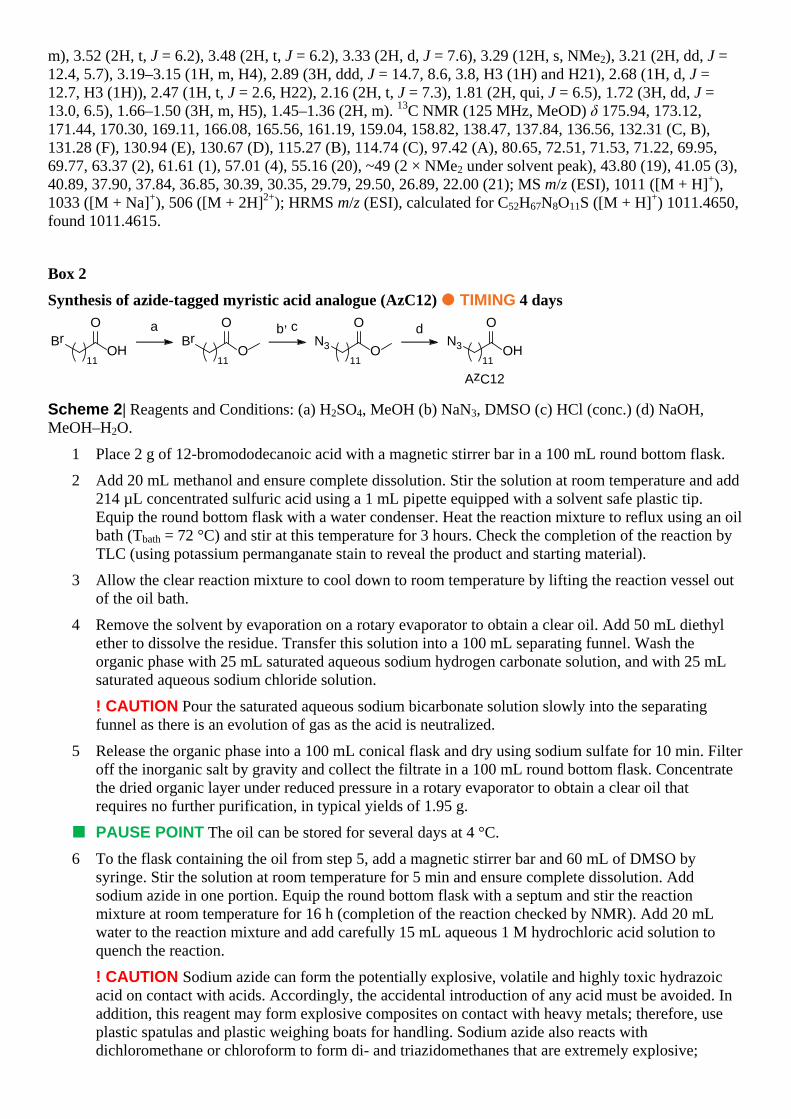
m), 3.52 (2H, t, J = 6.2), 3.48 (2H, t, J = 6.2), 3.33 (2H, d, J = 7.6), 3.29 (12H, s, NMe2), 3.21 (2H, dd, J = 12.4, 5.7), 3.19–3.15 (1H, m, H4), 2.89 (3H, ddd, J = 14.7, 8.6, 3.8, H3 (1H) and H21), 2.68 (1H, d, J = 12.7, H3 (1H)), 2.47 (1H, t, J = 2.6, H22), 2.16 (2H, t, J = 7.3), 1.81 (2H, qui, J = 6.5), 1.72 (3H, dd, J = 13.0, 6.5), 1.66–1.50 (3H, m, H5), 1.45–1.36 (2H, m). 13C NMR (125 MHz, MeOD) δ 175.94, 173.12, 171.44, 170.30, 169.11, 166.08, 165.56, 161.19, 159.04, 158.82, 138.47, 137.84, 136.56, 132.31 (C, B), 131.28 (F), 130.94 (E), 130.67 (D), 115.27 (B), 114.74 (C), 97.42 (A), 80.65, 72.51, 71.53, 71.22, 69.95, 69.77, 63.37 (2), 61.61 (1), 57.01 (4), 55.16 (20), ~49 (2 × NMe2 under solvent peak), 43.80 (19), 41.05 (3), 40.89, 37.90, 37.84, 36.85, 30.39, 30.35, 29.79, 29.50, 26.89, 22.00 (21); MS m/z (ESI), 1011 ([M + H]+), 1033 ([M + Na]+), 506 ([M + 2H]2+); HRMS m/z (ESI), calculated for C52H67N8O11S ([M + H]+) 1011.4650, found 1011.4615.
Box 2
Synthesis of azide-tagged myristic acid analogue (AzC12) TIMING 4 days
N3 OH
O
11
AzC12
N3 O
O
11
BrO
O
11
BrOH
O
11
a b, c d
Scheme 2| Reagents and Conditions: (a) H2SO4, MeOH (b) NaN3, DMSO (c) HCl (conc.) (d) NaOH, MeOH–H2O.
1 Place 2 g of 12-bromododecanoic acid with a magnetic stirrer bar in a 100 mL round bottom flask.
2 Add 20 mL methanol and ensure complete dissolution. Stir the solution at room temperature and add 214 µL concentrated sulfuric acid using a 1 mL pipette equipped with a solvent safe plastic tip. Equip the round bottom flask with a water condenser. Heat the reaction mixture to reflux using an oil bath (Tbath = 72 °C) and stir at this temperature for 3 hours. Check the completion of the reaction by TLC (using potassium permanganate stain to reveal the product and starting material).
3 Allow the clear reaction mixture to cool down to room temperature by lifting the reaction vessel out of the oil bath.
4 Remove the solvent by evaporation on a rotary evaporator to obtain a clear oil. Add 50 mL diethyl ether to dissolve the residue. Transfer this solution into a 100 mL separating funnel. Wash the organic phase with 25 mL saturated aqueous sodium hydrogen carbonate solution, and with 25 mL saturated aqueous sodium chloride solution.
! CAUTION Pour the saturated aqueous sodium bicarbonate solution slowly into the separating funnel as there is an evolution of gas as the acid is neutralized.
5 Release the organic phase into a 100 mL conical flask and dry using sodium sulfate for 10 min. Filter off the inorganic salt by gravity and collect the filtrate in a 100 mL round bottom flask. Concentrate the dried organic layer under reduced pressure in a rotary evaporator to obtain a clear oil that requires no further purification, in typical yields of 1.95 g.
PAUSE POINT The oil can be stored for several days at 4 °C.
6 To the flask containing the oil from step 5, add a magnetic stirrer bar and 60 mL of DMSO by syringe. Stir the solution at room temperature for 5 min and ensure complete dissolution. Add sodium azide in one portion. Equip the round bottom flask with a septum and stir the reaction mixture at room temperature for 16 h (completion of the reaction checked by NMR). Add 20 mL water to the reaction mixture and add carefully 15 mL aqueous 1 M hydrochloric acid solution to quench the reaction.
! CAUTION Sodium azide can form the potentially explosive, volatile and highly toxic hydrazoic acid on contact with acids. Accordingly, the accidental introduction of any acid must be avoided. In addition, this reagent may form explosive composites on contact with heavy metals; therefore, use plastic spatulas and plastic weighing boats for handling. Sodium azide also reacts with dichloromethane or chloroform to form di- and triazidomethanes that are extremely explosive;

therefore, these solvents should never be used as substitutes in this reaction. Finally, make sure to place the whole apparatus behind a weighted safety shield and close the sash of the fume hood.
! CAUTION The quenching reaction is highly exothermic. Stir the reaction mixture at room temperature and cautiously add the aqueous 1 M hydrochloric acid solution dropwise by syringe.
7 Wait for the mixture to cool down to room temperature and transfer this mixture into a 250 mL separating funnel. Extract the aqueous solution with three 50 mL portions of ethyl acetate. Combine the organic layers and wash with three 50 mL portions of water, two 50 mL portions of saturated aqueous sodium chloride solution.
8 Release the organic phase into a 250 mL conical flask and dry using sodium sulfate for 10 min. Filter off the inorganic salt by gravity and collect the filtrate in a 250 mL round bottom flask.
9 Concentrate the dried organic layer under reduced pressure in a rotary evaporator to obtain a yellow oil. 1.96 g of methyl 12-bromododecanoate were typically obtained.
PAUSE POINT The crude product can be stored overnight at 4 °C.
10 Add sufficient amounts of a mixture of 1 part diethyl ether and 9 parts cyclohexane to a beaker containing ~100 g silica so that a homogenous gel develops under stirring. Pour that mixture into a glass column with a 6 cm diameter. Open the tap and apply pressure (using a set of bellows or slight inert gas pressure) to ensure compact packing.
11 Dissolve the product from step 9 in 50 mL of ethyl acetate and then add ~10 g of dry silica gel. Remove the solvent by evaporation on a rotary evaporator to provide a fine white powder.
12 Pour the white powder into the top of the flash chromatography column and carefully cover with 1 cm sand. Fill the column with a mixture of 1 part diethyl ether and 9 parts cyclohexane. Elute the column under pressure and collect fractions (12–15 mL fractions).
13 Detect the product using thin-layer chromatography (Rf = 0.2) and potassium permanganate stain.
14 Combine and then concentrate the fractions containing the product under reduced pressure in a rotary evaporator to yield a colourless oil. Dry the product for at least 20 min under high vacuum. 1.51 g of a clear oil was typically obtained.
PAUSE POINT The oil can be stored for several days at 4 °C.
15 To the flask containing the oil from step 14, add a magnetic stirrer bar and add 5.9 mL of 2 M sodium hydroxide aqueous solution by syringe. Add 3–5 mL of methanol to obtain a homogeneous solution. Stir at room temperature for 24 hours (completion of the reaction checked by TLC (cyclohexane–diethylether 9:1) using potassium permanganate stain, starting material Rf = 0.20, product Rf = 0.05).
16 Remove the methanol by evaporation on a rotary evaporator so that only the aqueous phase remains. Stir at room temperature, add 20 mL ethyl acetate and then acidify the aqueous layer by adding carefully aqueous 2 M hydrochloric acid solution (10 mL).
! CAUTION Pour the aqueous 2 M hydrochloric acid solution into the reaction vessel slowly as there is an evolution of gas as the base is neutralized.
17 Separate the two layers and extract the aqueous phase with two 50 mL portions of ethyl acetate. Combine the organic extracts and wash with 50 mL water and 50 mL saturated aqueous sodium chloride solution.
18 Release the organic phase into a 250 mL conical flask and dry using sodium sulfate for 10 min. Filter off the inorganic salt by gravity and collect the filtrate in a 250 mL round bottom flask.
19 Concentrate the dried organic layer under reduced pressure in a rotary evaporator to obtain a colorless oil that crystallizes to a white solid upon cooling. ~1.32 g of AzC12 was typically obtained.
PAUSE POINT The product can be stored for several months at –20 °C.
? TROUBLESHOOTING An oil is obtained. This can happen when hydrochloric acid is present with the final product. Dissolve the crude product in 50 mL ethyl acetate and wash the organic layer

with three portions of 50 mL water, then wash with 50 mL saturated aqueous sodium chloride solution, dry using sodium sulfate and remove the volatiles by rotary evaporation.
Typical Results AzC12 was isolated as a white solid (1.32 g, 93%). 1H NMR (400 MHz, CDCl3) δ 3.23 (2H, t, J = 7.0 Hz, -CH2N3), 2.33 (2H, t, J = 7.5 Hz, CH2CO2H), 1.55–1.72 (4H, m, CH2CH2CO2H and CH2CH2N3), 1.24–1.43 (14H, m, (CH2)6). 13C NMR (101 MHz, CDCl3) δ 179.71 (C=O), 51.70, 34.14, 29.64 (2C), 29.57, 29.41, 29.34, 29.24, 29.04, 26.91, 24.87. MS m/z (ESI), 240 ([M – H]–); HRMS m/z (ESI), calculated for C12H23N3O2 ([M–H]–) 240.1712, found 240.1719.
Transformation of E. coli TIMING 2 days 1 Thaw a frozen 100 μL aliquot of chemically competent E. coli on ice.
2 Transfer to a 15 mL centrifuge tube and add 0.5 μL (50 ng) of DNA stock as desired (CaNMT, PfAFR1 or both for dual transformations), swirl the tubes gently and incubate the cells on ice for a further 30 min.
3 During this time, pre-warm SOC medium in a 42 °C water bath for use in step 62.
4 Heat-shock each tube of cells in the 42 °C water bath for exactly 1 min. Return the cells to the ice and incubate for 2 min.
CRITICAL STEP Heat-shock must not be carried out for more than 1 min, otherwise poor transformation efficiency will result.
5 Add 900 μL pre-warmed (42 °C) SOC medium to each tube of transformed E. coli and incubate at 37 °C for 1 hour.
6 During this time, use a microwave oven to melt the agar by heating for 2 minutes, in 20 s bursts, removing the bottle each time and swirling the contents to ensure even heating. Once the agar is completely molten, close and leave to cool to ~50 °C, after which the appropriate antibiotic (from the stock solutions) should be added:
A For single CaNMT transformations add 160 µL of ampicillin to 80 mL agar.
B For single PfARF1 transformations add 80 µL to 80 mL agar.
C For double transformations add 160 µL of ampicillin and 80 µL kanamycin.
! CAUTION Make sure that the lid is completely removed from the agar before heating in the microwave otherwise pressure build up from the heating can cause the bottle to explode.
7 Gently swirl the bottles of agar to ensure even distribution of the antibiotic(s), but not so as to introduce bubbles, and then pour enough into plastic culture dishes to cover the bottom. Pour 2 dishes for each transformation (for experiments with AzC12 and with myristic acid only as a control).
8 Once step 5 is completed, take a 100 µL aliquot of the bacteria, add to an agar dish (containing the appropriate antibiotic) and spread evenly using a disposable cell spreader. Repeat this for each transformation, in duplicate, so that you have:
A 2 × CaNMT single transformation
B 2 × PfARF1 single transformation
C 2 × Double transformations
9 Transfer these dishes to an incubator and incubate at 37 °C overnight.
Feeding bacteria and lysis TIMING 2 days 10 Remove the dishes from the incubator and inspect the colonies.
PAUSE POINT Dishes containing transformed E. Coli can be wrapped in cling film and stored at 4 °C for several weeks.

? TROUBLESHOOTING Discrete colonies of E. Coli should be growing against the antibiotic selection. If there are no colonies in a particular dish, repeat the transformation as required. If the colonies are too dense, such that a single colony cannot be picked off, scrape a small area of the cells and use this to inoculate an overnight culture (with antibiotic selection). Plate out a series of volumes of this overnight culture (e.g. 10, 25, 50, and 100 µL), against the appropriate antibiotic selection, to find a suitable colony density.
11 Add 10 mL of LB broth into each of six 50 mL centrifuge tubes. Pick single colonies from one of the 6 transformations and inoculate a 10 mL aliquot of LB broth. Repeat this until all 6 aliquots of broth have been inoculated with a different colony:
A 2 × CaNMT single transformation
B 2 × PfARF1 single transformation
C 2 × Double transformations
12 Transfer these cultures to the orbital shaker incubator and grow the bacteria at 37 °C until an OD600 of 0.5–0.6 has been obtained.
13 During this time, add 35 µL IPTG stock solution (1 M aqueous) to 350 µL myristic acid stock solution (50 mM in DMSO) to make the myristic acid feeding stock. Add 35 µL IPTG stock solution (1 M aqueous) to 350 µL AzC12 stock solution (50 mM in DMSO) to make the AzC12 feeding stock.
14 Once step 12 has been completed, add a 110 µL aliquot of the appropriate feeding mix to each culture:
Add the myristic acid feeding mix to
A 1 × CaNMT single transformation
B 1 × PfARF1 single transformation
C 1 × Double transformations
Add the AzC12 feeding mix to
D 1 × CaNMT single transformation
E 1 × PfARF1 single transformation
F 1 × Double transformations
15 Return the cultures to the orbital shaker incubator and incubate for 4 h.
16 Transfer the culture tubes to a refrigerated centrifuge and pellet the cells by centrifugation for 15 min at 6500 g at 4 °C. Decant the supernatant from the cells and let the uncapped tubes stand upside down on tissue paper for ~2 minutes to allow the last of the broth to be removed.
17 Add 5 mL of PBS to each tube and vortex gently ensuring complete resuspension of the cells. Centrifuge for 15 min at 6500 g at 4 °C. Decant the PBS from the cells and let the uncapped tubes stand upside down on tissue paper for ~2 minutes to allow the last of the liquid to be removed.
18 Repeat step 17 twice, for a total of three PBS washes.
PAUSE POINT The pelleted cells can be stored at –80 °C for several weeks.
19 To each of the cell pellets, add 300 μL of lysis buffer, pipette the mixture up and down to ensure that all of the pellet is suspended and then transfer to a 1.5 mL micro centrifuge tube and place in ice.
20 Incubate the tubes on ice for 20 minutes before centrifugation at 17,000 g, 4 °C for 20 min.
21 Remove the soluble fraction and transfer to a new 1.5 mL microcentrifuge tube and return to the ice.
22 Determine the total protein concentration using the DCTM assay according to the manufacturer’s instructions (Standards: BSA in lysis buffer). Typical concentrations achieved are in the range 1.5–3 mg mL–1.

PAUSE POINT The lysates can be stored at –20 °C for several weeks.
Click chemistry on bacterial lysates and gel imaging TIMING 3 days 23 Calculate the volume of lysate required for 100 μg of total protein. E.g. if the total protein
concentration is determined to be 2.15 mg mL–1, 46 μL of lysate are required. Subtract this value from 97 μL to determine the volume of lysis buffer to use to normalize the reaction volume (in the worked example this would be 51 μL).
24 For each sample to undergo click chemistry, aliquot the desired volume of lysis buffer (calculated in step 23) into a 2 mL microcentrifuge tube. Add to each tube the appropriate volume of lysate to give 100 μg of protein (calculated in step 23) and place the tubes in ice. Each tube should contain 100 μg protein in 97 μL lysis buffer.
25 To a 0.65 μL microcentrifuge tube, add (for 6 samples) 8.13 μL of CuSO4 stock, 3.25 μL of capture reagent stock and 6.5 μL of TBTA stock. Vortex the mixture briefly and let stand for 2 minutes. Add 3.25 μL of TCEP and vortex the mixture for 10 s.
26 Add 3.25 μL of this mixture to each lysate sample, place the tubes on the multi-vortex mixer and vortex for 1 h.
27 Add 1 mL of ice cold MeOH to each tube and transfer to a –80 °C freezer and leave overnight.
PAUSE POINT The protein precipitations can be stored at –80 °C for several days.
28 Pellet the precipitated proteins by centrifugation at 17,000 g, 4 °C for 20 min.
29 Carefully decant the liquid and place the tubes, inverted, on a piece of tissue paper and let stand for 2 minutes. Add 1 mL of MeOH to each sample and vortex the tube thoroughly to resuspend the pellet. Pellet the proteins again by centrifugation at 17,000 g, 4 °C for 20 min.
30 Repeat step 29 twice for a total of 3 MeOH washes.
31 Decant the final MeOH wash and leave the tubes inverted on tissue paper for 10 min.
32 Add 20 μL 4% SDS in PBS to each protein pellet and sonicate until a clear solution is obtained.
33 Dilute the SDS to 0.4% final by adding 180 μL 1 × PBS to each sample. Take a 22.5 μL sample from each tube, transfer to a 0.65 mL microcentrifuge tube, add 7.5 μL 4 × SLB and boil for 10 min in the heat block. Retain these samples for running on a gel in step 39.
34 For the 6 samples, take a total of 120 μL Dynabeads and, using the magnetic rack, wash the beads 3 times with 120 μL PBS. Add a final 120 μL of PBS and split the beads in to 6 × 1.5 mL microcentrifuge tubes (20 μL per tube) and, using the rack, remove the buffer.
35 For each sample, take the solution remaining from step 33 and place on an aliquot of the washed beads. Place the bead suspensions on the multi-vortexer and vortex for 1 hour.
36 Return the tubes to the magnetic rack, remove the supernatants and, for each sample, transfer to clean 0.65 mL microcentrifuge tube. Take a 22.5 μL sample from each tube, transfer to a 0.65 mL microcentrifuge tube, add 7.5 μL 4 × SLB and boil for 10 min in the heat block. Retain these samples for running on a gel in step 39.
37 Wash each aliquot of beads in the following sequence (using 50 μL for each wash step): 3 × with 0.2% SDS in PBS, 2 × 4 M guanidine hydrochloride, 2 × 4 M urea and 2 × PBS. Then to each tube of beads add 60 μL of 1 × SLB and boil for 10 min.
38 Centrifuge each of the boiled samples briefly to ensure that all the solution is at the bottom of each tube.
39 Prepare three 12% Bis-Tris gels with 10 wells, assemble the gel running tanks and fill to a sufficient level with 1 × MES buffer. Load 30 μL of each of the 18 samples and Precision Plus marker as shown below:

Mw 1 2 3 4 5 6 Mw
Where: Mw = Precision Plus1
1 = CaNMT only, myristate fed
2 = PfARF1 only, myristate fed
3 = Dual expression, myristate fed
4 = CaNMT only, AzC12 fed
5 = PfARF1 only, AzC12 fed
6 = Dual expression, AzC12 fed
Gel 1 = Click reaction samples
Gel 2 = Supernatant samples
Gel 3 = Boiled beads
40 Run the gels at 180 V until the blue dye front has started to exit the gels (~40 min).
41 Remove the gels from the cassettes, being careful to remove the stacking gel and place in gel soaking solution. Place the gel containers on an orbital shaker and gently agitate for 30 minutes. After this time wash the gels thoroughly with water.
42 Scan the soaked gels using filters for Cy3 (for TAMRA) and Cy5 (for the molecular weight markers).
? TROUBLESHOOTING If the gel image is obscured by swirls of fluorescence (see Figure 4) soak the gel in gel soaking solution and reimage.
43 Transfer the gels to a tray containing the blue silver protein stain and shake for 1 h. After this, shake the gels in water until sufficiently destained for scanning (typically ~3 h).
44 Scan the gels for total protein content.

Figure 3| Typical results. Gels a–c Coomassie® stained, gels d–f in-gel fluorescence. Gels a and d show proteins after click chemistry, b and e show proteins remaining in the supernatant after pull-down onto Streptavidin-coupled magnetic beads and gels c and f show the proteins eluted from the beads. Lanes 1–3, myristate fed, NMT only, ARF only and both. Lanes 4–6, AzC12 fed, NMT only, ARF only and both. * In gels c and f the extraneous bands at ~13 kDa correspond to monomeric streptavidin, stripped from the beads during elution.

Figure 3| Troubleshooting gels (gel used for illustrative purposes only). Example gel (a) not soaked, B) soaked in gel soaking solution for 2 × 5 min, C) soaked in gel soaking solution for 2 × 30 min.
ACKNOWLEDGMENTS We are grateful to BBSRC (grant BB/D02014X/1 to EWT), EPSRC and the Imperial College Institute of Chemical Biology (PhD studentship to MHW) and Cancer Research UK (grant C29637/A 10711 to EWT) for funding this work.
AUTHOR CONTRIBUTIONS W.P.H. conducted the initial syntheses, carried out the bacterial, click chemistry and SDS-PAGE/imaging work, and prepared the first draft of the manuscript. M.H.W. refined the design and synthesis of the trifunctional capture reagent, and optimized the click chemistry conditions. E.T. refined the synthesis of and prepared the azido-myristate analogue, and was also involved in manuscript preparation. E.W.T. supervised all of the work and prepared the final version of the manuscript.
COMPETING FINANCIAL INTERESTS The authors declare no competing financial interests.
1 Saxon, E. & Bertozzi, C. R. Chemical and biological strategies for engineering cell surface glycosylation. Annu. Rev. Cell Dev. Biol. 17, 1-23, (2001).
2 Bertozzi, C. R. & Kiessling, L. L. Chemical glycobiology. Science 291, 2357-2364, (2001). 3 Saxon, E. & Bertozzi, C. R. Cell surface engineering by a modified Staudinger reaction. Science 287,
2007-2010, (2000). 4 Riz, I., Hawley, T. S. & Hawley, R. G. Lentiviral fluorescent protein expression vectors for
biotinylation proteomics. Methods Mol. Biol. 699, 431-447, (2011). 5 de Boer, E. et al. Efficient biotinylation and single-step purification of tagged transcription factors in
mammalian cells and transgenic mice. Proc. Natl. Acad. Sci. USA 100, 7480-7485, (2003). 6 Thyagarajan, A. & Ting, A. Y. Imaging activity-dependent regulation of neurexin-neuroligin
interactions using trans-synaptic enzymatic biotinylation. Cell 143, 456-469, (2010). 7 Puthenveetil, S., Liu, D. S., White, K. A., Thompson, S. & Ting, A. Y. Yeast display evolution of a
kinetically efficient 13-amino acid substrate for lipoic acid ligase. J. Am. Chem. Soc. 131, 16430-16438, (2009).
8 Berry, A. F. et al. Rapid multilabel detection of geranylgeranylated proteins by using bioorthogonal ligation chemistry. Chembiochem 11, 771-773, (2010).
9 Sprung, R. et al. Tagging-via-substrate strategy for probing O-GlcNAc modified proteins. J. Proteome Res. 4, 950-957, (2005).
10 Kho, Y. et al. A tagging-via-substrate technology for detection and proteomics of farnesylated proteins. Proc. Natl. Acad. Sci. USA 101, 12479-12484, (2004).
11 Heal, W. P. et al. Bioorthogonal chemical tagging of protein cholesterylation in living cells. Chem. Commun. 47, 4081–4083, (2011).

12 Lee, S. E. et al. Synthesis and reactivity of novel gamma-phosphate modified ATP analogues. Bioorg. Med. Chem. Lett. 19, 3804-3807, (2009).
13 Kwon, S. J., Choi, E. Y., Seo, J. B. & Park, O. K. Isolation of the Arabidopsis phosphoproteome using a biotin-tagging approach. Mol. Cells 24, 268-275, (2007).
14 Elphick, L. M., Lee, S. E., Gouverneur, V. & Mann, D. J. Using chemical genetics and ATP analogues to dissect protein kinase function. ACS Chem. Biol. 2, 299-314, (2007).
15 Alaimo, P. J., Shogren-Knaak, M. A. & Shokat, K. M. Chemical genetic approaches for the elucidation of signaling pathways. Curr. Opin. Chem. Biol. 5, 360-367, (2001).
16 Heal, W. P. & Tate, E. W. Getting a chemical handle on protein post-translational modification. Org. Biomol. Chem. 8, 731-738, (2010).
17 Charron, G. et al. Robust Fluorescent Detection of Protein Fatty-Acylation with Chemical Reporters. J. Am. Chem. Soc. 131, 4967-4975, (2009).
18 Kang, R. et al. Neural palmitoyl-proteomics reveals dynamic synaptic palmitoylation. Nature 456, 904-909, (2008).
19 Heal, W. P., Wickramasinghe, S. R., Leatherbarrow, R. J. & Tate, E. W. N-Myristoyl transferase-mediated protein labelling in vivo. Org. Biomol. Chem. 6, 2308-2315, (2008).
20 Heal, W. P. et al. Site-specific N-terminal labelling of proteins in vitro and in vivo using N-myristoyl transferase and bioorthogonal ligation chemistry. Chem. Commun., 480-482, (2008).
21 Hang, H. C. et al. Chemical probes for the rapid detection of Fatty-acylated proteins in Mammalian cells. J. Am. Chem. Soc. 129, 2744-2745, (2007).
22 Roth, A. F. et al. Global analysis of protein palmitoylation in yeast. Cell 125, 1003-1013, (2006). 23 Price, H. P. et al. Myristoyl-CoA:protein N-myristoyltransferase, an essential enzyme and potential
drug target in kinetoplastid parasites. J. Biol. Chem. 278, 7206-7214, (2003). 24 Wright, M. H., Heal, W. P., Mann, D. J. & Tate, E. W. Protein myristoylation in health and disease.
J. Chem. Biol., (2009). 25 Kishore, N. S. et al. The substrate specificity of Saccharomyces cerevisiae myristoyl- CoA:protein
N-myristoyltransferase. Analysis of myristic acid analogs containing oxygen, sulfur, double bonds, triple bonds, and/or an aromatic residue. J. Biol. Chem. 266, 8835-8855, (1991).
26 Heuckeroth, R. O. et al. Novel fatty acyl substrates for myristoyl-CoA:protein N-myristoyl- transferase. J. Lipid Res. 31, 1121-1129, (1990).
27 Moorhouse, A. D. & Moses, J. E. Click chemistry and medicinal chemistry: a case of "cyclo-addiction". ChemMedChem 3, 715-723, (2008).
28 Kolb, H. C., Finn, M. G. & Sharpless, K. B. Click Chemistry: Diverse Chemical Function from a Few Good Reactions. Angew. Chem. Int. Ed. Engl. 40, 2004-2021, (2001).
29 Huisgen, R. 1,3-Diploar Cycloadditions. Proc. Chem. Soc., 357-396, (1961). 30 Rush, J. S. & Bertozzi, C. R. New aldehyde tag sequences identified by screening formylglycine
generating enzymes in vitro and in vivo. J. Am. Chem. Soc. 130, 12240-12241, (2008). 31 Sambrook, J. & Russell, D. W. The condensed protocols from Molecular cloning : a laboratory
manual. (Cold Spring Harbor Laboratory Press, 2006). 32 Candiano, G. et al. Blue silver: a very sensitive colloidal Coomassie G-250 staining for proteome
analysis. Electrophoresis 25, 1327-1333, (2004).