Embed Size (px)
Citation preview

Multifunctional Drug Treatment in Neurotrauma
Bogdan Stoica, Kimberly Byrnes, and Alan I. Faden
Department of Neuroscience, Georgetown University Medical Center, Washington, DC 20057
Summary: Although the concepts of secondary injury andneuroprotection after neurotrauma are experimentally well sup-ported, clinical trials of neuroprotective agents in traumaticbrain injury or spinal cord injury have been disappointing. Moststrategies to date have used drugs directed toward a singlepathophysiological mechanism that contributes to early ne-crotic cell death. Given these failures, recent research has in-creasingly focused on multifunctional (i.e., multipotential, plu-
ripotential) agents that target multiple injury mechanisms,particularly those that occur later after the insult. Here wereview two such approaches that show particular promise inexperimental neurotrauma: cell cycle inhibitors and small cy-clized peptides. Both show extended therapeutic windows fortreatment and appear to share at least one important target. KeyWords: Neurotrauma, neuroprotection, treatment, cell cycleinhibitors, small cyclized peptides.
INTRODUCTION
Trauma to the CNS causes both direct tissue damageand more delayed biochemical changes that lead to cellloss (secondary injury), demyelination, and related func-tional deficits.1 Initiation of such biochemical cascadesoccurs from minutes to weeks after the insult. Numerousfactors associated with delayed tissue loss have beenidentified from experimental studies of traumatic braininjury (TBI) and spinal cord injury (SCI); these includeproducts of lipid degradation, disrupted ionic homeosta-sis, altered neurotransmitter release and receptor func-tion, and inflammatory and immune changes.1–3 To-gether, these biochemical and associated metaboliceffects result in loss of neuronal and oligodendroglialcells, reactive astrogliosis, and proliferation/activation ofmicroglia.4,5
Most neuroprotective strategies have been directed atindividual components of this delayed reactive cascade,such as reducing free radical-induced actions, excitotox-icity, or inflammation. Whereas many such strategieshave proven effective in experimental animal models ofTBI or SCI, they have shown little or no neuroprotectiveactions in humans.2 However, the majority of clinicalneuroprotective approaches to date have been directed atreducing neuronal necrosis, which is a relatively early
event that is largely completed within 6 to 8 h.6 Yet onlya relative minority of patients with neurotrauma can havetreatment initiated within this time period. In addition,most therapies have aimed at modifying single compo-nents of the complex secondary injury cascade, eventhough it is recognized that many autodestructive bio-chemical changes occur in parallel. Use of multiple drugtreatments, each directed to a different secondary injurycomponent, has rarely been attempted (even experimen-tally) in neurotrauma,7 although multifactorial combina-tion drug approaches have long been standard therapy forcertain infectious diseases and cancers. However, even ifsuch combination treatments showed promise in animalmodels, the methodological difficulties and costs associ-ated with such multi-drug comparison studies in treatingclinical neurotrauma would likely prove prohibitive.
An alternative approach would be to identify singleagents that can modify diverse secondary injury cas-cades. A number of such multifunctional or multipoten-tial treatments has been proposed and successfully testedin experimental neurotrauma models. These have in-cluded naturally occurring substances, such as thyro-tropin-releasing hormone (TRH), progesterone, heatshock protein, neurotrophic factors, and erythropoietin;drugs developed for other disorders such as statins orantibiotics; and agents developed through rational drugdesign.2
We have developed two multifunctional treatment ap-proaches that have proved to be remarkably effective forthe treatment of TBI and/or SCI. One was developed
Address correspondence and reprint requests to: Bogdan A. Stoica,M.D., Department of Neuroscience, Research Building, Room WG18,3970 Reservoir Rd, NW, Georgetown University School of Medicine,Washington, DC 20057. E-mail: [email protected].
Neurotherapeutics: The Journal of the American Society for Experimental NeuroTherapeutics
Vol. 6, 14–27, January 2009 © The American Society for Experimental NeuroTherapeutics, Inc.14
through a rational drug design program and was based onthe tripeptide hormone TRH. The other has adapteddrugs used extensively in experimental oncology withtargets based on data developed from extensive genom-ics profiling in experimental TBI and SCI.
TRH AND NOVEL TRH ANALOGUES
In the early 1980s, we demonstrated that TRH, whenused at higher than physiological concentrations, mark-edly improved outcome after experimental SCI, with atherapeutic window of at least 24 h.8,9 TRH inhibitsmultiple secondary injury factors or processes, includingdeclines of blood flow and bioenergetics, lipid degrada-
tion products such as peptidyl leukotriene and plateletactivating factor, ionic dyshomeostasis (Na�, K�,Ca��, Mg��), endogenous opioids, and excitotox-ins.10–12 Subsequently, we found that TRH analoguesthat modified either the N-terminal or the middle aminoacid of the tri-peptide hormone pyroglutamyl-histidyl-prolineamide were even more effective than TRH, withlonger biological half-lives and fewer undesirable phys-iological actions. Such analogues proved highly effectivein improving functional recovery and reducing lesionvolume after experimental SCI or TBI.13–17 The neuro-protective actions of TRH and TRH analogues in exper-imental neurotrauma have subsequently been confirmed
FIG. 1. Comparison of thyrotropin-releasing hormone (TRH) and various substituted TRH analogues that retain the endocrine, auto-nomic, and analeptic actions of TRH. Modifications of the N-terminus retain neuroprotective activity, whereas modifications of theC-terminus do not. �, positive effect; �, no effect. Reprinted with permission from Faden AI, et al. Ann NY Acad Sci 2005;1053:472-481.
DRUG TREATMENT IN NEUROTRAUMA 15
Neurotherapeutics, Vol. 6, No. 1, 2009
by many laboratories.18–22 Moreover, a small clinicaltrial of TRH suggested protective effects after SCI.23
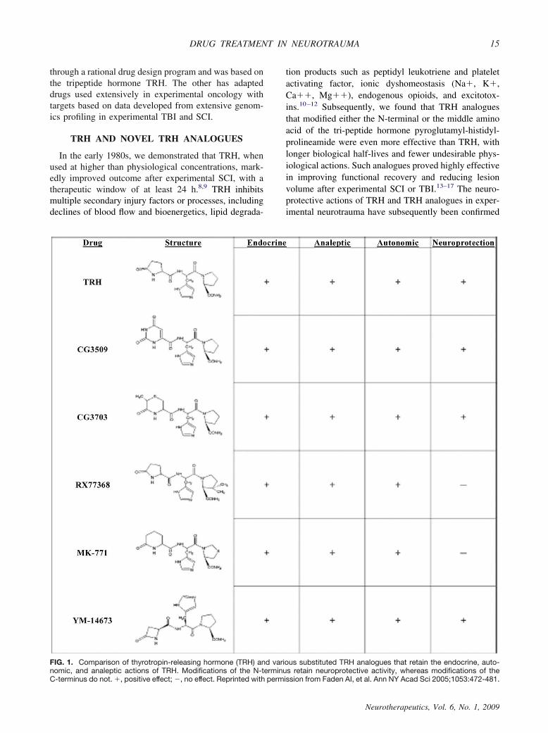
TRH is metabolized through two major pathways: en-dopeptidase cleavage of pyroglutamyl to produce cyclo-histidyly-proline diketopiperazine (CHP) or deamidationyielding the free acid form of TRH.24,25 Various TRHanalogues have been developed that modify one of itsamino acids (FIG. 1).26 Pyroglutamyl substitutions limitendopeptidase-mediated metabolism, resulting in com-pounds that have far longer biological half-lives thanTRH (6–8 h vs 5 min); some of these are also morepotent than TRH in terms of CNS activity. For example,YM-14673 is longer acting than TRH (8–36 times) andmuch more potent (10–100 times).15 However, N-termi-nal substitutions retain the other physiological actions ofTRH (i.e., endocrine, autonomic, and analeptic). Wehave also evaluated modifications of the histidyl residue(i.e., imidazole substitution); certain substitutions re-duced the cardiovascular and/or endocrine activity whilemaintaining the neuroprotective actions of TRH (FIG.2).27 Critically, modification of the C-terminus results incompounds devoid of neuroprotective activity, although
they retain endocrine, autonomic, and analeptic activitysimilar to TRH.
Based on these observations, we developed dual-sub-stituted TRH analogues (i.e., modifications at both theN-terminal and histidyl moieties). Such compounds (53a,57a) have limited endocrine, autonomic, and analepticeffects while preserving or enhancing the neuroprotec-tive actions (FIG. 2).16,26 Compound 53a is at least twoorders of magnitude more hydrophobic than either TRHor YM-14673, based on their partition coefficients be-tween n-octanol and water (logP); thus it should haveenhanced cellular permeability to the CNS.16
CYCLIC DIPEPTIDES
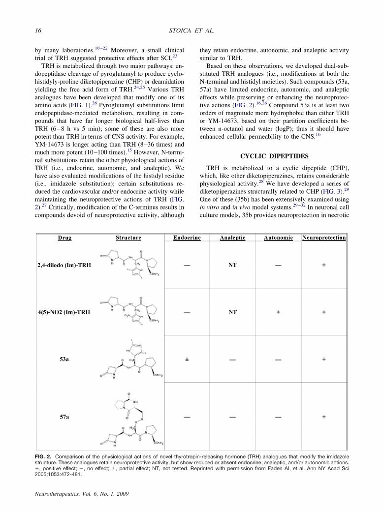
TRH is metabolized to a cyclic dipeptide (CHP),which, like other diketopiperazines, retains considerablephysiological activity.28 We have developed a series ofdiketopiperazines structurally related to CHP (FIG. 3).29
One of these (35b) has been extensively examined usingin vitro and in vivo model systems.29–32 In neuronal cellculture models, 35b provides neuroprotection in necrotic
FIG. 2. Comparison of the physiological actions of novel thyrotropin-releasing hormone (TRH) analogues that modify the imidazolestructure. These analogues retain neuroprotective activity, but show reduced or absent endocrine, analeptic, and/or autonomic actions.�, positive effect; �, no effect; �, partial effect; NT, not tested. Reprinted with permission from Faden AI, et al. Ann NY Acad Sci2005;1053:472-481.
STOICA ET AL.16
Neurotherapeutics, Vol. 6, No. 1, 2009
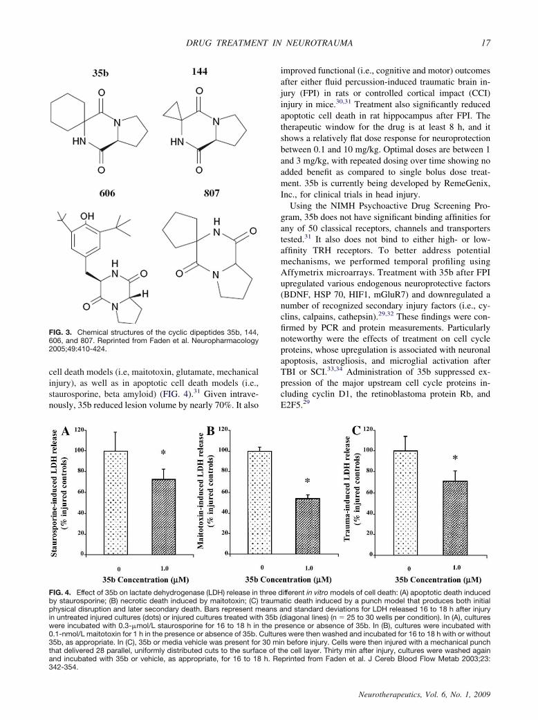
cell death models (i.e, maitotoxin, glutamate, mechanicalinjury), as well as in apoptotic cell death models (i.e.,staurosporine, beta amyloid) (FIG. 4).31 Given intrave-nously, 35b reduced lesion volume by nearly 70%. It also
improved functional (i.e., cognitive and motor) outcomesafter either fluid percussion-induced traumatic brain in-jury (FPI) in rats or controlled cortical impact (CCI)injury in mice.30,31 Treatment also significantly reducedapoptotic cell death in rat hippocampus after FPI. Thetherapeutic window for the drug is at least 8 h, and itshows a relatively flat dose response for neuroprotectionbetween 0.1 and 10 mg/kg. Optimal doses are between 1and 3 mg/kg, with repeated dosing over time showing noadded benefit as compared to single bolus dose treat-ment. 35b is currently being developed by RemeGenix,Inc., for clinical trials in head injury.
Using the NIMH Psychoactive Drug Screening Pro-gram, 35b does not have significant binding affinities forany of 50 classical receptors, channels and transporterstested.31 It also does not bind to either high- or low-affinity TRH receptors. To better address potentialmechanisms, we performed temporal profiling usingAffymetrix microarrays. Treatment with 35b after FPIupregulated various endogenous neuroprotective factors(BDNF, HSP 70, HIF1, mGluR7) and downregulated anumber of recognized secondary injury factors (i.e., cy-clins, calpains, cathepsin).29,32 These findings were con-firmed by PCR and protein measurements. Particularlynoteworthy were the effects of treatment on cell cycleproteins, whose upregulation is associated with neuronalapoptosis, astrogliosis, and microglial activation afterTBI or SCI.33,34 Administration of 35b suppressed ex-pression of the major upstream cell cycle proteins in-cluding cyclin D1, the retinoblastoma protein Rb, andE2F5.29
FIG. 3. Chemical structures of the cyclic dipeptides 35b, 144,606, and 807. Reprinted from Faden et al. Neuropharmacology2005;49:410-424.
FIG. 4. Effect of 35b on lactate dehydrogenase (LDH) release in three different in vitro models of cell death: (A) apoptotic death inducedby staurosporine; (B) necrotic death induced by maitotoxin; (C) traumatic death induced by a punch model that produces both initialphysical disruption and later secondary death. Bars represent means and standard deviations for LDH released 16 to 18 h after injuryin untreated injured cultures (dots) or injured cultures treated with 35b (diagonal lines) (n � 25 to 30 wells per condition). In (A), cultureswere incubated with 0.3-�mol/L staurosporine for 16 to 18 h in the presence or absence of 35b. In (B), cultures were incubated with0.1-nmol/L maitotoxin for 1 h in the presence or absence of 35b. Cultures were then washed and incubated for 16 to 18 h with or without35b, as appropriate. In (C), 35b or media vehicle was present for 30 min before injury. Cells were then injured with a mechanical punchthat delivered 28 parallel, uniformly distributed cuts to the surface of the cell layer. Thirty min after injury, cultures were washed againand incubated with 35b or vehicle, as appropriate, for 16 to 18 h. Reprinted from Faden et al. J Cereb Blood Flow Metab 2003;23:342-354.
DRUG TREATMENT IN NEUROTRAUMA 17
Neurotherapeutics, Vol. 6, No. 1, 2009
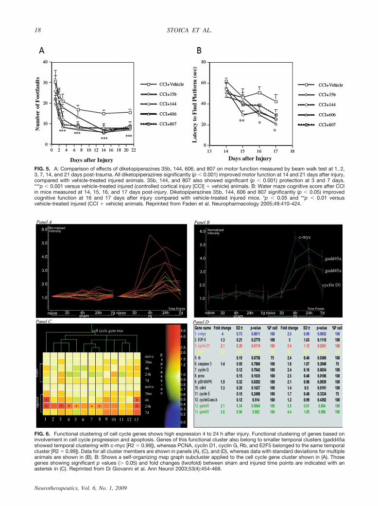
FIG. 5. A: Comparison of effects of diketopiperazines 35b, 144, 606, and 807 on motor function measured by beam walk test at 1, 2,3, 7, 14, and 21 days post-trauma. All diketopiperazines significantly (p � 0.001) improved motor function at 14 and 21 days after injury,compared with vehicle-treated injured animals. 35b, 144, and 807 also showed significant (p � 0.001) protection at 3 and 7 days.***p � 0.001 versus vehicle-treated injured (controlled cortical injury [CCI] � vehicle) animals. B: Water maze cognitive score after CCIin mice measured at 14, 15, 16, and 17 days post-injury. Diketopiperazines 35b, 144, 606 and 807 significantly (p � 0.05) improvedcognitive function at 16 and 17 days after injury compared with vehicle-treated injured mice. *p � 0.05 and **p � 0.01 versusvehicle-treated injured (CCI � vehicle) animals. Reprinted from Faden et al. Neuropharmacology 2005;49:410-424.
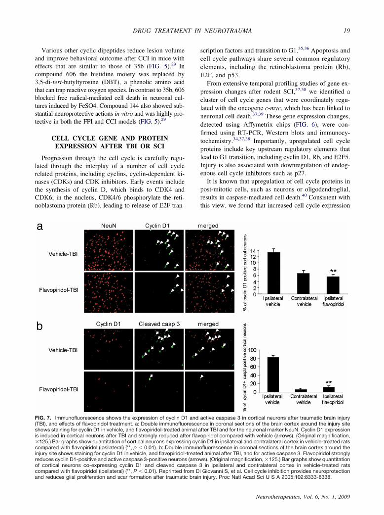
FIG. 6. Functional clustering of cell cycle genes shows high expression 4 to 24 h after injury. Functional clustering of genes based oninvolvement in cell cycle progression and apoptosis. Genes of this functional cluster also belong to smaller temporal clusters (gadd45ashowed temporal clustering with c-myc [R2 � 0.99]), whereas PCNA, cyclin D1, cyclin G, Rb, and E2F5 belonged to the same temporalcluster [R2 � 0.99]). Data for all cluster members are shown in panels (A), (C), and (D), whereas data with standard deviations for multipleanimals are shown in (B). B: Shows a self-organizing map graph subcluster applied to the cell cycle gene cluster shown in (A). Thosegenes showing significant p values (� 0.05) and fold changes (twofold) between sham and injured time points are indicated with anasterisk in (C). Reprinted from Di Giovanni et al. Ann Neurol 2003;53(4):454-468.
STOICA ET AL.18
Neurotherapeutics, Vol. 6, No. 1, 2009
Various other cyclic dipeptides reduce lesion volumeand improve behavioral outcome after CCI in mice witheffects that are similar to those of 35b (FIG. 5).29 Incompound 606 the histidine moiety was replaced by3,5-di-tert-butyltyrosine (DBT), a phenolic amino acidthat can trap reactive oxygen species. In contrast to 35b, 606blocked free radical-mediated cell death in neuronal cul-tures induced by FeSO4. Compound 144 also showed sub-stantial neuroprotective actions in vitro and was highly pro-tective in both the FPI and CCI models (FIG. 5).29
CELL CYCLE GENE AND PROTEINEXPRESSION AFTER TBI OR SCI
Progression through the cell cycle is carefully regu-lated through the interplay of a number of cell cyclerelated proteins, including cyclins, cyclin-dependent ki-nases (CDKs) and CDK inhibitors. Early events includethe synthesis of cyclin D, which binds to CDK4 andCDK6; in the nucleus, CDK4/6 phosphorylate the reti-noblastoma protein (Rb), leading to release of E2F tran-
scription factors and transition to G1.35,36 Apoptosis andcell cycle pathways share several common regulatoryelements, including the retinoblastoma protein (Rb),E2F, and p53.
From extensive temporal profiling studies of gene ex-pression changes after rodent SCI,37,38 we identified acluster of cell cycle genes that were coordinately regu-lated with the oncogene c-myc, which has been linked toneuronal cell death.37,39 These gene expression changes,detected using Affymetrix chips (FIG. 6), were con-firmed using RT-PCR, Western blots and immunocy-tochemistry.34,37,38 Importantly, upregulated cell cycleproteins include key upstream regulatory elements thatlead to G1 transition, including cyclin D1, Rb, and E2F5.Injury is also associated with downregulation of endog-enous cell cycle inhibitors such as p27.
It is known that upregulation of cell cycle proteins inpost-mitotic cells, such as neurons or oligodendroglial,results in caspase-mediated cell death.40 Consistent withthis view, we found that increased cell cycle expression
FIG. 7. Immunofluorescence shows the expression of cyclin D1 and active caspase 3 in cortical neurons after traumatic brain injury(TBI), and effects of flavopiridol treatment. a: Double immunofluorescence in coronal sections of the brain cortex around the injury siteshows staining for cyclin D1 in vehicle, and flavopiridol-treated animal after TBI and for the neuronal marker NeuN. Cyclin D1 expressionis induced in cortical neurons after TBI and strongly reduced after flavopiridol compared with vehicle (arrows). (Original magnification,125.) Bar graphs show quantitation of cortical neurons expressing cyclin D1 in ipsilateral and contralateral cortex in vehicle-treated ratscompared with flavopiridol (ipsilateral) (**, p � 0.01). b: Double immunofluorescence in coronal sections of the brain cortex around theinjury site shows staining for cyclin D1 in vehicle, and flavopiridol-treated animal after TBI, and for active caspase 3. Flavopiridol stronglyreduces cyclin D1-positive and active caspase 3-positive neurons (arrows). (Original magnification, 125.) Bar graphs show quantitationof cortical neurons co-expressing cyclin D1 and cleaved caspase 3 in ipsilateral and contralateral cortex in vehicle-treated ratscompared with flavopiridol (ipsilateral) (**, P � 0.01). Reprinted from Di Giovanni S, et al. Cell cycle inhibition provides neuroprotectionand reduces glial proliferation and scar formation after traumatic brain injury. Proc Natl Acad Sci U S A 2005;102:8333-8338.
DRUG TREATMENT IN NEUROTRAUMA 19
Neurotherapeutics, Vol. 6, No. 1, 2009
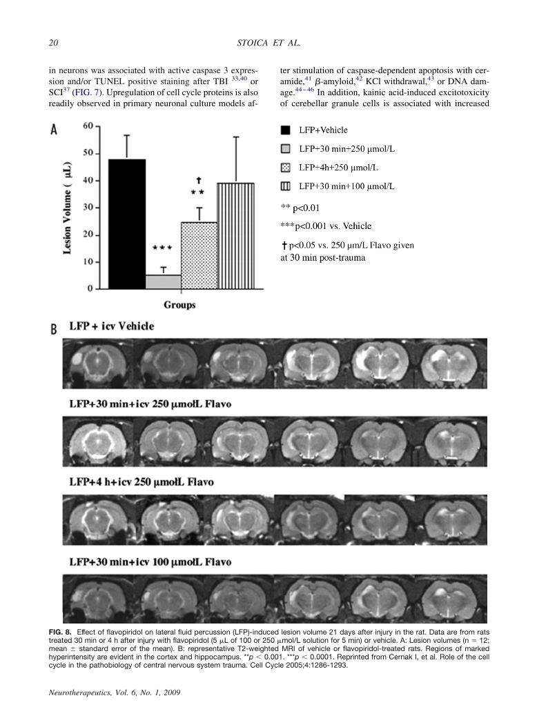
in neurons was associated with active caspase 3 expres-sion and/or TUNEL positive staining after TBI 33,40 orSCI37 (FIG. 7). Upregulation of cell cycle proteins is alsoreadily observed in primary neuronal culture models af-
ter stimulation of caspase-dependent apoptosis with cer-amide,41 �-amyloid,42 KCl withdrawal,43 or DNA dam-age.44–46 In addition, kainic acid-induced excitotoxicityof cerebellar granule cells is associated with increased
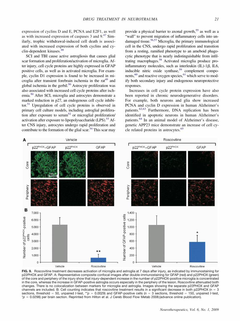
FIG. 8. Effect of flavopiridol on lateral fluid percussion (LFP)-induced lesion volume 21 days after injury in the rat. Data are from ratstreated 30 min or 4 h after injury with flavopiridol (5 �L of 100 or 250 �mol/L solution for 5 min) or vehicle. A: Lesion volumes (n � 12;mean � standard error of the mean). B: representative T2-weighted MRI of vehicle or flavopiridol-treated rats. Regions of markedhyperintensity are evident in the cortex and hippocampus. **p � 0.001. ***p � 0.0001. Reprinted from Cernak I, et al. Role of the cellcycle in the pathobiology of central nervous system trauma. Cell Cycle 2005;4:1286-1293.
STOICA ET AL.20
Neurotherapeutics, Vol. 6, No. 1, 2009
expression of cyclins D and E, PCNA and E2F1, as wellas with increased expression of caspases 3 and 9.47 Sim-ilarly, trophic withdrawal-induced cell death is associ-ated with increased expression of both cyclins and cy-clin-dependent kinases.48
SCI and TBI cause active astrogliosis that causes glialscar formation and proliferation/activation of microglia. Af-ter injury, cell cycle proteins are highly expressed in GFAPpositive cells, as well as in activated microglia. For exam-ple, cyclin D1 expression is found to be increased in mi-croglia after transient forebrain ischemia in the rat49 andglobal ischemia in the gerbil.50 Astrocyte proliferation wasalso associated with increased cell cycle proteins after isch-emia.50 After SCI, microglia and astrocytes demonstrate amarked reduction in p27, an endogenous cell cycle inhibi-tor.51 Upregulation of cell cycle proteins is observed inprimary cell culture models, including astroglial prolifera-tion after exposure to serum52 or microglial proliferation/activation after exposure to lipopolysaccharide (LPS).53 Af-ter CNS injury, astrocytes undergo rapid proliferation andcontribute to the formation of the glial scar.54 This scar may
provide a physical barrier to axonal growth,55 as well as a“wall” to prevent migration of inflammatory cells into un-damaged tissue.56,57 Microglia, the primary immunologicalcell in the CNS, undergo rapid proliferation and transitionfrom a resting, ramified phenotype to an ameboid phago-cytic phenotype that is nearly indistinguishable from infil-trating macrophages.58 Activated microglia produce pro-inflammatory molecules, such as interleukin (IL)-1�, IL6,inducible nitric oxide synthase,59 complement compo-nents,60 and reactive oxygen species,61 which serve to mod-ify both secondary injury and endogenous neuroprotectiveresponses.
Increases in cell cycle protein expression have alsobeen reported in chronic neurodegenerative disorders.For example, both neurons and glia show increasedPCNA and cyclin D expression in human Alzheimer’spatients.62,63 Furthermore, DNA replication has beenidentified in apoptotic neurons in human Alzheimer’spatients.64 In an animal model of Alzheimer’s disease,genetic APP23 mice demonstrate an increase of cell cy-cle related proteins in astrocytes.65
FIG. 9. Roscovitine treatment decreases activation of microglia and astroglia at 7 days after injury, as indicated by immunostaining forp22PHOX and GFAP. A: Representative composite confocal images after double-immunostaining for GFAP (red) and p22PHOX (green)of the core and periphery of the injury show that injury-dependent increase in the number of p22PHOX-positive microglia is concentratedin the core, whereas the increase in GFAP-positive astroglia occurs especially in the periphery of the lesion. Roscovitine attenuated bothchanges. There is no colocalization between markers for microglia and astroglia. Images showing the separate p22PHOX and GFAPchannels are included. B: Cell counting indicates that roscovitine treatment results in a significant decrease in both p22PHOX (n � 3sections, threshold � 50, unpaired t-test, **p � 0.0029) and GFAP-positive cells (n � 3 sections, threshold � 150, unpaired t-test,*p � 0.0298) per brain section. Reprinted from Hilton et al. J Cereb Blood Flow Metab 2008;(advance online publication).
DRUG TREATMENT IN NEUROTRAUMA 21
Neurotherapeutics, Vol. 6, No. 1, 2009
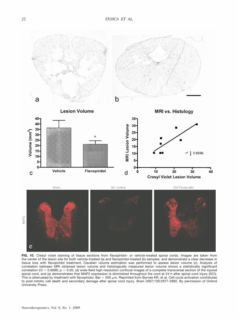
FIG. 10. Cresyl violet staining of tissue sections from flavopiridol- or vehicle-treated spinal cords. Images are taken fromthe center of the lesion site for both vehicle-treated (a) and flavopiridol-treated (b) samples, and demonstrate a clear decrease intissue loss with flavopiridol treatment. Cavalieri volume estimation was performed to assess lesion volume (c). Analysis ofcorrelation between MRI obtained lesion volume and histologically measured lesion volume shows a statistically significantcorrelation (r2 � 0.6696; p � 0.05; (d) wide-field high-resolution confocal images of a complete transversal section of the injuredspinal cord, and (e) demonstrates that MAP2 expression is diminished throughout the cord at 24 h after spinal cord injury (SCI).This is attenuated by treatment with flavopiridol. Bar � 500 �m. Reprinted from Byrnes KR, et al. Cell cycle activation contributesto post-mitotic cell death and secondary damage after spinal cord injury. Brain 2007;130:2977-2992. By permission of OxfordUniversity Press.
STOICA ET AL.22
Neurotherapeutics, Vol. 6, No. 1, 2009
DRUG TREATMENT IN NEUROTRAUMA 23
Neurotherapeutics, Vol. 6, No. 1, 2009
INHIBITION OF CELL CYCLECell cycle inhibitors have been developed and exten-
sively evaluated in experimental cancer models, and sev-eral have been tested in humans. The best characterizedand studied among these are flavopiridol, a semi-syn-thetic flavonoid derived from rohitukin bark,66 and thepurine analogues roscovitine and olomoucine.67 Fla-vopiridol blocks all the CDKs and also inhibits the tran-scription of cyclin D1.68,69 In contrast, the purine ana-logues preferentially inhibit CDK2 and CDK5, althoughat higher concentrations these may inhibit other ki-nases.70
Each of these agents shows neuroprotection in vitro,such as against etoposide-induced neuronal apoptosis33
or apoptosis of cerebellar granule cells after KCl with-drawal.43 Moreover, olomoucine inhibits hypoxia-in-duced neuronal cell death in culture,71 whereas flavopiri-dol inhibits kainite-mediated or colchicines-mediatedapoptotic cell death.72,73 A recent study in our laboratorydetermined that inhibition of multiple cyclin-dependentkinases reduces etoposide-induced neuronal apoptosis,including CDK1 and CDK4.74
Cell cycle inhibitors show inhibitory effects on theproliferation and activation of mitotic cells, such as mi-croglia and astrocytes in vitro. For example, stimulationof microglia with LPS induces proliferation. Pre-treat-ment of microglia with cell cycle inhibitors, such asflavopiridol or roscovitine, for 1 h prior to the addition ofLPS results in a significant suppression of microglialproliferation33 and nitric oxide production.74 Impor-tantly, roscovitine treatment of microglial cells stimu-lated with LPS reduced microglial-induced neurotoxi-city.74 Similarly, proliferation of astrocytes induced bythe addition of 10% serum was completely inhibited byflavopiridol.33
In vivo, cell cycle inhibition using pharmacologicalapproaches has shown neuroprotective effects. For ex-ample, early treatment with flavopiridol, administeredcentrally, showed remarkable neuroprotection after FPIin rats.40 Lesion volume was reduced by approximately70% and chronic behavioral recovery (motor and cogni-tive) was indistinguishable from sham-injured controls.Caspase-mediated neuronal cell death after TBI was
nearly completely attenuated. In addition to neuroprotec-tion, significant effects on mitotic cells were also ob-served. GFAP expression and markers of microglial ac-tivation were markedly reduced. These changes wereassociated with near complete suppression of cell cycleproteins in neurons, astroglia, and microglia, respecti-vely.40 Delayed administration of flavopiridol was sim-ilarly found to have neuroprotective effects. In a fol-low-up study, flavopiridol was administered centrally at30 min or 4 h after FPI, or systemically (intraperitone-ally) at 24 h after FPI;33 each of these treatments resultedin markedly reduced lesion volumes that were approxi-mately 90%, 50%, and 60%, respectively (FIG. 8).
The more specific cell cycle inhibitor roscovitine,which does not have potentially confusing effects ongene transcription, has similar actions after FPI. Admin-istration of roscovitine 30 min after FPI resulted inhighly significant reductions in lesion volume and im-proved behavioral outcome (motor and cognitive). Thiscell cycle inhibitor also reduced astrogliosis and pro-duced a marked inhibition of microglial activation-re-lated inflammation74 (FIG. 9).
Research in SCI confirms the strong beneficial effectsof treatment with cell cycle inhibitors. We have shownthat flavopiridol treatment, centrally administered bymini-osmotic pump beginning 30 min post-trauma andcontinuing over 7 days, significantly improved motorrecovery and reduced lesion volume at 28 days.34 Treat-ment-reduced cell cycle protein induction in neurons andastrocytes; this reduction was associated with decreasedcleaved caspase-3 labeling in neurons and oligodendro-cytes, as well as reduction in glial scar. Neuronal loss,measured by MAP-2 staining, was alleviated by fla-vopiridol treatment, and tissue loss was significantly re-duced overall. Further treatment with the cell-cycleinhibitor markedly limited microglial activation and as-sociated inflammatory factors (FIGS. 10 and 11). Thecell cycle inhibitor olomoucine has also been shown todecrease lesion volume and improve function afterSCI.75 Reductions in microglial-related inflammation76
and astrocytic scar75 were found with olomoucine treat-ment, supporting the beneficial effect of cell-cycle inhi-bition after SCI. Preliminary work using cyclin D1
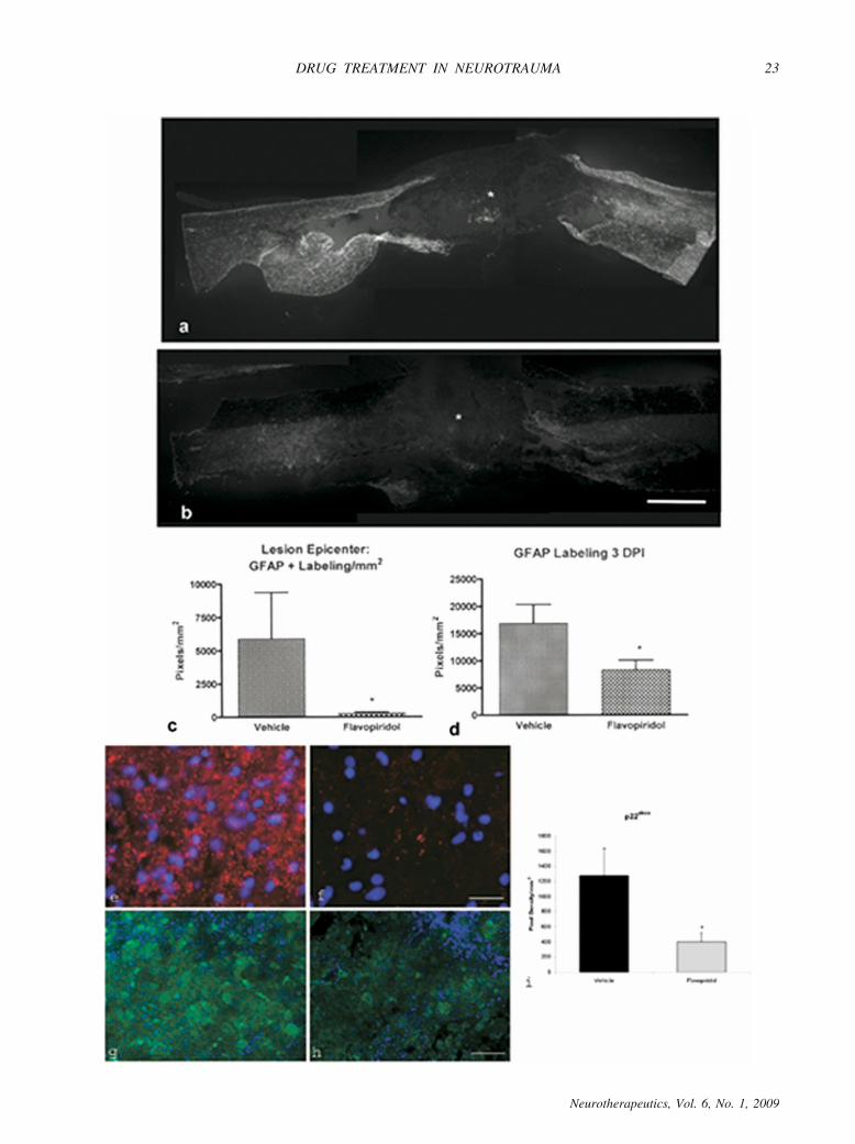
FIG. 11. Astrocyte and microglial marker immunohistochemistry after injury and treatment. Immunohistochemistry performed forastrocytes (a, b), osteopontin (e, f), and p22phox (g, h) at 3 and 28 days post-injury. Heavy astrocytic (GFAP) labeling was foundsurrounding the lesion site (*) at 28 days after SCI in vehicle-treated tissue (shown as a mosaic image of the entire 10-mm cord sectionsurrounding the lesion epicenter); (a) however, little GFAP labeling was found in samples that had received flavopiridol continuousinfusion (b). Quantitation of the proportional area of GFAP labeling in the spinal cord (through the 1 cm surrounding the lesion site)showed a significant decrease in GFAP labeling at 28 (c) and 3 (d) days post-injury in flavopiridol-treated tissue (p � 0.05; n � 3/groupat 3rd day; 10/group at 28 days). Immunolabeling for osteopontin and p22phox, factors expressed by microglia, was also decreased byflavopiridol treatment (f, h) in comparison to vehicle (e, g). All images are obtained from 1 mm rostral to the lesion epicenter, in the centerregion of a transverse spinal cord section. Quantitation of p22phox labeling in the spinal cord (through the 1 cm surrounding the lesionsite) showed a significant decrease in labeling at 28 days post-injury in flavopiridol-treated tissue (i; *p � 0.05). Bar � 1 mm (a, b); 50�m (e, f); 100 �m (g, h). Reprinted from Byrnes KR, et al. Cell cycle activation contributes to post-mitotic cell death and secondarydamage after spinal cord injury. Brain 2007;130:2977-2992. By permission of Oxford University Press.
STOICA ET AL.24
Neurotherapeutics, Vol. 6, No. 1, 2009
knockout mice subjected to SCI has shown decreasedlesion volumes in knockouts as compared to wild typecontrols, consistent with the pharmacological inhibitionstudies.34
Research in experimental cerebral ischemia is highlyconsistent with the TBI and SCI studies. Dominant neg-ative CDK4/5 animals show reduced neuronal cell deathafter focal or global brain ischemia,77 as does treatmentwith CDK inhibitors.78,79 Moreover, after focal cerebralischemia, cyclin D1 knockouts or animals treated witholomoucine show reduced astrocyte proliferation. Exci-totoxic cell death after kainic acid administration is at-tenuated by treatment with anti-sense oligonucleotidesdirected against CDK4 or cyclin D1.
SUMMARY
Activation of cell cycle proteins in the CNS causesproliferation of mitotic cells, such as astroglia or micro-glia, but induces apoptosis in post-mitotic cells, such asneurons or oligodendroglia. Acute injuries to the CNS,including TBI and SCI, cause upregulation of many cell-cycle proteins in both mitotic and post-mitotic cells.These changes cause neuronal and oligodendroglial celldeath, astroglial scar formation and proliferation/activa-tion of microglia, with the release of associated inflam-matory factors. Treatment with cell-cycle inhibitors re-sults in striking neuroprotection, likely related to itsmultifunctional actions on these diverse cell types. Be-cause cell-cycle proteins have such diverse effects, evenselective inhibitors of these pathways may serve as mul-tifunctional neuroprotective agents.
Another approach to multi-potential drug treatment ofCNS injury is to use compounds that modulate differentsignal transduction pathways that are involved in sec-ondary injury. TRH is a naturally occurring brain hor-mone, which when used at higher than physiologicallevels as a drug can inhibit many factors and mechanismsimplicated in delayed cell death. Thus, TRH and TRHanalogues can improve blood flow and bio-energeticstate; limit loss of ionic homeostasis; reduce lipid deg-radation; and inhibit the actions of endogenous opioids,leukotrienes, platelet-activating factor, and possibly glu-tamate.29,32 The neuroprotective effects do not appear tobe mediated by TRH receptors, as they occur at supra-physiological doses and can be dissociated from theother physiological effects of TRH (i.e., endocrine, an-aleptic, autonomic).
Diketopiperazines that are structurally related to ametabolic product of TRH have marked neuroprotectiveactivity, but do not act on either high- or low-affinityTRH receptors. They also have diverse multifunctionalneuroprotective actions. As with cell-cycle inhibitors, theprototype compound 35b inhibits the activation of manycell-cycle proteins after injury. But they also reduce
other known secondary injury factors, including calpainsand cathepsins, while upregulating several well-estab-lished endogenous neuroprotective factors includingbrain derived neurotrophic factor, heat shock protein 70,and hypoxia-inducible factor 1. Each of the latter factorshas considerable protective activity in animal models.These findings underscore the attractiveness of multi-functional drug approaches for the treatment of neuro-trauma and other neurodegenerative disorders.
Acknowledgments: This work has been supported byNational Institutes of Health grants 5R01NS052568 and5R01NS054221 to author (AIF).
REFERENCES
1. Yakovlev AG, Faden AI. Mechanisms of neural cell death: impli-cations for development of neuroprotective treatment strategies.NeuroRX 2004;1:5–16.
2. Faden AI, Stoica B. Neuroprotection: challenges and opportunities.Arch Neurol 2007;64:794–800.
3. Faden AI. Neuroprotection and traumatic brain injury: theoreticaloption or realistic proposition. Curr Opin Neurol 2002;15:707–712.
4. Dumont RJ, Okonkwo DO, Verma S, et al. Acute spinal cordinjury, part I: pathophysiologic mechanisms. Clin Neuropharmacol2001;24:254–264.
5. Tator CH. Experimental and clinical studies of the pathophysiol-ogy and management of acute spinal cord injury. J Spinal CordMed 1996;19:206–214.
6. Olney JW. Excitotoxin-mediated neuron death in youth and oldage. Prog Brain Res 1990;86:37–51.
7. Faden AI. Comparison of single and combination drug treatmentstrategies in experimental brain trauma. J Neurotrauma 1993;10:91–100.
8. Faden AI, Jacobs TP, Holaday JW. Thyrotropin-releasing hormoneimproves neurologic recovery after spinal trauma in cats. N EnglJ Med 1981;305:1063–1067.
9. Faden AI, Jacobs TP, Smith MT. Thyrotropin-releasing hormonein experimental spinal injury: dose response and late treatment.Neurology 1984;34:1280–1284.
10. Faden AI, Yum SW, Lemke M, Vink R. Effects of TRH-analogtreatment on tissue cations, phospholipids and energy metabolismafter spinal cord injury. J Pharmacol Exp Ther 1990;255:608–614.
11. Lux WE, Jr., Feuerstein G, Faden AI. Alteration of leukotriene D4hypotension by thyrotropin releasing hormone. Nature 1983;302:822–824.
12. Feuerstein G, Lux WE, Ezra D, Hayes EC, Snyder F, Faden AI.Thyrotropin-releasing hormone blocks the hypotensive effects ofplatelet-activating factor in the unanesthetized guinea pig. J Car-diovasc Pharmacol 1985;7:335–340.
13. McIntosh TK, Vink R, Faden AI. An analogue of thyrotropin-releasing hormone improves outcome after brain injury: 31P-NMRstudies. Am J Physiol 1988;254:R785–792.
14. McIntosh TK, Vink R, Faden AI. Beneficial effect of the TRHanalog CG-3703 on outcome and survival following traumaticbrain injury in rats. Prog Clin Biol Res 1988;264:415–420.
15. Faden AI. TRH analog YM-14673 improves outcome followingtraumatic brain and spinal cord injury in rats: dose-response stud-ies. Brain Res 1989;486:228–235.
16. Faden AI, Fox GB, Fan L, et al. Novel TRH analog improvesmotor and cognitive recovery after traumatic brain injury in ro-dents. Am J Physiol 1999;277:R1196–1204.
17. Faden AI, Sacksen I, Noble LJ. Structure-activity relationships ofTRH analogs in rat spinal cord injury. Brain Res 1988;448:287–293.
18. Wang GL, Zhu C. Effects of thyrotropin-releasing hormone onacute experimental traumatic head injury in cats. Chin Med J(Engl) 1991;104:939–944.
DRUG TREATMENT IN NEUROTRAUMA 25
Neurotherapeutics, Vol. 6, No. 1, 2009
19. Tanaka K, Ogawa N, Asanuma M, Kondo Y. Thyrotropin releasinghormone prevents abnormalities of cortical acetylcholine andmonoamines in mice following head injury. Regul Pept 1997;70:173–178.
20. Arias MJ. Treatment of experimental spinal cord injury with TRH,naloxone, and dexamethasone. Surg Neurol 1987;28:335–338.
21. Behrmann DL, Bresnahan JC, Beattie MS. Modeling of acutespinal cord injury in the rat: neuroprotection and enhanced recov-ery with methylprednisolone, U-74006F and YM-14673. Exp Neu-rol 1994;126:61–75.
22. Puniak MA, Freeman GM, Agresta CA, Van Newkirk L, BaroneCA, Salzman SK. Comparison of a serotonin antagonist, opioidantagonist, and TRH analog for the acute treatment of experimen-tal spinal trauma. J Neurotrauma 1991;8:193–203.
23. Pitts LH, Ross A, Chase GA, Faden AI. Treatment with thyro-tropin-releasing hormone (TRH) in patients with traumatic spinalcord injuries. J Neurotrauma 1995;12:235–243.
24. Friedman TC, Wilk S. Delineation of a particulate thyrotropin-releasing hormone-degrading enzyme in rat brain by the use ofspecific inhibitors of prolyl endopeptidase and pyroglutamyl pep-tide hydrolase. J Neurochem 1986;46:1231–1239.
25. Coggins PJ, McDermott JR, Snell CR, Gibson AM. Thyrotrophinreleasing hormone degradation by rat synaptosomal peptidases:production of the metabolite His-Pro. Neuropeptides 1987;10:147–156.
26. Faden Knoblach SM, Movsesyan VA, Lea PMt, Cernak I. Novelneuroprotective tripeptides and dipeptides. Ann N Y Acad Sci2005;1053:472–481.
27. Faden AI, Labroo VM, Cohen LA. Imidazole-substituted ana-logues of TRH limit behavioral deficits after experimental braintrauma. J Neurotrauma 1993;10:101–108.
28. Prasad C. Limited proteolysis and physiological regulation: anexample from thyrotropin-releasing hormone metabolism. Thyroid1998;8:969–975.
29. Faden AI, Movsesyan VA, Knoblach SM, Ahmed F, Cernak I.Neuroprotective effects of novel small peptides in vitro and afterbrain injury. Neuropharmacology 2005;49:410–424.
30. Faden AI, Fox GB, Di X, et al. Neuroprotective and nootropicactions of a novel cyclized dipeptide after controlled cortical im-pact injury in mice. J Cereb Blood Flow Metab 2003;23:355–363.
31. Faden AI, Knoblach SM, Cernak I, et al. Novel diketopiperazineenhances motor and cognitive recovery after traumatic brain injuryin rats and shows neuroprotection in vitro and in vivo. J CerebBlood Flow Metab 2003;23:342–354.
32. Faden AI, Knoblach SM, Movsesyan VA, Cernak I. Novel smallpeptides with neuroprotective and nootropic properties. J Alzhei-mers Dis 2004;6:S93–97.
33. Cernak I, Stoica B, Byrnes KR, Di Giovanni S, Faden AI. Role ofthe cell cycle in the pathobiology of central nervous systemtrauma. Cell Cycle 2005;4:1286–1293.
34. Byrnes KR, Stoica BA, Fricke S, Di Giovanni S, Faden AI. Cellcycle activation contributes to post-mitotic cell death and second-ary damage after spinal cord injury. Brain 2007;130:2977–2992.
35. Kitagawa M, Higashi H, Jung HK, et al. The consensus motif forphosphorylation by cyclin D1-Cdk4 is different from that for phos-phorylation by cyclin A/E-Cdk2. Embo J 1996;15:7060–7069.
36. Sears RC, Nevins JR. Signaling networks that link cell prolifera-tion and cell fate. J Biol Chem 2002;277:11617–11620.
37. Di Giovanni S, Knoblach SM, Brandoli C, Aden SA, Hoffman EP,Faden AI. Gene profiling in spinal cord injury shows role of cellcycle in neuronal death. Ann Neurol 2003;53:454–468.
38. De Biase A, Knoblach SM, Di Giovanni S, et al. Gene expressionprofiling of experimental traumatic spinal cord injury as a functionof distance from impact site and injury severity. Physiol Genomics2005;22:368–381.
39. Kangas A, Nicholson DW, Holtta E. Involvement of CPP32/Caspase-3 in c-Myc-induced apoptosis. Oncogene 1998;16:387–398.
40. Di Giovanni S, Movsesyan V, Ahmed F, et al. Cell cycle inhibitionprovides neuroprotection and reduces glial proliferation and scarformation after traumatic brain injury. Proc Natl Acad Sci U S A2005;102:8333–8338.
41. Strazza M, Luddi A, Brogi A, et al. Activation of cell cycleregulatory proteins in the apoptosis of terminally differentiatedoligodendrocytes. Neurochem Res 2004;29:923–931.
42. Giovanni A, Wirtz-Brugger F, Keramaris E, Slack R, Park DS.Involvement of cell cycle elements, cyclin-dependent kinases,pRb, and E2F x DP, in B-amyloid-induced neuronal death. J BiolChem 1999;274:19011–19016.
43. Padmanabhan J, Park DS, Greene LA, Shelanski ML. Role of cellcycle regulatory proteins in cerebellar granule neuron apoptosis.J Neurosci 1999;19:8747–8756.
44. Otsuka Y, Tanaka T, Uchida D, et al. Roles of cyclin-dependentkinase 4 and p53 in neuronal cell death induced by doxorubicin oncerebellar granule neurons in mouse. Neurosci Lett 2004;365:180–185.
45. Kruman II, Wersto RP, Cardozo-Pelaez F, et al. Cell cycle acti-vation linked to neuronal cell death initiated by DNA damage.Neuron 2004;41:549–561.
46. Park DS, Morris EJ, Bremner R, et al. Involvement of retinoblas-toma family members and E2F/DP complexes in the death ofneurons evoked by DNA damage. J Neurosci 2000;20:3104–3114.
47. Park DS, Obeidat A, Giovanni A, Greene LA. Cell cycle regulatorsin neuronal death evoked by excitotoxic stress: implications forneurodegeneration and its treatment. Neurobiol Aging 2000;21:771–781.
48. Park DS, Levine B, Ferrari G, Greene LA. Cyclin dependent kinaseinhibitors and dominant negative cyclin dependent kinase 4 and 6promote survival of NGF-deprived sympathetic neurons. J Neuro-sci 1997;17:8975–8983.
49. Wiessner C, Brink I, Lorenz P, Neumann-Haefelin T, Vogel P,Yamashita K. Cyclin D1 messenger RNA is induced in microgliarather than neurons following transient forebrain ischaemia. Neu-roscience 1996;72:947–958.
50. Kato H, Takahashi A, Itoyama Y. Cell cycle protein expression inproliferating microglia and astrocytes following transient globalcerebral ischemia in the rat. Brain Res Bull 2003;60:215–221.
51. Shen A, Liu Y, Zhao J, et al. Temporal-spatial expressions ofp27kip1 and its phosphorylation on Serine-10 after acute spinalcord injury in adult rat: implications for post-traumatic glial pro-liferation. Neurochem Int 2008;52:1266–1275.
52. Tanaka T, Tatsuno I, Noguchi Y, et al. Activation of cyclin-dependent kinase 2 (Cdk2) in growth-stimulated rat astrocytes.Geranylgeranylated Rho small GTPase(s) are essential for the in-duction of cyclin E gene expression. J Biol Chem 1998;273:26772–26778.
53. Lee SC, Liu W, Brosnan CF, Dickson DW. GM-CSF promotesproliferation of human fetal and adult microglia in primary cul-tures. Glia 1994;12:309–318.
54. Hoke A, Silver J. Heterogeneity among astrocytes in reactive gli-osis. Perspect Dev Neurobiol 1994;2:269–274.
55. Silver J, Miller JH. Regeneration beyond the glial scar. Nat RevNeurosci 2004;5:146–156.
56. Ridet JL, Malhotra SK, Privat A, Gage FH. Reactive astrocytes:cellular and molecular cues to biological function. Trends Neurosci1997;20:570–577.
57. Faulkner JR, Herrmann JE, Woo MJ, Tansey KE, Doan NB, So-froniew MV. Reactive astrocytes protect tissue and preserve func-tion after spinal cord injury. J Neurosci 2004;24:2143–2155.
58. Raivich G, Jones LL, Werner A, Bluthmann H, Doetschmann T,Kreutzberg GW. Molecular signals for glial activation: pro- andanti-inflammatory cytokines in the injured brain. Acta NeurochirSuppl 1999;73:21–30.
59. Benveniste EN. Inflammatory cytokines within the central nervoussystem: sources, function, and mechanism of action. Am J Physiol1992;263:C1–16.
60. Lynch NJ, Willis CL, Nolan CC, et al. Microglial activation andincreased synthesis of complement component C1q precedesblood-brain barrier dysfunction in rats. Mol Immunol 2004;40:709–716.
61. Pawate S, Shen Q, Fan F, Bhat NR. Redox regulation of glialinflammatory response to lipopolysaccharide and interferon-gamma. J Neurosci Res 2004;77:540–551.
62. Wharton SB, Williams GH, Stoeber K, et al. Expression of Ki67,PCNA and the chromosome replication licensing protein Mcm2 in
STOICA ET AL.26
Neurotherapeutics, Vol. 6, No. 1, 2009

glial cells of the ageing human hippocampus increases with theburden of Alzheimer-type pathology. Neurosci Lett 2005;383:33–38.
63. Yang Y, Mufson EJ, Herrup K. Neuronal cell death is preceded bycell cycle events at all stages of Alzheimer’s disease. J Neurosci2003;23:2557–2563.
64. Yang Y, Geldmacher DS, Herrup K. DNA replication precedesneuronal cell death in Alzheimer’s disease. J Neurosci 2001;21:2661–2668.
65. Gartner U, Bruckner MK, Krug S, Schmetsdorf S, Staufenbiel M,Arendt T. Amyloid deposition in APP23 mice is associated withthe expression of cyclins in astrocytes but not in neurons. ActaNeuropathol 2003;106:535–544.
66. Newcomb EW. Flavopiridol: pleiotropic biological effects enhanceits anti-cancer activity. Anticancer Drugs 2004;15:411–419.
67. Meijer L, Raymond E. Roscovitine and other purines as kinaseinhibitors. From starfish oocytes to clinical trials. Acc Chem Res2003;36:417–425.
68. Dai Y, Grant S. Small molecule inhibitors targeting cyclin-dependentkinases as anticancer agents. Curr Oncol Rep 2004;6:123–130.
69. Swanton C. Cell-cycle targeted therapies. Lancet Oncol 2004;5:27–36.
70. Abraham RT, Acquarone M, Andersen A, et al. Cellular effects ofolomoucine, an inhibitor of cyclin-dependent kinases. Biol Cell1995;83:105–120.
71. Bossenmeyer-Pourie C, Chihab R, Schroeder H, Daval JL. Tran-sient hypoxia may lead to neuronal proliferation in the developingmammalian brain: from apoptosis to cell cycle completion. Neu-roscience 1999;91:221–231.
72. Jorda EG, Verdaguer E, Canudas AM, et al. Neuroprotective actionof flavopiridol, a cyclin-dependent kinase inhibitor, in colchicine-induced apoptosis. Neuropharmacology 2003;45:672–683.
73. Verdaguer E, Jimenez A, Canudas AM, et al. Inhibition of cellcycle pathway by flavopiridol promotes survival of cerebellar gran-ule cells after an excitotoxic treatment. J Pharmacol Exp Ther2004;308:609–616.
74. Hilton GD, Stoica BA, Byrnes KR, Faden AI. Roscovitinereduces neuronal loss, glial activation, and neurologic deficitsafter brain trauma. J Cereb Blood Flow Metab 2008;28:1845–1859.
75. Tian DS, Yu ZY, Xie MJ, Bu BT, Witte OW, Wang W. Suppres-sion of astroglial scar formation and enhanced axonal regenerationassociated with functional recovery in a spinal cord injury ratmodel by the cell cycle inhibitor olomoucine. J Neurosci Res2006;84:1053–1063.
76. Tian DS, Dong Q, Pan DJ, et al. Attenuation of astrogliosis bysuppressing of microglial proliferation with the cell cycle inhibitorolomoucine in rat spinal cord injury model. Brain Res 2007;1154:206–214.
77. Rashidian J, Iyirhiaro G, Aleyasin H, et al. Multiple cyclin-depen-dent kinases signals are critical mediators of ischemia/hypoxicneuronal death in vitro and in vivo. Proc Natl Acad Sci U S A2005;102:14080–14085.
78. Wang F, Corbett D, Osuga H, et al. Inhibition of cyclin-dependentkinases improves CA1 neuronal survival and behavioral perfor-mance after global ischemia in the rat. J Cereb Blood Flow Metab2002;22:171–182.
79. Osuga H, Osuga S, Wang F, et al. Cyclin-dependent kinases as atherapeutic target for stroke. Proc Natl Acad Sci U S A 2000;97:10254–10259.
DRUG TREATMENT IN NEUROTRAUMA 27
Neurotherapeutics, Vol. 6, No. 1, 2009