Embed Size (px)
Citation preview

Progress in Neuro-Psychopharmacology & Biological Psychiatry 48 (2014) 64–78
Contents lists available at ScienceDirect
Progress in Neuro-Psychopharmacology & BiologicalPsychiatry
j ourna l homepage: www.e lsev ie r .com/ locate /pnp
Review
Multifunctional aspects of allopregnanolone in stress andrelated disorders
Anjana Bali, Amteshwar Singh Jaggi ⁎Department of Pharmaceutical Sciences and Drug Research, Punjabi University Patiala, 147002, India
Abbreviations: HPA, Hypothalamus-pituitary- adrenalreleasing factor; mPFC, median prefrontal cortex; PBR, Pepostsynaptic current; NKCC1, Na+-K+-Cl- co-transporter;itaries; SON, Supraoptic nucleus; HVA, Homovanillic acid;⁎ Corresponding author at: Department of Pharmaceut
E-mail address: [email protected] (A.S. Jag
0278-5846/$ – see front matter © 2013 Elsevier Inc. All rihttp://dx.doi.org/10.1016/j.pnpbp.2013.09.005
a b s t r a c t
a r t i c l e i n f oArticle history:Received 20 June 2013Received in revised form 5 September 2013Accepted 5 September 2013Available online 14 September 2013
Keywords:AllopregnanoloneAnxietyDepressionGABAPanicStress
Allopregnanolone (3α-hydroxy-5α-pregnan-20-one) is a major cholesterol-derived neurosteroid in the centralnervous systemand is synthesized fromprogesterone by steroidogenic enzymes, 5α-reductase (the rate-limitingenzyme) and 3α-hydroxysteroid dehydrogenase. The pathophysiological role of allopregnanolone in neuropsy-chiatric disorders has been highlighted in several investigations. The changes in neuroactive steroid levels aredetected in stress and stress-related disorders including anxiety, panic and depression. The changes inallopregnanolone in response to acute stressor tend to restore the homeostasis by dampening the hyper-activated HPA axis. However, long standing stressors leading to development of neuropsychiatric disordersincluding depression and anxiety are associatedwith decrease in the allopregnanolone levels. GABAA receptor com-plex has been considered as the primary target of allopregnanolone andmajority of its inhibitory actions are medi-ated through GABA potentiation or direct activation of GABA currents. The role of progesterone receptors inproducing the late actions of allopregnanolone particularly in lordosis facilitation has also been described.Moreover,recent studies have also described the involvement of other multiple targets including brain-derived neurotrophicfactor (BDNF), glutamate, dopamine, opioids, oxytocin, and calcium channels. The present review discusses the var-ious aspects of allopregnanolone in stress and stress-related disorders including anxiety, depression and panic.
© 2013 Elsevier Inc. All rights reserved.
Contents
1. Introduction . . . . . . . . . . . . . . . . . . . . . . . . . . . . . . . . . . . . . . . . . . . . . . . . . . . . . . . . . . . . . . . 652. Allopregnanolone in stress . . . . . . . . . . . . . . . . . . . . . . . . . . . . . . . . . . . . . . . . . . . . . . . . . . . . . . . . 66
2.1. Acute stress . . . . . . . . . . . . . . . . . . . . . . . . . . . . . . . . . . . . . . . . . . . . . . . . . . . . . . . . . . . . 662.2. Chronic stress . . . . . . . . . . . . . . . . . . . . . . . . . . . . . . . . . . . . . . . . . . . . . . . . . . . . . . . . . . . 672.3. Hypothesis for differential levels of allopregnanolone in acute and chronic stresses . . . . . . . . . . . . . . . . . . . . . . . . . . . . 682.4. Allopregnanolone and depression . . . . . . . . . . . . . . . . . . . . . . . . . . . . . . . . . . . . . . . . . . . . . . . . . . 682.5. Allopregnanolone and anxiety . . . . . . . . . . . . . . . . . . . . . . . . . . . . . . . . . . . . . . . . . . . . . . . . . . . 692.6. Allopregnanolone and panic . . . . . . . . . . . . . . . . . . . . . . . . . . . . . . . . . . . . . . . . . . . . . . . . . . . . 692.7. Allopregnanolone and lordosis . . . . . . . . . . . . . . . . . . . . . . . . . . . . . . . . . . . . . . . . . . . . . . . . . . . 70
3. Mechanism of action . . . . . . . . . . . . . . . . . . . . . . . . . . . . . . . . . . . . . . . . . . . . . . . . . . . . . . . . . . . 703.1. GABAA receptor complex as primary target . . . . . . . . . . . . . . . . . . . . . . . . . . . . . . . . . . . . . . . . . . . . . . 70
3.1.1. Post-synaptic GABAA receptors . . . . . . . . . . . . . . . . . . . . . . . . . . . . . . . . . . . . . . . . . . . . . . . 703.1.2. Presynaptic GABA . . . . . . . . . . . . . . . . . . . . . . . . . . . . . . . . . . . . . . . . . . . . . . . . . . . . . 72
3.2. Other targets . . . . . . . . . . . . . . . . . . . . . . . . . . . . . . . . . . . . . . . . . . . . . . . . . . . . . . . . . . . 723.2.1. Progesterone receptors . . . . . . . . . . . . . . . . . . . . . . . . . . . . . . . . . . . . . . . . . . . . . . . . . . 723.2.2. Glutamate . . . . . . . . . . . . . . . . . . . . . . . . . . . . . . . . . . . . . . . . . . . . . . . . . . . . . . . . 723.2.3. BDNF . . . . . . . . . . . . . . . . . . . . . . . . . . . . . . . . . . . . . . . . . . . . . . . . . . . . . . . . . . 733.2.4. Opioids and oxytocin . . . . . . . . . . . . . . . . . . . . . . . . . . . . . . . . . . . . . . . . . . . . . . . . . . . 733.2.5. Dopamine . . . . . . . . . . . . . . . . . . . . . . . . . . . . . . . . . . . . . . . . . . . . . . . . . . . . . . . . 733.2.6. Calcium channels . . . . . . . . . . . . . . . . . . . . . . . . . . . . . . . . . . . . . . . . . . . . . . . . . . . . . 74
axis; BDNF, Brain-derived neurotrophic factor; TSST, Trier Social Stress test; SBBS, Steroidogenic stimulant; CRF, Corticotropinripheral benzodiazepine receptors; TSPO, Translocator protein; PVN, Paraventricular nucleus; sEPSCs, Spontaneous excitatory3α-HSD, 3α-hydroxysteroid dehydrogenase; AlloP, Allopregnanolone; NMDA, N-methyl-D-aspartate; NTS, Nucleus tractus sol-PKA, Protein kinase A; MAPK, Msitogen-activated protein kinases; VMH, Ventromedial hypothalamus.ical Sciences and Drug Research, Punjabi University Patiala, Patiala 147002, India. Tel.: +91 9501016036 (mobile).gi).
ghts reserved.

65A. Bali, A.S. Jaggi / Progress in Neuro-Psychopharmacology & Biological Psychiatry 48 (2014) 64–78
3.3. Signal transduction pathway through kinases . . . . . . . . . . . . . . . . . . . . . . . . . . . . . . . . . . . . . . . . . . . . 743.3.1. Protein kinase A (PKA) . . . . . . . . . . . . . . . . . . . . . . . . . . . . . . . . . . . . . . . . . . . . . . . . . . 743.3.2. Mitogen-activated protein kinases (MAPKs) . . . . . . . . . . . . . . . . . . . . . . . . . . . . . . . . . . . . . . . . . 74
4. Conclusion . . . . . . . . . . . . . . . . . . . . . . . . . . . . . . . . . . . . . . . . . . . . . . . . . . . . . . . . . . . . . . . 75Acknowledgment . . . . . . . . . . . . . . . . . . . . . . . . . . . . . . . . . . . . . . . . . . . . . . . . . . . . . . . . . . . . . . . 75References . . . . . . . . . . . . . . . . . . . . . . . . . . . . . . . . . . . . . . . . . . . . . . . . . . . . . . . . . . . . . . . . . . 75
1. Introduction
Stress has long been implicated in the etiology and pathophysiologyof chronic physical and mental health conditions including anxiety,panic and depression. Stressful stimuli disturb the homeostasis andevoke a spectrum of adaptive physiologic responses, including the acti-vation of autonomic function and the hypothalamic–pituitary–adrenal(HPA) axis (McEwen and Wingfield, 2003). Stress and anxiety areoften used interchangeably; however, stress is an antecedent and is acausative factor for the development of anxiety. Furthermore, anxietydisorders generally precede the development of depression suggestinga continuumbetween these disorderswith commonpathophysiologicalfeatures. Both of these disorders are the result of inappropriate adapta-tion to stressors; therefore, these have been termed as stress-relateddisorders with a causal role of HPA system dysregulation (Bali et al.,2013; Erhardt et al., 2006). The changes in neuroactive steroid levelsare detected in stress and stress-related disorders including anxiety,panic and depression (Paul and Purdy, 1992) suggesting the pathophys-iological role of neurosteroids in these neuropsychiatric disorders.Recently, Gunn et al. critically appraised the impact of brain-derivedneurosteroids on the stress response to acute and chronic challengeswith an aim to emphasize the therapeutic potential of neurosteroidfor the treatment of stress-associated disorders (Gunn et al., 2011).
Neurosteroids (neuroactive steroids) are endogenous steroids thatwere earlier defined to function as endocrine messengers, however,now these are defined as local neuromodulators which act in a para-crine, or autocrine manner to “fine tune” inhibitory transmission inthe central nervous system (Agís-Balboa et al., 2006). Within thebrain, the steroid synthesizing enzymes are mainly expressed incorticolimbic glutamatergic neurons of the cortex, hippocampus,olfactory bulb, amygdala and thalamus. Agís-Balboa and collabora-tors, and other groups of scientists demonstrated that neither 5α-Rtype I nor 3α-HSD mRNAs are expressed in glial fibrillary acidicprotein-positive glial cells (Agís-Balboa et al., 2006, 2007; Pinnaet al., 2008). This is in contrast with previous studies which reportedthat these steroidogenic enzymes are widely expressed in the glia(Kiyokage et al., 2005; Melcangi et al., 1993a,b). Melcangi et al. dem-onstrated that neurons, astrocytes and oligodendrocytes possesssignificant 5α-reductase activity and among these cells, neuronsexhibit significantly more 5α-reductase activity than oligodendro-cytes followed by glial cells (Melcangi et al., 1990, 1993a,b). However,these scientists described that 3α-reductase is predominantly expressedin the astrocytes, with very low yield in neurons (Melcangi et al., 1993a,b). In subsequent years, Gottfried-Blackmore and co-workers demon-strated the mRNA expression of 5α-reductase type 1 in mouse microglia(Gottfried-Blackmore et al., 2008). Neurosteroids are synthesized de novoin the central and peripheral nervous system from cholesterol or steroidalprecursors that are imported from the peripheral sources. Based on thestructure, neurosteroids aremainly classified intopregnaneneurosteroidsincluding allopregnanolone and allotetrahydrodeoxycorticosterone and,androstane neurosteroids including androstanediol, etiocholanoneand dehydroepiandrosterone. Progesterone (4-pregnene-3,20-dione) and deoxycorticosterone are the main precursors ofallopregnanolone and allotetrahydrodeoxycorticosterone, respec-tively (Reddy, 2003). Neuroactive steroids such as allopregnanoloneand allotetrahydrodeoxycorticosterone mainly exhibit inhibitory ac-tions and produce sedation, anxiolytic, and anticonvulsant actions.
On the other hand, sulfated neuroactive steroids such as pregneno-lone sulfate and dehydroepiandrosterone sulfate produce excitatoryactions to produce anxiogenic and proconvulsant actions. Furthermore,sulfated neurosteroids such as pregnenolone sulfate also serve asmemory-enhancing agents (Mathis et al., 1996). Neurosteroids includ-ing allopregnanolone modulate neuronal excitability by genomic (clas-sical intracellular steroid receptors) and non-genomic rapid actions (ionchannels and membrane receptors) (Reddy, 2003; Rupprecht et al.,1993).
Allopregnanolone is the major cholesterol-derived neurosteroid inthe brain and there have been evidences documenting that humanshave the higher concentrations of allopregnanolone as compared toany other neurosteroid or its isomer (Parizek et al., 2005; Porcu et al.,2009). It is synthesized from cholesterol-derived progesterone in atwo-step pathway requiring the enzymes 5α-reductase and 3α-hydroxysteroid dehydrogenase (Morrow, 2007). 5α-Reductase and3α-hydroxysteroid dehydrogenase are highly expressed and co-localized in a region-specific manner in the primary GABAergic andglutamatergic neurons such as pyramidal neurons, granular cells,reticulothalamic neurons, nucleus accumbens, purkinje cells andmedium spiny neurons of the striatum (Celotti et al., 1992; Melcangiet al., 1993a,b; Rupprecht, 1997; Kiyokage et al., 2005; Agís-Balboaet al., 2006, 2007; Magnaghi, 2007; Pinna et al., 2008). In periphery,allopregnanolone is mainly synthesized in the adrenal gland andgonads. However, the levels of these hormones are increased in thebrain in response to stress even in adrenalectomized and gonadecto-mized animals suggesting that these neurosteroids are also synthesizedin the brain (Paul and Purdy, 1992).
Numerous studies have documented the pathophysiological role ofallopregnanolone in stress, anxiety, and depression (Nin et al., 2011;Purdy et al., 1991) (Table 1). It has been reported that the concentrationof neurosteroids may reach 100 nM during the estrous cycle as well asduring acute stress (Purdy et al., 1991). The changes in the endogenousallopregnanolone levels have been observed to be closely related topremenstrual and post-partum dysphoric disorders. Preclinical studieshave also reported the influence of allopregnanolone in panic andlordosis behavior (González-Flores et al., 2010; Miryala et al., 2011).GABAA receptor complex has been considered as the primary target ofallopregnanolone and majority of its inhibitory actions are mediatedthrough GABA potentiation or direct activation of GABA currents(Shirayama et al., 2011; Singh and Kumar, 2008). Gunn and collabora-tors critically reviewed that the brain-derived neurosteroids influencethe stress response to acute and chronic challenges, both prenatallyand post-natally through adulthood by GABAA receptor interactions.There is a close relationship between early life stress experiences anddevelopment of highly debilitating psychiatric conditions includinganxiety, depression and drug addiction. Neurosteroids play an impor-tant role in the early neuronal development and, it is suggested that im-pairment in brain signaling due to deficiency of these GABAA receptormodulating neurosteroids may be responsible for the development ofearly life adversity-associated psychiatric conditions in adulthood. Fur-thermore, neurosteroid-induced enhancement of GABAergic inhibitioncontributes in inhibiting anxious phenotype in adulthood in responseto prenatal stress exposure and maternal separation in early lifehood(Gunn et al., 2011).
The role of progesterone receptors in ‘late actions’ of allopregnanoloneparticularly in lordosis facilitation has also been described (Miryala
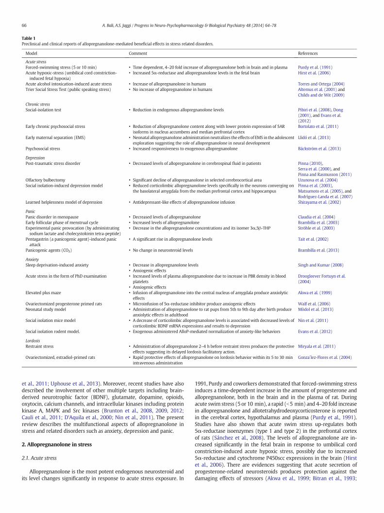
Table 1Preclinical and clinical reports of allopregnanolone-mediated beneficial effects in stress related disorders.
Model Comment References
Acute stressForced-swimming stress (5 or 10 min) • Time dependent, 4–20 fold increase of allopregnanolone both in brain and in plasma Purdy et al. (1991)Acute hypoxic-stress (umbilical cord constriction-induced fetal hypoxia)
• Increased 5α-reductase and allopregnanolone levels in the fetal brain Hirst et al. (2006)
Acute alcohol intoxication-induced acute stress • Increase of allopregnanolone in humans Torres and Ortega (2004)Trier Social Stress Test (public speaking stress) • No increase of allopregnanolone in humans Altemus et al. (2001) and
Childs and de Wit (2009)
Chronic stressSocial-isolation test • Reduction in endogenous allopregnanolone levels Pibiri et al. (2008), Dong
(2001), and Evans et al.(2012)
Early chronic psychosocial stress • Reduction of allopregnanolone content along with lower protein expression of 5ARisoforms in nucleus accumbens and median prefrontal cortex
Bortolato et al. (2011)
Early maternal separation (EMS) • Neonatal allopregnanolone administration neutralizes the effects of EMS in the adolescentexploration suggesting the role of allopregnanolone in neural development
Llidó et al. (2013)
Psychosocial stress • Increased responsiveness to exogenous allopregnanolone Bäckström et al. (2013)
DepressionPost-traumatic stress disorder • Decreased levels of allopregnanolone in cerebrospinal fluid in patients Pinna (2010),
Serra et al. (2000), andPinna and Rasmusson (2011)
Olfactory bulbectomy • Significant decline of allopregnanolone in selected cerebrocortical area Uzunova et al. (2004)Social isolation-induced depression model • Reduced corticolimbic allopregnanolone levels specifically in the neurons converging on
the basolateral amygdala from the median prefrontal cortex and hippocampusPinna et al. (2003),Matsumoto et al. (2005), andRodrìguez-Landa et al. (2007)
Learned helplessness model of depression • Antidepressant-like effects of allopregnanolone infusion Shirayama et al. (2002)
PanicPanic disorder in menopause • Decreased levels of allopregnanolone Claudia et al. (2004)Early follicular phase of menstrual cycle • Increased levels of allopregnanolone Brambilla et al. (2003)Experimental panic provocation (by administratingsodium lactate and cholecystokinin tetra-peptide)
• Decrease in the allopregnanolone concentrations and its isomer 3α,5β-THP Ströhle et al. (2003)
Pentagastrin (a panicogenic agent)-induced panicattack
• A significant rise in allopregnanolone levels Tait et al. (2002)
Panicogenic agents (CO2) • No change in neurosteroid levels Brambilla et al. (2013)
AnxietySleep deprivation-induced anxiety • Decrease in allopregnanolone levels
• Anxiogenic effectsSingh and Kumar (2008)
Acute stress in the form of PhD examination • Increased levels of plasma allopregnanolone due to increase in PBR density in bloodplatelets
• Anxiogenic effects
Droogleever Fortuyn et al.(2004)
Elevated plus maze • Infusion of allopregnanolone into the central nucleus of amygdala produce anxiolyticeffects
Akwa et al. (1999)
Ovariectomized progesterone primed rats • Microinfusion of 5α-reductase inhibitor produce anxiogenic effects Walf et al. (2006)Neonatal study model • Administration of allopregnanolone to rat pups from 5th to 9th day after birth produce
anxiolytic effects in adulthoodMòdol et al. (2013)
Social isolation mice model • A decrease of corticolimbic allopregnanolone levels is associated with decreased levels ofcorticolimbic BDNF mRNA expressions and results to depression
Nin et al. (2011)
Social isolation rodent model. • Exogenous administered AlloP-mediated normalization of anxiety-like behaviors Evans et al. (2012)
LordosisRestraint stress • Administration of allopregnanolone 2–4 h before restraint stress produces the protective
effects suggesting its delayed lordosis facilitatory action.Miryala et al. (2011)
Ovariectomized, estradiol-primed rats • Rapid protective effects of allopregnanolone on lordosis behavior within its 5 to 30 minintravenous administration
Gonza´lez-Flores et al. (2004)
66 A. Bali, A.S. Jaggi / Progress in Neuro-Psychopharmacology & Biological Psychiatry 48 (2014) 64–78
et al., 2011; Uphouse et al., 2013). Moreover, recent studies have alsodescribed the involvement of other multiple targets including brain-derived neurotrophic factor (BDNF), glutamate, dopamine, opioids,oxytocin, calcium channels, and intracellular kinases including proteinkinase A, MAPK and Src kinases (Brunton et al., 2008, 2009, 2012;Cauli et al., 2011; D'Aquila et al., 2000; Nin et al., 2011). The presentreview describes the multifunctional aspects of allopregnanolone instress and related disorders such as anxiety, depression and panic.
2. Allopregnanolone in stress
2.1. Acute stress
Allopregnanolone is the most potent endogenous neurosteroid andits level changes significantly in response to acute stress exposure. In
1991, Purdy and coworkers demonstrated that forced-swimming stressinduces a time-dependent increase in the amount of progesterone andallopregnanolone, both in the brain and in the plasma of rat. Duringacute swim stress (5 or 10 min), a rapid (b5 min) and 4–20 fold increasein allopregnanolone and allotetrahydrodeoxycorticosterone is reportedin the cerebral cortex, hypothalamus and plasma (Purdy et al., 1991).Studies have also shown that acute swim stress up-regulates both5α-reductase isoenzymes (type 1 and type 2) in the prefrontal cortexof rats (Sánchez et al., 2008). The levels of allopregnanolone are in-creased significantly in the fetal brain in response to umbilical cordconstriction-induced acute hypoxic stress, possibly due to increased5α-reductase and cytochrome P450scc expressions in the brain (Hirstet al., 2006). There are evidences suggesting that acute secretion ofprogesterone-related neurosteroids produces protection against thedamaging effects of stressors (Akwa et al., 1999; Bitran et al., 1993;

67A. Bali, A.S. Jaggi / Progress in Neuro-Psychopharmacology & Biological Psychiatry 48 (2014) 64–78
Drugan et al., 1989; Patchev et al., 1996). The rise in allopregnanolonelevels in response to acute stress is an endogenous protective mecha-nism to reduce excitotoxicity following acute stress (Hirst et al.,2006). Allopregnanolone downregulates the gene transcription of theHPA axis hormones including corticosterone release hormone and adre-nocorticotropic hormone to produce anti-stress and anxiolytic effects(Childs et al., 2010; Patchev et al., 1994, 1996).
On the other hand, no conclusive data exist with respect to plasmaallopregnanolone levels following acute stress in humans (Altemuset al., 2001; Childs and deWit, 2009; Girdler et al., 2001). Some studieshave shown the increased levels of allopregnanolone in humans inresponse to acute alcohol intoxication-induced acute stress (Torresand Ortega et al., 2004). Droogleever Fortuyn and co-workers describedthe increased levels of plasma allopregnanolone in healthy humanbeings in response to acute stress in the form of PhD examination(Droogleever Fortuyn et al., 2004). However, Altemus and coworkerssuggested that there is no increase in allopregnanolone in the group ofhuman subjects in response to the Trier Social Stress Test, a commonlyused stressor, which induces reliable physiological and psychologicalresponses to stress. The authors determined the effect of lactation onHPA axis in response to Trier Social Stress Test and evaluated the resultson the basis of different responses among three different groups(lactating, postpartum non-lactating and healthy women) in theearly follicular phase of the menstrual cycle. It was documented thatthere was no increase in allopregnanolone in human subjects inresponse to the Trier Social Stress Test (Altemus et al., 2001). Childsand de Wit also reported that the plasma levels of allopregnanoloneare not altered significantly in humans in response to Trier Social StressTest (a standardized public speaking stress procedure) (Childs and deWit, 2009).
Studies have also shown the significant rise in allopregnanolonein premenstrual dysphoric disorder women along with greaterallopregnanolone/progesterone ratio during the luteal phase. However,no relationship between symptom severity and allopregnanolone hasbeen established because relatively lower levels of allopregnanoloneare reported in premenstrual dysphoric disorder women with moresevere symptoms of anxiety and irritability (Girdler et al., 2001). It hasbeen reported that several GABAA receptor modulators includingallopregnanolone exhibit biphasic effects. At lower concentrations,allopregnanolone produces adverse anxiogenic effects; while it showsanxiolytic effects at higher concentrations. The severity of mood symp-toms in premenstrual dysphoric disorder is related to the serum con-centrations of allopregnanolone in a manner similar to an invertedU-shaped curve. The development of negativemood symptoms is corre-lated with the serum concentration of allopregnanolone that is similarto endogenous luteal phase levels of 1.5–2 nmol/l, while low and highconcentrations have less effect on mood (Andréen et al., 2006, 2009;Bäckström et al., 2011). The exact mechanisms responsible for differ-ence in allopregnanolone levels in these situations are not very clear.Unlike in animals, there are no well-established protocols of acutestress, except for Trier Social Stress Test (Kirschbaum et al., 1993), andvery often, the clinical situation mimicking the acute stress is consid-ered as the state of acute stress in human. The above described reportssuggesting the variable relationship of allopregnanolone with differentstressors may be due to very different nature of stressors in thesestudies. Acute alcohol intoxication is a putative systemic stressor, TSSTrepresents psychological stressor and premenstrual dysphoric disorderis a complex phenomenon involving important physiological and endo-crinological alterations. The difference in the allopregnanolone levels inTrier Social Stress Test and alcohol intoxication-induced acute stressmay be due to difference in severity of stress intensity. Acute alcoholbeing a strong stressor may increase the allopregnanolone levels inhumans, while the Trier social stress stressor (public speaking) oflower intensity is unable to elevate allopregnanolone significantly.However, due to lack of well-established stress specific protocols, thefindings from these indirectly stress-related studies may not yield
conclusive results regarding the relationship between allopregnanoloneand acute stress in humans.
2.2. Chronic stress
The chronic exposure to stress and high levels of corticosterone gen-erally decreases the allopregnanolone levels (opposite to acute stressconditions) (Serra et al., 2000, 2008). Almost all the studies pertainingto allopregnanolone and chronic stress have been performed in socialisolation-induced stress model. Pibiri and coworkers reported that thedecreased 5α-reductase type I mRNA expression and allopregnanolonelevels in neurons of the medial prefrontal cortex, hippocampus, andbasolateral amygdala are responsible for enhanced contextual fearresponses and impaired fear extinction in social isolation-inducedchronic stress inmice. Furthermore, authors demonstrated the develop-ment of a sort of similar behavioral changes in normal mice on SKF105111 (a potent 5α-reductase type I inhibitor) administration, whichsignificantly decreases the corticolimbic allopregnanolone levels(Pibiri et al., 2008). The same group of scientists demonstrated thatdownregulation of corticolimbic 5-α-reductase-type-I mRNA expres-sion in brain is responsible for enhanced fear response in anabolicandrogenic steroid treatment (a model of stress induction)-inducedbehavioral deficits. Furthermore, the normalization of corticolimbicallopregnanolone levels with allopregnanolone treatment (8 μmol/kg)attenuates fear response in mice due to anabolic androgenic steroidtreatment (Agís-Balboa et al., 2009).
In awell-established ratmodel of early chronic psychosocial stress ofisolation rearing, the significantly lower protein expression of 5-α-reductase isoforms (I and II) in the nucleus accumbens and medialprefrontal cortex brain regions along with the reduced content ofallopregnanolone in the brain, but not in the plasma, has been demon-strated (Bortolato et al., 2011). The micro-infusion of norfluoxetine, aselective brain steroidogenic stimulant (SBSS), into the basolateralamygdala increases the allopregnanolone levels in the corticolimbicarea to reduce aggression in socially-isolated mice (Nelson and Pinna,2011). In a social isolation model of chronic stress, a significant reduc-tion in endogenous allopregnanolone levels is correlated with thedevelopment of behavioral deficits. However, exogenous administra-tion of allopregnanolone has been shown to normalize the behavioralchanges and impaired hippocampal neurogenesis in a social isolationrodent model of chronic stress. Allopregnanolone mediated normaliza-tion of HPA dysfunction may be responsible for the noted beneficialeffects in chronic stress-associated deleterious changes (Evans et al.,2012). Neonatal administration of allopregnanolone (10 mg/kg s.c.) inrats during neonatal period (between postnatal day 5 and day 9) hasbeen shown to neutralize the behavioral abnormalities in adulthooddue to early maternal separation (Llidó et al., 2013). The mechanismsresponsible for persistent decrease in allopregnanolone along withreduced sensitivity and function of the GABAA receptors during chronicstress remain unclear (Turkmen et al., 2011).
All the studies showing the decreased levels of allopregnanolone inchronic stress are actually based on post-weaning social isolation.Although, most of the abnormal changes in these isolation modelshave been ascribed to isolation-induced stress induction, however, im-portant contribution of other aspects of social deprivation may not becompletely ruled out. In fact, rats reared on their own from weaninglack essential sensory inputs like social contact, olfactory informationand environmental stimuli with insufficient sensory information,which otherwise is required for the normal development of brain com-ponents including hippocampus, pituitary and hypothalamic system(Muchimapura and Marsden, 2004). The probable lack of normal HPAaxis in isolation stress subjected animals is supported by the bluntedrelease of CRF into the hypophysial portal systemanddecreased cortico-sterone release (Sanchez et al., 1998), which is in contrast to othermodels of chronic and chronic variable stresses that are characterizedby increased corticosterone release (Herman et al., 1995; Katz et al.,

68 A. Bali, A.S. Jaggi / Progress in Neuro-Psychopharmacology & Biological Psychiatry 48 (2014) 64–78
1981; Marin et al., 2007). Moreover, corticosterone release in responseto acute stress is much higher in chronically stressed animals(Armario et al., 1985). These findings support the fact that apart fromper se stress, other deprivation factors may also be involved in alteringallopregnanolone levels during chronic stress. However, apart fromabove described post-weaning social isolation-based studies, there areno studies describing the changes in allopregnanolone levels in chronicstress that may differentiate the direct effect of stress per se and otherassociated factors. In contrast to chronic stress studies in animals, avery recent study in humans has described the increased responsive-ness to exogenous allopregnanolone (measured in terms of saccadiceye velocity) of womenwith occupational psychosocial stress and burn-out syndrome than the healthy controls in an experimental setting dueto up-regulation of α4 and δ-subunits of GABAA receptor (Bäckströmet al., 2013).
2.3. Hypothesis for differential levels of allopregnanolone in acute andchronic stresses
While the levels of allopregnanolone are increased in response toacute stress (Hirst et al., 2006; Purdy et al., 1991), its levels are reducedduring repetitive exposures to stress (Pibiri et al., 2008). There is noexperimental study to explain the differential effects of acute andchronic stresses on the allopregnanolone levels. Based on the reportsof published reports, a hypothesis may be formulated to explain thesedifferential changes. During acute stress, the levels of allopregnanoloneare increased to normalize the hyper-activated HPA axis and restorehomeostasis (Akwa et al., 1999; Bitran et al., 1993). However, duringpersistent/repetitive stress exposures, tolerance may develop againstincreased allopregnanolone levels probably due to alteration in sen-sitivity of GABAA receptors. There have been a number of studiesshowing the development of tolerance to allopregnanolone andreduction in GABAA receptor sensitivity during repetitive stressexposures (Guidotti et al., 2001; Turkmen et al., 2011). In order toregain GABAA receptor sensitivity, changes may be triggered in thebody to reduce allopregnanolone levels possibly by attenuating up-regulated steroidogenic enzymes. Studies showing attenuation ofchronic stress-associated behavioral deficits with GABAA receptormodulators, including allopregnanolone (Agís-Balboa et al., 2009;Llidó et al., 2013; Nelson and Pinna, 2011) suggest the normal sensi-tivity of GABAA receptors in the brain. Accordingly, it may be hypoth-esized that the decreased levels of allopregnanolone during chronicstress are the compensatory changes to regain the reduced hyper-sensitivity of GABAA receptors. However, experimental studies areneeded to support this hypothesis regarding the differential effectsof acute and chronic stresses on allopregnanolone levels.
2.4. Allopregnanolone and depression
Studies have demonstrated that allopregnanolone levels are de-creased in the cerebrospinal fluid of patients with post-traumaticstress disorder and major unipolar depression (Pinna, 2010; Serraet al., 2000). In post-traumatic stress disorder, the cerebrospinalfluid levels of allopregnanolone were measured in premenopausalwomen with and without post-traumatic stress disorder. The levelsof allopregnanolone were 40 fmol/ml in non-post-traumatic stressdisorder subjects, while in the post-traumatic stress disordergroup allopregnanolone levels were ~40% of healthy group levels.The low levels of allopregnanolone in the cerebrospinal fluid ofpremenopausal women with post-traumatic stress disorder maycontribute to an imbalance in inhibitory versus excitatory neuro-transmission, resulting in increased re-experiencing traumatic eventsand depressive symptoms (Rasmusson et al., 2006). Studies have dem-onstrated that the bilateral olfactory bulbectomy (one of the most vali-dated models of depression) in rats produces a significant decline inallopregnanolone content in the selected cerebrocortical area, which in
turn is shown to be reversed by chronic treatment with an antide-pressant (Uzunova et al., 2004). It has been reported that socially-isolated mice express reduced corticolimbic allopregnanolone levelsspecifically in the neurons converging on the basolateral amygdala fromthe median prefrontal cortex and hippocampus (Matsumoto et al.,2007). The decreased brain allopregnanolone is due to downregulationof 5α-reductase type I expression, the rate-limiting enzyme in brainallopregnanolone biosynthesis. Dong and coworkers found that socialisolation produces a decrease in 5α-reductase type I mRNA expressionin the olfactory bulb (the brain region that contains a very large amountof this enzyme) (Dong, 2001). Recently, Drugan and coworkers docu-mented the possible role of allopregnanolone in the development ofstress-resilience. The authors described that resilient rats, exposed toinescapable tail shock, but not developing learned helplessness, exhibitaltered sensitivity to behavioral effects of GABA receptor antagonistsand reduced benzodiazepine receptor ligand binding. Based on this,the authors tentatively proposed that the development of resiliencemight involve enhanced activity of endogenous benzodiazepine-likecompound such as allopregnanolonewith allostericmodulatory activityof the GABAA receptor (Drugan et al., 2013). However, it is worth men-tioning that allopregnanolone is really not a benzodiazepine-like com-pound. In fact unlike benzodiazepines that show selectivity for GABAA
receptor subunits, allopregnanolone exhibits a promiscuous binding ata large number of GABAA receptor subunits with same affinity.
Exogenous allopregnanolone has been shown to exertantidepressant-like effects in rodent models of depression (Guidottiet al., 2001; Khisti and Chopde, 2000; Rodrìguez-Landa et al., 2007).Shirayama and coworkers examined the antidepressant effects ofallopregnanolone infusion into the cerebral ventricles, hippocam-pus, amygdala, nucleus accumbens, or prefrontal cortex in learnedhelplessness model of depression (Shirayama et al., 2002). Pinnaand coworkers described the up-regulation of neurosteroid biosyn-thesis as a pharmacological strategy to improve the behavioraldeficits in a putative mouse model of posttraumatic disorder anddemonstrated that fluoxetine and its congeners ameliorate the aggres-sive and anxiety-like behavior by increasing the allopregnanolone levelsin the corticolimbic region (Pinna and Rasmusson, 2011). In depression,an important role of posterior nucleus accumbens (a part of extendedamygdala, comprising the bed nucleus of the stria terminalis, the centralnucleus of amygdala and the shell of nucleus accumbens) has been de-scribed (Schlaepfer et al., 2008). Studies have shown the presence of5α-reductase in the nucleus accumbens and the expression of this en-zyme is sensitive to acute stress in the mPFC region (Sánchez et al.,2008), and to early chronic psychosocial stress in themPFC and nucleusaccumbens (Bortolato et al., 2011). Furthermore, systemic administra-tion and intra-accumbal infusion of allopregnanolone in ovariectomizedrats has been shown to significantly reduce the immobility time in theforced swim test, an animal model of depression (Molina-Hernandezet al., 2005).
Clinical studies have reported the changes in neuroactive steroidconcentrations in response to antidepressant drug treatment providingthe support that neurosteroids play an important role in the pathologyof depression. After threeweek treatmentwith antidepressants of threedifferent classes (desipramine, fluoxetine, sertraline and venlafaxine),the reduced levels of allopregnanolone are normalized in depressivepatients (Uzunova et al., 1998). Fluoxetine normalizes the reducedlevels of allopregnanolone in the brain, increases the responsivenessof GABAA receptors to GABA mimetic drugs and alleviates aggressivebehavior in socially-isolated mice (Pinna et al., 2004). It has been sug-gested that fluoxetine-induced increase in allopregnanolone is due toa change of the activity of enzyme 3α-hydroxy steroid dehydrogenase.The development of resistance to selective serotonin reuptake inhibi-tors in patients with depression and post traumatic disorder has beenmainly attributed to blockade of allopregnanolone synthesis in thebrain (Pinna and Rasmusson, 2011). A significant fall in neurosteroidlevels, including allopregnanolone after postpartum (0.30 ng/ml) as

69A. Bali, A.S. Jaggi / Progress in Neuro-Psychopharmacology & Biological Psychiatry 48 (2014) 64–78
compared to during pregnancy (26.69 ng/ml) has been linked withthe development of postpartum behavioral disorder. Furthermorein these subjects, a reduced resting-state functional connectivitywithin corticolimbic regions such as anterior cingulate cortex, andbilateral amygdala, hippocampus and dorsolateral prefrontal corti-ces was also demonstrated (Deligiannidis et al., 2013).
On the contrary, various studies have also suggested that normal-ization of allopregnanolone level is neither required nor sufficientfor clinical response in depression. Non-pharmacological treatmentof depressed persons such as those with partial sleep deprivation(Schüle et al., 2003), repetitive transcranial magnetic stimulation(Plewnia and Padberg, 2012), and electroconvulsive therapy (Baghaiet al., 2012) did not alter allopregnanolone levels. Furthermore, it hasalso been reported that mirtazapine treatment of five weeks in-creases the levels of allopregnanolone in responders as well as innon-responders of anti-depressant treatment (Schüle et al., 2007)suggesting no significant role of allopregnanolone in antidepressanttherapy.
2.5. Allopregnanolone and anxiety
There have been a number of studies documenting the anxiolytic andsedative properties of allopregnanolone (Bitran et al., 1993;Mòdol et al.,2011; Soderpalm et al., 2004). Singh and Kumar investigated the effectsof allopregnanolone on sleep deprivation-induced anxiety-like behaviorand oxidative damage in mice. Pretreatment with allopregnanolone(10 mg/kg) significantly improved the locomotor activity, weight loss,and anxiety-like behavior, restored reduced glutathione and catalaseactivity, and attenuated elevated lipid peroxidation (Singh and Kumar,2008). Infusion of allopregnanolone, but not pregnenolone sulfate, intothe central nucleus of the amygdala has been shown to produceanxiolytic-like effects in two rodent models of anxiety indicatingthat the amygdala (regulating fear and anxiety) may be involved inmediating the anxiolytic-effects of neurosteroids (Akwa et al.,1999). Microinfusion of a 5α-reductase inhibitor into the amygdalaof progesterone-primed ovariectomized rats has been shown toproduce the anxiogenic-like effects in the open-field, plus-maze,and defensive freezing tests (Walf et al., 2006). Microinfusion ofallopregnanolone into the lateral septum has been shown to attenu-ate the conflict test in Wistar rats (Molina-Hernandez et al., 2003).
Engin and Treit demonstrated that allopregnanolone produces fullanxiolytic effects when microinfused into the amygdala (in elevatedplus maze and shock-probe burying tests), partial anxiolytic effectswhen microinfused into the mPFC (only in elevated plus maze test)and no anxiolytic-like effects when microinfused into the dorsal hippo-campus (in none of the tests) (Engin and Treit, 2007). In contrast,there are a number of other studies suggesting the key role of hippo-campus in mediating anxiolytic actions of allopregnanolone. Bitranand coworkers demonstrated that intrahippocampal infusion ofallopregnanolone precursor (pregnanolone) produces anxiolyticeffects in both elevated plus-maze and shock-probe burying para-digms (Bitran et al., 1999). The same group of scientists demonstratedthat stimulation of intrahippocampal allopregnanolone synthesisproduces the anxiolytic effects in the plus-maze and shock-probeburying tests. Moreover, administration of 5α-reductase inhibitoris shown to attenuate the anxiolytic effects by inhibiting the synthe-sis of intrahippocampal allopregnanolone (Bitran et al., 2004).Recently, Mòdol and co-workers described the anxiolytic actions ofallopregnanolone and mentioned the important role of the dorsal(CA1) hippocampus in producing several behavioral effects ofneurosteroids such as exploration, anxiety, learning and memory.An intra-hippocampal administration of allopregnanolone is shown toenhance the exploration in terms of an increase in the total and theinner numbers of head-dips, percentage of entries into the open armsof the elevated plus maze with no effects on aversive learning retention(Mòdol et al., 2011).
While the acute application of allopregnanolone has been shownto produce the anxiolytic effects (Akwa et al., 1999; Bitran et al.,1993; Mòdol et al., 2011), the prolonged continuous exposure (48–72 h) of this neurosteroid produces time-dependent anxiogeniceffects (Gulinello and Smith, 2003; Gulinello et al., 2001). Thechanges in the actions of allopregnanolone have been mainly attributedto changes in the structural composition of GABAA receptors (discussedin Mechanism of action). Using magnetic resonance imaging technique,a clinical study examined the effects of allopregnanolone on emotionregulating neurocircuitry. The neuroimaging evidence demonstratesthat pregnenolone (400 mg)-induced increase in allopregnanolonelevels reduces the amygdala and insula activity (brain regions asso-ciated with generation of negative emotions), and increases the dor-sal medial prefrontal cortex activity (regions linked with regulatoryprocesses) during emotion appraisal task. Furthermore, there is anenhanced connectivity between the amygdala and dorsal medial pre-frontal cortex alongwith reduced self-reported anxiety, thus, projectingallopregnanolone as a major target for pharmacologic intervention inthe treatment of anxiety disorders (Sripada et al., 2013).
The group of Darbra and others demonstrated that the changes inallopregnanolone levels in neonates alter the morphology of the brainstructures including hippocampus. In fact, allopregnanolone modulatesthe important processes in the hippocampus during the developmentalphase of the postnatal period, when the adult pattern of inhibitorytransmission is being established (Darbra and Pallarès, 2009; Darbraet al., 2013). These scientists described the development of various com-plex changes related to behavior in adulthood depending on the doseand frequency of allopregnanolone administration in neonates. Asingle acute administration of allopregnanolone (10 mg/kg) on thefifth postnatal day increased the novelty-directed locomotor activity,decreased habituation in the open field and altered GABAA receptorresponse to midazolam (1 mg/kg) and flumazenil (10 mg/kg) duringthe adulthood (Darbra and Pallarès, 2009). The postnatal administrationof allopregnanolone (20 mg/kg) to rat pups once a day from the 5th tothe 9th day after birth is shown to induce an anxiolytic-like profile inadulthood in elevated plus maze anxiety model (Darbra and Pallarès,2009). However, injection of allopregnanolone (20 mg/kg s.c.) in neo-nates, from postnatal day 5 to postnatal day 9, is shown to suppress theanxiolytic effects of intrahippocampal allopregnanolone administration(0.2 μg/0.5 μl) in the elevated plus maze test (Mòdol et al., 2013)suggesting the complex role of neonatal allopregnanolone in thematura-tion of hippocampal function and behavior.
The significant rise in allopregnanolone levels during anxiety hasbeen correlated with increase in the density of peripheral benzodiaz-epine receptors (PBRs). The translocator protein (TSPO), earlierknown as PBR, is an 18 kDa protein and is mainly located on theouter mitochondrial membrane. It interacts with steroidogenicacute regulatory protein to transport cholesterol to first steroidogen-ic enzyme (P450scc), which transforms cholesterol into variousneurosteroids including allopregnanolone. A positive correlationbetween the plasma allopregnanolone and TSPO density on bloodplatelets in healthy human beings subjected to acute stress has beenreported (Droogleever Fortuyn et al., 2004). Another study demonstrat-ed that the anxiolytic effects of etifoxine (a TSPO ligand) are abolishedin the presence offinasteride, an inhibitor of 5α-reductase that convertsprogesterone into allopregnanolone (Verleye et al., 2005). Adminis-tration of other TSPO ligands has also been shown to produce anxio-lytic effects even in humans by promoting neurosteroidogenesis(Nothdurfter et al., 2012; Schüle et al., 2011) suggesting that activa-tion of TSPO in the brain increases the cerebral production ofallopregnanolone to produce anxiolytic effects.
2.6. Allopregnanolone and panic
The spontaneous panic attacks are characterized by increasedlevels of allopregnanolone in the plasma that may possibly be due

70 A. Bali, A.S. Jaggi / Progress in Neuro-Psychopharmacology & Biological Psychiatry 48 (2014) 64–78
to activation of compensatory mechanisms to counter the occur-rence of spontaneous panic attacks (Ströhle et al., 2002). Other studieshave also shown that neurosteroids including allopregnanolone arehyper-secreted, particularly during the early follicular phase of men-strual cycle in women with panic disorder to reduce the HPA hyperac-tivity and attenuate anxiety (Brambilla et al., 2003). The reports ofincreased allopregnanolone levels in panic disorder patients are oppo-site of those seen in patients with major depression in which reductionin the levels of allopregnanolone and its isomer has been demonstrated(Romeo et al., 1998; Ströhle et al., 2000). The observed differences inneuroactive steroid composition in patients with panic disorder mayresult in greater GABAA receptor-mediated neuronal activity. An alter-ation in GABAA receptor modulating neuroactive steroid compositionmay possibly be due to reduced sensitivity of benzodiazepines inpatients with panic disorder (Roy-Byrne et al., 1990).
On the other hand, studies have also shown a decrease in theallopregnanolone levels in patients with panic disorder. A case con-trol clinical study to establish the relationship between the panic dis-order in menopause and allopregnanolone levels demonstrated theinverse relationship between allopregnanolone levels and develop-ment of psychological symptoms (assessed by climacteric symptomsquestionnaire) (Claudia et al., 2004). A clinical study demonstratedthe effectiveness of 2 month paroxetine treatment in attenuatingpanic disorder in men and attributed the beneficial effects toincreased allopregnanolone, not progesterone and tetrahydro-deoxycorticosterone levels in the plasma (Brambilla et al., 2005). Incomparison to spontaneous pain attacks, the experimentally-inducedpanic attacks in patients with panic disorder have yielded variableresults. Strohle and coworkers demonstrated a decrease in the con-centrations of allopregnanolone during experimental panic provoca-tion by administrating sodium lactate and cholecystokinin tetra-peptide in patients with panic disorder, but not in normal humans(Ströhle et al., 2003). The exact mechanisms responsible for reduc-tion in allopregnanolone are unknown; however, it may be due tofailure in the compensatory mechanism to maintain or increaseallopregnanolone levels in response to stress. On the other hand, Taitand coworkers documented a significant rise in allopregnanolone levelsin response to pentagastrin (a panicogenic agent) challenge (Tait et al.,2002); while Brambilla and coworkers demonstrated no change inneurosteroid levels in response to panicogenic agents (CO2) (Brambillaet al., 2013). It is difficult to propose the possible reasons for the differentresults in these studies. It may be possible that the extent of panic induc-tion varies with the different panicogenic agents that may result in vari-able release pattern of allopregnanolone.
2.7. Allopregnanolone and lordosis
Numerous studies have documented that stress alters the lordosisbehavior (female sexual behavior) (Meisel et al., 1979) and the involve-ment of allopregnanolone in affecting hormonal and behavioral eventsduring ovulation has also been described (Genazzani et al., 1995).Miryala and co-workers investigated the protective effects ofallopregnanolone against lordosis-inhibiting effects of restraint inovariectomized Fischer female rats. Administration of allopregnanolone(4.0 mg/kg), two days after priming the ovariectomized female ratswith estradiol benzoate (10 μg), is shown to reduce the effects ofrestraint stress on lordosis inhibition. Pretreatment with indomethacin(3α-hydroxysteroid dehydrogenase inhibitor) does not attenuate theprotective effects of allopregnanolone suggesting that allopregnanoloneper se, not its metabolite dihydroprogesterone, is responsible for its lor-dosis facilitatory actions. The authors demonstrated that administrationof allopregnanolone 2–4 h before restraint produces the protectionsuggesting its delayed actions (Miryala et al., 2011). On the otherhand, earlier studies have demonstrated the rapid protective effects ofallopregnanolone on lordosis behaviorwithin its 5 to 30 min intravenous
administration (González-Flores et al., 2010) suggesting its rapid as wellas delayed protective effects.
3. Mechanism of action
3.1. GABAA receptor complex as primary target
3.1.1. Post-synaptic GABAA receptorsThe majority of inhibitory actions of allopregnanolone against
neuronal excitability are ascribed to positive modulation of GABAA
receptors (Fig. 1). At nanomolar concentrations, allopregnanolonepotentiates the GABAA receptor-mediated inhibitory currents (Akket al., 2005; Stell et al., 2003); while at higher concentrations (μM), itdirectly opens GABAA receptors in the absence of ligand binding(Belelli and Lambert, 2005; Hosie et al., 2006). Pinna and collaboratorsdemonstrated for the first time that endogenous corticolimbic storesof allopregnanolone play a pivotal neurophysiological role in facilitatingthe fine-tuning of the action of GABA at GABAA receptors (Pinna et al.,2000). The effects of muscimol, benzodiazepines, and pentobarbital(GABAA receptor agonists), were abolished in the presence of 5α-reductase and 3α-hydroxysteroidoxidoreductase inhibitors suggestingthat brain-produced allopregnanolone regulates the potency of theGABAA receptors (Guidotti et al., 2001; Matsumoto et al., 1999; Pinnaet al., 2000). Various other experimental studies have also shown thatpositive allostericmodulation of GABA receptors is crucial formediatingthe beneficial effects of allopregnanolone. Administration of flumazenil(a GABA receptor antagonist) and picrotoxin (noncompetitiveantagonist for the GABAA receptor chloride channels) has beenshown to attenuate allopregnanolone-mediated anxiolytic and anti-oxidant effects and,muscimol (a GABAmimetic) has been shown to en-hance anti-anxiety and anti-oxidant effects of allopregnanolone in sleepdeprivation-induced anxiety model. These findings suggest thatallopregnanolone induces its protective effects by GABAergic modula-tion at various recognition sites on the GABA–benzodiazepine receptorcomplex (Singh and Kumar, 2008). Shirayama and coworkers have re-ported that co-infusion of flumazenil with allopregnanolone into thehippocampal CA3 region, but not into the central amygdala, blocks theantidepressant-like effects of allopregnanolone suggesting that it exertsantidepressant-like effects in the CA3 region of hippocampus throughthe GABAergic system (Shirayama et al., 2011).
The role of GABAergic system in mediating allopregnanolone's ac-tion during acute stress is very well defined. It has been demonstratedthat acute stress-induced increase in allopregnanolone tends to normal-ize HPA axis by increasing GABAergic neurotransmission in CRH-releasing parvocellular neurons of hypothalamus paraventricular nucle-us (PVN) (Miklos and Kovacs, 2002), which is a key brain regioninvolved in initiating the neuroendocrine and autonomic responses tostressor (Ulrich-Lai and Herman, 2009). The PVN region receives con-siderable GABAergic inputs from the dorsal hypothalamus, preopticarea and areas of the extended amygdala, making it a key target regionfor the actions of allopregnanolone. Experimental studies showing anincrease in plasma corticosterone with microinjection of bicuculline(Hewitt et al., 2009) and reduction in corticosterone with microinjec-tion of muscimol in PVN suggest that GABAA receptor-mediated inhibi-tion is important for regulating HPA axis activity. Accordingly, it hasbeen proposed that rapid elevation of neurosteroid levels during acutestress normalizes the HPA axis over activity by enhancing theGABAergic inhibition in the PVN region.
The role of GABAA receptors located on different parts of limbicsystem (e.g. septum, hippocampus, amygdala) has been implicated forthe anxiolytic effects of benzodiazepines. Similarly, intracerebralmicroinfusion of allopregnanolone-induced anxiolytic effects hasalso been attributed to potentiation/activation of GABAA receptorsin these areas (Akwa et al., 1999; Engin and Treit, 2007; Mòdolet al., 2011). Nucleus accumbens is a critical brain region controllingthe various aspects of depression (Schlaepfer et al., 2008). It has been

Fig. 1. Mechanisms involved in multifunctional actions of allopregnanolone in stress and related disorders.
71A. Bali, A.S. Jaggi / Progress in Neuro-Psychopharmacology & Biological Psychiatry 48 (2014) 64–78
reported that 95% of the neurons in the nucleus accumbens are themedium spiny GABAergic projection neurons (Gangarossa et al.,2013). Therefore, allopregnanolone-mediated beneficial effects indepressive patients may be attributed to increased GABAergic trans-mission in nucleus accumbens and related brain regions. In premen-strual disorder, it has been proposed that rather than the differencesin neurosteroid levels, it is the difference in sensitivity of GABAA
receptors towards allopregnanolone that underlies in the developmentof premenstrual disorders (Turkmen et al., 2011). These patients arefound to be less responsive to GABAA receptor modulators includingallopregnanolone during the luteal phase, not during the follicularphase (when the GABA steroids are absent) (Nyberg et al., 2004;Sundstrom et al., 1998).
The GABAA receptor is a pentameric structure and different subunitsare organized pseudo-symmetrically around the central channel. Thereare 19 subunits (α1–6, β1–3, γ1–3, δ, ε, θ, π, ρ1–3) that are divided intodifferent subfamilies based on their amino acid homology (Olsen andSieghart, 2009). The major isoform of GABAA receptor comprises twoα1 subunits, two β2 subunits, and a single γ2 subunit (McKernan andWhiting, 1996) and the general arrangement (around the centralpore) is βαγβα counterclockwise, when viewed from the outside ofthe cell (Baumann et al., 2002). This isoform is ubiquitously presentthroughout the brain and is predominantly present within the synapse.The other pre-synaptically and extra-synaptically localized δ-containingGABAA receptors, also termed as δ-GABAA receptors, display a morerestricted distribution and are mainly present in the cerebellum, den-tate gyrus, thalamus, striatum, and cortex regions (Wei et al., 2003;Wisden et al., 1992). These two types of GABAA receptors are function-ally differentiated on thebasis of the type of inhibitory currentmediatedby these receptors i.e., persistent tonic inhibitory currents (Itonic), orconventional inhibitory postsynaptic currents (Iphasic) in the centralnervous system. GABAA receptors mediating Iphasic are activated bybrief exposure to a high concentration of the neurotransmitter, whilethe receptorsmediating Itonic are activated by low concentration of neu-rotransmitter (Farrant and Nusser, 2005; Semyanov et al., 2004).
δ-GABAA receptors generally mediate Itonic and are more sensitiveto neurosteroids than γ-GABAA receptors that mediate Iphasic (Brownet al., 2002; Farrant and Nusser, 2005; Stell et al., 2003). Despite thehigher sensitivity of δ-GABAA receptors for allopregnanolone, the latterhas been shown to act at a wide variety of GABAA receptor subtypeswith broad spectrum of action in the CNS (Mitchell et al., 2008). Theaction of allopregnanolone on synaptic and extrasynaptic δ-GABAA
receptors is typically manifested as prolongation of the decay time ofinhibitory postsynaptic currents (IPSCs) and increase in the tonic con-ductance. Administration of 5α-reductase inhibitor, SKF 105111, hasbeen shown to reduce the decay time of spontaneous IPSCs (sIPSCs)recorded from the cortical neurons of mice (Puia et al., 2003). Apartfrom GABAA facilitatory or activating function, allopregnanolone in-creases the expression levels of GABAA receptor subunits α2, α3, α4and δ in the central nervous system including lumbosacral spinal cord(Peng et al., 2009).
Though, there are a number of reports suggesting the anxiolyticactions of allopregnanolone (Akwa et al., 1999; Bitran et al., 1993;Mòdol et al., 2011), still studies have shown the development of anxietywith its administration (Gulinello and Smith, 2003; Gulinello et al.,2001; Smith, 2012). The development of anxiety in these conditionshas been attributed to changes in the structural composition of GABAA
receptors. Gulinello and coworkers described that relative long continu-ous exposure of allopregnanolone (48 h or above) increases the expres-sion of the α4 subunits of the GABAA receptor in the hippocampus.Pharmacologically, these α4-containing GABAA receptors are very dis-tinct and are insensitive to the benzodiazepine agonists such as loraze-pam, but are positively modulated by the benzodiazepine antagonist,flumazenil. An increase in functional α4-containing GABAA receptorsmay be responsible for altered activity of allopregnanolone and thus,the anxiogenic effects may be produced instead of usual anxiolyticeffects (Gulinello and Smith, 2003; Gulinello et al., 2001). Recently,Smith and coworkers also ascribed allopregnanolone-induced devel-opment of anxiety during the pubertal period (post-natal day ∼35–44) of female mice to increased expression of α4βδ-GABA receptors

72 A. Bali, A.S. Jaggi / Progress in Neuro-Psychopharmacology & Biological Psychiatry 48 (2014) 64–78
(stress-sensitive target for steroid allopregnanolone) on the den-drites of CA1 pyramidal cells at the onset of puberty in the hippo-campus (Smith, 2012).
Two distinct binding sites for allopregnanolone have been identifiedon the GABAA receptors and binding of allopregnanolone on theseseparate sites may either potentiate GABA or directly increase GABAcurrents. The binding of allopregnanolone to α-subunits of trans-membrane domains is responsible for GABA potentiating effects, whileit's binding to interfacial residues between α and β subunits mediatesthe direct activation (Hosie et al., 2006; Hosie et al., 2006). Hosieand co-workers demonstrated that significant receptor activation byallopregnanolone relies on occupancy of both the domains (Hosieet al., 2006). However using concatenated receptors, it has beenshown that the actions of allopregnanolone may be enhanced even inthe presence of a single functional GABA site and regardless whetherit interacts withα subunit from the same or the other β–α pair. Accord-ingly it has been proposed that occupation of either site withallopregnanolone potentiates the opening of the GABAA receptor(Bäckström et al., 2011). The putative steroid binding site is located inthe membrane-spanning regions of the α subunit of the receptor,extending from the α1Gln241 residue in the M1 membrane-spanningregion to the residues α1Asn407 and α1Tyr410 in the M4 domain.The α1Gln241 and α1Asn407 binding sites are crucial for mediatingthe potentiation responses; while αThr236 and βTyr284 mediate di-rect activation of GABAA receptors (Hosie et al., 2006). However, it isnoteworthy thatα1Gln241 mutation disrupts the GABA potentiatingeffects of allopregnanolone without altering the actions of 3α,5β,3-hydroxymethylpregnan-20-one (analog of pregnanolone). On theother hand, the mutation of additional residue adjacent to the ste-roid binding pocket, α1Ser240 to leucine disrupts the GABA potenti-ating effect of 3α,5β,3-hydroxymethylpregnan-20-one, but not ofallopregnanolone (Akk et al., 2008).
3.1.2. Presynaptic GABAIn contrast to inhibitory role of allopregnanolone on postsynaptic
GABA receptors, a recent study has demonstrated that allopregnanoloneincreases the frequency of glutamatergic spontaneous excitatory post-synaptic currents (sEPSCs) in isolated hilar neurons due to activation/potentiation of presynaptically located GABAA receptors. Theseexcitatory actions were completely blocked by a non-competitiveGABAA receptor blocker, tetrodotoxin (Na+ channel blocker) orCd2+ (Ca2+ channel blocker). It suggests that allopregnanolone-induced activation of presynaptic GABAA receptors induces presyn-aptic depolarization and increases Ca2+ influx into the presynapticnerve terminals via voltage dependent sodium and calcium chan-nels to increase the spontaneous release of glutamate. Furthermore,allopregnanolone-induced increase in sEPSC frequency was alsosignificantly reduced in the presence of bumetanide, a specificNKCC1 (Na+–K+–Cl− co-transporter type 1) blocker (with no in-hibitory effect on GABAA receptors) (Jang et al., 2001) suggestingthat bumetanide-sensitive NKCC1 contributes to allopregnanolone-induced presynaptic depolarization of glutamatergic nerve terminalsprojecting to hilar neurons. Furthermore, the authors employed Υ-cyclodextrin (a sequestering agentwhichbinds to several neurosteroids),picrotoxin and tetrodotoxin to demonstrate that allopregnanolone ton-ically activates and/or potentiates presynaptic GABAA receptors to affectspontaneous glutamate release onto the hilar neurons. The hippocam-pus is an important region controlling stress and memory, and themossy fibers originating from the DG granule cells innervate the hilarneurons. The majority of sEPSCs recorded from the isolated hilar neu-rons actually originate from the mossy fibers. Furthermore, the mossyfibers project to more inhibitory interneurons than excitatory neu-rons in DG and CA3 regions. Therefore, it may be proposed thatallopregnanolone activates presynaptic GABAA receptors to modu-late glutamatergic neurotransmission to affect the excitability of
DG–hilus–CA3 network, and may contribute in producing beneficialeffects in behavioral disorders (Kim et al., 2011).
3.2. Other targets
Apart from well-defined GABAA modulatory function of allo-pregnanolone, various other targets are also influencedbyneurosteroidsto produce behavioral changes. The exact mechanism i.e. whetherallopregnanolone directly modulates these targets or indirectly throughGABAA receptor is investigational.
3.2.1. Progesterone receptorsIt has been described that allopregnanolone produces its beneficial
effects in lordosis facilitation by activating the intracellular progesteronereceptors. A recent study has also described that allopregnanolone-mediated restoration of restraint stress (of 5 min)-induced decline inlordosis behavior is abolished in the presence of RU486 (progesteronereceptor antagonist) suggesting that progesterone receptors are in-volved in allopregnanolone's ability to restore lordosis behavior duringrestraint stress (Uphouse et al., 2013). Studies showing the attenuationof late protective effects of allopregnanolone in the presence of RU486suggest that its delayed actions require indirect activation of progester-one receptors (Miryala et al., 2011; Uphouse et al., 2013). However,Wirth and coworkers demonstrated that the blockade of progesteronereceptor does not diminish the anxiolytic response of allopregnanoloneindicating that activation of intracellular progesterone receptors is notinvolved in producing anxiolytic response (Wirth, 2011). These findingssuggest that more studies are needed to study the role of progesteronereceptors in producing beneficial effects of allopregnanolone.
3.2.2. GlutamateStudies have shown that allopregnanolonemodulates glutamatergic
neurotransmission to control the various aspects related to behavior(Fig. 1). Hu and coworkers demonstrated that allopregnanolonedecreases stimulus-evoked glutamate release in the medial prefrontalcortex without affecting the basal glutamatergic neurotransmission(Hu et al., 2007). Pinna and coworkers demonstrated that decreasedallopregnanolone levels during anxiety may enhance the glutamatergicneurotransmission to produce characteristic symptoms of anxiety. Inresponse to anabolic androgen steroid treatment, a selective reductionin 5-α reductase type I mRNA expression is demonstrated in theglutamatergic neuronal populations of the basolateral amygdala (70%)and CA3 glutamatergic pyramidal neurons and DG granule cells (30%)(Pinna et al., 2008). The reduction in allopregnanolone content in theselective glutamatergic neuronal population of the corticolimbic andcorticothalamic circuitsmay enhance the glutamatergic neurotransmis-sion to produce anxiety and emotional disorders (Pinna et al., 2008).Based on studies showing the modulation of glutamate–NO–cGMPpathway by GABAergic neurotransmission, the role of allopregnanolonein modulating the functions of the glutamate–NO–cGMP pathway hasbeen proposed. Cauli and coworkers demonstrated that administrationof allopregnanolone through the micro-dialysis probe increases thebasal cGMP levels, but completely antagonizes N-methyl-D-aspartate(NMDA)-induced increase in cGMP in the cerebellum suggesting thatallopregnanolone may serve as NMDA antagonist (Cauli et al., 2011).Kim and coworkers also reported an increase in glutamate release inthe presence of allopregnanolone in isolated hilar neurons. Shirayamaand coworkers reported that coinfusion of (+)MK801 (NMDA receptorantagonist) blocks the antidepressant effects of allopregnanolone in thecentral amygdala suggesting that allopregnanolone exerts antidepres-sant actions by activating the glutamatergic transmission in the centralregion of amygdala (Shirayama et al., 2011). Kim and coworkers de-scribed that the hilar neurons of the mossy fibers project to more inhib-itory interneurons than excitatory neurons in the DG and CA3 regionsand therefore, increase in excitatory glutamatergic neurotransmission

73A. Bali, A.S. Jaggi / Progress in Neuro-Psychopharmacology & Biological Psychiatry 48 (2014) 64–78
in these neurons activate inhibitory GABAergic system to attenuate be-havioral disorders (Kim et al., 2011).
3.2.3. BDNFBDNF is a member of the “neurotrophin” family and is actively in-
volved in regulating the stress response and behavioral dysfunctionassociated with anxiety and depression. It is present in very highconcentrations in the hippocampus and cerebral cortex, and itsdownregulation has been positively correlated with the develop-ment of depressive symptoms. The decreased BDNF expression hasbeen reported in the post-mortem brain and in the blood cells ofdepressed patients (Gonul et al., 2005; Karege et al., 2005; Piccinniet al., 2008). Furthermore, BDNF infusion into the hippocampus in-duces antidepressant-like effects (Shirayama et al., 2002) and micelacking BDNF fail to respond to antidepressants. The various studieshave attempted to correlate the allopregnanolone levels and BDNFexpression in depressive conditions (Fig. 1). It has been shown thatallopregnanolone levels and BDNFmRNA expression are downregulatedin the same brain areas including the medial frontal cortex, hippocam-pus, and basolateral amygdala regions (Nelson and Pinna, 2011;Pibiriet al., 2008). Nin and co-workers demonstrated that in socially isolatedmice, a decrease of corticolimbic allopregnanolone levels is associat-ed with the decreased levels of corticolimbic BDNF mRNA expres-sions (Nin et al., 2011). Exogenous allopregnanolone or S-norfluoxetine at concentration sufficient to increase the level ofallopregnanolone in corticolimbic region is shown to normalizeBDNF mRNA expression in the corticolimbic region along with im-provement in dendritic spine morphology and behavioral deficitsin social isolation model (Nin et al., 2011). These treatments alsoprevented the reinstatement of fear memory following extinctionsuggesting that allopregnanolone- or S-norfluoxetine-induced BDNFupregulation in the corticolimbic region is responsible for fear extinctionprocessing (Nelson and Pinna, 2011; Pibiri et al., 2008). Recently,Evans and coworkers demonstrated that exogenously administeredallopregnanolone-mediated normalization of depressive/anxiety-likebehaviors in a chronic stress model is secondary to increased BDNF ex-pression in the brain (Evans et al., 2012).
3.2.4. Opioids and oxytocinThe role of endogenous opioids inmodulating anxiety and emotions
is very well defined (Sauriyal et al., 2011). Preclinical studies haveshown that the non-selective opioid receptor antagonist (naloxone)increases; while morphine decreases the ultrasonic vocalizationsamong infants separated from their mother, and among rats exposedto a predator (Blanchard et al., 1991; Kalin et al., 1988). Brunton and co-workers demonstrated that allopregnanolone may produce its anti-stress effect through opioid-dependent mechanism. In late pregnancy,an adaptation to different stressors, including IL-1β, is characterizedby reduced activation of pPVN CRH neurons, and attenuated ACTH andcorticosterone response (Brunton et al., 2008). Administration of finas-teride during late pregnancy is shown to attenuate stress adaptation,accordingly, the role of increased expressions of 5-α reductase and3-α reductase in the nucleus tractus solitarius (NTS) and PVN leadingto increased allopregnanolone generation has been described instress adaptation during late pregnancy. The other studies havealso shown the increased levels of allopregnanolone in the brain dur-ing pregnancy suggesting its role in attenuating stress-induced HPAaxis overactivity (Concas et al., 1999). The participation of endogenousopioids in allopregnanolone-mediated stress adaptation is demonstratedby results showing the restoration of the HPA axis response to stressor(IL-1β) in late pregnancy with naloxone. Furthermore, finasteridedecreased proenkephalin-A mRNA expression in the NTS regionsuggesting that allopregnanolone-induced HPA axis inhibition duringlate pregnancy may be mediated through upregulation of endogenousopioids. An earlier study by the same authors demonstrated an increasein expression of μ-opioid receptors in the NTS region in late pregnancy
suggesting the key role of opioids in stress adaptation (Brunton et al.,2005). Furthermore, the combined administration of naloxone andfinasteride does not produce any additive or synergistic effectssuggesting inter-linkage between allopregnanolone and opioid mecha-nisms (Brunton et al., 2009).
The same group of scientists has recently described that finaste-ride potentiates IL-1β-evoked oxytocin secretion in late pregnantrats suggesting that allopregnanolone-mediated activation of GABAA
receptors may decrease oxytocin to produce adaptation. Furthermore,treatment with naloxone is shown to greatly enhance IL-1β evokedoxytocin response to late pregnancy, and finasteride did not enhancethis effect, suggesting that allopregnanolone and endogenous opioidmechanisms do not act independently. It has been proposed thatallopregnanolone-mediated enhancement of opioid expression mayinhibit the oxytocin secretion to induce adaptation to immunogenicchallenge in late pregnancy (Brunton et al., in press). Themagnocellularneurosecretory cells of the supraoptic nucleus (SON) projecting to theneurohypophysis release oxytocin into the bloodstream and GABA isthe major neurotransmitter modulating neuronal excitability in SON(Brussaard et al., 1999; Randle and Renaud, 1987). Thus, GABA facilita-tory actions of allopregnanolonemay be responsible for reduced oxyto-cin secretion and adaptation process (Jo et al., 2011) (Fig. 1).
Brunton and coworkers described that allopregnanolone-mediateddecrease in oxytocin may be involved in the development of stressadaptation in late pregnancy (Brunton et al., in press). Although, somestudies have reported the anxiogenic role of oxytocin (Hoge et al.,2008), the majority of studies have shown the anxiolytic and stressadaptive function of oxytocin. An increased systemic/brain oxytocinconcentration in response to stress tends to restore the homeostasis asstudies have shown that it attenuates anxiety and stress and inducesthe feeling of well-being (Onaka et al., 2012; Uvnas-Moberg andPetersson, 2005). The nursingmothers are calmer and less anxious dur-ing stressful situations, possibly due to high brain oxytocin activity(Slattery and Neumann, 2008).
3.2.5. DopamineStudies have shown that reduction in dopamine levels leads to
development of anxiety and increase in dopaminergic activity hasbeen shown to produce anti-anxiety and anti-depressant effects(D'Aquila et al., 2000; Jahng et al., 2012). The intraperitoneal injectionof apomorphine (dopamine D1/D2 receptor agonist) has been shownto produce anxiolytic-like behaviors (Reza Zarrindast et al., 2013). Theselective deletion of CRHR1 in the midbrain dopaminergic neuronsleading to reduced dopamine release in the prefrontal cortex is shownto result in development of anxiety-like behavior (Refojo et al., 2013).A recent clinical study has demonstrated decreased cerebrospinal fluidlevels of homovanillic acid (HVA), a metabolite of dopamine, in patientssuffering from posttraumatic stress disorder suggesting that increasingdopaminergic neurotransmission may be a potential therapy forposttraumatic stress disorder (Geracioti et al., 2013). D'Aquila and co-workers demonstrated that allopregnanolone produces antidepressant-like effects in female Sprague–Dawley rats in forced swimming testduring estrus and diestrus stages by dopamine-dependent mechanism.Administration of raclopride (D2 receptor antagonist), but not SCH23390 (D1 antagonist), is shown to attenuate allopregnanolone-inducedincrease in swimming behavior (antidepressant action) showing an im-portant role of dopamine D2-like receptor activation inmediating antide-pressant effects of allopregnanolone (D'Aquila et al., 2010). The tardivedyskinesia-related study has shown that allopregnanolone is capable ofreversing the abnormal movements in a disease state by increasing thelevels of dopamine (Bishnoi et al., 2008). Allopregnanolone has beenshown to dose-dependently increase the dopamine release in the nucleusaccumbens (Rouge-Pont et al., 2002). The mechanisms involved inallopregnanolone-mediated increase in dopamine neurotransmis-sion are not fully explored. It may be possible that allopregnanolonedirectly increases the biosynthesis of dopamine by inducing

74 A. Bali, A.S. Jaggi / Progress in Neuro-Psychopharmacology & Biological Psychiatry 48 (2014) 64–78
dopamine synthesizing enzyme, tyrosine hydroxylase (Adeosunet al., 2012; Charalampopoulos et al., 2005). Allopregnanolone-mediated increase in dopaminergic neurotransmission may also beattributed to decreased metabolism of dopamine in the brain(Muneoka et al., 2009).
However, the effect of allopregnanolone and dopaminergic neu-rotransmission seems to be more complex. Motzo and coworkersdemonstrated that administration of allopregnanolone (10 to 15 μg)completely prevents foot-shock-induced increase in extracellular dopa-mine concentrations in the nucleus accumbens and cerebral cortex(Motzo et al., 1996). Furthermore, there have also been studies showingthe increase in dopaminergic neurotransmission due to reduced allo-pregnanolone content (Bortolato et al., 2011;Dazzi et al., 2002). Bortolatoand coworkers demonstrated the decreased allopregnanolone and in-creased dopamine levels in rat nucleus accumbens andmedial prefrontalcortex in isolation-rearing-induced chronic stress model. Furthermore,administration of finasteride was shown to enhance the dopamine levelssuggesting that allopregnanolone may be actually involved in reducingthe dopaminergic neurotransmission (Bortolato et al., 2011). Therefore,detailed studies are needed to explore the relationship between allo-pregnanolone and dopamine in modulating behavior.
3.2.6. Calcium channelsStudies have shown that allopregnanolonemay also indirectlymod-
ulate L-type calcium channels to reduce the neuronal excitability(Fig. 1). Hu and coworkers demonstrated that allopregnanolone-mediated inhibitory effects on glutamatergic neurotransmission in themedial prefrontal cortex are mediated through inhibition of L-type cal-cium channels. Administration of L-type calcium channel blocker, butnot of N- and P/Q-types, was shown to attenuate allopregnanolone-mediated inhibitory effects on presynaptic glutamate release in themedial prefrontal cortex (Hu et al., 2007). Earl and Tietz investigatedthe effects of allopregnanolone on whole-cell Ba2+ currents throughrecombinant neuronal Cav1.2 and Cav1.3 L-calcium channels expressedwith β(3) and α(2)δ-1 in HEK293T cells and demonstrated allo-pregnanolone as a potent inhibitor of L-calcium channels (IC50,∼10 μM) and its inhibition was correlated with reduced neuronalexcitability (Earl and Tietz, 2011). Cav1.2 and Cav1.3 channels arethe predominant forms of calcium channels expressed in the brainand Cav1.2 constitutes about 80% of total L-type calcium channelsin the brain (Vacher et al., 2008). The various studies have shownthe up-regulation of these channels in the brain region in responseto chronic stress (Maigaard et al., 2012; Mamczarz et al., 1999). TheCav1.3−/− mice are demonstrated to display less immobility in theforced swim and in the tail suspension tests suggesting the criticalrole of these channels in development of depression- and anxiety-like behaviors (Busquet et al., 2010). The pharmacological agentscapable of blocking L-type channels are capable of attenuatingstress-induced behavioral deficits (Bali et al., 2013; Kumar et al.,2012; Saade et al., 2003). Therefore, it is possible to suggest thatallopregnanolone-mediated blockade of L-type calcium channelsmay also be an important contributing factor for attenuating anxietyand depressive behaviors.
3.3. Signal transduction pathway through kinases
Kinases (a class of enzymes that phosphorylates other proteins) areessential regulators of stress-induced structural and behavioral plastic-ity and the signaling mechanisms of allopregnanolone are regulatedby these kinases including protein kinase A (PKA) and mitogen-activated protein kinases (MAPK) including SRC kinases.
3.3.1. Protein kinase A (PKA)Hu and coworkers demonstrated that allopregnanolone-mediated
inhibitory effects on glutamatergic neurotransmission in the medialprefrontal cortex are mediated through protein kinase A inhibition
(Fig. 1). The authors reported no significant effect of allopregnanoloneon spontaneous glutamate release, but described reduced stimulus(depolarizing agent and electrical)-evoked presynaptic glutamaterelease in the medial prefrontal cortex suggesting that the inhibitoryeffects of allopregnanolone on glutamate neurotransmission are neuro-nal activity dependent (Hu et al., 2007; Tauboll et al., 1993). Furthermore,it was shown that allopregnanolone significantly attenuates depolarizingagent-induced PKA activation suggesting that allopregnanolonedecreases the presynaptic glutamate release by activating PKA. Theother studies have shown the key role of PKA activation as an up-stream event of stimulus evoked glutamate release (Grilli et al.,2004; Leenders and Sheng, 2005). The authors further demonstratedthat rather than direct inhibitory effects of allopregnanolone on PKAactivation, an indirect pathway is involved as allopregnanolone didnot produce any effect on forskolin (a direct PKA activator)-inducedPKA activation. Furthermore, it was reported that allopregnanoloneinhibited extracellular calcium influx-evoked PKA activation, buthad no effect on intracellular calcium store release-evoked PKA acti-vation. Furthermore, L-type calcium channel blockers, but not N- andP/Q-types, attenuated allopregnanolone-mediated inhibitory effectson PKA activation and presynaptic glutamate release suggesting thatallopregnanolone inhibits the stimulus-evoked glutamate release ina PKA dependent mechanism via inhibition of L-type calcium chan-nels on the medial PFC (Hu et al., 2007).
3.3.2. Mitogen-activated protein kinases (MAPKs)The critical role of MAPK signal transduction in mediating lordosis
facilitatory effects of progesterone and allopregnanolone has been dem-onstrated (González-Flores et al., 2010). A study revealed that infusionof MAPK inhibitor, PD98059, into the third ventricle of ovariectomizedrats decreases allopregnanolone-induced lordosis and proceptivitysuggesting that MAPK signaling is required for the facilitatory actionsof allopregnanolone on the female sexual behavior. It was proposedthat MAPK-dependent phosphorylation of progesterone receptorscontributes to allopregnanolone-mediated facilitatory action as degra-dation of progesterone receptors with 26s proteasome pathway hasbeen reported to decrease the actions of progestins (González-Floreset al., 2004; Etgen and Acosta-Martinez, 2003). Administration ofPD98059 and RU86 was shown to inhibit cGMP-induced lordosis be-havior suggesting that progesterone receptors and MAPK are requiredfor cGMP-mediated facilitatory actions. Based on the previous studies,it was hypothesized that cGMPmay activate PKG to phosphorylate pro-gesterone receptors indirectly through MAPK, while progestins couldactivate Ras and Rap proteins to phosphorylate progesterone receptorsvia MAPK pathway (Manna and Stocco, 2011) to induce female sexualbehavior.
Flores and coworkers demonstrated that Src/MAPK signalingpathway is involved in progesterone and its ring A reduced metabolites(including allopregnanolone) in facilitation of the lordosis andproceptive behaviors in ovariectomized, estradiol primed rats. ICVinfusion and direct bilateral administration of Src inhibitor, PPA2,into the ventromedial hypothalamus (VMH) significantly attenuatedprogesterone and its ring A reduced metabolite (allopregnanolone)-induced estrous behavior suggesting that allopregnanolone-mediatedactivation of Src MAPK pathway is a key process in the VMH neuronsto facilitate estrous behavior (Fig. 1). Furthermore, it was shown thatring A reduced metabolites (allopregnanolone) are more potent thanprogesterone in stimulating Src, which may be responsible for highereffectiveness of allopregnanolone (as compared to progesterone) tostimulate the estrous behavior in the estrogen primed rats (Beyer,1986). A study of Blackmore and coworkers also demonstrated thehigher activity of allopregnanolone in mediating calcium increase inplatelets in a Src signal transduction pathway as compared to progester-one (Blackmore, 2008). The higher efficacy of allopregnanolone thanprogesterone suggests thatmechanisms other than progesterone recep-tor activation are also involved in lordosis facilitation behavior. It was

75A. Bali, A.S. Jaggi / Progress in Neuro-Psychopharmacology & Biological Psychiatry 48 (2014) 64–78
proposed that allopregnanolone may activate MAPK (Basta-Kaim et al.,2007) which in turn may phosphorylate progesterone receptors(Rupprecht et al., 1993) to stimulate Src kinase that in turn maybe involved in MAPK activation to facilitate lordosis behavior(González-Flores et al., 2010).
4. Conclusion
Both preclinical as well as clinical studies have documented thediverse actions of allopregnanolone on stress and stress-associatedbehavioral disorders includingdepression, anxiety and panic. The differ-ent neuropsychiatric disorders are characterized by alterations inallopregnanolone levels in the brain and plasma with an aim to restorethe homeostasis and produce neuroprotection. The decreased levels ofallopregnanolone during chronic stress are attributed to the compensa-tory changes to regain the reduced hypersensitivity of GABAA receptors.However mores studies are needed to support the above hypothesis.GABAA receptor complex has been documented as the primary targetof allopregnanolone. However, the roles of progesterone receptors,glutamate, dopamine, opioids, oxytocin and voltage gated calciumchannels are described for their anti-anxiety, anti-depressant and anti-stress effects. Allopregnanolone influences the neural activity throughactivation of different kinases including protein kinase A (PKA), MAPK,and Src kinase systems. However, whether these targets may be in-volved independently or act in concert with GABAA receptors is notclear. Nevertheless, there is a need to establish the interrelationshipamong these different targets as well as to explore the new targets forunderstanding the mechanisms involved in allopregnanolone's multi-functional aspects in the brain.
Acknowledgment
The authors are grateful to the Department of Pharmaceutical Sci-ences andDrug Research, Punjabi University, Patiala, India for supportingthis study and providing technical facilities for the work.
References
Adeosun SO, Hou X, Jiao Y, et al. Allopregnanolone reinstates tyrosine hydroxylase immu-noreactive neurons and motor performance in an MPTP-lesioned mouse model ofParkinson's disease. PLoS One 2012;7:e50040.
Agís-Balboa RC, Pinna G, Zhubi A, Maloku E, Veldic M, Costa E, et al. Characterization ofbrain neurons that express enzymes mediating neurosteroid biosynthesis. Proc NatlAcad Sci U S A 2006;103:14602–7.
Agís-Balboa RC, Pinna G, Pibiri F, Kadriu B, Costa E, Guidotti A. Down-regulation ofneurosteroid biosynthesis in corticolimbic circuits mediates social isolation-inducedbehavior in mice. Proc Natl Acad Sci U S A 2007;104:18736–41.
Agís-Balboa RC, Pibiri F, NelsonM, Pinna G. Enhanced fear responses in mice treated withanabolic androgenic steroids. Neuroreport 2009;20:617–21.
Akk G, Shu HJ, Wang C, Steinbach JH, Zorumski CF, Covey DF, et al. Neurosteroid access tothe GABAA receptor. J Neurosci 2005;25:11605–13.
Akk G, Li P, Bracamontes J, Reichert DE, Covey DF, Steinbach JH. Mutations of the GABA-Areceptor 1 subunit M1 domain reveal unexpected complexity for modulation by neu-roactive steroids. Mol Pharmacol 2008;74:614–27.
Akwa Y, Purdy RH, Koob GF, Britton KT. The amygdala mediates the anxiolytic-like effectof the neurosteroid allopregnanolone in rat. Behav Brain Res 1999;106:119–25.
Altemus M, Redwine LS, Leong YM, Frye CA, Porges SW, Carter CS. Responses to laborato-ry psychosocial stress in postpartum women. Psychosom Med 2001;63:814–21.
Andréen L, Sundström-Poromaa I, Bixo M, Nyberg S, Bäckström T. Allopregnanolone con-centration andmood—a bimodal association in postmenopausal women treatedwithoral progesterone. Psychopharmacology (Berl) 2006;187:209–21.
Andréen L, Nyberg S, Turkmen S, van Wingen G, Fernández G, Bäckström T. Sex steroidinduced negative mood may be explained by the paradoxical effect mediated byGABAA modulators. Psychoneuroendocrinology 2009;34:1121–32.
Armario A, Restrepo C, Castellanos JM, Balasch J. Dissociation between adrenocorticotro-pin and corticosterone responses to restraint after previous chronic exposure tostress. Life Sci 1985;36:2085–92.
Bäckström T, Haage D, Löfgren M, Johansson IM, Strömberg J, Nyberg S, et al. Paradoxicaleffects of GABA-Amodulators may explain sex steroid induced negative mood symp-toms in some persons. Neuroscience 2011;191:46–54.
Bäckström T, Bixo M, Nyberg S, Savic I. Increased neurosteroid sensitivity — an explana-tion to symptoms associated with chronic work related stress in women?Psychoneuroendocrinology 2013;38:1078–89.
Baghai TC, Lieb M, Rupprecht R. Electroconvulsive therapy— indications and practical usefor pharmacotherapy resistant depressive disorders. Fortschr Neurol Psychiatr2012;80:720–31.
Bali A, Gupta S, Singh N, Jaggi AS. Implicating the role of plasma membrane localized cal-cium channels and exchangers in stress-induced deleterious effects. Eur J Pharmacol2013;714:229–38.
Basta-Kaim A, Budziszewska B, Jaworska-Feil L, Leśkiewicz M, Tetich M, Otczyk M, et al. Ef-fects of neurosteroids on glucocorticoid receptor-mediated gene transcription in LMCATcells—a possible interaction with psychotropic drugs. Eur Neuropsychopharmacol2007;17:37–45.
Baumann SW, Baur R, Sigel E. Forced subunit assembly in α1β2ϒ2 GABAA receptors.Insight into the absolute arrangement. J Biol Chem 2002;277:46020–5.
Belelli D, Lambert JJ. Neurosteroids: endogenous regulators of the GABAA receptor. NatRev Neurosci 2005;6:565–75.
Beyer C, Gonza´lez-Mariscal G. Elevation in hypothalamic cyclic AMP as a common factorin the facilitation of lordosis in rodents: a working hypothesis. Ann NY Acad Sci1986;474:270–81.
Bishnoi M, Chopra K, Kulkarni SK. Progesterone attenuates neuroleptic-induced orofacialdyskinesia via the activity of its metabolite, allopregnanolone, a positive GABAA mod-ulating neurosteroid. Prog Neuropsychopharmacol Biol Psychiatry 2008;32:451–61.
Bitran D, Purdy RH, Kellogg. Anxiolytic effect of progesterone is associated with increasesin cortical allopregnanolone and GABAA receptor function. Pharmacol Biochem Behav1993;45:423–8.
Bitran D, Dugan M, Renda P, Ellis R, Foley M. Anxiolytic effects of the neuroactive steroidpregnanolone (3 alpha-OH-5 beta-pregnan-20-one) after microinjection in the dor-sal hippocampus and lateral septum. Brain Res 1999;850:217–24.
Bitran D, Foley M, Audette D, Leslie N, Frye CA. Activation of peripheral mitochondrialbenzodiazepine receptors in the hippocampus stimulates allopregnanolone synthesisand produces anxiolytic-like effects in the rat. Psychopharmacology 2004;151:64–71.
Blackmore PF. Progesterone metabolites rapidly stimulate calcium influx in human plate-lets by a src-dependent pathway. Steroids 2008;73:738–50.
Blanchard DC, Weatherspoon A, Shepherd J, Rodgers RJ, Weiss SM, Blanchard RJ. “Para-doxical” effects of morphine on antipredator defense reactions in wild and laboratoryrats. Pharmacol Biochem Behav 1991;40:819–28.
Bortolato M, Devoto P, Roncada P, Frau R, Flore G, Saba P, et al. Isolation rearing-inducedreduction of brain 5α-reductase expression: relevance to dopaminergic impairments.Neuropharmacology 2011;60:1301–8.
Brambilla F, Biggio G, Pisu MG, Bellodi L, Perna G, Bogdanovich-Djukic V, et al.Neurosteroid secretion in panic disorder. Psychiatr Res 2003;118:107–16.
Brambilla F, Mellado C, Alciati A, Pisu MG, Purdy RH, Zanone S, et al. Plasma concentra-tions of anxiolytic neuroactive steroids in men with panic disorder. Psychiatry Res2005;135:185–90.
Brambilla F, Perini G, Serra M, Pisu MG, Zanone S, Toffanin T, et al. Changes in neuroactivesteroid secretion associated with CO2-induced panic attacks in normal individuals.Psychoneuroendocrinology 2013;10:2234–42.
Brown N, Kerby J, Bonnert TP, Whiting PJ, Wafford KA. Pharmacological characterizationof a novel cell line expressing human α4β3δ GABAA receptors. Br J Pharmacol2002;136:965–74.
Brunton PJ, Meddle SL, Ma S, Ochedalski T, Douglas AJ, Russell JA. Endogenous opioids andattenuated hypothalamic-pituitary-adrenal axis responses to immune challenge inpregnant rats. J Neurosci. 2005;25:5117–26.
Brunton PJ, Arunachalam S, Russel JA. Control of neurohypophysial hormone secretion,blood osmolality and volume in pregnancy. J Physiol Pharmacol 2008;59:27–45.
Brunton PJ, McKay AJ, Ochedalski T, Piastowska A, Rebas E, Lachowicz A, et al. Central opi-oid inhibition of neuroendocrine stress responses in pregnancy in the rat is inducedby the neurosteroid allopregnanolone. J Neurosci 2009;29:6449–60.
Brunton PJ, Bales J, Russell JA. Allopregnanolone and induction of endogenous opioid in-hibition of oxytocin responses to immune stress in pregnant rats. J Neuroendocrinol2012;24:690–700.
Brunton PJ, Russell JA, Hirst JJ. Allopregnanolone in the brain: Protecting pregnancy and birthoutcomes. Prog Neurobiol. 2013. http://dx.doi.org/10.1016/j.pneurobio.2013.08.005.(in press).
Brussaard AB, Devay P, Leyting-Vermeulen JL, Kits KS. Changes in properties andneurosteroid regulation of GABAergic synapses in the supraoptic nucleus duringthe mammalian female reproductive cycle. J Physiol 1999;516:513–24.
Busquet P, Nguyen NK, Schmid E, Tanimoto N, Seeliger MW, Ben-Yosef T, et al. CaV1.3L-type Ca2+ channels modulate depression-like behaviour in mice independent ofdeaf phenotype. Int J Neuropsychopharmacol 2010;13:499–513.
Cauli O, González-Usano A, Agustí A, Felipo V. Differential modulation of the glutamate–nitric oxide–cyclic GMP pathway by distinct neurosteroids in cerebellum in vivo.Neuroscience 2011;190:27–36.
Celotti F, Melcangi RC, Martini L. The 5 alpha-reductase in the brain: molecular aspectsand relation to brain function. Front Neuroendocrinol 1992;13:163–215.
Charalampopoulos I, Dermitzaki E, Vardouli L, Tsatsanis C, Stournaras C, Margioris AN,et al. Dehydroepiandrosterone sulfate and allopregnanolone directly stimulate cate-cholamine production via induction of tyrosine hydroxylase and secretion by affect-ing actin polymerization. Endocrinology 2005;146:3309–18.
Childs E, de Wit H. Hormonal, cardiovascular, and subjective responses to acute stress insmokers. Psychopharmacology (Berl) 2009;203:1–12.
Childs E, Van Dam NT, de Wit H. Effects of acute progesterone administration upon re-sponses to acute psychosocial stress in men. Exp Clin Psychopharmacol 2010;18:78–86.
Claudia P, Andrea C, Chiara C, Stefano L, Giuseppe M, Vincenzo DL, et al. Panic disorder inmenopause: a case control study. Maturitas 2004;48:147–54.
Concas A, Follesa P, Barbaccia ML, Purdy RH, Biggio G. Physiological modulation of GABAAreceptor plasticity by progesterone metabolites. Eur J Pharmacol 1999;375:225–35.

76 A. Bali, A.S. Jaggi / Progress in Neuro-Psychopharmacology & Biological Psychiatry 48 (2014) 64–78
D'Aquila PS, Collu M, Gessa GL, Serra G. The role of dopamine in the mechanism of actionof antidepressant drugs. Eur J Pharmacol 2000;405:365–73.
D'Aquila PS, Canu S, Sardella M, Spanu C, Serra G, Franconi F. Dopamine is involvedin the antidepressant-like effect of allopregnanolone in the forced swimmingtest in female rats. Behav Pharmacol 2010;21(1):21–8. http://dx.doi.org/10.1097/FBP.0b013e32833470a7. [Feb].
Darbra S, Pallarès M. Neonatal allopregnanolone increases novelty-directed locomotionand disrupts behavioural responses to GABAA receptor modulators in adulthood. IntJ Dev Neurosci 2009;27:617–25.
Darbra S, Modol L, Vallée M, Pallarès M. Neonatal neurosteroid levels are determinant inshaping adult prepulse inhibition response to hippocampal allopregnanolone in rats.Psychoneuroendocrinology 2013;S0306-4530(12):00420–9.
Dazzi L, Serra M, Vacca G, Ladu S, Latrofa A, Trapani G, et al. Depletion of corticalallopregnanolone potentiates stress-induced increase in cortical dopamine output.Brain Res 2002;932:135–9.
Deligiannidis KM, Sikoglu EM, Shaffer SA, Frederick B, Svenson AE, Kopoyan A, Kosma CA,Rothschild AJ, Moore CM. GABAergic neuroactive steroids and resting-state function-al connectivity in postpartum depression: a preliminary study. J Psychiatr Res.2013;47:816–28.
Dong E. Brain 5α-dihydroprogesterone and allopregnanolone synthesis in amousemodelof protracted social isolation. Proc Natl Acad Sci U S A 2001;98:2849–54.
Droogleever Fortuyn HA, van Broekhoven F, Span PN, Bäckström T, Zitman FG, Verkes RJ.Effects of PhD examination stress on allopregnanolone and cortisol plasma levels andperipheral benzodiazepine receptor density. Psychoneuroendocrinology 2004;29:1341–4.
Drugan RC, Morrow AL, Weizman R, Weizman A, Deutsch SI, Crawley JN, et al.Stress-induced behavioral depression in the rat is associated with a decrease in GABAreceptor-mediated chloride ion flux and brain benzodiazepine receptor occupancy.Brain Res 1989;487:45–51.
Earl DE, Tietz EI. Inhibition of recombinant L-type voltage-gated calcium channels by posi-tive allosteric modulators of GABAA receptors. J Pharmacol Exp Ther 2011;337:301–11.
Engin E, Treit D. The anxiolytic-like effects of allopregnanolone vary as a function of intra-cerebral microinfusion site: the amygdala, medial prefrontal cortex, or hippocampus.Behav Pharmacol 2007;18:461–70.
Erhardt A, Ising M, Unschuld PG, Kern N, Lucae S, Pütz B, et al. Regulation of thehypothalamic–pituitary–adrenocortical system in patients with panic disorder.Neuropsychopharmacology 2006;31:2515–22.
Etgen AM, Acosta-Martinez M. Participation of growth factor signal transduction pathwaysin estradiol facilitation of female reproductive behavior. Endocrinology 2003;144:3828–35.
Evans J, Sun Y, McGregor A, Connor B. Allopregnanolone regulates neurogenesis anddepressive/anxiety-like behaviour in a social isolation rodent model of chronic stress.Neuropharmacology 2012;63:1315–26.
Farrant M, Nusser Z. Variations on an inhibitory theme: phasic and tonic activation ofGABAA receptors. Nat Rev Neurosci 2005;6:215–29.
Gangarossa G, Espallergues J, de Kerchove d'Exaerde A, El Mestikawy S, Gerfen CR,Hervé D, et al. Distribution and compartmental organization of GABAergicmedium-sized spiny neurons in the mouse nucleus accumbens. Front NeuralCircuits 2013;7:22.
Genazzani AR, Palumbo MA, de Micheroux AA, Artini PG, Criscuolo M, Ficarra G, et al. Ev-idence for a role for the neurosteroid allopregnanolone in the modulation of repro-ductive function in female rats. Eur J Endocrinol 1995;133(3):375–80. [Sep].
Geracioti Jr TD, Jefferson-Wilson L, Strawn JR, Baker DG, Dashevsky BA, Horn PS, et al. Ef-fect of traumatic imagery on cerebrospinal fluid dopamine and serotoninmetabolitesin posttraumatic stress disorder. J Psychiatr Res 2013;47:995–8.
Girdler SS, Straneva PA, Light KC, Pedersen CA, Morrow AL. Allopregnanolone levels andreactivity to mental stress in premenstrual dysphoric disorder. Biol Psychiatry2001;49:788–97.
Gonul AS, Akdeniz F, Taneli F, Donat OEker C, Vahip S. Effect of treatment on serumbrain-derived neurotrophic factor levels in depressed patients. Eur Arch PsychiatryClin Neurosci 2005;255:381–6.
González-Flores O, Guerra-Araiza C, Cerbo´n M, Camacho-Arroyo I, Etgen AM. The 26Sproteasome participates in the sequential inhibition of estrous behavior induced byprogesterone in rats. Endocrinology 2004;145:2328–36.
González-Flores O, Beyer C, Gómora-Arrati P, García-Juárez M, Lima-Hernández FJ,Soto-Sánchez A, et al. A role for Src kinase in progestin facilitation of estrous behaviorin estradiol-primed female rats. Horm Behav 2010;58:223–9.
Gottfried-Blackmore A, Sierra A, Jellinck PH, McEwen BS, Bulloch K. Brain microgliaexpress steroid-converting enzymes in the mouse. J Steroid Biochem Mol Biol2008;109:96–107.
Grilli M, Raiteri L, Pittaluga A. Somatostatin inhibits glutamate release from mousecerebrocortical nerve endings through presynaptic sst2 receptors linked to theadenylyl cyclase–protein kinase A pathway. Neuropharmacology 2004;46:388–96.
Guidotti A, Dong E, Matsumoto K, Pinna G, Rasmusson AM, Costa E. The socially-isolatedmouse: a model to study the putative role of allopregnanolone and 5alpha-dihydroprogesterone in psychiatric disorders. Brain Res Rev 2001;37:110–5.
Gulinello M, Smith SS. Anxiogenic effects of neurosteroid exposure: sex differences and al-tered GABAA receptor pharmacology in adult rats. J Pharmacol Exp Ther 2003;305:541–8.
Gulinello M, Gong QH, Li X, Smith SS. Short-term exposure to a neuroactive steroid in-creases alpha4 GABAA receptor subunit levels in association with increased anxietyin the female rat. Brain Res 2001;910:55–66.
Gunn BG, Brown AR, Lambert JJ, Belelli D. Neurosteroids and GABAA receptor interactions:a focus on stress, 5:131; 2011. http://dx.doi.org/10.3389/fnins.2011.00131 [Epub2011 Dec 5].
Herman JP, Adams D, Prewitt C. Regulatory changes in neuroendocrine stress-integrativecircuitry produced by a variable stress paradigm. Neuroendocrinology 1995;61:180–90.
Hewitt SA, Wamsteeker JI, Kurz EU, Bains JS. Altered chloride homeostasis removes syn-aptic inhibitory constraint of the stress axis. Nat Neu- rosci 2009;12:438–43.
Hirst JJ, Yawno T, Nguyen P, Walker DW. Stress in pregnancy activates neurosteroid pro-duction in the fetal brain. Neuroendocrinology 2006;84:264–74.
Hoge EA, Pollack MH, Kaufman RE, Zak PJ, Simon NM. Oxytocin levels in social anxietydisorder. CNS Neurosci Ther 2008;14:165–70.
Hosie AM,Wilkins ME, da Silva HM, Smart TG. Endogenous neurosteroids regulate GABAA
receptors through two discrete transmembrane sites. Nature 2006;444:486–9.Hu XJ, Wang FH, Stenfors C, Ogren SO, Kehr J. Effects of the 5-HT1B receptor antagonist
NAS-181 on extracellular levels of acetylcholine, glutamate and GABA in the frontalcortex and ventral hippocampus of awake rats: a microdialysis study. EurNeuropsychopharmacol 2007;17:580–6.
Jahng JW, Yoo SB, Kim JY, Kim BT, Lee JH. Increased mesohippocampal dopaminergic ac-tivity and improved depression-like behaviors in maternally separated rats followingrepeated fasting/refeeding cycles. J Obes 2012;2012:497101.
Jang IS, Jeong HJ, Akaike N. Contribution of the Na-K-Cl co-transporter on GABAAreceptor-mediated presynaptic depolarization in excitatory nerve terminals. JNeurosci. 2001;21:5962–72.
Jo JY, Jeong JA, Pandit S, Stern JE, Lee SK, Ryu PD, et al. Neurosteroid modulation ofbenzodiazepine-sensitive GABAA tonic inhibition in supraoptic magnocellular neu-rons. Am J Physiol Regul Integr Comp Physiol 2011;300:1578–87.
Kalin NH, Shelton SE, Barksdale CM. Opiate modulation of separation induced distress innon-human primates. Brain Res 1988;440:285–92.
Karege F, Bondolfi G, Gervasoni N, Schwald M, Aubry JM, Bertschy G. Low brain-derivedneurotrophic factor (BDNF) levels in serum of depressed patients probably resultsfrom lowered platelet BDNF release unrelated to platelet reactivity. Biol Psychiatry2005;57:1068–72.
Katz RJ, Roth KA, Carroll BJ. Acute and chronic stress effects on open field activity inthe rat: implications for a model of depression. Neurosci Biobehav Rev 1981;5:247–51.
Khisti RT, Chopde CT. Serotonergic agents modulate antidepressant-like effect ofthe neurosteroid 3alpha-hydroxy-5alpha-pregnan-20-one in mice. Brain Res2000;26(865):291–300.
Kim BG, Cho JH, Choi IS, Lee MG, Jang IS. Modulation of presynaptic GABAA receptors byendogenous neurosteroids. Br J Pharmacol 2011;164:1698–710.
Kirschbaum C, Pirke KM, Hellhammer DH. The ‘Trier Social Stress Test’—a tool for investi-gating psychobiological stress responses in a laboratory setting. Neuropsychobiology1993;28:76–81.
Kiyokage E, Toida K, Suzuki-Yamamoto T, Ishimura K. Localization of 5alpha-reductase inthe rat main olfactory bulb. J Comp Neurol 2005;493:381–95.
Kumar N, Singh N, Jaggi AS. Anti-stress effects of cilnidipine and nimodipine in immobi-lization subjected mice. Physiol Behav 2012;105:1148–55.
Leenders AG, Sheng ZH. Modulation of neurotransmitter release by the secondmessenger-activated protein kinases: implications for presynaptic plasticity. PharmacolTher 2005;105:69–84.
Llidó A, Mòdol L, Darbra S, Pallarès M. Interaction between neonatal allopregnanolone ad-ministration and earlymaternal separation: effects on adolescent and adult behaviorsin male rat. Horm Behav 2013;63:577–85.
Magnaghi V. GABA and neuroactive steroid interactions in glia: new roles for old players?Curr Neuropharmacology 2007;5:47–64.
Maigaard K, Hageman I, Jørgensen A, Jørgensen MB, Wörtwein G. Electroconvulsive stim-ulations prevent chronic stress-induced increases in L-type calcium channel mRNAsin the hippocampus and basolateral amygdala. Neurosci Lett 2012;516:24–8.
Mamczarz J, Budziszewska B, Antkiewicz-Michaluk L, Vetulani J. The Ca2+ channel block-ade changes the behavioral and biochemical effects of immobilization stress.Neuropsychopharmacology 1999;20:248–54.
Manna PR, Stocco DM. The role of specific mitogen-activated protein kinase signalingcascades in the regulation of steroidogenesis. J Signal Transduct 2011;2011:821615.
Marin MT, Cruz FC, Planeta CS. Chronic restraint or variable stresses differently affect thebehavior, corticosterone secretion and body weight in rats. Physiol Behav 2007;90:29–35.
Mathis C, Vogel E, Cagniard B, Criscuolo F, Ungerer A. The neurosteroid pregnenolone sul-fate blocks deficits induced by a competitive NMDA antagonist in active avoidanceand lever-press learning tasks in mice. Neuropharmacology 1996;358:1057–64.
Matsumoto K, Uzunova V, Pinna G, Taki K, Uzunov DP, Watanabe H, et al. Permissive roleof brain allopregnanolone content in the regulation of pentobarbital-induced rightingreflex loss. Neuropharmacology 1999;38:955–63.
Matsumoto K, Puia G, Dong E, Pinna G. GABAA receptor neurotransmission dysfunction ina mouse model of social isolation-induced stress: possible insights into anon-serotonergic mechanism of action of SSRIs in mood and anxiety disorders. Stress2005;10:3–12.
Matsumoto K, Puia G, Dong E, Pinna G. GABA(A) receptor neurotransmission dysfunctionin a mouse model of social isolation-induced stress: possible insights into anon-serotonergicmechanism of action of SSRIs inmood and anxiety disorders. Stress.2007;10:3–12.
McEwen BS, Wingfield JC. The concept of allostasis in biology and biomedicine. HormBehav 2003;43:2–15.
McKernan RM, Whiting PJ. Which GABAA-receptor subtypes really occur in the brain?Trends Neurosci 1996;19:139–43.
Meisel RL, Dohanich GP, Ward IL. Effects of prenatal stress on avoidance acquisition,open-field performance and lordotic behavior in male rats. Physiol Behav 1979;22:527–30.

77A. Bali, A.S. Jaggi / Progress in Neuro-Psychopharmacology & Biological Psychiatry 48 (2014) 64–78
Melcangi RC, Celotti F, Ballabio M, Castano P, Massarelli R, Poletti A, et al. 5alpha-reductase activity in isolated and cultured neuronal and glial cells of the rat.Brain Res 1990;516:229–36.
Melcangi RC, Celotti F, Castano P, Martini L. Differential localisation of the 5α-reductaseand the 3α-hydroxysteroid dehy-drogenase in neuronal and glial cell cultures. Endo-crinology 1993a;132:1252–9.
Melcangi RC, Celotti F, Castano P, Martini L. Differential localization of the 5 alpha-reductaseand the 3 alpha-hydroxysteroid dehydrogenase in neuronal and glial cultures. Endocri-nology 1993b;132:1252–9.
Miklos IH, Kovacs KJ. GABAergic innervation of corticotropin-releasing hormone(CRH)-secreting parvocellular neurons and its plasticity as demonstrated by quanti-tative immunoelectron microscopy. Neuroscience 2002;113:581–92.
Miryala CS, Hassell J, Adams S, Hiegel C, Uzor N, Uphouse L. Mechanisms responsible forprogesterone's protection against lordosis-inhibiting effects of restraint II. Role ofprogesterone metabolites. Horm Behav 2011;60:226–32.
Mitchell EA, Herd MB, Gunn BG, Lambert JJ, Belelli D. Neurosteroid modulation of GABAAreceptors: molecular determinants and significance in health and disease.Neurochem Int 2008;52:588–95.
Mòdol L, Darbra S, Pallarès M. Neurosteroids infusion into the CA1 hippocampal region onexploration, anxiety-like behaviour and aversive learning. Behav Brain Res 2011;222:223–9.
Mòdol L, Darbra S, Vallèe M, Pallarès M. Alteration of neonatal allopregnanolone levelsaffects exploration, anxiety, aversive learning and adult behavioural response tointrahippocampal neurosteroids. Behav Brain Res 2013;241:96–104.
Molina-Hernandez M, Tellez-Alcantara NP, Garcia JP, Lopez JIO, Jaramillo MT.Anti-conflict-like actions of intralateral septal infusions of allopregnanolone inWistarrats. Pharmacol Biochem Behav 2003;75:397–404.
Molina-Hernandez M, Tellez- Alcantara NP, Garcia JP, Lopez JI, Jaramillo MT.Antidepressant-like actions of intra-accumbens infusions of allopregnanolone inovariectomized Wistar rats. Pharmacol Biochem Behav 2005;80:401–9.
Morrow AL. Recent developments in the significance and therapeutic relevance of neuro-active steroids — introduction to the special issue. Pharmacol Ther 2007;116:1–6.
Motzo C, Porceddu ML, Maira G, Flore G, Concas A, Dazzi L, et al. Inhibition of basal andstress-induced dopamine release in the cerebral cortex and nucleus accumbens offreely moving rats by the neurosteroid allopregnanolone. J Psychopharmacol1996;10:266–72.
Muchimapura S, Marsden CA. The effect of social isolation rearing on the development ofthe hippocampus and serotonergic function. 2004;17:1–8.
Muneoka K, Kuwagata M, Shirayama Y, Ogawa T, Shioda S. Biphasic effects of neonatalallopregnanolone on striatal dopamine metabolism. Neuroreport 2009;20:860–3.
Nelson M, Pinna G. S-norfluoxetine microinfused into the basolateral amygdala increasesallopregnanolone levels and reduces aggression in socially isolated mice. Neurophar-macology 2011;60:1154–9.
Nin MS, Martinez LA, Pibiri F, Nelson M, Pinna G. Neurosteroids reduce socialisolation-induced behavioral deficits: a proposed link with neurosteroid-mediatedupregulation of BDNF expression. Front Endocrinol (Lausanne) 2011;2.
Nothdurfter C, Baghai TC, Schüle C, Rupprecht R. Translocator protein (18 kDa) (TSPO) asa therapeutic target for anxiety and neurologic disorders. Eur Arch Psychiatry ClinNeurosci 2012;262:107–12.
Nyberg S, Wahlstrom G, Backstrom T, Sundstrom Poromaa I. Altered sensitivity to alcoholin the late luteal phase among patients with premenstrual dysphoric disorder.Psychoneuroendocrinology 2004;29:767–77.
Olsen RW, SieghartW. GABAA receptors: subtypes provide diversity of function and phar-macology. Neuropharmacology 2009;56:141–8.
Onaka T, Takayanagi Y, Yoshida M. Roles of oxytocin neurones in the control of stress, en-ergy metabolism, and social behaviour. J Neuroendocrinol 2012;24:587–98.
Parizek A, Hill M, Kancheva R, Havlikova H, Kancheva L, Cindr J, et al. Neuroactivepregnanolone isomers during pregnancy. J Clin Endocrinol Metab 2005;90:395–403.
Patchev VK, Shoaib M, Holsboer F, Almeida OF. The neurosteroid tetrahydroprogesteronecounteracts corticotropin-releasing hormone-induced anxiety and alters the releaseand gene expression of corticotropin-releasing hormone in the rat hypothalamus.Neuroscience 1994;62:265–71.
Patchev VK, Hassan AH, Holsboer DF, Almeida OF. The neurosteroid tetrahydroprogesteroneattenuates the endocrine response to stress and exerts glucocorticoid-like effects on va-sopressin gene transcription in the rat hypothalamus. Neuropsychopharmacology1996;15:533–40.
Paul SM, Purdy RH. Neuroactive steroids. FASEB J 1992;6:2311–22.Peng HY, Chen GD, Lee SD, Lai CY, Chiu CH, Cheng CL, et al. Neuroactive steroids inhibit
spinal reflex potentiation by selectively enhancing specific spinal GABAA receptorsubtypes. Pain 2009;143:12–20.
Pibiri F, Nelson M, Guidotti A, Costa E, Pinna G. Decreased corticolimbic allopregnanoloneexpression during social isolation enhances contextual fear: a model relevant forposttraumatic stress disorder. Proc Natl Acad Sci U S A 2008;105:5567–72.
Piccinni A, Marazziti D, Catena M, Domenici L, Del Debbio A, Bianchi C, et al. Plasma andserum brain-derived neurotrophic factor (BDNF) in depressed patients during 1 yearof antidepressant treatments. J Affect Disord 2008;105:279–83.
Pinna G. In a mouse model relevant for post-traumatic stress disorder, selective brain ste-roidogenic stimulants (SBSS) improve behavioral deficits by normalizingallopregnanolone biosynthesis. Behav Pharmacol 2010;21:438–50.
Pinna G, Rasmusson AM. Upregulation of neurosteroid biosynthesis as a pharmacologicalstrategy to improve behavioral deficits in a putative mouse model of PTSD. JNeuroendocrinol 2011. http://dx.doi.org/10.1111/j.1365-2826.2011.02234.
Pinna G, Uzunova V, Matsumoto K, Puia G, Mienville JM, Costa E, et al. Brainallopregnanolone regulates the potency of the GABAA receptor agonist muscimol.Neuropharmacology 2000;39:440–8.
Pinna G, Dong E, Matsumoto K, Costa E, Guidotti A. In socially isolated mice, the reversalof brain allopregnanolone down-regulation mediates the anti-aggressive action offluoxetine. Proc Natl Acad Sci U S A 2003;100:2035–40.
Pinna G, Costa E, Guidotti A. Fluoxetine and norfluoxetine stereospecifically facilitate pen-tobarbital sedation by increasing neurosteroids. Proc Natl Acad Sci U S A 2004;101:6222–5.
Pinna G, Agís-Balboa RC, Pibiri F, Nelson M, Guidotti A, Costa E. Neurosteroid biosynthesisregulates sexually dimorphic fear and aggressive behavior in mice. Neurochem Res2008;33:1990–2007.
Plewnia C, Padberg F. Transcranial and invasive brain stimulation for depression.Nervenarzt 2012;83:1006–12.
Porcu P, O'Buckley TK, Alward SE, Marx CE, Shampine LJ, Girdler SS, et al. Simultaneousquantification of GABAergic 3alpha,5alpha/3alpha,5beta neuroactive steroids inhuman and rat serum. Steroids 2009;74:463–73.
Puia G, Mienville JM, Matsumoto K, Takahata H, Watanabe H, Costa E, et al. On the puta-tive physiological role of allopregnanolone on GABAA receptor function. Neurophar-macology 2003;44:49–55.
Purdy RH, Morrow AL, Moore Jr PH, PaulS M. Stress-induced elevations of gamma-aminobutyric acid type A receptor-active steroids in the rat brain. Proc Natl Acad SciU S A 1991;88:4553–7.
Randle JC, Renaud LP. Actions of gamma-aminobutyric acid on rat supraoptic nucleusneurosecretory neurones in vitro. J Physiol 1987;387:629–47.
Rasmusson AM, Pinna G, Paliwal P, Weisman D, Gottschalk C, Charney D, et al. Decreasedcerebrospinal fluid allopregnanolone levels in women with posttraumatic stress dis-order. Biol Psychiatry 2006;60:704–13.
Reddy DS. Pharmacology of endogenous neuroactive steroids. Crit Rev Neurobiol2003;15:197–234.
Refojo D, Schweizer M, Kuehne C, Ehrenberg S, Thoeringer C, Vogl AM, et al.Glutamatergic and dopaminergic neurons mediate anxiogenic and anxiolytic effectsof CRHR1. Science 2013;333:1903–7.
Reza Zarrindast M, Eslimi Esfahani D, Oryan S, Nasehi M, Torabi Nami M. Effects of dopa-mine receptor agonist and antagonists on cholestasis-induced anxiolytic-like behav-iors in rats. Eur J Pharmacol 2013;702:25–31.
Rodrìguez-Landa JF, Contreras CM, Bernal-Morales B, Gutièrrez-Garcìa AG, Saavedra M.Allopregnanolone reduces immobility in the forced swimming test and increasesthe firing rate of lateral septal neurons through actions on the GABAA receptor inthe rat. J Psychopharmacol 2007;21:76–84.
Romeo E, Ströhle A, Spalletta G, di Michele F, Hermann B, Holsboer F, et al. Effects of an-tidepressant treatment on neuroactive steroids in major depression. Am J Psychiatry1998;155:910–3.
Rouge-Pont F, Mayo W, Marinelli M, Gingra M, LeMoal M, Piazza PV. The neuros teroidallopregnanolone increases dopamine release and dopaminergic response to mor-phine in the rat nucleus accumbens. Eur J Neurosci 2002;16:169–73.
Roy-Byrne PP, Cowley DS, Greenblatt DJ, Shader RI, Hommer D. Reduced benzodiazepinesensitivity in panic disorder. Arch Gen Psychiatry 1990;47(6):534–8. [Jun].
Rupprecht R. The neuropsychopharmacological potential of neuroactive steroids. JPsychiatr Res 1997;31:297–314.
Rupprecht R, Reul JM, Trapp T, van Steensel B, Wetzel C, Damm K, et al. Progesteronereceptor-mediated effects of neuroactive steroids. Neuron 1993;11:523–30.
Saade S, Balleine BW,Minor TR. The L-type calcium channel blocker nimodipinemitigates“learned helplessness” in rats. Pharmacol Biochem Behav 2003;74:269–78.
Sanchez MM, Aguado F, Sanchez-Toscano F, Saphier D. Neuroendocrine and immu-nocytochemical demonstrations of decreased hypothalamo–pituitary–adrenalaxis responsiveness to restraint stress after long-term social isolation. Endocri-nology 1998;139:579–87.
Sánchez P, Torres JM, Gavete P, Ortega E. Effects of swim stress on mRNA and proteinlevels of steroid 5α-reductase isozymes in prefrontal cortex of adult male rats.Neurochem Int 2008;52:426–31.
Sauriyal DS, Jaggi AS, Singh N. Extending pharmacological spectrum of opioids beyond an-algesia: multifunctional aspects in different pathophysiological states. Neuropeptides2011;45:175–88.
Schlaepfer TE, Cohen MX, Frick C, Kosel M, Brodesser D, Axmacher N, et al. Deep brainstimulation to reward circuitry alleviates anhedonia in refractory major depression.Neuropsychopharmacology 2008;33:368–77.
Schüle C, di Michele F, Baghai T, Romeo E, Bernardi G, Zwanzger P, et al. Influence of sleepdeprivation on neuroactive steroids in major depression. Neuropsychopharmacology2003;28:577–81.
Schüle C, Baghai TC, di Michele F, Eser D, Pasini A, Schwarz M, et al. Effects of combi-nation treatment with mood stabilizers and mirtazapine on plasma concentra-tions of neuroactive steroids in depressed patients. Psychoneuroendocrinology2007;32:669–80.
Schüle C, Eser D, Baghai TC, Nothdurfter C, Kessler JS, Rupprecht R. Neuroactive steroids inaffective disorders: target for novel antidepressant or anxiolytic drugs. Neuroscience2011;191:55–77. http://dx.doi.org/10.1016/j.neuroscience.2011.03.025. [Sep 15,Epub 2011 Mar 23].
Semyanov A, Walker MC, Kullmann DM, Silver RA. Tonically active GABAA receptors:modulating gain and maintaining the tone. Trends Neurosci 2004;27:262–9.
Serra M, Pisu MG, Littera M, Papi G, Sanna E, Tuveri F, et al. Social isolation-induced de-creases in both the abundance of neuroactive steroids and GABAA receptor functionin rat brain. J Neurochem 2000;75:732–40.
Serra M, Pisu MG, Mostallino MC, Sanna E, Biggio G. Changes in neuroactive steroid con-tent during social isolation stress modulate GABAA receptor plasticity and function.Brain Res Rev 2008;57:520–30.
Shirayama Y, Chen AC, Nakagawa S, Russell DS, Duman RS. Brain-derived neurotrophicfactor produces antidepressant effects in behavioral models of depression. J Neurosci2002;22:3251–61.

78 A. Bali, A.S. Jaggi / Progress in Neuro-Psychopharmacology & Biological Psychiatry 48 (2014) 64–78
Shirayama Y, Muneoka K, Fukumoto M, Tadokoro S, Fukami G, Hashimoto K, et al. Infu-sions of allopregnanolone into the hippocampus and amygdala, but not into the nu-cleus accumbens and medial prefrontal cortex, produce antidepressant effects on thelearned helplessness rats. Hippocampus 2011;21:1105–13.
Singh A, Kumar A. Possible GABAergic modulation in the protective effect ofallopregnanolone on sleep deprivation-induced anxiety-like behavior and oxidativedamage in mice. Methods Find Exp Clin Pharmacol 2008;30:681–709.
Slattery DA, Neumann ID. No stress please! Mechanisms of stress hyporesponsiveness ofthe maternal brain. J Physiol 2008;586:377–85.
Smith SS. The influence of stress at puberty on mood and learning: role of the α(4)βδGABAA receptor. Neuroscience 2012;S0306–4522(12):00992-X. http://dx.doi.org/10.1016/j.neuroscience.2012.09.065.
Soderpalm AH, Lindsey S, Purdy RH, Hauger R, Wit de H. Administration of progesteroneproduces mild sedative-like effects in men and women. Psychoneuroendocrinology2004;29:339–54.
Sripada RK, Marx CE, King AP, Rampton JC, Ho SS, Liberzon I. Allopregnanolone elevationsfollowing pregnenolone administration are associated with enhanced activation ofemotion regulation neurocircuits. Biol Psychiatry 2013;S0006-3223(12):01093-1.http://dx.doi.org/10.1016/j.biopsych.2012.12.008.
Stell BM, Brickley SG, Tang CY, Farrant M, Mody I. Neuroactive steroids reduce neuronal ex-citability by selectively enhancing tonic inhibition mediated by d subunit-containingGABAA receptors. Proc Natl Acad Sci U S A 2003;100:14439–44.
Ströhle A, Romeo E, di Michele F, Pasini A, Hermann B, Gajewsky G, et al. Induced panicattacks shift gamma-aminobutyric acid type A receptor modulatory neuroactive ste-roid composition in patients with panic disorder: preliminary results. Arch Gen Psy-chiatry 2003;60:161–8.
Ströhle A, Pasini A, Romeo E, Hermann B, Spalletta G, di Michele F, et al. Fluoxetine de-creases concentrations of 3α,5α-tetrahydrodeoxycorticosterone (THDOC) in majordepression. J Psychiatr Res 2000;34:183–6.
Ströhle A, Romeo E, di Michele F, Pasini A, Yassouridis A, Holsboer F, et al. GABAA
receptor-modulating neuroactive steroid composition in patients with panic disorderbefore and during paroxetine treatment. Am J Psychiatry 2002;159:145–7.
Sundstrom I, Andersson A, Nyberg S, Ashbrook D, Purdy RH, Backstrom T. Patients withpremenstrual syndrome have a different sensitivity to a neuroactive steroid duringthe menstrual cycle compared to control subjects. Neuroendocrinology 1998;67:126–38.
Tait GR, McManus K, Bellavance F, Lara N, ChrapkoW, Le Mellédo JM. Neuroactive steroidchanges in response to challenge with the panicogenic agent pentagastrin.Psychoneuroendocrinology 2002;27:417–29.
Tauboll E, Ottersen OP, Gjerstad L. The progesterone metabolite 5 alpha-pregnan-3alpha-ol-20-one reduces K+-induced GABA and glutamate release from identifiednerve terminals in rat hippocampus: a semiquantitative immunocytochemicalstudy. Brain Res 1993;623:329–33.
Torres JM, Ortega E. Alcohol intoxication increases allopregnanolone levels in male ado-lescent humans. Psychopharmacology (Berl) 2004;172:352–5. [Mar].
Turkmen S, Backstrom T, Wahlstrom G, Andreen L, Johansson IM. Tolerance toallopregnanolonewith focus on the GABA-A receptor. Br J Pharmacol 2011;162:311–27.
Ulrich-Lai YM, Herman JP. Neural regulation of endocrine and autonomic stress responses.Nat Rev Neurosci 2009;10:397–409.
Uphouse L, Adams S, Miryala CS, Hassell J, Hiegel C. RU486 blocks effects ofallopregnanolone on the response to restraint stress. Pharmacol BiochemBehav 2013;103(3):568–72. http://dx.doi.org/10.1016/j.pbb.2012.09.024. [Jan,Epub 2012 Oct 6].
Uvnas-Moberg K, Petersson M. Oxytocin, a mediator of anti-stress, well-being, social in-teraction, growth and healing. Z Psychosom Med Psychother 2005;51:57–80.
Uzunova V, Sheline Y, Davis JM, Rasmusson A, Uzunov DP, Costa E, et al. Increase in thecerebrospinal fluid content of neurosteroids in patients with unipolar major depres-sion who are receiving fluoxetine or fluvoxamine. Proc Natl Acad Sci U S A 1998;95:3239–44.
Uzunova V,Wrynn AS, Kinnunen A, Ceci M, Kohler C, Uzunov DP. Chronic antidepressantsreverse cerebrocortical allopregnanolone decline in the olfactory-bulbectomized rat.Eur J Pharmacol 2004;486:31–4.
Vacher H, Mohapatra DP, Trimmer JS. Localization and targeting of voltage-dependent ionchannels in mammalian central neurons. Physiol Rev 2008;88:1407–47.
Verleye M, Akwa Y, Liere P, Ladurelle N, Pianos A, Eychenne B, et al. The anxiolyticetifoxine activates the peripheral benzodiazepine receptor and increases theneurosteroid levels in rat brain. Pharmacol Biochem Behav 2005;82:712–20.
Walf AA, Sumida K, Frye CA. Inhibiting 5 alpha-reductase in the amygdala attenuates an-tianxiety and antidepressive behavior of naturally receptive and hormone-primedovariectomized rats. Psychopharmacology 2006;186:302–11.
Wei W, Zhang N, Peng Z, Houser CR, Mody I. Perisynaptic localization of deltasubunit-containing GABAA receptors and their activation by GABA spillover in themouse dentate gyrus. J Neurosci 2003;23:10650–61.
Wirth MM. Beyond the HPA axis: progesterone-derived neuroactive steroids in humanstress and emotion. Front Endocrinol (Lausanne) 2011;2:19.
Wisden W, Laurie DJ, Monyer H, Seeburg PH. The distribution of 13 GABAA receptorsubunit mRNAs in the rat brain. I. Telencephalon, diencephalon, mesencephalon. JNeurosci 1992;12:1040–62.