Embed Size (px)
Citation preview
Multifaceted Interfaces of Bacterial Competition
Reed M. Stubbendieck,a,b Paul D. Straighta,b
Interdisciplinary Program in Genetics, Texas A&M University, College Station, Texas, USAa; Department of Biochemistry & Biophysics, Texas A&M University, CollegeStation, Texas, USAb
Microbial communities span many orders of magnitude, ranging in scale from hundreds of cells on a single particle of soil tobillions of cells within the lumen of the gastrointestinal tract. Bacterial cells in all habitats are members of densely populatedlocal environments that facilitate competition between neighboring cells. Accordingly, bacteria require dynamic systems to re-spond to the competitive challenges and the fluctuations in environmental circumstances that tax their fitness. The assemblageof bacteria into communities provides an environment where competitive mechanisms are developed into new strategies for sur-vival. In this minireview, we highlight a number of mechanisms used by bacteria to compete between species. We focus on recentdiscoveries that illustrate the dynamic and multifaceted functions used in bacterial competition and discuss how specific mecha-nisms provide a foundation for understanding bacterial community development and function.
Microbes compete to survive in naturally mixed communitiesand diverse environments. Microbial communities colonize
niches as different as the surface of our teeth to the soils beneathour feet. The taxonomic diversity of organisms within these com-munities is a complex function of differing nutrients, niches, andinteractions between species. In general, the abiotic influences oncommunities are identified through analysis of the chemical, spa-tial, and other relevant parameters that define local environments.Abiotic factors are varied, affecting microbial growth in manyways, and can often be manipulated in the laboratory to under-stand their influence on microbial communities. The interactionsbetween species, on the other hand, are functions of a particularcommunity and are a challenge to identify and resolve. Somebroad categorization provides guidelines for outcomes expectedduring interaction between species. Specifically, when nonneutralinteractions occur between species, they are at times cooperative,but this appears to be the exception to the rule (1). More com-monly, competition between species appears to define the inter-actions that may predominate in microbial communities.
Competition is categorized into two modes, exploitative andinterference (2). Exploitative competition is passive in the sensethat one organism depletes its surroundings of nutrients, therebypreventing competitors from gaining access to those resources. Incontrast, interference competition invokes antagonistic factorsproduced to impede competitors (3). In microbial systems, com-petition is typically framed in the context of growth limitation orinhibition due to exploitation and interference. However, whilespecies may be sensitive or resistant to growth inhibitory activities,they also may engage in antibiotic synthesis, motility, sporulation,predatory functions, and biofilm formation in response to com-petition. Although not universal among all bacteria, these physi-ological changes represent the diversity of mechanisms to enhancethe competitive fitness of bacterial species equipped with them.The ability of individual species to employ a spectrum of compet-itive mechanisms and responses to challenges may be essential totheir survival in communities of diverse organisms, where com-petitive stress may take many forms. To better understand theforces that enable bacteria to thrive in communities, we considernumerous competitive functions that determine the relative fit-ness of different bacteria within a community.
Direct studies on natural communities, such as those in soils or
plant and animal hosts, are notoriously difficult, because they arecomplex and variable. Also, explanting environmental isolates tothe laboratory creates additional complications. For instance,many organisms do not grow under standard laboratory condi-tions. Recent technological advances, such as the iChip (4), enablethe growth of many previously uncultured bacteria, but in situmanipulation of whole bacterial communities remains challeng-ing. A frequently used approach to study microbial communityinteractions is to culture two or more species together under de-fined conditions. By investigating microbial interactions in de-fined formats, culture-based studies can provide powerful mech-anistic insights into competitive functions.
In recent years, competition studies between bacteria havecontributed to a more informed view of the competitive mecha-nisms used by different species. We focus this minireview onmechanisms of interference and exploitation competition be-tween species involving specialized metabolites, enzymes, andfunctions associated with the cell envelope, highlighting interac-tion outcomes that differ from growth inhibition by classical an-tibiotics. The cell envelope forms the barrier between a bacterialcell and its surroundings, which include competing bacteria. Wewill parse different competitive mechanisms into those that occuracross the envelope due to the exchange of diffusible factors andthose that require contact between cell envelopes, either directlyor via their embedded proteins.
INTERFERENCE AND EXPLOITATION AT A DISTANCESpecialized metabolites. Competition between species is oftenmediated through bioactive metabolites synthesized by competi-tors. Specialized metabolites (SMs) are molecules produced bybacteria that are not involved in primary metabolism but are in-volved in other biological processes. Many specialized metabolites
Accepted manuscript posted online 31 May 2016
Citation Stubbendieck RM, Straight PD. 2016. Multifaceted interfaces of bacterialcompetition. J Bacteriol 198:2145–2155. doi:10.1128/JB.00275-16.
Editor: W. Margolin, University of Texas Medical School at Houston
Address correspondence to Paul D. Straight, [email protected].
Copyright © 2016, American Society for Microbiology. All Rights Reserved.
MINIREVIEW
crossmark
August 2016 Volume 198 Number 16 jb.asm.org 2145Journal of Bacteriology
on Septem
ber 12, 2018 by guesthttp://jb.asm
.org/D
ownloaded from
were previously called “secondary” metabolites because theirpresence is dispensable under laboratory conditions, and theirproduction often occurs during the late stages of growth (5).However, SMs may be essential for some bacteria to persist in theenvironment (6) or under competitive stress. In the context ofcompetitive interactions, SMs of primary interest are those affect-ing the growth and development of competing bacteria. For in-stance, antibiotics provide some of the clearest mechanistic in-sights into chemical interactions between competing species ofbacteria. However, considering their measurable biological activ-ities at subinhibitory concentrations, even the empirical roles ofantibiotics in nature are subject to debate (7–10). Overall, thebiological functions of SMs are numerous and, arguably, largelyunknown. We will focus, therefore, on several illuminating exam-ples where bacteria use antibiotics and other SMs in precisely tar-geted mechanisms that affect competing organisms in ways otherthan inhibition of growth. The ability of bacteria to respond dy-namically to a range of chemical stresses may have profound ef-fects on their fitness in competitive multispecies communities.
Exploitation competition due to SMs. In some cases, clearlyself-serving functions of SMs indirectly lead to the exploitation ofresources, yielding a competitive advantage. Exploitation compe-tition occurs when one organism disrupts the growth of its com-petitors by using a shared limited resource (11). Exploitation oftenoccurs when one bacterial species alters its external environmentthrough various metabolic functions and prohibits the growth ofother bacterial species (3). This exploitation can arise from directconsumption of nutrients, buildup of toxic waste products, or theactivity of SMs. An example of SM-mediated exploitation is foundin siderophores, which are SMs produced for the capture of iron(12). Iron is essential for cytochromes and iron-sulfur proteins,and competition for iron is driven by its availability. Siderophoresare one mechanism to chelate external iron, which is then im-ported as a complex into the producer cells (13). Siderophoreproduction thus increases the bioavailability of iron and simulta-neously depletes the supply available to competitors. The signifi-cance of iron is underscored by the numerous examples of sidero-phore-mediated competition in different environments, includingcompetition for colonization of the light organ in Hawaiianbobtail squid by different strains of Vibrio fischeri (14) and be-tween the human opportunistic pathogens Staphylococcus aureusand Pseudomonas aeruginosa (15). Bacteria also acquire iron fromtheir environment and engage in exploitation competition by us-ing other iron uptake systems, including transporters (16). How-ever, because siderophores are extracellular SMs, they are alsosubject to piracy by other species, posing a competitive risk to theproducing organism (e.g., see references 17 and 18). These exam-ples of siderophore-mediated interactions illustrate the potentialcomplexity of specialized metabolites and exploitative interac-tions that are probably pervasive in nutrient-limited environ-ments.
Interference competition due to SMs. (i) Antibiotic activitywithout antibiosis. The classic view of antibiotics and other SMsas weapons has guided their isolation and characterization sincetheir discovery. In the process of discovery, antibiotic moleculesare isolated from bacterial strains grown in the laboratory andtested for growth inhibition of target organisms (19). This ap-proach has been effective for identifying the majority of antibiot-ics, but it has left gaps in our understanding of the ecologicalfunctions of these molecules. For instance, concentrations of an-
tibiotics sufficient to inhibit growth may be rare in natural envi-ronments (20, 21). Do antibiotics at lower-than-inhibitory con-centrations have functions relevant to competitive interactions?This question has inspired investigation into the effects of subin-hibitory concentrations of antibiotics on bacteria, where a widerange of responses have been observed among organisms exposedto different antibiotics. For example, subinhibitory concentra-tions of jadomycin B cause Streptomyces coelicolor to prematurelysporulate and produce a pigmented antibiotic, prodigiosin (22).Subinhibitory concentrations of kanamycin induce the expressionof type VI secretion genes in P. aeruginosa (23). Numerous otherantibiotics induce global transcriptional responses (reviewed indepth in reference 24). Cellular stresses from subinhibitory anti-biotic concentrations may trigger these responses as early warningsystems of chemical warfare. Alternatively, the natural functionsof some antibiotics and SMs may be reflected in the subinhibitoryresponses of competitors, independent of inhibitory activity (10).Clearly delineated mechanisms of concentration-dependent ac-tivities and responses during competition are needed to under-stand the roles of antibiotics and other SMs in community dy-namics.
(ii) Multifunctional metabolites. Bacteria produce manySMs, representing an enormous chemical diversity with poorlyunderstood function (20). Although antibiotic activity is the mostcommon activity ascribed to SMs, many antibiotics also have ef-fects on bacterial competitors that are independent of growth in-hibition (see above). There are numerous reports detailing theeffects of SMs on the multicellular development of a bacterialspecies. For example, the soil bacterium Pseudomonas protegensproduces 2,4-diacetylphloroglucinol, an SM with antifungal activ-ity that is used in biocontrol (25). The cellular differentiation ofBacillus subtilis is inhibited by 2,4-diacetylphloroglucinol whencultured with P. protegens (26). In contrast, B. subtilis biofilm for-mation is stimulated by the antifungal nystatin (27) and by pep-tide antibiotics (28). Bacillaene is a B. subtilis-produced SM thatwas originally identified as an antibiotic inhibitor of protein syn-thesis (29). Bacillaene also interferes with prodigiosin productionin Streptomyces coelicolor and Streptomyces lividans without inhib-iting growth (30, 31).
Another mechanism for SM interference in competitor devel-opment is to derail normal signaling processes. For example, somemarine bacteria produce SMs that interfere with quorum sensingand thus disrupt subsequent downstream processes reliant oncommunication between competitor cells (32, 33). One challengeis to understand the fitness benefits of such modulatory activitiesin competitive interactions between bacteria. However, in manycases, the connection between SMs and the responses they elicit incompetitors is unknown. Model systems using two or more bac-teria cultured together have been developed to investigate howSMs and other factors influence competitive fitness under con-trolled settings.
Model systems of SM-mediated competition between spe-cies. Multispecies model systems are advantageous because theyopen the door to the diversity of competitive functions used by asingle organism, including the production of multiple SMs anddifferent patterns of response to competitor SMs. Soil bacteriaprovide an illustrative example of diverse competitive functions.Species of Streptomyces are ubiquitous in the soil and renownedfor their capacity to synthesize SMs (34). In addition, Streptomycesspecies undergo developmental phases of their life cycle, including
Minireview
2146 jb.asm.org August 2016 Volume 198 Number 16Journal of Bacteriology
on Septem
ber 12, 2018 by guesthttp://jb.asm
.org/D
ownloaded from
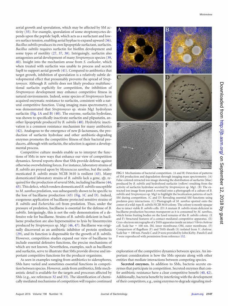
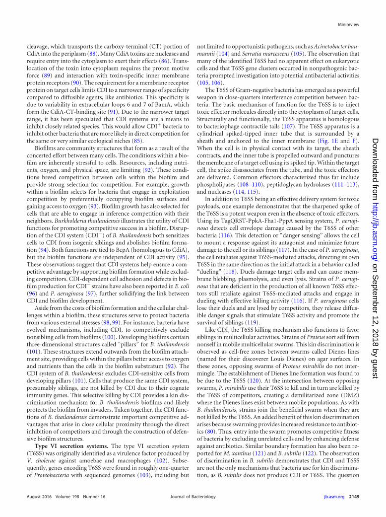
aerial growth and sporulation, which may be affected by SM ac-tivity (35). For example, sporulation of some streptomycetes de-pends upon the peptide SapB, which acts as a surfactant and low-ers surface tension, enabling aerial hyphae to expand upward (36).Bacillus subtilis produces its own lipopeptide surfactant, surfactin.Bacillus subtilis requires surfactin for biofilm development andsome types of motility (27, 37, 38). Intriguingly, surfactin alsoantagonizes aerial development of many Streptomyces species (39,40). Insight into the mechanism arose from S. coelicolor, whichwhen treated with surfactin was unable to process and secreteSapB to support aerial growth (41). Compared to antibiotics thattarget growth, inhibition of sporulation is a relatively subtle de-velopmental effect that presumably prevents the spread of Strep-tomyces. Although B. subtilis does not likely produce multifunc-tional surfactin explicitly for competition, the inhibition ofStreptomyces development may enhance competitive fitness innatural environments. Indeed, some species of Streptomyces haveacquired enzymatic resistance to surfactin, consistent with a nat-ural competitive function. Using imaging mass spectrometry, itwas demonstrated that Streptomyces sp. strain Mg1 hydrolyzessurfactin (Fig. 1A and B) (40). The enzyme, surfactin hydrolase,was shown to specifically inactivate surfactin and plipastatin, an-other lipopeptide produced by B. subtilis (40). Hydrolytic inacti-vation is a common resistance mechanism for many antibiotics(42). Analogous to the emergence of new �-lactamases, the pro-duction of surfactin hydrolase and other antibiotic-degradingenzymes promotes the competitive fitness of their bacterial pro-ducers, although with surfactin, the selection is against a develop-mental process.
Competitive culture models enable us to interpret the func-tions of SMs in new ways that enhance our view of competitiondynamics. Several reports show that SMs provide defense againstotherwise overwhelming forces. For instance, laboratory strains ofB. subtilis are preyed upon by Myxococcus xanthus, but the undo-mesticated B. subtilis strain NCIB 3610 is resilient (43). Manydomesticated laboratory strains of B. subtilis lack a gene, sfp, re-quired for the production of several SMs, including bacillaene (44,45). This defect, which renders domesticated B. subtilis susceptibleto M. xanthus predation, was subsequently shown to be specific tothe loss of bacillaene production (43) (Fig. 1C and D). Indeed,exogenous application of bacillaene protected sensitive strains ofB. subtilis and Escherichia coli from predation. Thus, under thepressure of predation, bacillaene is essential for the defense of B.subtilis. Intriguingly, this is not the only demonstration of a de-fensive role for bacillaene. Strains of B. subtilis deficient in bacil-laene production are also hypersensitive to lysis by linearmycinsproduced by Streptomyces sp. Mg1 (46, 47). Bacillaene was origi-nally discovered as an antibiotic inhibitor of protein synthesis(29), and its function is dispensable for the growth of B. subtilis.However, competition studies expand our view of bacillaene toinclude essential defensive functions, the precise mechanisms ofwhich are not known. Nevertheless, examples, such as bacillaeneand surfactin, serve to illustrate that SMs provide diverse and im-portant competitive functions for the producer organisms.
As seen in examples ranging from antibiotics to siderophores,SMs have varied and sometimes essential functions in competi-tion between species. However, aside from antibiotics, little mech-anistic detail is available for the targets and processes affected bySMs (e.g., see references 32 and 33). The identification of chemi-cally mediated mechanisms of competition will require continued
exploration of the competitive dynamics between species. An im-portant consideration is how the SMs operate along with otherentities that mediate interactions between competing species.
Secreted enzymes. In addition to SMs, bacteria secrete en-zymes that participate in competition. Secreted enzymes that con-fer antibiotic resistance have a clear competitive benefit (40, 42).Additionally, bacteria benefit by interfering with the developmentof their competitors, e.g., using enzymes to degrade signaling mol-
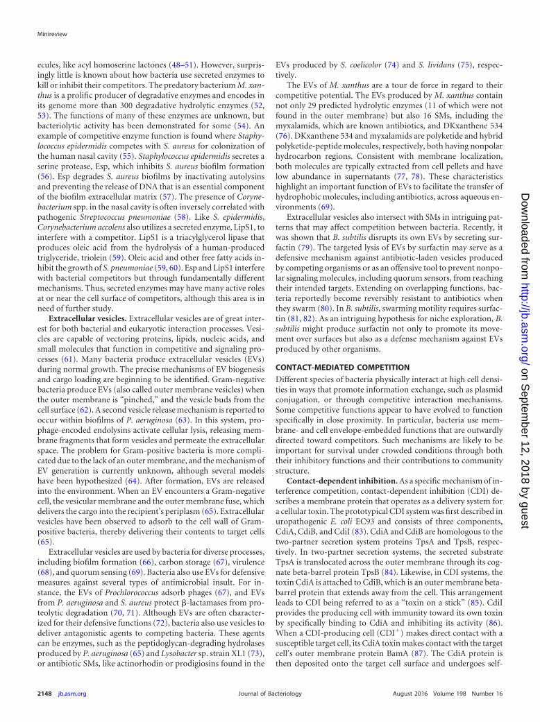
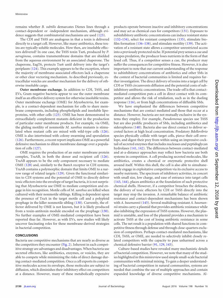
FIG 1 Mechanisms of bacterial competition. (A and B) Detection of patternsof SM production and degradation through imaging mass spectrometry. (A)False-colored extracted ion image showing the distribution of surfactin (blue)produced by B. subtilis and hydrolyzed surfactin (yellow) resulting from theactivity of surfactin hydrolase secreted by Streptomyces sp. Mg1. (B) The ex-tracted ion image from panel A overlaid onto a photograph of a culture of B.subtilis and Streptomyces sp. Mg1 to highlight the localization patterns of eachSM during competition. (C and D) Revealing essential SM functions usingpredator-prey interactions. (C) Photograph of M. xanthus spotted onto thecenter of a wild-type B. subtilis NCIB 3610 colony. The colony is mostly opaquedue to intact viable B. subtilis cells. (D) A mutant B. subtilis strain deficient inbacillaene production becomes transparent as it is consumed by M. xanthus,which forms fruiting bodies on the lysed remains of the B. subtilis colony. (Eand F) Structural features of a contact-mediated competitive apparatus. (E)Cryo-electron micrographs of a T6SS apparatus inside an intact Vibrio choleraecell. Scale bar � 100 nm. IM, inner membrane; OM, outer membrane. (F)Comparison of flagellum (F) and T6SS sheath (S) isolated from V. cholerae.Scale bar � 100 nm. Panels C and D were provided by John Kirby. Panels E andF were reproduced with permission from reference 152.
Minireview
August 2016 Volume 198 Number 16 jb.asm.org 2147Journal of Bacteriology
on Septem
ber 12, 2018 by guesthttp://jb.asm
.org/D
ownloaded from
ecules, like acyl homoserine lactones (48–51). However, surpris-ingly little is known about how bacteria use secreted enzymes tokill or inhibit their competitors. The predatory bacterium M. xan-thus is a prolific producer of degradative enzymes and encodes inits genome more than 300 degradative hydrolytic enzymes (52,53). The functions of many of these enzymes are unknown, butbacteriolytic activity has been demonstrated for some (54). Anexample of competitive enzyme function is found where Staphy-lococcus epidermidis competes with S. aureus for colonization ofthe human nasal cavity (55). Staphylococcus epidermidis secretes aserine protease, Esp, which inhibits S. aureus biofilm formation(56). Esp degrades S. aureus biofilms by inactivating autolysinsand preventing the release of DNA that is an essential componentof the biofilm extracellular matrix (57). The presence of Coryne-bacterium spp. in the nasal cavity is often inversely correlated withpathogenic Streptococcus pneumoniae (58). Like S. epidermidis,Corynebacterium accolens also utilizes a secreted enzyme, LipS1, tointerfere with a competitor. LipS1 is a triacylglycerol lipase thatproduces oleic acid from the hydrolysis of a human-producedtriglyceride, triolein (59). Oleic acid and other free fatty acids in-hibit the growth of S. pneumoniae (59, 60). Esp and LipS1 interferewith bacterial competitors but through fundamentally differentmechanisms. Thus, secreted enzymes may have many active rolesat or near the cell surface of competitors, although this area is inneed of further study.
Extracellular vesicles. Extracellular vesicles are of great inter-est for both bacterial and eukaryotic interaction processes. Vesi-cles are capable of vectoring proteins, lipids, nucleic acids, andsmall molecules that function in competitive and signaling pro-cesses (61). Many bacteria produce extracellular vesicles (EVs)during normal growth. The precise mechanisms of EV biogenesisand cargo loading are beginning to be identified. Gram-negativebacteria produce EVs (also called outer membrane vesicles) whenthe outer membrane is “pinched,” and the vesicle buds from thecell surface (62). A second vesicle release mechanism is reported tooccur within biofilms of P. aeruginosa (63). In this system, pro-phage-encoded endolysins activate cellular lysis, releasing mem-brane fragments that form vesicles and permeate the extracellularspace. The problem for Gram-positive bacteria is more compli-cated due to the lack of an outer membrane, and the mechanism ofEV generation is currently unknown, although several modelshave been hypothesized (64). After formation, EVs are releasedinto the environment. When an EV encounters a Gram-negativecell, the vesicular membrane and the outer membrane fuse, whichdelivers the cargo into the recipient’s periplasm (65). Extracellularvesicles have been observed to adsorb to the cell wall of Gram-positive bacteria, thereby delivering their contents to target cells(65).
Extracellular vesicles are used by bacteria for diverse processes,including biofilm formation (66), carbon storage (67), virulence(68), and quorum sensing (69). Bacteria also use EVs for defensivemeasures against several types of antimicrobial insult. For in-stance, the EVs of Prochlorococcus adsorb phages (67), and EVsfrom P. aeruginosa and S. aureus protect �-lactamases from pro-teolytic degradation (70, 71). Although EVs are often character-ized for their defensive functions (72), bacteria also use vesicles todeliver antagonistic agents to competing bacteria. These agentscan be enzymes, such as the peptidoglycan-degrading hydrolasesproduced by P. aeruginosa (65) and Lysobacter sp. strain XL1 (73),or antibiotic SMs, like actinorhodin or prodigiosins found in the
EVs produced by S. coelicolor (74) and S. lividans (75), respec-tively.
The EVs of M. xanthus are a tour de force in regard to theircompetitive potential. The EVs produced by M. xanthus containnot only 29 predicted hydrolytic enzymes (11 of which were notfound in the outer membrane) but also 16 SMs, including themyxalamids, which are known antibiotics, and DKxanthene 534(76). DKxanthene 534 and myxalamids are polyketide and hybridpolyketide-peptide molecules, respectively, both having nonpolarhydrocarbon regions. Consistent with membrane localization,both molecules are typically extracted from cell pellets and havelow abundance in supernatants (77, 78). These characteristicshighlight an important function of EVs to facilitate the transfer ofhydrophobic molecules, including antibiotics, across aqueous en-vironments (69).
Extracellular vesicles also intersect with SMs in intriguing pat-terns that may affect competition between bacteria. Recently, itwas shown that B. subtilis disrupts its own EVs by secreting sur-factin (79). The targeted lysis of EVs by surfactin may serve as adefensive mechanism against antibiotic-laden vesicles producedby competing organisms or as an offensive tool to prevent nonpo-lar signaling molecules, including quorum sensors, from reachingtheir intended targets. Extending on overlapping functions, bac-teria reportedly become reversibly resistant to antibiotics whenthey swarm (80). In B. subtilis, swarming motility requires surfac-tin (81, 82). As an intriguing hypothesis for niche exploration, B.subtilis might produce surfactin not only to promote its move-ment over surfaces but also as a defense mechanism against EVsproduced by other organisms.
CONTACT-MEDIATED COMPETITION
Different species of bacteria physically interact at high cell densi-ties in ways that promote information exchange, such as plasmidconjugation, or through competitive interaction mechanisms.Some competitive functions appear to have evolved to functionspecifically in close proximity. In particular, bacteria use mem-brane- and cell envelope-embedded functions that are outwardlydirected toward competitors. Such mechanisms are likely to beimportant for survival under crowded conditions through boththeir inhibitory functions and their contributions to communitystructure.
Contact-dependent inhibition. As a specific mechanism of in-terference competition, contact-dependent inhibition (CDI) de-scribes a membrane protein that operates as a delivery system fora cellular toxin. The prototypical CDI system was first described inuropathogenic E. coli EC93 and consists of three components,CdiA, CdiB, and CdiI (83). CdiA and CdiB are homologous to thetwo-partner secretion system proteins TpsA and TpsB, respec-tively. In two-partner secretion systems, the secreted substrateTpsA is translocated across the outer membrane through its cog-nate beta-barrel protein TpsB (84). Likewise, in CDI systems, thetoxin CdiA is attached to CdiB, which is an outer membrane beta-barrel protein that extends away from the cell. This arrangementleads to CDI being referred to as a “toxin on a stick” (85). CdiIprovides the producing cell with immunity toward its own toxinby specifically binding to CdiA and inhibiting its activity (86).When a CDI-producing cell (CDI�) makes direct contact with asusceptible target cell, its CdiA toxin makes contact with the targetcell’s outer membrane protein BamA (87). The CdiA protein isthen deposited onto the target cell surface and undergoes self-
Minireview
2148 jb.asm.org August 2016 Volume 198 Number 16Journal of Bacteriology
on Septem
ber 12, 2018 by guesthttp://jb.asm
.org/D
ownloaded from
cleavage, which transports the carboxy-terminal (CT) portion ofCdiA into the periplasm (88). Many CdiA toxins are nucleases andrequire entry into the cytoplasm to exert their effects (86). Trans-location of the toxin into cytoplasm requires the proton motiveforce (89) and interaction with toxin-specific inner membraneprotein receptors (90). The requirement for a membrane receptorprotein on target cells limits CDI to a narrower range of specificitycompared to diffusible agents, like antibiotics. This specificity isdue to variability in extracellular loops 6 and 7 of BamA, whichform the CdiA-CT-binding site (91). Due to the narrower targetrange, it has been speculated that CDI systems are a means toinhibit closely related species. This would allow CDI� bacteria toinhibit other bacteria that are more likely in direct competition forthe same or very similar ecological niches (85).
Biofilms are community structures that form as a result of theconcerted effort between many cells. The conditions within a bio-film are inherently stressful to cells. Resources, including nutri-ents, oxygen, and physical space, are limiting (92). These condi-tions breed competition between cells within the biofilm andprovide strong selection for competition. For example, growthwithin a biofilm selects for bacteria that engage in exploitationcompetition by preferentially occupying biofilm surfaces andgaining access to oxygen (93). Biofilm growth has also selected forcells that are able to engage in inference competition with theirneighbors. Burkholderia thailandensis illustrates the utility of CDIfunctions for promoting competitive success in a biofilm. Disrup-tion of the CDI system (CDI�) of B. thailandensis both sensitizescells to CDI from isogenic siblings and abolishes biofilm forma-tion (94). Both functions are tied to BcpA (homologous to CdiA),but the biofilm functions are independent of CDI activity (95).These observations suggest that CDI systems help ensure a com-petitive advantage by supporting biofilm formation while exclud-ing competitors. CDI-dependent cell adhesion and defects in bio-film production for CDI� strains have also been reported in E. coli(96) and P. aeruginosa (97), further solidifying the link betweenCDI and biofilm development.
Aside from the costs of biofilm formation and the cellular chal-lenges within a biofilm, these structures serve to protect bacteriafrom various external stresses (98, 99). For instance, bacteria haveevolved mechanisms, including CDI, to competitively excludenonsibling cells from biofilms (100). Developing biofilms containthree-dimensional structures called “pillars” for B. thailandensis(101). These structures extend outwards from the biofilm attach-ment site, providing cells within the pillars better access to oxygenand nutrients than the cells in the biofilm substratum (92). TheCDI system of B. thailandensis excludes CDI-sensitive cells fromdeveloping pillars (101). Cells that produce the same CDI system,presumably siblings, are not killed by CDI due to their cognateimmunity genes. This selective killing by CDI provides a kin dis-crimination mechanism for B. thailandensis biofilms and likelyprotects the biofilm from invaders. Taken together, the CDI func-tions of B. thailandensis demonstrate important competitive ad-vantages that arise in close cellular proximity through the directinhibition of competitors and through the construction of defen-sive biofilm structures.
Type VI secretion systems. The type VI secretion system(T6SS) was originally identified as a virulence factor produced byV. cholerae against amoebae and macrophages (102). Subse-quently, genes encoding T6SS were found in roughly one-quarterof Proteobacteria with sequenced genomes (103), including but
not limited to opportunistic pathogens, such as Acinetobacter bau-mannii (104) and Serratia marcescens (105). The observation thatmany of the identified T6SS had no apparent effect on eukaryoticcells and that T6SS gene clusters occurred in nonpathogenic bac-teria prompted investigation into potential antibacterial activities(105, 106).
The T6SS of Gram-negative bacteria has emerged as a powerfulweapon in close-quarters interference competition between bac-teria. The basic mechanism of function for the T6SS is to injecttoxic effector molecules directly into the cytoplasm of target cells.Structurally and functionally, the T6SS apparatus is homologousto bacteriophage contractile tails (107). The T6SS apparatus is acylindrical spiked-tipped inner tube that is surrounded by asheath and anchored to the inner membrane (Fig. 1E and F).When the cell is in physical contact with its target, the sheathcontracts, and the inner tube is propelled outward and puncturesthe membrane of a target cell using its spiked tip. Within the targetcell, the spike disassociates from the tube, and the toxic effectorsare delivered. Common effectors characterized thus far includephospholipases (108–110), peptidoglycan hydrolases (111–113),and nucleases (114, 115).
In addition to T6SS being an effective delivery system for toxicpayloads, one example demonstrates that the sharpened spike ofthe T6SS is a potent weapon even in the absence of toxic effectors.Using its TagQRST-PpkA-Fha1-PppA sensing system, P. aerugi-nosa detects cell envelope damage caused by the T6SS of otherbacteria (116). This detection or “danger sensing” allows the cellto mount a response against its antagonist and minimize futuredamage to the cell or its siblings (117). In the case of P. aeruginosa,the cell retaliates against T6SS-mediated attacks, directing its ownT6SS in the same direction as the initial attack in a behavior called“dueling” (118). Duels damage target cells and can cause mem-brane blebbing, plasmolysis, and even lysis. Strains of P. aerugi-nosa that are deficient in the production of all known T6SS effec-tors still retaliate against T6SS-mediated attacks and engage indueling with effective killing activity (116). If P. aeruginosa cellslose their duels and are lysed by competitors, they release diffus-ible danger signals that stimulate T6SS activity and promote thesurvival of siblings (119).
Like CDI, the T6SS killing mechanism also functions to favorsiblings in multicellular activities. Strains of Proteus sort self fromnonself in mobile multicellular swarms. This kin discrimination isobserved as cell-free zones between swarms called Dienes lines(named for their discoverer Louis Dienes) on agar surfaces. Inthese zones, opposing swarms of Proteus mirabilis do not inter-mingle. The establishment of Dienes line formation was found tobe due to the T6SS (120). At the intersection between opposingswarms, P. mirabilis use their T6SS to kill and in turn are killed bythe T6SS of competitors, creating a demilitarized zone (DMZ)where the Dienes lines exist between mobile populations. As withB. thailandensis, strains join the beneficial swarm when they arenot killed by the T6SS. An added benefit of this kin discriminationarises because swarming provides increased resistance to antibiot-ics (80). Thus, entry into the swarm promotes competitive fitnessof bacteria by excluding unrelated cells and by enhancing defenseagainst antibiotics. Similar boundary formation has also been re-ported for M. xanthus (121) and B. subtilis (122). The observationof discrimination in B. subtilis demonstrates that CDI and T6SSare not the only mechanisms that bacteria use for kin discrimina-tion, as B. subtilis does not produce CDI or T6SS. The question
Minireview
August 2016 Volume 198 Number 16 jb.asm.org 2149Journal of Bacteriology
on Septem
ber 12, 2018 by guesthttp://jb.asm
.org/D
ownloaded from
remains whether B. subtilis demarcates Dienes lines through acontact-dependent or -independent mechanism, although evi-dence suggests that combinatorial mechanisms are used (123).
The CDI and T6SS are analogous in that a toxin is delivereddirectly to a target cell. However, like many antibiotics, these tox-ins are typically soluble molecules. How then, are insoluble effec-tors delivered? In one case, the T6SS toxin Tse6, produced by P.aeruginosa, contains transmembrane domains that are shieldedfrom the aqueous environment by an associated chaperone. Thechaperone, EagT6, protects Tse6 until delivery into the target’speriplasm (124). This example appears to be the exception, wherethe majority of membrane-associated effectors lack a chaperoneor other clear vectoring mechanism. As described previously, ex-tracellular vesicles are another mechanism for the delivery of oth-erwise insoluble cargo.
Outer membrane exchange. In addition to CDI, T6SS, andEVs, Gram-negative bacteria appear to use the outer membraneitself as an effective delivery system for otherwise insoluble toxins.Outer membrane exchange (OME) for Myxobacteria, for exam-ple, is a contact-dependent mechanism for cells to share mem-brane components, including phospholipids and insoluble lipo-proteins, with other cells (125). OME has been demonstrated toextracellularly complement mutants deficient in the productionof particular outer membrane products. For example, via OME,the gliding motility of nonmotile M. xanthus mutants is stimu-lated when mutant cells are mixed with wild-type cells (126).OME is also intertwined with colony swarming and sporulation(126). Furthermore, a recent report implicates OME as a powerfuldefensive mechanism to dilute membrane damage over a popula-tion of cells (127).
OME requires the production of an outer membrane proteincomplex, TraAB, in both the donor and recipient cell (126).TraAB appears to be the only component necessary to mediateOME (128) and, similarly to the BamA receptor in CDI systems,TraAB contains a polymorphic domain that limits OME to a nar-row range of related targets (129). Given the functional similari-ties to CDI systems and the potential of OME to directly delivertoxic effectors into the envelope of target bacteria, it is not surpris-ing that Myxobacteria use OME to mediate competition and en-gage in kin recognition. Motile cells of M. xanthus are killed whencultured with their nonmotile siblings. Killing is dependent uponthe presence of TraA in the target motile cell and a polyploidprophage in the killer nonmotile sibling (130). Currently, the ef-fector delivered by OME is not known, but it is likely producedfrom a toxin-antitoxin module encoded on the prophage (130).No further examples of OME-mediated competition have beenreported thus far. However, as with EVs, new studies will likelyuncover fascinating roles for these membrane-derived strategiesin bacterial competition.
CONCLUSIONS
Bacteria use competitive mechanisms that are nearly as diverse asthe competitors they encounter (Fig. 2). Inherent in each compet-itive strategy are advantages and disadvantages. When bacteria usesecreted effectors, like antibiotics, enzymes, or vesicles, they areable to compete while minimizing the risks of direct damage dur-ing contact-mediated competition. Once a cell exports its compet-itive molecules across its envelope, those molecules are subject todiffusion, which diminishes their inhibitory effect on competitorsat a distance. However, many of these metabolically expensive
products operate between inactive and inhibitory concentrationsand may act as chemical cues for competitors (131). Exposure tosubinhibitory antibiotic concentrations can induce resistant states(132–134), select for resistant competitors (135), stimulate bio-film formation (136–138), and stimulate motility (139). The acti-vation of a resistant state allows a competitor unrestricted accessinto a previously protected niche. If potential prey senses a cue andescapes predation, the producer loses nutrients in the form of thatlysed cell. Thus, if a competitor senses a cue, the producer maysuffer the consequences for competitive fitness. However, it is alsoimportant to note that our current understanding of the responseto subinhibitory concentrations of antibiotics and other SMs inthe context of bacterial communities is limited and requires fur-ther investigation. The direct delivery of toxins into a target cell byCDI or T6SS circumvents diffusion and the potential costs of sub-inhibitory antibiotic concentrations. The trade-off is that contact-mediated competition puts a cell in direct contact with its com-petitor and allows the risk of retaliation, such as in the duelingresponse (116), or from high concentrations of diffusible SMs.
We have emphasized the differences between competitivemechanisms that are contact mediated and those that occur at adistance. However, bacteria are not mutually exclusive in the sys-tems they employ. For example, Pseudomonas species use T6SSbut are also prolific producers of SMs, including antibiotics andsiderophores (140). Bacteria also use direct contact to deliver se-creted factors at high local concentration. Predatory Bdellovibriospecies physically collide with target cells, pierce their cell enve-lope, and digest their prey from within using an impressive cock-tail of secreted enzymes that includes nucleases and peptidoglycanhydrolases (141, 142). The differences between contact-mediatedand at-a-distance approaches may reflect how bacteria use bothsystems in competition. A cell producing secreted molecules, likeantibiotics, creates a chemical or enzymatic protective shellaround itself. Within this shell, the cell is also able to simultane-ously engage in exploitative competition via its exclusive access tonearby nutrients. The spectrum of inhibitory activities, in concertwith small size, low charge, and ease of entrance into target cells(143, 144), places antibiotics at the foundation of such protectivechemical shells. However, if a competitor breaches the defenses,the delivery of toxic effectors by CDI or T6SS directly into thetarget may stop the invasion. A remarkable balance of antibioticresistance and contact-dependent mechanisms has been shownwith A. baumannii (145). Several multidrug-resistant A. bauman-nii strains carry a plasmid that provides antibiotic resistance whilealso inhibiting the expression of T6SS systems. However, the plas-mid is unstable, and loss of the plasmid provides a mechanism toactivate T6SS at the cost of losing antibiotic resistance in somecells. The net result is a population with shared functions in com-petitive fitness through defense and through close-quarters exclu-sion of competitors. Perhaps contact-mediated mechanisms, likeCDI, T6SS, or OME, are needed to selectively inhibit closely re-lated competitors with the capacity to pass unharmed across achemical defensive barrier (91, 129, 145).
Culture-based studies have revealed many mechanistic detailsof bacterial competition. However, we note that many of the stud-ies highlighted in this minireview used simple small-scale bacterialcommunities with minimal mixing. To gain a deeper understand-ing of bacterial competition in natural communities, systems areneeded that combine the use of multiple approaches and containexpanded knowledge of diverse competitive mechanisms. Al-
Minireview
2150 jb.asm.org August 2016 Volume 198 Number 16Journal of Bacteriology
on Septem
ber 12, 2018 by guesthttp://jb.asm
.org/D
ownloaded from
though beyond the scope of this minireview, mathematical mod-eling is a powerful approach to understand how bacterial commu-nities are formed and maintained (e.g., see references 146 and147). Mathematical approaches stand to become more powerfulas they incorporate diverse competitive outcomes in addition tokilling or survival. For instance, what effects does T6SS-mediatedretaliation have in a modeled competition? How does SM-medi-ated developmental inhibition affect a community? What are theconsequences of exposure for cells outside the inhibitory ranges ofSMs? Using controlled experiments in the laboratory, new mech-anistic details of competition are being identified, despite limita-tions to our understanding of these mechanisms in natural envi-ronments. The genomes of many antibiotic-producing bacteriacontain silent SM gene clusters that are not expressed under lab-oratory conditions (148). Likewise, many studies with CDI andT6SS require artificial expression conditions (149, 150). Theseobstacles are a central focus of current efforts to understand
competitive mechanisms. Meanwhile, models that bettermimic the native environment are being developed to providea clearer view of bacterial interactions under natural condi-tions (e.g., see references 86, 115, and 151) The examples aboveand many more innovative studies are expanding our views ofthe interactive interfaces between two bacterial species. Theemerging challenge is to build these interfaces into networks,which will represent the many facets of competition withinmicrobial communities.
ACKNOWLEDGMENTS
We thank Stefan Pukatzki for helpful comments on the manuscript. Wethank John Kirby for providing images. We thank Patrick Lane (ScEYEnceStudios) for graphical enhancement of Fig. 2. We thank the Texas A&MUniversity Center for Mass Spectrometry for assistance in imaging massspectrometry.
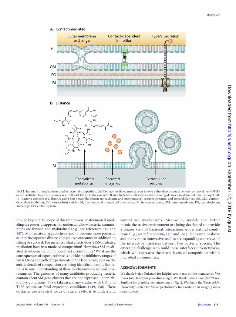
FIG 2 Summary of mechanisms used in bacterial competition. (A) Contact-mediated mechanisms involve either direct contact between cell envelopes (OME)or are facilitated by protein complexes (CDI and T6SS). In the case of CDI and T6SS, toxic effectors (square or wedged circle) are delivered into the target cell.(B) Bacteria compete at a distance using SMs (examples shown are bacillaene and streptomycin), secreted enzymes, and extracellular vesicles. CDI, contact-dependent inhibition; EVs, extracellular vesicles; M, membrane; MT, target cell membrane; IM, inner membrane; OM, outer membrane; PG, peptidoglycan;T6SS, type VI secretion system.
Minireview
August 2016 Volume 198 Number 16 jb.asm.org 2151Journal of Bacteriology
on Septem
ber 12, 2018 by guesthttp://jb.asm
.org/D
ownloaded from
FUNDING INFORMATIONThis work, including the efforts of Paul D. Straight, was funded by NSF |NSF Office of the Director (OD) (MCB-1253215). This work, includingthe efforts of Paul D. Straight, was funded by Welch Foundation (A-1796).
This work was partially supported by Texas A&M AgriLife.
REFERENCES1. Foster KR, Bell T. 2012. Competition, not cooperation, dominates
interactions among culturable microbial species. Curr Biol 22:1845–1850. http://dx.doi.org/10.1016/j.cub.2012.08.005.
2. Birch LC. 1957. The meanings of competition. Am Nat 91:5–18. http://dx.doi.org/10.1086/281957.
3. Hibbing ME, Fuqua C, Parsek MR, Peterson SB. 2010. Bacterial com-petition: surviving and thriving in the microbial jungle. Nat Rev Micro-biol 8:15–25. http://dx.doi.org/10.1038/nrmicro2259.
4. Ling LL, Schneider T, Peoples AJ, Spoering AL, Engels I, Conlon BP,Mueller A, Schäberle TF, Hughes DE, Epstein S, Jones M, Lazarides L,Steadman VA, Cohen DR, Felix CR, Fetterman KA, Millett WP, NittiAG, Zullo AM, Chen C, Lewis K. 2015. A new antibiotic kills pathogenswithout detectable resistance. Nature 517:455– 459. http://dx.doi.org/10.1038/nature14098.
5. Davies J. 2013. Specialized microbial metabolites: functions and origins.J Antibiot 66:361–364. http://dx.doi.org/10.1038/ja.2013.61.
6. Price-Whelan A, Dietrich LEP, Newman DK. 2006. Rethinking “sec-ondary” metabolism: physiological roles for phenazine antibiotics. NatChem Biol 2:71–78. http://dx.doi.org/10.1038/nchembio764.
7. Davies J. 2006. Are antibiotics naturally antibiotics? J Ind MicrobiolBiotechnol 33:496 – 499. http://dx.doi.org/10.1007/s10295-006-0112-5.
8. Yim G, Wang HH, Davies J. 2006. The truth about antibiotics. Int J MedMicrobiol 296:163–170. http://dx.doi.org/10.1016/j.ijmm.2006.01.039.
9. Yim G, Wang HH, Davies J. 2007. Antibiotics as signalling molecules.Philos Trans R Soc Lond B Biol Sci 362:1195–1200. http://dx.doi.org/10.1098/rstb.2007.2044.
10. Romero D, Traxler MF, López D, Kolter R. 2011. Antibiotics as signalmolecules. Chem Rev 111:5492–5505. http://dx.doi.org/10.1021/cr2000509.
11. Park T. 1954. Experimental studies of interspecies competition II. Tem-perature, humidity, and competition in two species of Tribolium. PhysiolZool 27:177–238.
12. Hider RC, Kong X. 2010. Chemistry and biology of siderophores. NatProd Rep 27:637– 657. http://dx.doi.org/10.1039/b906679a.
13. Winkelmann G. 2002. Microbial siderophore-mediated transport.Biochem Soc Trans 30:691– 696. http://dx.doi.org/10.1042/bst0300691.
14. Lee KH, Ruby EG. 1994. Competition between Vibrio fischeri strainsduring initiation and maintenance of a light organ symbiosis. J Bacteriol176:1985–1991.
15. Harrison F, Paul J, Massey RC, Buckling A. 2008. Interspecific com-petition and siderophore-mediated cooperation in Pseudomonas aerugi-nosa. ISME J 2:49 –55. http://dx.doi.org/10.1038/ismej.2007.96.
16. Andrews SC, Robinson AK, Rodríguez-Quiñones F. 2003. Bacterialiron homeostasis. FEMS Microbiol Rev 27:215–237. http://dx.doi.org/10.1016/S0168-6445(03)00055-X.
17. Traxler MF, Seyedsayamdost MR, Clardy J, Kolter R. 2012. Interspe-cies modulation of bacterial development through iron competition andsiderophore piracy. Mol Microbiol 86:628 – 644. http://dx.doi.org/10.1111/mmi.12008.
18. Galet J, Deveau A, Hôtel L, Frey-Klett P, Leblond P, Aigle B. 2015.Pseudomonas fluorescens pirates both ferrioxamine and ferricoelichelinsiderophores from Streptomyces ambofaciens. Appl Environ Microbiol81:3132–3141. http://dx.doi.org/10.1128/AEM.03520-14.
19. Lewis K. 2013. Platforms for antibiotic discovery. Nat Rev Drug Discov12:371–387. http://dx.doi.org/10.1038/nrd3975.
20. Davies J, Ryan KS. 2012. Introducing the parvome: bioactive com-pounds in the microbial world. ACS Chem Biol 7:252–259. http://dx.doi.org/10.1021/cb200337h.
21. Bernier SP, Surette MG. 2013. Concentration-dependent activity ofantibiotics in natural environments. Front Microbiol 4:20.
22. Wang W, Ji J, Li X, Wang J, Li S, Pan G, Fan K, Yang K. 2014.Angucyclines as signals modulate the behaviors of Streptomyces coeli-color. Proc Natl Acad Sci U S A 111:5688 –5693. http://dx.doi.org/10.1073/pnas.1324253111.
23. Jones C, Allsopp L, Horlick J, Kulasekara H, Filloux A. 2013. Subin-
hibitory concentration of kanamycin induces the Pseudomonas aerugi-nosa type VI secretion system. PLoS One 8:e81132. http://dx.doi.org/10.1371/journal.pone.0081132.
24. Davies J, Spiegelman GB, Yim G. 2006. The world of subinhibitoryantibiotic concentrations. Curr Opin Microbiol 9:445– 453. http://dx.doi.org/10.1016/j.mib.2006.08.006.
25. Haas D, Défago G. 2005. Biological control of soil-borne pathogens byfluorescent pseudomonads. Nat Rev Microbiol 3:307–319. http://dx.doi.org/10.1038/nrmicro1129.
26. Powers MJ, Sanabria-Valentín E, Bowers AA, Shank EA. 2015. Inhi-bition of cell differentiation in Bacillus subtilis by Pseudomonas protegens.J Bacteriol 197:2129 –2138. http://dx.doi.org/10.1128/JB.02535-14.
27. López D, Fischbach MA, Chu F, Losick R, Kolter R. 2009. Structurallydiverse natural products that cause potassium leakage trigger multicel-lularity in Bacillus subtilis. Proc Natl Acad Sci U S A 106:280 –285. http://dx.doi.org/10.1073/pnas.0810940106.
28. Bleich R, Watrous JD, Dorrestein PC, Bowers AA, Shank EA. 2015.Thiopeptide antibiotics stimulate biofilm formation in Bacillus subtilis.Proc Natl Acad Sci U S A 112:3086 –3091. http://dx.doi.org/10.1073/pnas.1414272112.
29. Patel PS, Huang S, Fisher S, Pirnik D, Aklonis C, Dean L, Meyers E,Fernandes P, Mayerl F. 1995. Bacillaene, a novel inhibitor of procaryoticprotein synthesis produced by Bacillus subtilis: production, taxonomy,isolation, physico-chemical characterization and biological activity. JAntibiot (Tokyo) 48:997–1003. http://dx.doi.org/10.7164/antibiotics.48.997.
30. Straight PD, Fischbach MA, Walsh CT, Rudner DZ, Kolter R. 2007. Asingular enzymatic megacomplex from Bacillus subtilis. Proc Natl AcadSci U S A 104:305–310. http://dx.doi.org/10.1073/pnas.0609073103.
31. Vargas-Bautista C, Rahlwes K, Straight P. 2014. Bacterial competitionreveals differential regulation of the pks genes by Bacillus subtilis. J Bac-teriol 196:717–728. http://dx.doi.org/10.1128/JB.01022-13.
32. Teasdale ME, Liu J, Wallace J, Akhlaghi F, Rowley DC. 2009. Second-ary metabolites produced by the marine bacterium Halobacillus salinusthat inhibit quorum sensing-controlled phenotypes in Gram-negativebacteria. Appl Environ Microbiol 75:567–572. http://dx.doi.org/10.1128/AEM.00632-08.
33. Kwan JC, Meickle T, Ladwa D, Teplitski M, Paul V, Luesch H. 2011.Lyngbyoic acid, a “tagged” fatty acid from a marine cyanobacterium,disrupts quorum sensing in Pseudomonas aeruginosa. Mol Biosyst7:1205–1216. http://dx.doi.org/10.1039/c0mb00180e.
34. Chater KF. 2006. Streptomyces inside-out: a new perspective on the bac-teria that provide us with antibiotics. Philos Trans R Soc Lond B Biol Sci361:761–768. http://dx.doi.org/10.1098/rstb.2005.1758.
35. Flärdh K, Buttner MJ. 2009. Streptomyces morphogenetics: dissectingdifferentiation in a filamentous bacterium. Nat Rev Microbiol 7:36 – 49.http://dx.doi.org/10.1038/nrmicro1968.
36. Kodani S, Hudson ME, Durrant MC, Buttner MJ, Nodwell JR, WilleyJM. 2004. The SapB morphogen is a lantibiotic-like peptide derived fromthe product of the developmental gene ramS in Streptomyces coelicolor.Proc Natl Acad Sci U S A 101:11448 –11453. http://dx.doi.org/10.1073/pnas.0404220101.
37. Kearns DB, Losick R. 2003. Swarming motility in undomesticated Ba-cillus subtilis. Mol Microbiol 49:581–590.
38. López D, Vlamakis H, Losick R, Kolter R. 2009. Paracrine signaling ina bacterium. Genes Dev 23:1631–1638. http://dx.doi.org/10.1101/gad.1813709.
39. Straight PD, Willey JM, Kolter R. 2006. Interactions between Strep-tomyces coelicolor and Bacillus subtilis: role of surfactants in raising aer-ial structures. J Bacteriol 188:4918 – 4925. http://dx.doi.org/10.1128/JB.00162-06.
40. Hoefler BC, Gorzelnik KV, Yang JY, Hendricks N, Dorrestein PC,Straight PD. 2012. Enzymatic resistance to the lipopeptide surfactin asidentified through imaging mass spectrometry of bacterial competition.Proc Natl Acad Sci U S A 109:13082–13087. http://dx.doi.org/10.1073/pnas.1205586109.
41. Gaskell AA, Giovinazzo JA, Fonte V, Willey JM. 2012. Multi-tierregulation of the streptomycete morphogenetic peptide SapB. MolMicrobiol 84:501–515. http://dx.doi.org/10.1111/j.1365-2958.2012.08041.x.
42. Wright GD. 2005. Bacterial resistance to antibiotics: enzymatic degra-dation and modification. Adv Drug Deliv Rev 57:1451–1470. http://dx.doi.org/10.1016/j.addr.2005.04.002.
Minireview
2152 jb.asm.org August 2016 Volume 198 Number 16Journal of Bacteriology
on Septem
ber 12, 2018 by guesthttp://jb.asm
.org/D
ownloaded from
43. Müller S, Strack SN, Hoefler BC, Straight PD, Kearns DB, Kirby JR.2014. Bacillaene and sporulation protect Bacillus subtilis from predationby Myxococcus xanthus. Appl Environ Microbiol 80:5603–5610. http://dx.doi.org/10.1128/AEM.01621-14.
44. Mootz HD, Finking R, Marahiel MA. 2001. 4=-Phosphopantetheinetransfer in primary and secondary metabolism of Bacillus subtilis. J BiolChem 276:37289 –37298. http://dx.doi.org/10.1074/jbc.M103556200.
45. McLoon AL, Guttenplan SB, Kearns DB, Kolter R, Losick R. 2011.Tracing the domestication of a biofilm-forming bacterium. J Bacteriol193:2027–2034. http://dx.doi.org/10.1128/JB.01542-10.
46. Barger SR, Hoefler BC, Cubillos-Ruiz A, Russell WK, Russell DH,Straight PD. 2012. Imaging secondary metabolism of Streptomyces sp.Mg1 during cellular lysis and colony degradation of competing Bacillussubtilis. Antonie Van Leeuwenhoek 102:435– 445.
47. Stubbendieck RM, Straight PD. 2015. Escape from lethal bacterial com-petition through coupled activation of antibiotic resistance and a mobi-lized subpopulation. PLoS Genet 11:e1005722. http://dx.doi.org/10.1371/journal.pgen.1005722.
48. Kang BR, Lee JH, Ko SJ, Lee YH, Cha JS, Cho BH, Kim YC. 2004.Degradation of acyl-homoserine lactone molecules by Acinetobacter sp.strain C1010. Can J Microbiol 50:935–941. http://dx.doi.org/10.1139/w04-083.
49. Park SY, Kang HO, Jang HS, Lee JK, Koo BT, Yum DY. 2005. Identifi-cation of extracellular N-acylhomoserine lactone acylase from a Streptomy-ces sp. and its application to quorum quenching. Appl Environ Microbiol71:2632–2641. http://dx.doi.org/10.1128/AEM.71.5.2632-2641.2005.
50. Sio CF, Otten LG, Cool RH, Diggle SP, Braun PG, Bos R, Daykin M,Cámara M, Williams P, Quax WJ. 2006. Quorum quenching by anN-acyl-homoserine lactone acylase from Pseudomonas aeruginosa PAO1.Infect Immun 74:1673–1682. http://dx.doi.org/10.1128/IAI.74.3.1673-1682.2006.
51. Medina-Martínez MS, Uyttendaele M, Rajkovic A, Nadal P, DebevereJ. 2007. Degradation of N-acyl-L-homoserine lactones by Bacillus cereusin culture media and pork extract. Appl Environ Microbiol 73:2329 –2332. http://dx.doi.org/10.1128/AEM.01993-06.
52. Goldman BS, Nierman WC, Kaiser D, Slater SC, Durkin AS, Eisen JA,Ronning CM, Barbazuk WB, Blanchard M, Field C, Halling C, HinkleG, Iartchuk O, Kim HS, Mackenzie C, Madupu R, Miller N, Shvarts-beyn A, Sullivan SA, Vaudin M, Wiegand R, Kaplan HB. 2006.Evolution of sensory complexity recorded in a Myxobacterial genome.Proc Natl Acad Sci U S A 103:15200 –15205. http://dx.doi.org/10.1073/pnas.0607335103.
53. Berleman JE, Kirby JR. 2009. Deciphering the hunting strategy of abacterial wolfpack. FEMS Microbiol Rev 33:942–957. http://dx.doi.org/10.1111/j.1574-6976.2009.00185.x.
54. Sudo S, Dworkin M. 1972. Bacteriolytic enzymes produced by Myxo-coccus xanthus. J Bacteriol 110:236 –245.
55. Lina G, Boutite F, Tristan A, Bes M, Etienne J, Vandenesch F. 2003.Bacterial competition for human nasal cavity colonization: role of staph-ylococcal agr alleles. Appl Environ Microbiol 69:18 –23. http://dx.doi.org/10.1128/AEM.69.1.18-23.2003.
56. Iwase T, Uehara Y, Shinji H, Tajima A, Seo H, Takada K, Agata T,Mizunoe Y. 2010. Staphylococcus epidermidis Esp inhibits Staphylococcusaureus biofilm formation and nasal colonization. Nature 465:346 –349.http://dx.doi.org/10.1038/nature09074.
57. Chen C, Krishnan V, Macon K, Manne K, Narayana SVL, SchneewindO. 2013. Secreted proteases control autolysin-mediated biofilm growthof Staphylococcus aureus. J Biol Chem 288:29440 –29452. http://dx.doi.org/10.1074/jbc.M113.502039.
58. Laufer AS, Metlay JP, Gent JF, Fennie KP, Kong Y, Pettigrew MM.2011. Microbial communities of the upper respiratory tract and otitismedia in children. mBio 2(1):e00245-10. http://dx.doi.org/10.1128/mBio.00245-10.
59. Bomar L, Brugger SD, Yost BH, Davies SS, Lemon KP. 2016. Coryne-bacterium accolens releases antipneumococcal free fatty acids from hu-man nostril and skin surface triacylglycerols. mBio 7(1):e01725-15. http://dx.doi.org/10.1128/mBio.01725-15.
60. Speert DP, Wannamaker LW, Gray ED, Clawson CC. 1979. Bacteri-cidal effect of oleic acid on group A streptococci: mechanism of action.Infect Immun 26:1202–1210.
61. Berleman J, Auer M. 2013. The role of bacterial outer membrane vesiclesfor intra- and interspecies delivery. Environ Microbiol 15:347–354. http://dx.doi.org/10.1111/1462-2920.12048.
62. Schwechheimer C, Sullivan CJ, Kuehn MJ. 2013. Envelope control ofouter membrane vesicle production in Gram-negative bacteria. Bio-chemistry 52:3031–3040. http://dx.doi.org/10.1021/bi400164t.
63. Turnbull L, Toyofuku M, Hynen AL, Kurosawa M, Pessi G, Petty NK,Osvath SR, Cárcamo-Oyarce G, Gloag ES, Shimoni R, Omasits U, ItoS, Yap X, Monahan LG, Cavaliere R, Ahrens CH, Charles IG, NomuraN, Eberl L, Whitchurch CB. 2016. Explosive cell lysis as a mechanism forthe biogenesis of bacterial membrane vesicles and biofilms. Nat Com-mun 7:11220. http://dx.doi.org/10.1038/ncomms11220.
64. Brown L, Wolf JM, Prados-Rosales R, Casadevall A. 2015. Throughthe wall: extracellular vesicles in Gram-positive bacteria, mycobacte-ria and fungi. Nat Rev Microbiol 13:620 – 630. http://dx.doi.org/10.1038/nrmicro3480.
65. Kadurugamuwa JL, Beveridge TJ. 1996. Bacteriolytic effect of mem-brane vesicles from Pseudomonas aeruginosa on other bacteria includingpathogens: conceptually new antibiotics. J Bacteriol 178:2767–2774.
66. Schooling SR, Beveridge TJ. 2006. Membrane vesicles: an overlookedcomponent of the matrices of biofilms. J Bacteriol 188:5945–5957. http://dx.doi.org/10.1128/JB.00257-06.
67. Biller SJ, Schubotz F, Roggensack SE, Thompson AW, Summons RE,Chisholm SW. 2014. Bacterial vesicles in marine ecosystems. Science343:183–186. http://dx.doi.org/10.1126/science.1243457.
68. Kuehn MJ, Kesty NC. 2005. Bacterial outer membrane vesicles and thehost-pathogen interaction. Genes Dev 19:2645–2655. http://dx.doi.org/10.1101/gad.1299905.
69. Mashburn LM, Whiteley M. 2005. Membrane vesicles traffic signals andfacilitate group activities in a prokaryote. Nature 437:422– 425. http://dx.doi.org/10.1038/nature03925.
70. Ciofu O, Beveridge TJ, Kadurugamuwa J, Walther-Rasmussen J,Høiby N. 2000. Chromosomal �-lactamase is packaged into membranevesicles and secreted from Pseudomonas aeruginosa. J Antimicrob Che-mother 45:9 –13. http://dx.doi.org/10.1093/jac/45.1.9.
71. Lee J, Lee E-Y, Kim S-H, Kim D-K, Park K-S, Kim KP, Kim Y-K, RohT-Y, Gho YS. 2013. Staphylococcus aureus extracellular vesicles carrybiologically active �-lactamase. Antimicrob Agents Chemother 57:2589 –2595. http://dx.doi.org/10.1128/AAC.00522-12.
72. Kulkarni HM, Nagaraj R, Jagannadham MV. 2015. Protective role of E.coli outer membrane vesicles against antibiotics. Microbiol Res 181:1–7.http://dx.doi.org/10.1016/j.micres.2015.07.008.
73. Vasilyeva NV, Tsfasman IM, Suzina NE, Stepnaya OA, Kulaev IS.2008. Secretion of bacteriolytic endopeptidase L5 of Lysobacter sp. XL1into the medium by means of outer membrane vesicles. FEBS J 275:3827–3835.
74. Schrempf H, Koebsch I, Walter S, Engelhardt H, Meschke H. 2011.Extracellular Streptomyces vesicles: amphorae for survival and defence.Microb Biotechnol 4:286 –299. http://dx.doi.org/10.1111/j.1751-7915.2011.00251.x.
75. Schrempf H, Merling P. 2015. Extracellular Streptomyces lividans vesi-cles: composition, biogenesis and antimicrobial activity. Microb Bio-technol 8:644 – 658. http://dx.doi.org/10.1111/1751-7915.12274.
76. Berleman JE, Allen S, Danielewicz MA, Remis JP, Gorur A, Cunha J,Hadi MZ, Zusman DR, Northen TR, Witkowska HE, Auer M. 2014.The lethal cargo of Myxococcus xanthus outer membrane vesicles. FrontMicrobiol 5:474.
77. Meiser P, Bode HB, Müller R. 2006. The unique DKxanthene secondarymetabolite family from the myxobacterium Myxococcus xanthus is re-quired for developmental sporulation. Proc Natl Acad Sci U S A 103:19128 –19133. http://dx.doi.org/10.1073/pnas.0606039103.
78. Gerth K, Jansen R, Reifenstahl G, Höfle G, Irschik H, Kunze B,Reichenbach H, Thierbach G. 1983. The myxalamids, new antibioticsfrom Myxococcus xanthus (Myxobacterales). I Production, physico-chemical and biological properties, and mechanism of action J Antibiot(Tokyo) 36:1150 –1156.
79. Brown L, Kessler A, Cabezas-Sanchez P, Luque-Garcia JL, CasadevallA. 2014. Extracellular vesicles produced by the Gram-positive bacteriumBacillus subtilis are disrupted by the lipopeptide surfactin. Mol Microbiol93:183–198. http://dx.doi.org/10.1111/mmi.12650.
80. Butler MT, Wang Q, Harshey RM. 2010. Cell density and mobilityprotect swarming bacteria against antibiotics. Proc Natl Acad Sci U S A107:3776 –3781. http://dx.doi.org/10.1073/pnas.0910934107.
81. Kearns DB, Chu F, Rudner R, Losick R. 2004. Genes governing swarm-ing in Bacillus subtilis and evidence for a phase variation mechanism
Minireview
August 2016 Volume 198 Number 16 jb.asm.org 2153Journal of Bacteriology
on Septem
ber 12, 2018 by guesthttp://jb.asm
.org/D
ownloaded from
controlling surface motility. Mol Microbiol 52:357–369. http://dx.doi.org/10.1111/j.1365-2958.2004.03996.x.
82. van Gestel J, Vlamakis H, Kolter R. 2015. From cell differentiation tocell collectives: Bacillus subtilis uses division of labor to migrate. PLoSBiol 13:e1002141. http://dx.doi.org/10.1371/journal.pbio.1002141.
83. Aoki SK, Pamma R, Hernday AD, Bickham JE, Braaten BA, Low DA.2005. Contact-dependent inhibition of growth in Escherichia coli. Sci-ence 309:1245–1248. http://dx.doi.org/10.1126/science.1115109.
84. Jacob-Dubuisson F, Locht C, Antoine R. 2001. Two-partner secretionin Gram-negative bacteria: a thrifty, specific pathway for large virulenceproteins. Mol Microbiol 40:306 –313. http://dx.doi.org/10.1046/j.1365-2958.2001.02278.x.
85. Aoki SK, Poole SJ, Hayes CS, Low DA. 2011. Toxin on a stick: modularCDI toxin delivery systems play roles in bacterial competition. Virulence2:356 –359. http://dx.doi.org/10.4161/viru.2.4.16463.
86. Aoki SK, Diner EJ, de Roodenbeke CT, Burgess BR, Poole SJ,Braaten BA, Jones AM, Webb JS, Hayes CS, Cotter PA, Low DA.2010. A widespread family of polymorphic contact-dependent toxindelivery systems in bacteria. Nature 468:439 – 442. http://dx.doi.org/10.1038/nature09490.
87. Aoki SK, Malinverni JC, Jacoby K, Thomas B, Pamma R, Trinh BN,Remers S, Webb J, Braaten BA, Silhavy TJ, Low DA. 2008. Contact-dependent growth inhibition requires the essential outer membrane pro-tein BamA (YaeT) as the receptor and the inner membrane transportprotein AcrB. Mol Microbiol 70:323–340. http://dx.doi.org/10.1111/j.1365-2958.2008.06404.x.
88. Webb JS, Nikolakakis KC, Willett JLE, Aoki SK, Hayes CS, Low DA.2013. Delivery of CdiA nuclease toxins into target cells during contact-dependent growth inhibition. PLoS One 8:e57609. http://dx.doi.org/10.1371/journal.pone.0057609.
89. Ruhe ZC, Nguyen JY, Beck CM, Low DA, Hayes CS. 2014. Theproton-motive force is required for translocation of CDI toxins acrossthe inner membrane of target bacteria. Mol Microbiol 94:466 – 481. http://dx.doi.org/10.1111/mmi.12779.
90. Willett JLE, Gucinski GC, Fatherree JP, Low DA, Hayes CS. 2015.Contact-dependent growth inhibition toxins exploit multiple indepen-dent cell-entry pathways. Proc Natl Acad Sci U S A 112:11341–11346.http://dx.doi.org/10.1073/pnas.1512124112.
91. Ruhe ZC, Wallace AB, Low DA, Hayes CS. 2013. Receptor poly-morphism restricts contact-dependent growth inhibition to membersof the same species. mBio 4(4):e00480-13. http://dx.doi.org/10.1128/mBio.00480-13.
92. Stewart PS, Franklin MJ. 2008. Physiological heterogeneity in biofilms.Nat Rev Microbiol 6:199 –210. http://dx.doi.org/10.1038/nrmicro1838.
93. Kim W, Racimo F, Schluter J, Levy SB, Foster KR. 2014. Importanceof positioning for microbial evolution. Proc Natl Acad Sci U S A 111:E1639 –E1647. http://dx.doi.org/10.1073/pnas.1323632111.
94. Anderson MS, Garcia EC, Cotter PA. 2012. The Burkholderia bcpAIOBgenes define unique classes of two-partner secretion and contact depen-dent growth inhibition systems. PLoS Genet 8:e1002877. http://dx.doi.org/10.1371/journal.pgen.1002877.
95. Garcia EC, Anderson MS, Hagar JA, Cotter PA. 2013. BurkholderiaBcpA mediates biofilm formation independently of interbacterial con-tact-dependent growth inhibition. Mol Microbiol 89:1213–1225. http://dx.doi.org/10.1111/mmi.12339.
96. Ruhe ZC, Townsley L, Wallace AB, King A, Van der Woude MW, LowDA, Yildiz FH, Hayes CS. 2015. CdiA promotes receptor-independentintercellular adhesion. Mol Microbiol 98:175–192. http://dx.doi.org/10.1111/mmi.13114.
97. Mercy C, Ize B, Salcedo SP, de Bentzmann S, Bigot S. 2016.Functional characterization of Pseudomonas contact dependentgrowth inhibition (CDI) systems. PLoS One 11:e0147435. http://dx.doi.org/10.1371/journal.pone.0147435.
98. Mah TF, O’Toole GA. 2001. Mechanisms of biofilm resistance to anti-microbial agents. Trends Microbiol 9:34 –39. http://dx.doi.org/10.1016/S0966-842X(00)01913-2.
99. Nadell CD, Bassler BL. 2011. A fitness trade-off between local compe-tition and dispersal in Vibrio cholerae biofilms. Proc Natl Acad Sci U S A108:14181–14185. http://dx.doi.org/10.1073/pnas.1111147108.
100. Rendueles O, Ghigo JM. 2015. Mechanisms of competition in biofilmcommunities. Microbiol Spectr 3:1–18.
101. Anderson MS, Garcia EC, Cotter PA. 2014. Kind discrimination andcompetitive exclusion mediated by contact-dependent growth inhibition
systems shape biofilm community structure. PLoS Pathog 10:e1004076.http://dx.doi.org/10.1371/journal.ppat.1004076.
102. Pukatzki S, Ma AT, Sturtevant D, Krastins B, Sarracino D, NelsonWC, Heidelberg JF, Mekalanos JJ. 2006. Identification of a conservedbacterial protein secretion system in Vibrio cholerae using the Dictyoste-lium host model system. Proc Natl Acad Sci U S A 103:1528 –1533. http://dx.doi.org/10.1073/pnas.0510322103.
103. Bingle LE, Bailey CM, Pallen MJ. 2008. Type VI secretion: a beginner’sguide. Curr Opin Microbiol 11:3– 8. http://dx.doi.org/10.1016/j.mib.2008.01.006.
104. Carruthers MD, Nicholson PA, Tracy EN, Munson RS, Jr. 2013.Acinetobacter baumannii utilizes a type VI secretion system for bacterialcompetition. PLoS One 8:e59388. http://dx.doi.org/10.1371/journal.pone.0059388.
105. Murdoch SL, Trunk K, English G, Fritsch MJ, Pourkarimi E, Coul-thurst SJ. 2011. The opportunistic pathogen Serratia marcescens utilizestype VI secretion to target bacterial competitors. J Bacteriol 193:6057–6069. http://dx.doi.org/10.1128/JB.05671-11.
106. Hood RD, Singh P, Hsu F, Güvener T, Carl MA, Trinidad RRS,Silverman JM, Ohlson BB, Hicks KG, Plemel RL, Li M, Schwarz S,Wang WY, Merz AJ, Goodlett DR, Mougous JD. 2010. A type VIsecretion system of Pseudomonas aeruginosa targets a toxin to bacteria.Cell Host Microbe 7:25–37. http://dx.doi.org/10.1016/j.chom.2009.12.007.
107. Leiman PG, Basler M, Ramagopal UA, Bonanno JB, Sauder JM,Pukatzki S, Burley SK, Almo SC, Mekalanos JJ. 2009. Type VI secretionapparatus and phage tail-associated protein complexes share a commonevolutionary origin. Proc Natl Acad Sci U S A 106:4154 – 4159. http://dx.doi.org/10.1073/pnas.0813360106.
108. Russell AB, LeRoux M, Hathazi K, Agnello DM, Ishikawa T, WigginsPA, Wai SN, Mougous JD. 2013. Diverse type VI secretion phospho-lipases are functionally plastic antibacterial effectors. Nature 496:508 –512. http://dx.doi.org/10.1038/nature12074.
109. Jiang F, Waterfield NR, Yang J, Yang G, Jin Q. 2014. A Pseudomonasaeruginosa type VI secretion phospholipase D effector targets both pro-karyotic and eukaryotic cells. Cell Host Microbe 15:600 – 610. http://dx.doi.org/10.1016/j.chom.2014.04.010.
110. Flaugnatti N, Le TTH, Canaan S, Aschtgen M-S, Nguyen VS, BlangyS, Kellenberger C, Roussel A, Cambillau C, Cascales E, Journet L.2016. A phospholipase A1 antibacterial type VI secretion effector inter-acts directly with the C-terminal domain of the VgrG spike protein fordelivery. Mol Microbiol 99:1099 –1118. http://dx.doi.org/10.1111/mmi.13292.
111. Russell AB, Hood RD, Bui NK, LeRoux M, Vollmer W, Mougous JD.2011. Type VI secretion delivers bacteriolytic effectors to target cells.Nature 475:343–347. http://dx.doi.org/10.1038/nature10244.
112. Russell AB, Singh P, Brittnacher M, Bui NK, Hood RD, Carl MA,Agnello DM, Schwarz S, Goodlett DR, Vollmer W, Mougous JD. 2012.A widespread bacterial type VI secretion effector superfamily identifiedusing a heuristic approach. Cell Host Microbe 11:538 –549. http://dx.doi.org/10.1016/j.chom.2012.04.007.
113. Chou S, Bui NK, Russell AB, Lexa KW, Gardiner TE, LeRoux M,Vollmer W, Mougous JD. 2012. Structure of a peptidoglycan ami-dase effector targeted to Gram-negative bacteria by the type VI secre-tion system. Cell Rep 1:656 – 664. http://dx.doi.org/10.1016/j.celrep.2012.05.016.
114. Koskiniemi S, Lamoureux JG, Nikolakakis KC, t’Kint de RoodenbekeC, Kaplan MD, Low DA, Hayes CS. 2013. Rhs proteins from diversebacteria mediate intercellular competition. Proc Natl Acad Sci U S A110:7032–7037. http://dx.doi.org/10.1073/pnas.1300627110.
115. Ma L-S, Hachani A, Lin J-S, Filloux A, Lai E-M. 2014. Agrobacteriumtumefaciens deploys a superfamily of type VI secretion DNase effectors asweapons for interbacterial competition in planta. Cell Host Microbe 16:94 –104. http://dx.doi.org/10.1016/j.chom.2014.06.002.
116. Basler M, Ho BT, Mekalanos JJ. 2013. Tit-for-tat: type VI secretionsystem counterattack during bacterial cell-cell interactions. Cell 152:884 – 894. http://dx.doi.org/10.1016/j.cell.2013.01.042.
117. LeRoux M, Peterson SB, Mougous JD. 2015. Bacterial danger sensing.J Mol Biol 427:3744 –3753. http://dx.doi.org/10.1016/j.jmb.2015.09.018.
118. Basler M, Mekalanos JJ. 2012. Type 6 secretion dynamics within andbetween bacterial cells. Science 337:815. http://dx.doi.org/10.1126/science.1222901.
119. LeRoux M, Kirkpatrick RL, Montauti EI, Tran BQ, Peterson SB,
Minireview
2154 jb.asm.org August 2016 Volume 198 Number 16Journal of Bacteriology
on Septem
ber 12, 2018 by guesthttp://jb.asm
.org/D
ownloaded from
Harding BN, Whitney JC, Russell AB, Traxler B, Goo YA, GoodlettDR, Wiggins PA, Mougous JD. 2015. Kin cell lysis is a danger signalthat activates antibacterial pathways of Pseudomonas aeruginosa. eLife4:1– 65.
120. Alteri CJ, Himpsl SD, Pickens SR, Lindner JR, Zora JS, Miller JE, ArnoPD, Straight SW, Mobley HLT. 2013. Multicellular bacteria deploy thetype VI secretion system to preemptively strike neighboring cells. PLoSPathog 9:e1003608. http://dx.doi.org/10.1371/journal.ppat.1003608.
121. Vos M, Velicer GJ. 2009. Social conflict in centimeter-and global-scale populations of the bacterium Myxococcus xanthus. Curr Biol 19:1763–1767. http://dx.doi.org/10.1016/j.cub.2009.08.061.
122. Stefanic P, Kraigher B, Lyons NA, Kolter R, Mandic-Mulec I. 2015. Kindiscrimination between sympatric Bacillus subtilis isolates. Proc NatlAcad Sci U S A 112:14042–14047. http://dx.doi.org/10.1073/pnas.1512671112.
123. Lyons NA, Kraigher B, Stefanic P, Mandic-Mulec I, Kolter R. 2016. Acombinatorial kin discrimination system in Bacillus subtilis. Curr Biol26:733–742. http://dx.doi.org/10.1016/j.cub.2016.01.032.
124. Whitney JC, Quentin D, Sawai S, LeRoux M, Harding BN, LedvinaHE, Tran BQ, Robinson H, Goo YA, Goodlett DR, Raunser S, Mou-gous JD. 2015. An interbacterial NAD(P)(�) glycohydrolase toxin re-quires elongation factor Tu for delivery to target cells. Cell 163:607– 619.http://dx.doi.org/10.1016/j.cell.2015.09.027.
125. Nudleman E, Wall D, Kaiser D. 2005. Cell-to-cell transfer of bacterialouter membrane lipoproteins. Science 309:125–127. http://dx.doi.org/10.1126/science.1112440.
126. Pathak DT, Wei X, Bucuvalas A, Haft DH, Gerloff DL, Wall D. 2012.Cell contact-dependent outer membrane exchange in Myxobacteria: ge-netic determinants and mechanism. PLoS Genet 8:e1002626. http://dx.doi.org/10.1371/journal.pgen.1002626.
127. Vassallo C, Pathak DT, Cao P, Zuckerman DM, Hoiczyk E, Wall D.2015. Cell rejuvenation and social behaviors promoted by LPS exchangein Myxobacteria. Proc Natl Acad Sci U S A 112:E2939 –E2946. http://dx.doi.org/10.1073/pnas.1503553112.
128. Dey A, Wall D. 2014. A genetic screen in Myxococcus xanthus identifiesmutants that uncouple outer membrane exchange from a downstreamcellular response. J Bacteriol 196:4324 – 4332. http://dx.doi.org/10.1128/JB.02217-14.
129. Pathak DT, Wei X, Dey A, Wall D. 2013. Molecular recognition by apolymorphic cell surface receptor governs cooperative behaviors in bac-teria. PLoS Genet 9:e1003891. http://dx.doi.org/10.1371/journal.pgen.1003891.
130. Dey A, Vassallo CN, Conklin AC, Pathak DT, Troselj V, Wall D. 2016.Sibling rivalry in Myxococcus xanthus is mediated by kin recognition anda polyploid prophage. J Bacteriol 198:994 –1004. http://dx.doi.org/10.1128/JB.00964-15.
131. Goh E-B, Yim G, Tsui W, McClure J, Surette MG, Davies J. 2002.Transcriptional modulation of bacterial gene expression by subinhibi-tory concentrations of antibiotics. Proc Natl Acad Sci U S A 99:17025–17030. http://dx.doi.org/10.1073/pnas.252607699.
132. Chen L, He S, Li C, Ryu J. 2009. Sublethal kanamycin induced crossresistance to functionally and structurally unrelated antibiotics. J ExpMicrobiol Immunol 13:53–57.
133. Han TH, Lee J-H, Cho MH, Wood TK, Lee J. 2011. Environmentalfactors affecting indole production in Escherichia coli. Res Microbiol 162:108 –116. http://dx.doi.org/10.1016/j.resmic.2010.11.005.
134. Roch M, Clair P, Renzoni A, Reverdy M-E, Dauwalder O, Bes M,Martra A, Freydière A-M, Laurent F, Reix P, Dumitrescu O, Vanden-esch F. 2014. Exposure of Staphylococcus aureus to subinhibitory con-centrations of �-lactam antibiotics induces heterogeneous vancomycin-intermediate Staphylococcus aureus. Antimicrob Agents Chemother 58:5306 –5314. http://dx.doi.org/10.1128/AAC.02574-14.
135. Gullberg E, Cao S, Berg OG, Ilbäck C, Sandegren L, Hughes D,Andersson DI. 2011. Selection of resistant bacteria at very low antibiotic
concentrations. PLoS Pathog 7:e1002158. http://dx.doi.org/10.1371/journal.ppat.1002158.
136. Hoffman LR, D’Argenio DA, MacCoss MJ, Zhang Z, Jones RA, MillerSI. 2005. Aminoglycoside antibiotics induce bacterial biofilm formation.Nature 436:1171–1175. http://dx.doi.org/10.1038/nature03912.
137. Marr AK, Overhage J, Bains M, Hancock REW. 2007. The Lon proteaseof Pseudomonas aeruginosa is induced by aminoglycosides and is in-volved in biofilm formation and motility. Microbiology 153:474 – 482.http://dx.doi.org/10.1099/mic.0.2006/002519-0.
138. Kaplan JB, Izano EA, Gopal P, Karwacki MT, Kim S, Bose JL, BaylesKW, Horswill AR. 2012. Low levels of �-lactam antibiotics induce ex-tracellular DNA release and biofilm formation in Staphylococcus aureus.mBio 3(4):e00198-12. http://dx.doi.org/10.1128/mBio.00198-12.
139. Graff JR, Forschner-Dancause SR, Menden-Deuer S, Long RA,Rowley DC. 2013. Vibrio cholerae exploits sub-lethal concentrationsof a competitor-produced antibiotic to avoid toxic interactions. FrontMicrobiol 4:8.
140. Boruah HPD, Kumar BSD. 2002. Biological activity of secondary me-tabolites produced by a strain of Pseudomonas fluorescens. Folia Micro-biol (Praha) 47:359 –363. http://dx.doi.org/10.1007/BF02818690.
141. Lerner TR, Lovering AL, Bui NK, Uchida K, Aizawa S, Vollmer W,Sockett RE. 2012. Specialized peptidoglycan hydrolases sculpt theintra-bacterial niche of predatory Bdellovibrio and increase popula-tion fitness. PLoS Pathog 8:e1002524. http://dx.doi.org/10.1371/journal.ppat.1002524.
142. Lambert C, Sockett RE. 2013. Nucleases in Bdellovibrio bacteriovoruscontribute towards efficient self-biofilm formation and eradication ofpreformed prey biofilms. FEMS Microbiol Lett 340:109 –116. http://dx.doi.org/10.1111/1574-6968.12075.
143. Chopra I. 1988. Molecular mechanisms involved in the transport ofantibiotics into bacteria. Parasitology 96(Suppl):S25–S44.
144. Livermore DM. 1990. Antibiotic uptake and transport by bacteria. ScandJ Infect Dis Suppl 74:15–22.
145. Weber BS, Ly PM, Irwin JN, Pukatzki S, Feldman MF. 2015. Amultidrug resistance plasmid contains the molecular switch for type VIsecretion in Acinetobacter baumannii. Proc Natl Acad Sci U S A 112:9442–9447. http://dx.doi.org/10.1073/pnas.1502966112.
146. Kurtz ZD, Müller CL, Miraldi ER, Littman DR, Blaser MJ, BonneauRA. 2015. Sparse and compositionally robust inference of microbial eco-logical networks. PLoS Comput Biol 11:e1004226. http://dx.doi.org/10.1371/journal.pcbi.1004226.
147. Coyte KZ, Schluter J, Foster KR. 2015. The ecology of the microbiome:networks, competition, and stability. Science 350:663– 666. http://dx.doi.org/10.1126/science.aad2602.
148. Ochi K, Hosaka T. 2013. New strategies for drug discovery: activation ofsilent or weakly expressed microbial gene clusters. Appl Microbiol Bio-technol 97:87–98. http://dx.doi.org/10.1007/s00253-012-4551-9.
149. Beck CM, Morse RP, Cunningham DA, Iniguez A, Low DA, GouldingCW, Hayes CS. 2014. CdiA from Enterobacter cloacae delivers a toxicribosomal RNase into target bacteria. Structure 22:707–718. http://dx.doi.org/10.1016/j.str.2014.02.012.
150. Mougous JD, Cuff ME, Raunser S, Shen A, Zhou M, Gifford CA,Goodman AL, Joachimiak G, Ordoñez CL, Lory S, Walz T, JoachimiakA, Mekalanos JJ. 2006. A virulence locus of Pseudomonas aeruginosaencodes a protein secretion apparatus. Science 312:1526 –1530. http://dx.doi.org/10.1126/science.1128393.
151. Koch G, Yepes A, Förstner KU, Wermser C, Stengel ST, Modamio J,Ohlsen K, Foster KR, Lopez D. 2014. Evolution of resistance to alast-resort antibiotic in Staphylococcus aureus via bacterial competition.Cell 158:1060 –1071. http://dx.doi.org/10.1016/j.cell.2014.06.046.
152. Basler M, Pilhofer M, Henderson GP, Jensen GJ, Mekalanos JJ. 2012.Type VI secretion requires a dynamic contractile phage tail-like struc-ture. Nature 483:182–186. http://dx.doi.org/10.1038/nature10846.
Minireview
August 2016 Volume 198 Number 16 jb.asm.org 2155Journal of Bacteriology
on Septem
ber 12, 2018 by guesthttp://jb.asm
.org/D
ownloaded from