Embed Size (px)
Citation preview
NanoscaleAdvances
PAPER
Ope
n A
cces
s A
rtic
le. P
ublis
hed
on 3
0 N
ovem
ber
2020
. Dow
nloa
ded
on 4
/7/2
022
1:26
:31
AM
. T
his
artic
le is
lice
nsed
und
er a
Cre
ativ
e C
omm
ons
Attr
ibut
ion-
Non
Com
mer
cial
3.0
Unp
orte
d L
icen
ce.
View Article OnlineView Journal | View Issue
Multifaceted effe
School of Biological Sciences, Chungbuk
Seowon-Gu, Cheongju 28644, South Korea
chungbuk.ac.kr
† Electronic supplementary informa10.1039/d0na00665c
Cite this: Nanoscale Adv., 2021, 3, 528
Received 12th August 2020Accepted 26th November 2020
DOI: 10.1039/d0na00665c
rsc.li/nanoscale-advances
528 | Nanoscale Adv., 2021, 3, 528–5
cts of milk-exosomes (Mi-Exo) asa modulator of scar-free wound healing†
Gna Ahn, Yang-Hoon Kim * and Ji-Young Ahn *
Scar-free treatment is complex involving many cells in the human body but a very elaborate reaction. This
process demands regulation of various growth factors on behalf of TGFb3 around the damaged tissue, and it
is also important to protect cells from inflammatory reactions and oxidative stress to avoid abnormalities.
Here, we focused on bovine derived milk exosomes (Mi-Exo) and their scar-free healing potential. The
physiological properties (size and shape), biological markers (TSG101 and Bta-miR2478) and stability on
storage of Mi-Exo were analyzed. Mi-Exo exhibited significant NP (number of Mi-Exo particles)-
dependent scavenging activity in ABTS assay. In addition, Mi-Exo suppressed the expression of pro-
inflammatory mediators, IL-6 and TNFa, and pro-inflammatory chemokines, COX-2 and iNOS. This study
showed that cell migration was significantly inhibited in a Mi-Exo NP-dependent manner. We also
evaluated the expression of TGFb1 and TGFb3 on the basis of mRNA and protein levels. Furthermore, the
role of functional behavior of Mi-Exo in TGFb1 maturation was explored. This is the first study to
demonstrate that Mi-Exo may target the TGFb signaling pathway, which plays important roles in scar-
free wound healing.
1 Introduction
The restoration of damaged tissue is a complex but very elab-orate process that involves various cells. Four main process areinvolved: hemostasis, inammation, proliferation and remod-eling.1 Abnormalities in each process lead to delayed woundhealing or scar-formed wound healing.2 Scar is mainly formedby the overexpression of collagen and its excessive deposition.Therefore, it is important to regulate collagen synthesis andremodel tissue correctly. Commonly, this step is called scar-freehealing.3
Many cell growth factors such as the vascular endothelialgrowth factor (VEGF), transforming growth factor (TGF),platelet-derived growth factor (PDGF), and insulin growthfactor-1 (IGF-I) are involved in collagen synthesis.4–7 In partic-ular, the TGFb family members play the most important role inscar-free healing and facilitate cell proliferation and migration,differentiation, ECM production, and immune modulation.8
The TGFb family is divided into three isotypes: TGFb1, b2, andb3. TGFb1 and TGFb2 are related to the production of collagen,whereas TGFb3 participates in the anti-brotic process unre-lated to collagen synthesis, so that in scar-free healing studies,it is a very important factor.8,9 However, the overall wound
National University, 1 Chungdae-Ro,
. E-mail: [email protected]; jyahn@
tion (ESI) available. See DOI:
37
regeneration process can be ne-tuned by the complex charac-teristics of these factors.
The naturally occurring inammatory reaction is also a veryimportant process in scar-free healing.10,11 However, it isnecessary to quickly reduce the secretion of inammatoryfactors for tissue normalization and proceed to the proliferationphase in a short time, because the continuous inammatoryreaction can lead to scar formation.12 In conjunction withinstantaneous inammation, the tissue should not be damagedby oxidative stress, because when oxidative stress is exposed fora long time, it can induce impaired wound healing.13
More recently, immune cell derived exosomes have beenshown to confer immunosuppressive effects. Exosomes aredened as naturally released membrane particles from the cell,including prokaryotes and eukaryotes, the composition of lipidbilayers, and non-replicates.14 Exosomes originate in speciccompartments within the cell, termed multivesicular bodies(MVBs), and they are characterized by a size of 30–100 nm indiameter.15 An exosome contains a variety of substances, suchas proteins, miRNAs, RNA, and DNA that are the source of theparent cells.16,17 In particular, exosomes also play a direct orindirect role in cell-to-cell communication.18 According torecent studies, exosomes derived from human stem cells haveshown their ability as scar-free healing materials.19,20
Bovine milk is one of the most consumed safety foods, and itis very helpful for growth and immune activities, especially ininfants, because it contains a lot of nutrients.21 Accumulatingevidence shows that bovine milk derived bioactive exosomes(Mi-Exo) have been introduced as a therapeutic agent,
© 2021 The Author(s). Published by the Royal Society of Chemistry
Paper Nanoscale Advances
Ope
n A
cces
s A
rtic
le. P
ublis
hed
on 3
0 N
ovem
ber
2020
. Dow
nloa
ded
on 4
/7/2
022
1:26
:31
AM
. T
his
artic
le is
lice
nsed
und
er a
Cre
ativ
e C
omm
ons
Attr
ibut
ion-
Non
Com
mer
cial
3.0
Unp
orte
d L
icen
ce.
View Article Online
particularly focused on oral distribution, drug delivery, andcargo vehicles.22 However, to the best of our knowledge, theeffect of Mi-Exo on the prevention of scar formation has not yetbeen evaluated.
The aims of the present study were to provide an efficientextraction protocol for Mi-Exo, and investigate the effects of Mi-Exo as an antioxidant and anti-inammatory mediator, anduncover the potential scar-free wound healing mechanism,especially with regards to anti-cell migration behavior and TGFbfamily expression.
2 Materials and methods2.1 Mi-Exo isolation
Low-temperature pasteurized fat free milk (pFfM) waspurchased from a local market. Acetic acid (AA) was purchasedfrom Merck (USA). The Mi-Exo isolation was slightly modiedfrom a previous study.23 Briey, 40 mL pFfM was pre-warmed at37 �C for 10 min and ranged from 0.1% to 5.0%. AA (w/w for 40mL) was added to pFfM, and then the mixture was incubated atroom temperature (RT) for 5 min. The sample was centrifugedat 10 000 � g for 10 min at 4 �C, and then the collected super-natant called whey was ltered using a 0.22 mm bottle-topvacuum lter (Coring, USA). The ltered whey was ultra-centrifuged at 200 000 � g for 60 min at 4 �C (Beckman Coulter,USA), and washed in 10 mM Dulbecco's Phosphate-BufferedSaline (DPBS, Welgene, Korea) under the same conditions.The Mi-Exo pellet was dispersed in 10 mMDPBS buffer, and leat 4 �C for a day, to completely loosen the pellet. Then, it wasltered by using a 0.22 mm lter again to remove the precipitate,and the nal Mi-Exo product was used for this experiment, orstored in aliquots at �80 �C, until use.
2.2 Mi-Exo characterization
The nanoscale size and particle concentration of Mi-Exo weremeasured by using a qNano gold instrument (Izon, Australia).In the Cryo-EM image, a carbon-coated Cu mesh grid (electronmicroscopy science) was used. The grids were stored in liquidnitrogen, and then transferred to a cryo-specimen holder, andmaintained at �180 �C. Images were collected at a magnica-tion of 14 500� up to 25 000� on a Tecnai G2 F20 TWIN TMPoperating at 200 kV.
The exosomal protein concentration was measured by usinga BCA protein assay kit (Promega, USA). 30 mg protein wasloaded in a 10% SDS-PAGE gel for western blotting. A PVDFmembrane was treated with the tumor susceptibility gene-101(TSG101) antibody (Abcam, UK), and aer washing, the HRPconjugated anti-mouse IgG secondary antibody (Invitrogen,USA) was added. The membrane was further developed usingWesternBright™ ECL (Advansta, USA), and immediatelyimaged by using an Amersham Imager 600 (GE Healthcare, UK).
Mi-Exo microRNA was extracted using a manual protocol(Hybrid-R™ miRNA KIT, GeneAll, Korea). Then, the isolatedmicroRNA was amplied at 37 �C for 30 min using E. coli poly(A)polymerase (NEB) and cDNA synthesized using a TOPscript™cDNA Synthesis Kit (Enzynomics, Korea). Aer cDNA synthesis,
© 2021 The Author(s). Published by the Royal Society of Chemistry
real-time PCR (RT-PCR) was performed on a Mic Real-Time PCRSystem (Labgene, Switzerland). A GoTaq® qPCR amplicationkit (Promega, USA) was used. The reaction was initiated at 95 �Cfor 3 min, followed by 40 cycles at 95 �C for 15 s, and 60 �C for1 min. Table S1 of the ESI† lists all primers used in thisexperiment. Stability testing was performed at three differenttemperatures of 4 �C, 25 �C, and 37 �C under storage conditions,and the size distribution and Bta-miR-2478 Ct value werechecked.
2.3 ABTS assay
In vitro free radical scavenging assay of Mi-Exo was performedby an A2,20-azino-bis(3-ethylbenzothiazoline-6-sulphonic acid)(ABTS) scavenging test, according to previously describedprocedures.24 Briey, 7 mM ABTS and 2.45 mM potassiumpersulfate were mixed in a 1 : 1 ratio, and incubated overnight(O/N) in a dark room. Then, the mixed sample was diluted to 0.7OD, because of the high absorbance of ABTS solution. Finally,ABTS solution and Mi-Exo ranging from 108 to 1010 particleswere incubated at 37 �C for 15 min, and measured at 734 nm byusing an ELISA plate reader (SpectraMAX 190, MolecularDevices, USA). Positive control used the same procedure, butusing ascorbic acid (2 mg mL�1).
2.4 Cell uptake and toxicity assay
Dulbecco's Modied Eagle Medium (DMEM) was purchasedfrom Welgene (South Korea). Fetal Bovine Serum (FBS) waspurchased from Gibco (USA). All other materials werepurchased from Sigma-Aldrich. RAW264.7 (ATCC, number TIB-71) and IEC-18 (Korean Cell Line Bank, number 21589) cellswere cultured in DMEM supplemented 10% (v/v) FBS, andincubated at 37 �C in 5% CO2.
Mi-Exo was labeled with 2.5 mMDiO (Life Technologies, USA)and incubated at 37 �C for 20 min, and then concentrated at14 000 � g for 20 min at 4 �C using a 30k molecular weight cut-off (MWCO) centrifugal lter (Amicon® Ultra 0.5 mL Filters,Merck Millipore, USA).
Cell uptake assay was performed as below. The cells(RAW264.7 and IEC-18 cells, 3 � 104 cells per well) were placedonto a 10 mm cover slip in a 24-well plate, followed by incu-bation for 2 days. Aer the washing process, DiO-labeled Mi-Exowas added, and the cells were further cultured for 24 h (at 37 �Cin 5% CO2). The cells were xed with 4% chilled para-formaldehyde for 30 min, and washed 3 times in DPBS. Next,they were stained with DAPI for 5 min, and washed 3 timesagain. Mounting solution (Abcam, UK) was added to the cell-coated cover slip part and dried for O/N. The cell xation stepwas performed at RT. The cell uptake image was photographedusing a model LSM-880 with Airyscan and super-resolutionconfocal laser scanning microscopy (ZEISS, Germany).
2.5 Anti-inammation analysis
RAW264.7 cells were seeded into a 6-well plate (3 � 105 cells perwell), and incubated for 24 h. The cells were then washed withDPBS, and serum starvation was performed for 3 h. The exper-imental group was treated with Mi-Exo ranging from 108 to 1010
Nanoscale Adv., 2021, 3, 528–537 | 529
Nanoscale Advances Paper
Ope
n A
cces
s A
rtic
le. P
ublis
hed
on 3
0 N
ovem
ber
2020
. Dow
nloa
ded
on 4
/7/2
022
1:26
:31
AM
. T
his
artic
le is
lice
nsed
und
er a
Cre
ativ
e C
omm
ons
Attr
ibut
ion-
Non
Com
mer
cial
3.0
Unp
orte
d L
icen
ce.
View Article Online
particles per well for 1 h. 100 ng mL�1 of lipopolysaccharide(LPS, Sigma-Aldrich, USA) was added to each test well, and thecells were then incubated for 24 h. All incubation was per-formed in a 5% CO2 incubator at 37 �C. Treated cells werewashed 3 times with DPBS, and the cells were collected by usinga scraper, and then mRNA was extracted. mRNA extraction wasperformed according to the manual protocol of an extraction kit(Ribospin™ II, GeneAll, Korea). All conditions for RT-PCR aredescribed in Table S2 of the ESI.†
Cytokine ELISA and nitric oxide (NO) assays were the same asthe above procedure. Supernatants were centrifuged at 1000 � gfor 10 min, to remove cell debris. Cytokine of IL-6 and TNFawere detected by ELISA assay, according to the manual (SolarbioLife Science, China), and NO assay was performed by using a NOassay kit (Griess Reagent System, Promega, USA).
2.6 In vitro wound healing analysis
For the migration assay, IEC-18 cells were seeded into a 6-wellplate (3 � 105 cells per well), and then cultured until 90% ormore conuence is achieved. Aer scratching the center portionof the plate with a sterile tip, they were washed with DPBS.Then, 108 to 1010 particles of Mi-Exo were added to the wellplate. The status of the scratch wounds was monitored usinginverted microscopy at 24 h, and representative images werecollected (MC170 HD, Leica, Germany). The degree of migrationwas measured relative to the gap between cells through micro-scopic image data. The percentage of wound closing wascalculated using the following equation:
wound closingð%Þ ¼�AðcmÞ � BðcmÞ
AðcmÞ
�� 100 (A(cm): scarping
distance before treatment with Mi-Exo and B(cm): scarpingdistance aer 24 h treatment with Mi-Exo).
2.7 TGFb1 and TGFb3 detection in IEC-18 cells
IEC-18 cells were seeded into a 1004 dish (106 cells per well) andincubated for 24 h. The cells were then washed with DPBS, andserum starvation was performed for O/N. The experimentalgroup was treated withMi-Exo ranging from 108 to 1010 particlesper well, and then incubated for 24 h. 35 mg collected proteinwas loaded in a 10% SDS-PAGE gel for western blot analysis. APVDF membrane was treated with TGFb1, b-actin antibody(Abcam, UK) and TGFb3 (Invitrogen, USA). Aer washing, theHRP conjugated anti-rabbit IgG secondary antibody (Invitrogen,USA) was added. The detection step was the same as above 2.2.
2.8 Statistical analysis
All the data obtained in this study were analyzed by nonpara-metric tests (Mann–Whitney test). Values of p of less than 0.05were considered to be statistically signicant.
3 Results3.1 Optimized Mi-Exo isolation
We isolated Mi-Exo from low-temperature pasteurized fat freemilk (pFfM) to minimize internal composition degradation, and
530 | Nanoscale Adv., 2021, 3, 528–537
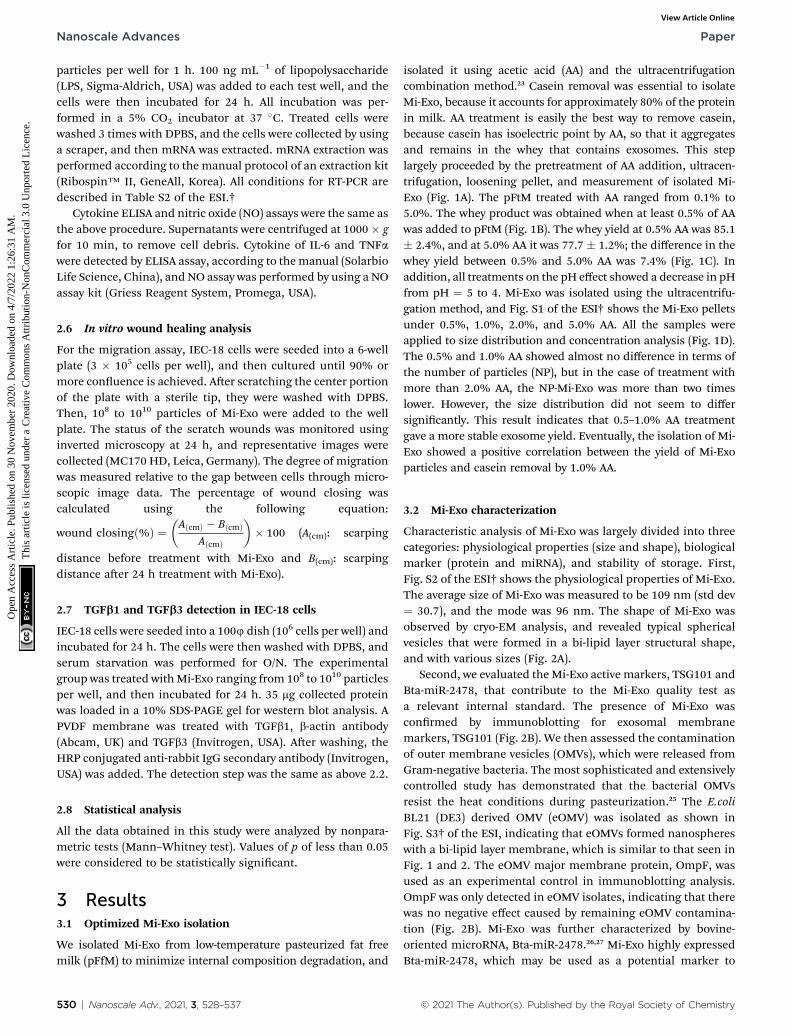
isolated it using acetic acid (AA) and the ultracentrifugationcombination method.23 Casein removal was essential to isolateMi-Exo, because it accounts for approximately 80% of the proteinin milk. AA treatment is easily the best way to remove casein,because casein has isoelectric point by AA, so that it aggregatesand remains in the whey that contains exosomes. This steplargely proceeded by the pretreatment of AA addition, ultracen-trifugation, loosening pellet, and measurement of isolated Mi-Exo (Fig. 1A). The pFtM treated with AA ranged from 0.1% to5.0%. The whey product was obtained when at least 0.5% of AAwas added to pFtM (Fig. 1B). The whey yield at 0.5% AA was 85.1� 2.4%, and at 5.0% AA it was 77.7 � 1.2%; the difference in thewhey yield between 0.5% and 5.0% AA was 7.4% (Fig. 1C). Inaddition, all treatments on the pH effect showed a decrease in pHfrom pH ¼ 5 to 4. Mi-Exo was isolated using the ultracentrifu-gation method, and Fig. S1 of the ESI† shows the Mi-Exo pelletsunder 0.5%, 1.0%, 2.0%, and 5.0% AA. All the samples wereapplied to size distribution and concentration analysis (Fig. 1D).The 0.5% and 1.0% AA showed almost no difference in terms ofthe number of particles (NP), but in the case of treatment withmore than 2.0% AA, the NP-Mi-Exo was more than two timeslower. However, the size distribution did not seem to differsignicantly. This result indicates that 0.5–1.0% AA treatmentgave a more stable exosome yield. Eventually, the isolation of Mi-Exo showed a positive correlation between the yield of Mi-Exoparticles and casein removal by 1.0% AA.
3.2 Mi-Exo characterization
Characteristic analysis of Mi-Exo was largely divided into threecategories: physiological properties (size and shape), biologicalmarker (protein and miRNA), and stability of storage. First,Fig. S2 of the ESI† shows the physiological properties of Mi-Exo.The average size of Mi-Exo was measured to be 109 nm (std dev¼ 30.7), and the mode was 96 nm. The shape of Mi-Exo wasobserved by cryo-EM analysis, and revealed typical sphericalvesicles that were formed in a bi-lipid layer structural shape,and with various sizes (Fig. 2A).
Second, we evaluated the Mi-Exo active markers, TSG101 andBta-miR-2478, that contribute to the Mi-Exo quality test asa relevant internal standard. The presence of Mi-Exo wasconrmed by immunoblotting for exosomal membranemarkers, TSG101 (Fig. 2B). We then assessed the contaminationof outer membrane vesicles (OMVs), which were released fromGram-negative bacteria. The most sophisticated and extensivelycontrolled study has demonstrated that the bacterial OMVsresist the heat conditions during pasteurization.25 The E.coliBL21 (DE3) derived OMV (eOMV) was isolated as shown inFig. S3† of the ESI, indicating that eOMVs formed nanosphereswith a bi-lipid layer membrane, which is similar to that seen inFig. 1 and 2. The eOMV major membrane protein, OmpF, wasused as an experimental control in immunoblotting analysis.OmpF was only detected in eOMV isolates, indicating that therewas no negative effect caused by remaining eOMV contamina-tion (Fig. 2B). Mi-Exo was further characterized by bovine-oriented microRNA, Bta-miR-2478.26,27 Mi-Exo highly expressedBta-miR-2478, which may be used as a potential marker to
© 2021 The Author(s). Published by the Royal Society of Chemistry
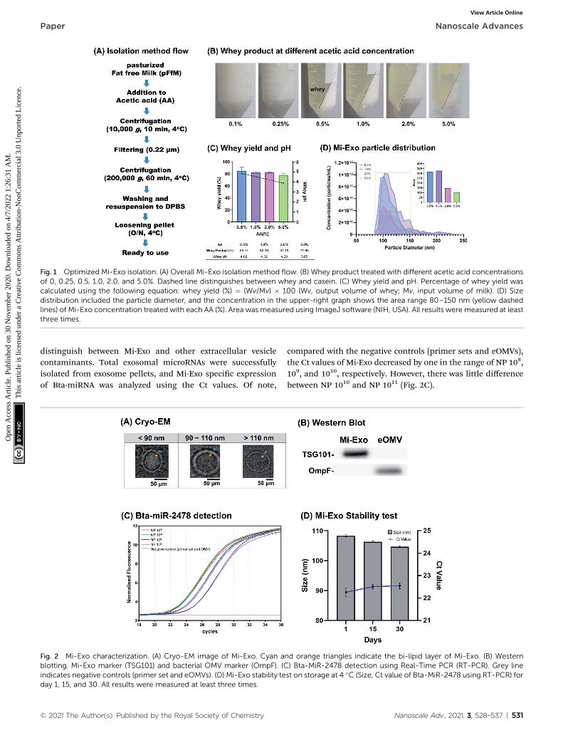
Fig. 1 Optimized Mi-Exo isolation. (A) Overall Mi-Exo isolation method flow. (B) Whey product treated with different acetic acid concentrationsof 0, 0.25, 0.5, 1.0, 2.0, and 5.0%. Dashed line distinguishes between whey and casein. (C) Whey yield and pH. Percentage of whey yield wascalculated using the following equation: whey yield (%) ¼ (Wv/Mv) � 100 (Wv, output volume of whey; Mv, input volume of milk). (D) Sizedistribution included the particle diameter, and the concentration in the upper-right graph shows the area range 80–150 nm (yellow dashedlines) of Mi-Exo concentration treated with each AA (%). Area was measured using ImageJ software (NIH, USA). All results were measured at leastthree times.
Paper Nanoscale Advances
Ope
n A
cces
s A
rtic
le. P
ublis
hed
on 3
0 N
ovem
ber
2020
. Dow
nloa
ded
on 4
/7/2
022
1:26
:31
AM
. T
his
artic
le is
lice
nsed
und
er a
Cre
ativ
e C
omm
ons
Attr
ibut
ion-
Non
Com
mer
cial
3.0
Unp
orte
d L
icen
ce.
View Article Online
distinguish between Mi-Exo and other extracellular vesiclecontaminants. Total exosomal microRNAs were successfullyisolated from exosome pellets, and Mi-Exo specic expressionof Bta-miRNA was analyzed using the Ct values. Of note,
Fig. 2 Mi-Exo characterization. (A) Cryo-EM image of Mi-Exo. Cyan ablotting. Mi-Exo marker (TSG101) and bacterial OMV marker (OmpF). (Cindicates negative controls (primer set and eOMVs). (D) Mi-Exo stability teday 1, 15, and 30. All results were measured at least three times.
© 2021 The Author(s). Published by the Royal Society of Chemistry
compared with the negative controls (primer sets and eOMVs),the Ct values of Mi-Exo decreased by one in the range of NP 108,109, and 1010, respectively. However, there was little differencebetween NP 1010 and NP 1011 (Fig. 2C).
nd orange triangles indicate the bi-lipid layer of Mi-Exo. (B) Western) Bta-MiR-2478 detection using Real-Time PCR (RT-PCR). Grey linest on storage at 4 �C (Size, Ct value of Bta-MiR-2478 using RT-PCR) for
Nanoscale Adv., 2021, 3, 528–537 | 531
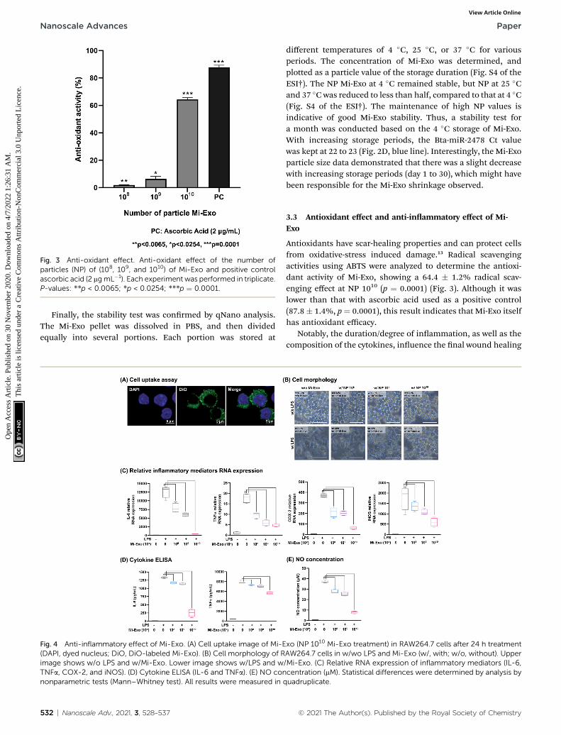
Fig. 3 Anti-oxidant effect. Anti-oxidant effect of the number ofparticles (NP) of (108, 109, and 1010) of Mi-Exo and positive controlascorbic acid (2 mgmL�1). Each experiment was performed in triplicate.P-values: **p < 0.0065; *p < 0.0254; ***p ¼ 0.0001.
Nanoscale Advances Paper
Ope
n A
cces
s A
rtic
le. P
ublis
hed
on 3
0 N
ovem
ber
2020
. Dow
nloa
ded
on 4
/7/2
022
1:26
:31
AM
. T
his
artic
le is
lice
nsed
und
er a
Cre
ativ
e C
omm
ons
Attr
ibut
ion-
Non
Com
mer
cial
3.0
Unp
orte
d L
icen
ce.
View Article Online
Finally, the stability test was conrmed by qNano analysis.The Mi-Exo pellet was dissolved in PBS, and then dividedequally into several portions. Each portion was stored at
Fig. 4 Anti-inflammatory effect of Mi-Exo. (A) Cell uptake image of Mi-E(DAPI, dyed nucleus; DiO, DiO-labeled Mi-Exo). (B) Cell morphology of Rimage shows w/o LPS and w/Mi-Exo. Lower image shows w/LPS and wTNFa, COX-2, and iNOS). (D) Cytokine ELISA (IL-6 and TNFa). (E) NO connonparametric tests (Mann–Whitney test). All results were measured in q
532 | Nanoscale Adv., 2021, 3, 528–537
different temperatures of 4 �C, 25 �C, or 37 �C for variousperiods. The concentration of Mi-Exo was determined, andplotted as a particle value of the storage duration (Fig. S4 of theESI†). The NP Mi-Exo at 4 �C remained stable, but NP at 25 �Cand 37 �C was reduced to less than half, compared to that at 4 �C(Fig. S4 of the ESI†). The maintenance of high NP values isindicative of good Mi-Exo stability. Thus, a stability test fora month was conducted based on the 4 �C storage of Mi-Exo.With increasing storage periods, the Bta-miR-2478 Ct valuewas kept at 22 to 23 (Fig. 2D, blue line). Interestingly, the Mi-Exoparticle size data demonstrated that there was a slight decreasewith increasing storage periods (day 1 to 30), which might havebeen responsible for the Mi-Exo shrinkage observed.
3.3 Antioxidant effect and anti-inammatory effect of Mi-Exo
Antioxidants have scar-healing properties and can protect cellsfrom oxidative-stress induced damage.13 Radical scavengingactivities using ABTS were analyzed to determine the antioxi-dant activity of Mi-Exo, showing a 64.4 � 1.2% radical scav-enging effect at NP 1010 (p ¼ 0.0001) (Fig. 3). Although it waslower than that with ascorbic acid used as a positive control(87.8� 1.4%, p¼ 0.0001), this result indicates that Mi-Exo itselfhas antioxidant efficacy.
Notably, the duration/degree of inammation, as well as thecomposition of the cytokines, inuence the nal wound healing
xo (NP 1010 Mi-Exo treatment) in RAW264.7 cells after 24 h treatmentAW264.7 cells in w/wo LPS and Mi-Exo (w/, with; w/o, without). Upper/Mi-Exo. (C) Relative RNA expression of inflammatory mediators (IL-6,centration (mM). Statistical differences were determined by analysis byuadruplicate.
© 2021 The Author(s). Published by the Royal Society of Chemistry
Paper Nanoscale Advances
Ope
n A
cces
s A
rtic
le. P
ublis
hed
on 3
0 N
ovem
ber
2020
. Dow
nloa
ded
on 4
/7/2
022
1:26
:31
AM
. T
his
artic
le is
lice
nsed
und
er a
Cre
ativ
e C
omm
ons
Attr
ibut
ion-
Non
Com
mer
cial
3.0
Unp
orte
d L
icen
ce.
View Article Online
outcome.11 In addition, it has been reported that LPS inducesthe morphological changes of RAW 264.7 cells, due to reorga-nization of the actin cytoskeleton, and produces inammatorymediators, such as IL-6, TNFa, COX2, iNOS, etc., in immunecells.28
To investigate the anti-inammatory activity of Mi-Exo, theintracellular uptake and immune-mediated morphologicalchanges were explored. Fig. 4A shows the confocal image thatreveals that the DiO-labeled Mi-Exo was internalized intoRAW264.7 cells. The cell morphology was then monitored byoptical microscopy and aer LPS treatment showed a change inthe attened spread cell (Fig. 4B). RAW 264.7 cells (3 � 105 cellsper mL) were incubated in the presence of Mi-Exo for 1 h in 6-well plates. LPS stimulation for 24 h induced lamellipodiaextension and the spreading of cells. However, the Mi-Exotreatment of RAW 264.7 cells prevented LPS-induced morpho-logical changes in a dose-dependent manner. In addition, theabove observation did not relate to RAW264.7 cell viability(Fig. S5A of the ESI†).
In order to conrm the anti-inammatory effects of Mi-Exo,the mRNA expression levels of the major inammatory media-tors IL-6, TNFa, COX2, and iNOS were examined. Fig. 4C resultsshow that all inammatory mediator RNA expression decreasedin a concentration-dependent manner by Mi-Exo. To support
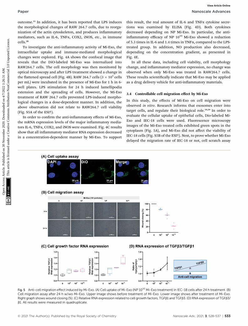
Fig. 5 Anti-cell migration effect induced by Mi-Exo. (A) Cell uptake of Mi-Cell migration assay after 24 h w/wo Mi-Exo. Upper image shows beforRight graph showswound closing (%). (C) Relative RNA expression relatedb1. All results were measured in quadruplicate.
© 2021 The Author(s). Published by the Royal Society of Chemistry
this result, the real amount of IL-6 and TNFa cytokine secre-tions was examined by ELISA (Fig. 4D). Both cytokinesdecreased depending on NP Mi-Exo. In particular, the anti-inammatory efficacy of NP 1010 Mi-Exo showed a reductionof 5.3 times in IL-6 and 1.4 times in TNFa, compared to the LPS-treated group. In addition, NO production also decreased,depending on the concentration gradient, as presented inFig. 4E.
In all these data, including cell viability, cell morphologychange, and inammatory mediator expression, no change wasobserved when only Mi-Exo was treated in RAW264.7 cells.These results scientically indicate that Mi-Exo may be appliedas a drug delivery vehicle for anti-inammatory materials.
3.4 Controllable cell migration effect by Mi-Exo
In this study, the effects of Mi-Exo on cell migration wereobserved in vitro. Research informs that exosomes enter intotarget cells, and regulate their biological role.19,29 In order toevaluate the cellular uptake of epithelial cells, Dio-labeled Mi-Exo and IEC-18 cells were used. Fluorescence microscopyimages of the Mi-Exo treated cells exhibited green spots in thecytoplasm (Fig. 5A), and Mi-Exo did not affect the viability ofIEC-18 cells (Fig. S5B of the ESI†). Next, to prove whether Mi-Exodelayed the migration rate of IEC-18 or not, cell scratch assay
Exo (NP 1010 Mi-Exo treatment) in IEC-18 cells after 24 h treatment. (B)e treatment of Mi-Exo. Lower image shows after treatment of Mi-Exo.to cell growth factors, TGFb1 and TGFb3. (D) RNA expression of TGFb3/
Nanoscale Adv., 2021, 3, 528–537 | 533
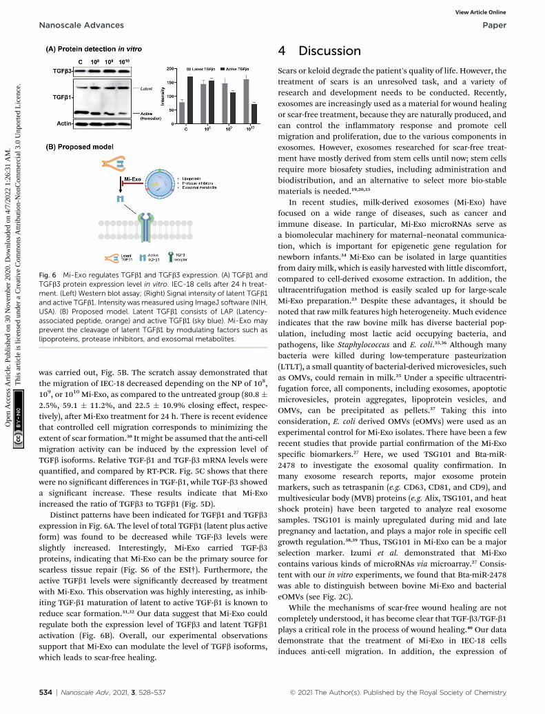
Fig. 6 Mi-Exo regulates TGFb1 and TGFb3 expression. (A) TGFb1 andTGFb3 protein expression level in vitro. IEC-18 cells after 24 h treat-ment. (Left) Western blot assay; (Right) Signal intensity of latent TGFb1and active TGFb1. Intensity was measured using ImageJ software (NIH,USA). (B) Proposed model. Latent TGFb1 consists of LAP (Latency-associated peptide, orange) and active TGFb1 (sky blue). Mi-Exo mayprevent the cleavage of latent TGFb1 by modulating factors such aslipoproteins, protease inhibitors, and exosomal metabolites.
Nanoscale Advances Paper
Ope
n A
cces
s A
rtic
le. P
ublis
hed
on 3
0 N
ovem
ber
2020
. Dow
nloa
ded
on 4
/7/2
022
1:26
:31
AM
. T
his
artic
le is
lice
nsed
und
er a
Cre
ativ
e C
omm
ons
Attr
ibut
ion-
Non
Com
mer
cial
3.0
Unp
orte
d L
icen
ce.
View Article Online
was carried out, Fig. 5B. The scratch assay demonstrated thatthe migration of IEC-18 decreased depending on the NP of 108,109, or 1010 Mi-Exo, as compared to the untreated group (80.8 �2.5%, 59.1 � 11.2%, and 22.5 � 10.9% closing effect, respec-tively), aer Mi-Exo treatment for 24 h. There is recent evidencethat controlled cell migration corresponds to minimizing theextent of scar formation.30 It might be assumed that the anti-cellmigration activity can be induced by the expression level ofTGFb isoforms. Relative TGF-b1 and TGF-b3 mRNA levels werequantied, and compared by RT-PCR. Fig. 5C shows that therewere no signicant differences in TGF-b1, while TGF-b3 showeda signicant increase. These results indicate that Mi-Exoincreased the ratio of TGFb3 to TGFb1 (Fig. 5D).
Distinct patterns have been indicated for TGFb1 and TGFb3expression in Fig. 6A. The level of total TGFb1 (latent plus activeform) was found to be decreased while TGF-b3 levels wereslightly increased. Interestingly, Mi-Exo carried TGF-b3proteins, indicating that Mi-Exo can be the primary source forscarless tissue repair (Fig. S6 of the ESI†). Furthermore, theactive TGFb1 levels were signicantly decreased by treatmentwith Mi-Exo. This observation was highly interesting, as inhib-iting TGF-b1 maturation of latent to active TGF-b1 is known toreduce scar formation.31,32 Our data suggest that Mi-Exo couldregulate both the expression level of TGFb3 and latent TGFb1activation (Fig. 6B). Overall, our experimental observationssupport that Mi-Exo can modulate the level of TGFb isoforms,which leads to scar-free healing.
534 | Nanoscale Adv., 2021, 3, 528–537
4 Discussion
Scars or keloid degrade the patient's quality of life. However, thetreatment of scars is an unresolved task, and a variety ofresearch and development needs to be conducted. Recently,exosomes are increasingly used as a material for wound healingor scar-free treatment, because they are naturally produced, andcan control the inammatory response and promote cellmigration and proliferation, due to the various components inexosomes. However, exosomes researched for scar-free treat-ment have mostly derived from stem cells until now; stem cellsrequire more biosafety studies, including administration andbiodistribution, and an alternative to select more bio-stablematerials is needed.19,20,33
In recent studies, milk-derived exosomes (Mi-Exo) havefocused on a wide range of diseases, such as cancer andimmune disease. In particular, Mi-Exo microRNAs serve asa biomolecular machinery for maternal–neonatal communica-tion, which is important for epigenetic gene regulation fornewborn infants.34 Mi-Exo can be isolated in large quantitiesfrom dairy milk, which is easily harvested with little discomfort,compared to cell-derived exosome extraction. In addition, theultracentrifugation method is easily scaled up for large-scaleMi-Exo preparation.23 Despite these advantages, it should benoted that rawmilk features high heterogeneity. Much evidenceindicates that the raw bovine milk has diverse bacterial pop-ulation, including most lactic acid occupying bacteria, andpathogens, like Staphylococcus and E. coli.35,36 Although manybacteria were killed during low-temperature pasteurization(LTLT), a small quantity of bacterial-derived microvesicles, suchas OMVs, could remain in milk.25 Under a specic ultracentri-fugation force, all components, including exosomes, apoptoticmicrovesicles, protein aggregates, lipoprotein vesicles, andOMVs, can be precipitated as pellets.37 Taking this intoconsideration, E. coli derived OMVs (eOMVs) were used as anexperimental control for Mi-Exo isolates. There have been a fewrecent studies that provide partial conrmation of the Mi-Exospecic biomarkers.27 Here, we used TSG101 and Bta-miR-2478 to investigate the exosomal quality conrmation. Inmany exosome research reports, major exosome proteinmarkers, such as tetraspanin (e.g. CD63, CD81, and CD9), andmultivesicular body (MVB) proteins (e.g. Alix, TSG101, and heatshock protein) have been targeted to analyze real exosomesamples. TSG101 is mainly upregulated during mid and latepregnancy and lactation, and plays a major role in specic cellgrowth regulation.38,39 Thus, TSG101 in Mi-Exo can be a majorselection marker. Izumi et al. demonstrated that Mi-Exocontains various kinds of microRNAs via microarray.27 Consis-tent with our in vitro experiments, we found that Bta-miR-2478was able to distinguish between bovine Mi-Exo and bacterialeOMVs (see Fig. 2C).
While the mechanisms of scar-free wound healing are notcompletely understood, it has become clear that TGF-b3/TGF-b1plays a critical role in the process of wound healing.40 Our datademonstrate that the treatment of Mi-Exo in IEC-18 cellsinduces anti-cell migration. In addition, the expression of
© 2021 The Author(s). Published by the Royal Society of Chemistry
Paper Nanoscale Advances
Ope
n A
cces
s A
rtic
le. P
ublis
hed
on 3
0 N
ovem
ber
2020
. Dow
nloa
ded
on 4
/7/2
022
1:26
:31
AM
. T
his
artic
le is
lice
nsed
und
er a
Cre
ativ
e C
omm
ons
Attr
ibut
ion-
Non
Com
mer
cial
3.0
Unp
orte
d L
icen
ce.
View Article Online
TGFb3 was elevated in response to Mi-Exo treatment, but thelevel of TGFb1 remained unchanged (Fig. 5B and C). Manystudies have reported that wound healing mainly follows theTGFb/Smad signaling pathway. Smad protein acts as a keytranscription factor of TGFb signaling, and plays a different roleof (1) receptor-activated Smad (Smad1, Smad2, Smad3, Smad5,and Smad8); (2) common mediator Smad (Smad4); and (3)inhibitory Smad (Smad6 and Smad7).41 Among these, Smad3protein is phosphorylated due to the activation of TGFbRI andTGFbRII, and phosphorylated Smad3 plays an important role incell growth and ECM formation. However, TGFb3 lowers theexpression level of Smad3, and increases the expression level ofSmad7, which decrease cell growth and ECM formation.2,41 Also,TGFb3 degrades collagen by promoting matrix metalloprotease-9 (MMP-9) expression, and this is speculated to slowly form anintercellular matrix between cells.42 Recent studies have sug-gested that TGFb1 promotes collagen synthesis, while othershave indicated that TGFb3 suppresses collagen deposition.These controversial results may be attributed to the intracel-lular molecular ratio of TGF-b3/TGF-b1 during different stagesof wound healing. Therefore, based on the ndings in thisstudy, we suggest that appropriate in vivo studies should beundertaken in the future to show the effectiveness of Mi-Exo inscarless modulation.
5 Conclusion
In summary, this study focused on Mi-Exo that is capable ofscar-free healing. First, we analyzed the isolation of Mi-Exo andanalyzed its characteristics: physiological properties (size andshape), biological markers (protein and miRNA), and stabilityon storage. Next, we demonstrated scar-free healing based onthree properties: antioxidant, anti-inammatory, andmolecularbalance of TGFb3 and TGFb1 through mRNA and proteinexpression. These results indicate that Mi-Exo can be utilized asa fascinating material that can minimize various scars orkeloids, including skin tissue damage, abrasion, acne extru-sion, and skin incision by surgery. Thus, Mi-Exo is expected toshow potential as a treatment material for various applications.
Author contributions
Investigation and data analysis, G. A and J.-Y. A.; experimentsdesign and result analysis, J.-Y. A. and Y.-H. K.; all authors haveread and agreed to the published version of the manuscript.
Conflicts of interest
The authors declare no conict of interest.
Acknowledgements
This work was supported by the National Research Foundationof Korea (NRF) grant funded by the Korea government (MEST)(NRF-2019R1A2C1010860) and Basic Science Research Programthrough the National Research Foundation of Korea (NRF)funded by the Ministry of Education (2020R1A6A1A06046235).
© 2021 The Author(s). Published by the Royal Society of Chemistry
References
1 S. Guo and L. A. Dipietro, J. Dent. Res., 2010, 89, 219–229.2 A. Han, B. Bandyopadhyay, P. Jayaprakash, I. Lua, D. Sahu,M. Chen, D. T. Woodley and W. Li, Biol. Open, 2012, 1,1169–1177.
3 K. P. Kras, Organogenesis, 2010, 6, 225–233.4 D. S. Steinbrech, B. J. Mehrara, D. Chau, N. M. Rowe,G. Chin, T. Lee, P. B. Saadeh, G. K. Gittes andM. T. Longaker, Ann. Plast. Surg., 1999, 42, 514–519;discussion 519–520.
5 M. Tang, W. Bian, L. Cheng, L. Zhang, R. Jin, W. Wang andY. Zhang, Int. J. Mol. Med., 2018, 41, 1487–1499.
6 J. P. Andrews, J. Marttala, E. Macarak, J. Rosenbloom andJ. Uitto, Matrix Biol., 2016, 51, 37–46.
7 Z. C. Hu, B. Tang, D. Guo, J. Zhang, Y. Y. Liang, D. Ma andJ. Y. Zhu, Clin. Exp. Dermatol., 2014, 39, 822–828.
8 M. K. Lichtman, M. Otero-Vinas and V. Falanga, WoundRepair Regen., 2016, 24, 215–222.
9 S. M. Karppinen, R. Heljasvaara, D. Gullberg, K. Tasanen andT. Pihlajaniemi, F1000Research, 2019, 8.
10 T. J. Koh and L. A. DiPietro, Expert Rev. Mol. Med., 2011, 13,e23.
11 S. A. Eming, T. Krieg and J. M. Davidson, J. Invest. Dermatol.,2007, 127, 514–525.
12 S. K. Shukla, A. K. Sharma, V. Gupta andM. H. Yashavarddhan, J. Tissue Viability, 2019, 28, 218–222.
13 S. D. Fitzmaurice, R. K. Sivamani and R. R. Isseroff, SkinPharmacol. Physiol., 2011, 24, 113–126.
14 C. Thery, K. W. Witwer, E. Aikawa, M. J. Alcaraz,J. D. Anderson, R. Andriantsitohaina, A. Antoniou, T. Arab,F. Archer, G. K. Atkin-Smith, D. C. Ayre, J. M. Bach,D. Bachurski, H. Baharvand, L. Balaj, S. Baldacchino,N. N. Bauer, A. A. Baxter, M. Bebawy, C. Beckham,A. Bedina Zavec, A. Benmoussa, A. C. Berardi, P. Bergese,E. Bielska, C. Blenkiron, S. Bobis-Wozowicz, E. Boilard,W. Boireau, A. Bongiovanni, F. E. Borras, S. Bosch,C. M. Boulanger, X. Breakeeld, A. M. Breglio,M. A. Brennan, D. R. Brigstock, A. Brisson,M. L. Broekman, J. F. Bromberg, P. Bryl-Gorecka, S. Buch,A. H. Buck, D. Burger, S. Busatto, D. Buschmann,B. Bussolati, E. I. Buzas, J. B. Byrd, G. Camussi,D. R. Carter, S. Caruso, L. W. Chamley, Y. T. Chang,C. Chen, S. Chen, L. Cheng, A. R. Chin, A. Clayton,S. P. Clerici, A. Cocks, E. Cocucci, R. J. Coffey, A. Cordeiro-da-Silva, Y. Couch, F. A. Coumans, B. Coyle, R. Crescitelli,M. F. Criado, C. D'Souza-Schorey, S. Das, A. DattaChaudhuri, P. de Candia, E. F. De Santana, O. De Wever,H. A. Del Portillo, T. Demaret, S. Deville, A. Devitt,B. Dhondt, D. Di Vizio, L. C. Dieterich, V. Dolo,A. P. Dominguez Rubio, M. Dominici, M. R. Dourado,T. A. Driedonks, F. V. Duarte, H. M. Duncan,R. M. Eichenberger, K. Ekstrom, S. El Andaloussi, C. Elie-Caille, U. Erdbrugger, J. M. Falcon-Perez, F. Fatima,J. E. Fish, M. Flores-Bellver, A. Forsonits, A. Frelet-Barrand,F. Fricke, G. Fuhrmann, S. Gabrielsson, A. Gamez-Valero,
Nanoscale Adv., 2021, 3, 528–537 | 535

Nanoscale Advances Paper
Ope
n A
cces
s A
rtic
le. P
ublis
hed
on 3
0 N
ovem
ber
2020
. Dow
nloa
ded
on 4
/7/2
022
1:26
:31
AM
. T
his
artic
le is
lice
nsed
und
er a
Cre
ativ
e C
omm
ons
Attr
ibut
ion-
Non
Com
mer
cial
3.0
Unp
orte
d L
icen
ce.
View Article Online
C. Gardiner, K. Gartner, R. Gaudin, Y. S. Gho, B. Giebel,C. Gilbert, M. Gimona, I. Giusti, D. C. Goberdhan,A. Gorgens, S. M. Gorski, D. W. Greening, J. C. Gross,A. Gualerzi, G. N. Gupta, D. Gustafson, A. Handberg,R. A. Haraszti, P. Harrison, H. Hegyesi, A. Hendrix,A. F. Hill, F. H. Hochberg, K. F. Hoffmann, B. Holder,H. Holthofer, B. Hosseinkhani, G. Hu, Y. Huang, V. Huber,S. Hunt, A. G. Ibrahim, T. Ikezu, J. M. Inal, M. Isin,A. Ivanova, H. K. Jackson, S. Jacobsen, S. M. Jay,M. Jayachandran, G. Jenster, L. Jiang, S. M. Johnson,J. C. Jones, A. Jong, T. Jovanovic-Talisman, S. Jung,R. Kalluri, S. I. Kano, S. Kaur, Y. Kawamura, E. T. Keller,D. Khamari, E. Khomyakova, A. Khvorova, P. Kierulf,K. P. Kim, T. Kislinger, M. Klingeborn, D. J. Klinke 2nd,M. Kornek, M. M. Kosanovic, A. F. Kovacs, E. M. Kramer-Albers, S. Krasemann, M. Krause, I. V. Kurochkin,G. D. Kusuma, S. Kuypers, S. Laitinen, S. M. Langevin,L. R. Languino, J. Lannigan, C. Lasser, L. C. Laurent,G. Lavieu, E. Lazaro-Ibanez, S. Le Lay, M. S. Lee,Y. X. F. Lee, D. S. Lemos, M. Lenassi, A. Leszczynska,I. T. Li, K. Liao, S. F. Libregts, E. Ligeti, R. Lim, S. K. Lim,A. Line, K. Linnemannstons, A. Llorente, C. A. Lombard,M. J. Lorenowicz, A. M. Lorincz, J. Lotvall, J. Lovett,M. C. Lowry, X. Loyer, Q. Lu, B. Lukomska, T. R. Lunavat,S. L. Maas, H. Malhi, A. Marcilla, J. Mariani, J. Mariscal,E. S. Martens-Uzunova, L. Martin-Jaular, M. C. Martinez,V. R. Martins, M. Mathieu, S. Mathivanan, M. Maugeri,L. K. McGinnis, M. J. McVey, D. G. Meckes Jr,K. L. Meehan, I. Mertens, V. R. Minciacchi, A. Moller,M. Moller Jorgensen, A. Morales-Kastresana, J. Morhayim,F. Mullier, M. Muraca, L. Musante, V. Mussack,D. C. Muth, K. H. Myburgh, T. Najrana, M. Nawaz,I. Nazarenko, P. Nejsum, C. Neri, T. Neri, R. Nieuwland,L. Nimrichter, J. P. Nolan, E. N. Nolte-'t Hoen, N. NorenHooten, L. O'Driscoll, T. O'Grady, A. O'Loghlen, T. Ochiya,M. Olivier, A. Ortiz, L. A. Ortiz, X. Osteikoetxea,O. Ostergaard, M. Ostrowski, J. Park, D. M. Pegtel,H. Peinado, F. Perut, M. W. Pfaffl, D. G. Phinney,B. C. Pieters, R. C. Pink, D. S. Pisetsky, E. Pogge vonStrandmann, I. Polakovicova, I. K. Poon, B. H. Powell,I. Prada, L. Pulliam, P. Quesenberry, A. Radeghieri,R. L. Raffai, S. Raimondo, J. Rak, M. I. Ramirez, G. Raposo,M. S. Rayyan, N. Regev-Rudzki, F. L. Ricklefs,P. D. Robbins, D. D. Roberts, S. C. Rodrigues, E. Rohde,S. Rome, K. M. Rouschop, A. Rughetti, A. E. Russell, P. Saa,S. Sahoo, E. Salas-Huenuleo, C. Sanchez, J. A. Saugstad,M. J. Saul, R. M. Schiffelers, R. Schneider, T. H. Schoyen,A. Scott, E. Shahaj, S. Sharma, O. Shatnyeva, F. Shekari,G. V. Shelke, A. K. Shetty, K. Shiba, P. R. Siljander,A. M. Silva, A. Skowronek, O. L. Snyder 2nd, R. P. Soares,B. W. Sodar, C. Soekmadji, J. Sotillo, P. D. Stahl,W. Stoorvogel, S. L. Stott, E. F. Strasser, S. Swi, H. Tahara,M. Tewari, K. Timms, S. Tiwari, R. Tixeira, M. Tkach,W. S. Toh, R. Tomasini, A. C. Torrecilhas, J. P. Tosar,V. Toxavidis, L. Urbanelli, P. Vader, B. W. van Balkom,S. G. van der Grein, J. Van Deun, M. J. van Herwijnen,K. Van Keuren-Jensen, G. van Niel, M. E. van Royen,
536 | Nanoscale Adv., 2021, 3, 528–537
A. J. van Wijnen, M. H. Vasconcelos, I. J. Vechetti Jr,T. D. Veit, L. J. Vella, E. Velot, F. J. Verweij, B. Vestad,J. L. Vinas, T. Visnovitz, K. V. Vukman, J. Wahlgren,D. C. Watson, M. H. Wauben, A. Weaver, J. P. Webber,V. Weber, A. M. Wehman, D. J. Weiss, J. A. Welsh,S. Wendt, A. M. Wheelock, Z. Wiener, L. Witte, J. Wolfram,A. Xagorari, P. Xander, J. Xu, X. Yan, M. Yanez-Mo, H. Yin,Y. Yuana, V. Zappulli, J. Zarubova, V. Zekas, J. Y. Zhang,Z. Zhao, L. Zheng, A. R. Zheutlin, A. M. Zickler,P. Zimmermann, A. M. Zivkovic, D. Zocco and E. K. Zuba-Surma, J. Extracell. Vesicles, 2018, 7, 1535750.
15 S. Sekhon, G. Ahn., G. Park, D. Park, S. Lee, J. Ahn andY. Kim, J. Toxicol. Environ. Health Sci., 2019, 11, 85–93.
16 S. A. Melo, L. B. Luecke, C. Kahlert, A. F. Fernandez,S. T. Gammon, J. Kaye, V. S. LeBleu, E. A. Mittendorf,J. Weitz, N. Rahbari, C. Reissfelder, C. Pilarsky,M. F. Fraga, D. Piwnica-Worms and R. Kalluri, Nature,2015, 523, 177–182.
17 C. de la Torre Gomez, R. V. Goreham, J. J. Bech Serra,T. Nann and M. Kussmann, Front. Genet., 2018, 9, 92.
18 G. Raposo and W. Stoorvogel, J. Cell Biol., 2013, 200, 373–383.
19 L. Wang, L. Hu, X. Zhou, Z. Xiong, C. Zhang,H. M. A. Shehada, B. Hu, J. Song and L. Chen, Sci. Rep.,2017, 7, 13321.
20 P. Hu, Q. Yang, Q. Wang, C. Shi, D. Wang, U. Armato,I. D. Pra and A. Chiarini, Int. J. Burns Trauma, 2019, 7, 38.
21 M. Yang, D. Song, X. Cao, R. Wu, B. Liu, W. Ye, J. Wu andX. Yue, Food Res. Int., 2017, 92, 17–25.
22 S. Manca, B. Upadhyaya, E. Mutai, A. T. Desaulniers,R. A. Cederberg, B. R. White and J. Zempleni, Sci. Rep.,2018, 8, 11321.
23 M. Somiya, Y. Yoshioka and T. Ochiya, J. Extracell. Vesicles,2018, 7, 1440132.
24 M. Ozgen, R. N. Reese, A. Z. Tulio Jr, J. C. Scheerens andA. R. Miller, J. Agric. Food Chem., 2006, 54, 1151–1157.
25 W. D. McCaig, A. Koller and D. G. Thanassi, J. Bacteriol.,2013, 195, 1120–1132.
26 M. J. C. van Herwijnen, T. A. P. Driedonks, B. L. Snoek,A. M. T. Kroon, M. Kleinjan, R. Jorritsma, C. M. J. Pieterse,E. Hoen and M. H. M. Wauben, Front. Nutr., 2018, 5, 81.
27 H. Izumi, M. Tsuda, Y. Sato, N. Kosaka, T. Ochiya,H. Iwamoto, K. Namba and Y. Takeda, J. Dairy Sci., 2015,98, 2920–2933.
28 R. J. Schutte, A. Parisi-Amon and W. M. Reichert, J. Biomed.Mater. Res., Part A, 2009, 88, 128–139.
29 A. Shabbir, A. Cox, L. Rodriguez-Menocal, M. Salgado andE. Van Badiavas, Stem Cells Dev., 2015, 24, 1635–1647.
30 N. Wang, F. Yao, K. Li, L. Zhang, G. Yin, M. Du and B. Wu,Int. J. Mol. Med., 2017, 39, 783–790.
31 D. J. Grainger, Arterioscler., Thromb., Vasc. Biol., 2004, 24,399–404.
32 S. Kojima, P. C. Harpel and D. B. Riin, J. Cell Biol., 1991,113, 1439–1445.
33 A. D. F. Ferreira and D. A. Gomes, Bioengineering, 2018, 6, 4.
© 2021 The Author(s). Published by the Royal Society of Chemistry
Paper Nanoscale Advances
Ope
n A
cces
s A
rtic
le. P
ublis
hed
on 3
0 N
ovem
ber
2020
. Dow
nloa
ded
on 4
/7/2
022
1:26
:31
AM
. T
his
artic
le is
lice
nsed
und
er a
Cre
ativ
e C
omm
ons
Attr
ibut
ion-
Non
Com
mer
cial
3.0
Unp
orte
d L
icen
ce.
View Article Online
34 J. Zempleni, A. Aguilar-Lozano, M. Sadri, S. Sukreet,S. Manca, D. Wu, F. Zhou and E. Mutai, J. Nutr., 2017, 147,3–10.
35 L. Quigley, R. McCarthy, O. O'Sullivan, T. P. Beresford,G. F. Fitzgerald, R. P. Ross, C. Stanton and P. D. Cotter, J.Dairy Sci., 2013, 96, 4928–4937.
36 V. Lafarge, J. C. Ogier, V. Girard, V. Maladen, J. Y. Leveau,A. Gruss and A. Delacroix-Buchet, Appl. Environ. Microbiol.,2004, 70, 5644–5650.
37 D. D. Taylor and S. Shah, Methods, 2015, 87, 3–10.
© 2021 The Author(s). Published by the Royal Society of Chemistry
38 K. B. Oh, M. J. Stanton, W. W. West, G. L. Todd andK. U. Wagner, Oncogene, 2007, 26, 5950–5959.
39 K. U. Wagner, A. Krempler, Y. Qi, K. Park, M. D. Henry,A. A. Triplett, G. Riedlinger, I. E. Rucker andL. Hennighausen, Mol. Cell. Biol., 2003, 23, 150–162.
40 R. W. D. Gilbert, M. K. Vickaryous and A. M. Viloria-Petit, J.Dev. Biol., 2016, 4, 21.
41 K. Jiang, G. Chun, Z. Wang, Q. Du, A. Wang and Y. Xiong,Mol. Med. Rep., 2016, 13, 3567–3573.
42 R. Hosokawa, K. Nonaka, M. Morifuji, L. Shum andM. Ohishi, J. Dent. Res., 2003, 82, 558–564.
Nanoscale Adv., 2021, 3, 528–537 | 537