Embed Size (px)
Citation preview
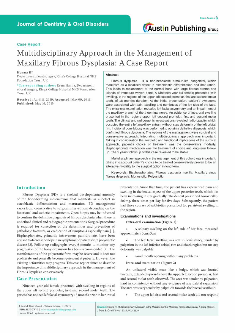
Citation: Hanna R. Multidisciplinary Approach in the Management of Maxillary Fibrous Dysplasia: A Case Report. J Dent & Oral Disord. 2019; 5(1): 1110.
J Dent & Oral Disord - Volume 5 Issue 1 - 2019ISSN: 2572-7710 | www.austinpublishinggroup.com Hanna. © All rights are reserved
Journal of Dentistry & Oral DisordersOpen Access
Abstract
Fibrous dysplasia is a non-neoplastic tumour-like congenital, which manifests as a localised defect in osteoblastic differentiation and maturation. This leads to replacement of the normal bone with large fibrous stroma and islands of immature woven bone. A Nineteen-year-old female presented with swelling, in the regions of the upper left second premolar, first and second molar teeth, of 18 months duration. At the initial presentation, patient’s symptoms were associated with pain, swelling and numbness of the left side of the face. The extra-oral examination revealed left facial asymmetry and an impairment of the maxillary branch of the trigeminal nerve. An evidence of intra-oral swelling presented in the regions upper left second premolar, first and second molar teeth, The clinical and radiographic investigations revealed radio-opacity, which occupied the entire left maxillary antram without step deformity of the left orbital rim. Incisional bony biopsy was performed to obtain a definitive diagnosis, which confirmed fibrous dysplasia. The options of the management were surgical and conservative approach. Integrating multidisciplinary approach was important. Taking in consideration the aesthetic and functional implications of the surgical approach, patient’s choice of treatment was the conservative modality. Bisphosphonate medication was the treatment of choice and long-term follow-up. The 5 years follow up of this case revealed to be stable.
Multidisciplinary approach in the management of this cohort was important, taking into account patient’s choice to be treated conservatively proven to be an alterative modality to the surgical option in long term.
Keywords: Bisphosphonates; Fibrous dysplasia maxilla; Maxillary sinus fibrous dysplasia; Monostotic; Polyostotic
IntroductionFibrous Dysplasia (FD) is a skeletal developmental anomaly
of the bone-forming mesenchyme that manifests as a defect in osteoblastic differentiation and maturation. FD management varies from conservative to surgical intervention, depending on the functional and esthetic impairments. Open biopsy may be indicated to confirm the definitive diagnosis of fibrous dysplasia when there is undefined clinical and radiographic presentations. Surgical procedure is required for correction of the deformities and prevention of pathologic fractures, or eradication of symptoms especially pain [1]. Bisphosphonates, primarily intravenous pamidronate, have been utilised to decrease bone pain in symptomatic patients with polyostotic disease [2]. Follow-up radiographs every 6 months to monitor any progression of the bony expansion has been recommended [2]. The manifestations of the polyostotic form may be severe and it does not proliferate and generally becomes quiescent at puberty. However, the existing deformities may progress. This case report aimed to describe the importance of multidisciplinary approach in the management of Fibrous Dysplasia conservatively.
Case PresentationNineteen-year-old female presented with swelling in regions of
the upper left second premolar, first and second molar teeth. The patient has noticed left facial asymmetry 18 months prior to her initial
Case Report
Multidisciplinary Approach in the Management of Maxillary Fibrous Dysplasia: A Case ReportHanna R*Department of oral surgery, King’s College Hospital NHS Foundation Trust, UK
*Corresponding author: Reem Hanna, Department of oral surgery, King’s College Hospital NHS Foundation Trust, UK
Received: April 13, 2019; Accepted: May 09, 2019; Published: May 16, 2019
presentation. Since that time, the patient has experienced pain and swelling in the buccal aspect of the upper posterior teeth, which has been increasing in size gradually. The dentist prescribed Amoxicillin, 500mg, three times per day for five days. Subsequently, the patient had three courses of antibiotics prescribed for persistent swelling in the region.
Examinations and investigationsExtra-oral examination (Figure 1)
• A solitary swelling on the left side of her face, measured approximately 3cm×3cm
• The left facial swelling was soft in consistency, tender by palpation in the left inferior orbital rim and cheek region but no step deformity was palpable.
• Good mouth opening without any problems.
Intra-oral examination (Figure 2)
An unilateral visible mass like a bulge, which was located buccally, extended upward above the upper left second premolar, first and second molar teeth observed. The area was tender by palpation, hard in consistency without any evidence of any palatal expansion. The area was very tender by palpation towards the buccal vestibule.
• The upper left first and second molar teeth did not respond
J Dent & Oral Disord 5(1): id1110 (2019) - Page - 02
Hanna R Austin Publishing Group
Submit your Manuscript | www.austinpublishinggroup.com
to Ethyl chloride. They were not tender on percussion.
• The mucosa was pinkish in colour without evidence of dehiscence.
• The palate and floor of the mouth were normal with no pathology.
• Neurological examination:
• The facial nerve were intact.
• Maxillary branch of the trigeminal nerve was impaired, numbness of the left side of the face along the distribution of the maxillary branch of the trigeminal nerve. The patient did not respond to sharp and soft stimuli.
• Full eye movements with no evidence of diplopia
• Imaging investigations
• Ultrasound was undertaken to exclude any foreign bodies.
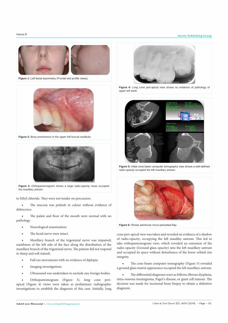
• Orthopantomogram (Figure 3), long cone peri-apical (Figure 4) views were taken as preliminary radiographic investigations to establish the diagnosis of this case. Initially, long
cone peri-apical view was taken and revealed an evidence of a shadow of radio-opacity, occupying the left maxillay antrum. This led to take orthopantomogram view, which revealed an extension of the radio-opacity (Ground-glass opacity) into the left maxillary antrum and occupied its space without disturbance of the lower orbital rim integrity.
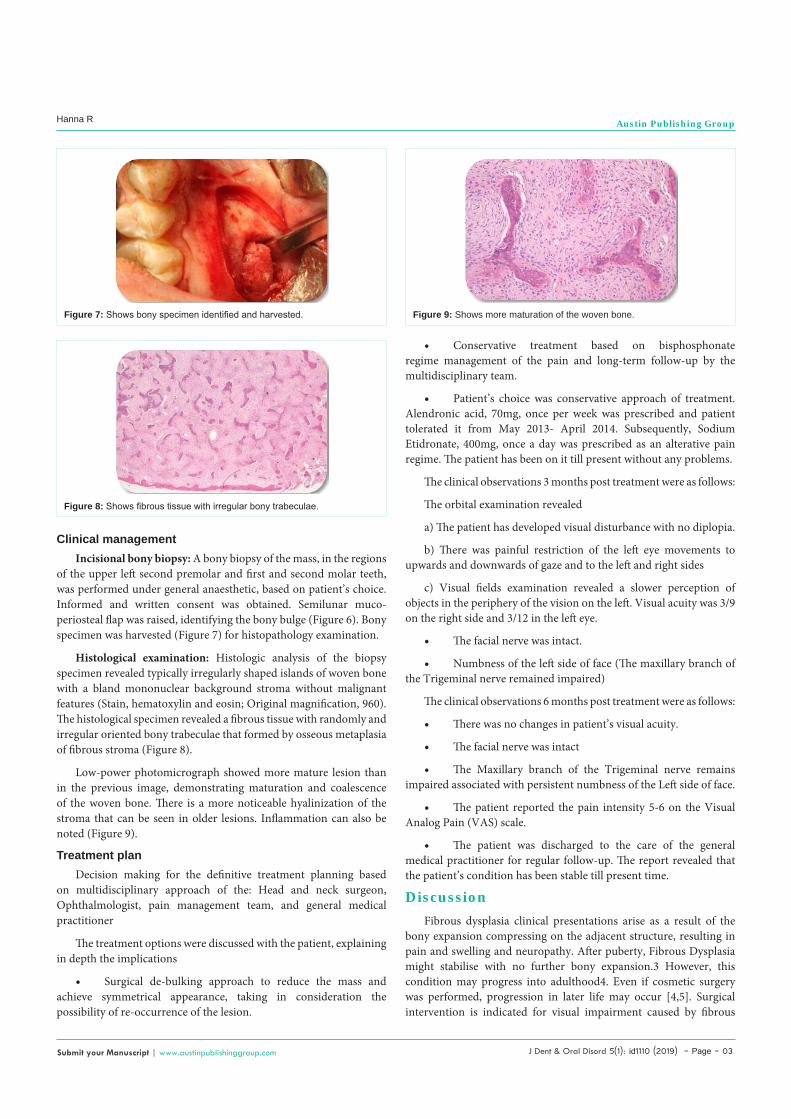
• The cone beam computer tomography (Figure 5) revealed a ground glass matrix appearance occupied the left maxillary antrum.
• The differential diagnoses were as follows; fibrous dysplasia, intra-osseous meningioma, Paget’s disease, or giant cell tumour. The decision was made for incisional bony biopsy to obtain a definitive diagnosis.
Figure 1: Left facial asymmetry (Frontal and profile views).
Figure 2: Bony prominence in the upper left buccal vestibule.
Figure 3: Orthopantomogram shows a large radio-opacity mass occupied the maxillary antram.
Figure 4: Long cone peri-apical view shows no evidence of pathology of upper left teeth.
Figure 5: Initial cone beam computer tomography view shows a well-defined radio-opacity occupied the left maxillary antram.
Figure 6: Shows semilunar muco-periosteal flap.
J Dent & Oral Disord 5(1): id1110 (2019) - Page - 03
Hanna R Austin Publishing Group
Submit your Manuscript | www.austinpublishinggroup.com
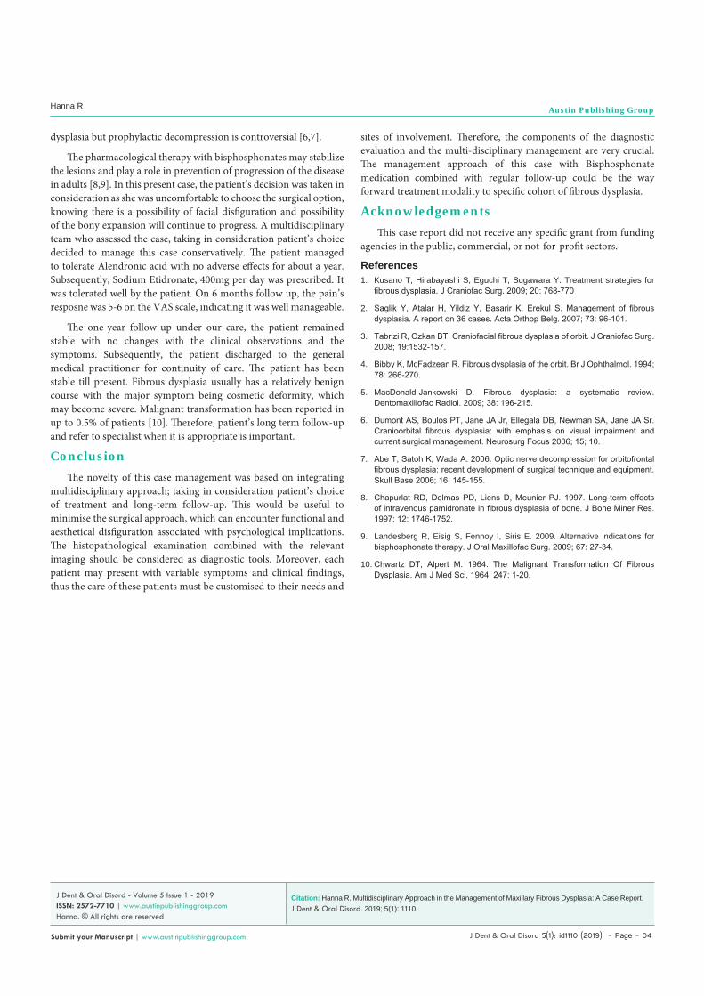
Clinical management Incisional bony biopsy: A bony biopsy of the mass, in the regions
of the upper left second premolar and first and second molar teeth, was performed under general anaesthetic, based on patient’s choice. Informed and written consent was obtained. Semilunar muco-periosteal flap was raised, identifying the bony bulge (Figure 6). Bony specimen was harvested (Figure 7) for histopathology examination.
Histological examination: Histologic analysis of the biopsy specimen revealed typically irregularly shaped islands of woven bone with a bland mononuclear background stroma without malignant features (Stain, hematoxylin and eosin; Original magnification, 960). The histological specimen revealed a fibrous tissue with randomly and irregular oriented bony trabeculae that formed by osseous metaplasia of fibrous stroma (Figure 8).
Low-power photomicrograph showed more mature lesion than in the previous image, demonstrating maturation and coalescence of the woven bone. There is a more noticeable hyalinization of the stroma that can be seen in older lesions. Inflammation can also be noted (Figure 9).
Treatment planDecision making for the definitive treatment planning based
on multidisciplinary approach of the: Head and neck surgeon, Ophthalmologist, pain management team, and general medical practitioner
The treatment options were discussed with the patient, explaining in depth the implications
• Surgical de-bulking approach to reduce the mass and achieve symmetrical appearance, taking in consideration the possibility of re-occurrence of the lesion.
• Conservative treatment based on bisphosphonate regime management of the pain and long-term follow-up by the multidisciplinary team.
• Patient’s choice was conservative approach of treatment. Alendronic acid, 70mg, once per week was prescribed and patient tolerated it from May 2013- April 2014. Subsequently, Sodium Etidronate, 400mg, once a day was prescribed as an alterative pain regime. The patient has been on it till present without any problems.
The clinical observations 3 months post treatment were as follows:
The orbital examination revealed
a) The patient has developed visual disturbance with no diplopia.
b) There was painful restriction of the left eye movements to upwards and downwards of gaze and to the left and right sides
c) Visual fields examination revealed a slower perception of objects in the periphery of the vision on the left. Visual acuity was 3/9 on the right side and 3/12 in the left eye.
• The facial nerve was intact.
• Numbness of the left side of face (The maxillary branch of the Trigeminal nerve remained impaired)
The clinical observations 6 months post treatment were as follows:
• There was no changes in patient’s visual acuity.
• The facial nerve was intact
• The Maxillary branch of the Trigeminal nerve remains impaired associated with persistent numbness of the Left side of face.
• The patient reported the pain intensity 5-6 on the Visual Analog Pain (VAS) scale.
• The patient was discharged to the care of the general medical practitioner for regular follow-up. The report revealed that the patient’s condition has been stable till present time.
DiscussionFibrous dysplasia clinical presentations arise as a result of the
bony expansion compressing on the adjacent structure, resulting in pain and swelling and neuropathy. After puberty, Fibrous Dysplasia might stabilise with no further bony expansion.3 However, this condition may progress into adulthood4. Even if cosmetic surgery was performed, progression in later life may occur [4,5]. Surgical intervention is indicated for visual impairment caused by fibrous
Figure 7: Shows bony specimen identified and harvested.
Figure 8: Shows fibrous tissue with irregular bony trabeculae.
Figure 9: Shows more maturation of the woven bone.
J Dent & Oral Disord 5(1): id1110 (2019) - Page - 04
Hanna R Austin Publishing Group
Submit your Manuscript | www.austinpublishinggroup.com
dysplasia but prophylactic decompression is controversial [6,7].
The pharmacological therapy with bisphosphonates may stabilize the lesions and play a role in prevention of progression of the disease in adults [8,9]. In this present case, the patient’s decision was taken in consideration as she was uncomfortable to choose the surgical option, knowing there is a possibility of facial disfiguration and possibility of the bony expansion will continue to progress. A multidisciplinary team who assessed the case, taking in consideration patient’s choice decided to manage this case conservatively. The patient managed to tolerate Alendronic acid with no adverse effects for about a year. Subsequently, Sodium Etidronate, 400mg per day was prescribed. It was tolerated well by the patient. On 6 months follow up, the pain’s resposne was 5-6 on the VAS scale, indicating it was well manageable.
The one-year follow-up under our care, the patient remained stable with no changes with the clinical observations and the symptoms. Subsequently, the patient discharged to the general medical practitioner for continuity of care. The patient has been stable till present. Fibrous dysplasia usually has a relatively benign course with the major symptom being cosmetic deformity, which may become severe. Malignant transformation has been reported in up to 0.5% of patients [10]. Therefore, patient’s long term follow-up and refer to specialist when it is appropriate is important.
ConclusionThe novelty of this case management was based on integrating
multidisciplinary approach; taking in consideration patient’s choice of treatment and long-term follow-up. This would be useful to minimise the surgical approach, which can encounter functional and aesthetical disfiguration associated with psychological implications. The histopathological examination combined with the relevant imaging should be considered as diagnostic tools. Moreover, each patient may present with variable symptoms and clinical findings, thus the care of these patients must be customised to their needs and
sites of involvement. Therefore, the components of the diagnostic evaluation and the multi-disciplinary management are very crucial.The management approach of this case with Bisphosphonate medication combined with regular follow-up could be the way forward treatment modality to specific cohort of fibrous dysplasia.
AcknowledgementsThis case report did not receive any specific grant from funding
agencies in the public, commercial, or not-for-profit sectors.
References1. Kusano T, Hirabayashi S, Eguchi T, Sugawara Y. Treatment strategies for
fibrous dysplasia. J Craniofac Surg. 2009; 20: 768-770
2. Saglik Y, Atalar H, Yildiz Y, Basarir K, Erekul S. Management of fibrous dysplasia. A report on 36 cases. Acta Orthop Belg. 2007; 73: 96-101.
3. Tabrizi R, Ozkan BT. Craniofacial fibrous dysplasia of orbit. J Craniofac Surg. 2008; 19:1532-157.
4. Bibby K, McFadzean R. Fibrous dysplasia of the orbit. Br J Ophthalmol. 1994; 78: 266-270.
5. MacDonald-Jankowski D. Fibrous dysplasia: a systematic review. Dentomaxillofac Radiol. 2009; 38: 196-215.
6. Dumont AS, Boulos PT, Jane JA Jr, Ellegala DB, Newman SA, Jane JA Sr. Cranioorbital fibrous dysplasia: with emphasis on visual impairment and current surgical management. Neurosurg Focus 2006; 15; 10.
7. Abe T, Satoh K, Wada A. 2006. Optic nerve decompression for orbitofrontal fibrous dysplasia: recent development of surgical technique and equipment. Skull Base 2006; 16: 145-155.
8. Chapurlat RD, Delmas PD, Liens D, Meunier PJ. 1997. Long-term effects of intravenous pamidronate in fibrous dysplasia of bone. J Bone Miner Res. 1997; 12: 1746-1752.
9. Landesberg R, Eisig S, Fennoy I, Siris E. 2009. Alternative indications for bisphosphonate therapy. J Oral Maxillofac Surg. 2009; 67: 27-34.
10. Chwartz DT, Alpert M. 1964. The Malignant Transformation Of Fibrous Dysplasia. Am J Med Sci. 1964; 247: 1-20.
Citation: Hanna R. Multidisciplinary Approach in the Management of Maxillary Fibrous Dysplasia: A Case Report. J Dent & Oral Disord. 2019; 5(1): 1110.
J Dent & Oral Disord - Volume 5 Issue 1 - 2019ISSN: 2572-7710 | www.austinpublishinggroup.com Hanna. © All rights are reserved