Embed Size (px)
Citation preview

molecules
Article
Comprehensive Chemical Profiling andMultidirectional Biological Investigation of Two WildAnthemis Species (Anthemis tinctoria var. Pallida andA. cretica subsp. tenuiloba): Focus onNeuroprotective Effects
Giustino Orlando 1,†, Gokhan Zengin 2,† , Claudio Ferrante 1,* , Maurizio Ronci 3 ,Lucia Recinella 1, Ismail Senkardes 4, Reneta Gevrenova 5, Dimitrina Zheleva-Dimitrova 5,Annalisa Chiavaroli 1, Sheila Leone 1, Simonetta Di Simone 1, Luigi Brunetti 1,Carene Marie Nancy Picot-Allain 6, Mohamad Fawzi Mahomoodally 6 ,Kouadio Ibrahime Sinan 2 and Luigi Menghini 1
1 Department of Pharmacy, University “G. d’Annunzio” of Chieti-Pescara, 66100 Chieti, Italy2 Department of Biology, Faculty of Science, Selcuk University, Konya 42130, Turkey3 Department of Medical, Oral and Biotechnological Sciences, University “G. d’Annunzio” of Chieti-Pescara,
66100 Chieti, Italy4 Department of Pharmaceutical Botany, Faculty of Pharmacy, Marmara University, Istanbul 34668, Turkey5 Department of Pharmacognosy, Faculty of Pharmacy, Medical University of Sofia, 1431 Sofia, Bulgaria6 Department of Health Sciences, Faculty of Science, University of Mauritius, Réduit 80837, Mauritius* Correspondence: [email protected]; Tel.: +39-0871-355-4755† These authors contributed equally to this work.
Academic Editors: Raffaele Capasso and Lorenzo Di Cesare MannelliReceived: 1 July 2019; Accepted: 14 July 2019; Published: 16 July 2019
�����������������
Abstract: Ethyl acetate (EA), methanol (MeOH), and aqueous extracts of aerial parts of Anthemistinctoria var. pallida (ATP) and A. cretica subsp. tenuiloba (ACT) were investigated for their phenoland flavonoid content, antioxidant, and key enzyme inhibitory potentials. All extracts displayedantiradical effects, with MeOH and aqueous extracts being a superior source of antioxidants. On theother hand, EA and MeOH extracts were potent against AChE and BChE. Enzyme inhibitory effectsagainst tyrosinase and α-glucosidase were observed, as well. We also studied Anthemis extracts inan ex vivo experimental neurotoxicity paradigm. We assayed extract influence on oxidative stressand neurotransmission biomarkers, including lactate dehydrogenase (LDH) and serotonin (5-HT),in isolated rat cortex challenged with K+ 60 mM Krebs-Ringer buffer (excitotoxicity stimulus). Anuntargeted proteomic analysis was finally performed in order to explore the putative mechanismin the brain. The pharmacological study highlighted the capability of ACT water extract to bluntK+ 60 mM increase in LDH level and 5-HT turnover, and restore physiological activity of specificproteins involved in neuron morphology and neurotransmission, including NEFMs, VAMP-2, andPKCγ, thus further supporting the neuroprotective role of ACT water extract.
Keywords: Anthemis; oxidative stress; neurotransmission; proteomic; phytomedicine
1. Introduction
Anthemis L is the second largest genus in Asteraceae family including more than 210 species,which are distributed in western Eurasia, Mediterranean and a small part of eastern Africa. Accordingto the Flora of Turkey, the Anthemideae are divided into three subgenera (Anthemis, Maruta and Cota)and the subgenus Anthemis includes four sections; Hiorthia, Anthemis, Maruta, and Chia [1–3]. In Turkey,
Molecules 2019, 24, 2582; doi:10.3390/molecules24142582 www.mdpi.com/journal/molecules

Molecules 2019, 24, 2582 2 of 25
the genus is represented by 81 taxa belonging to 51 species, 29 (54%) of which are endemic. Speciesbelonging to Anthemis genus are commonly referred to as “Papatya”, in Turkey [1,4,5].
The species belonging to Anthemis genus are known to possess various biological properties andhave found broad use in pharmaceutics, cosmetics, and food chemistry. The flowers of Anthemis speciesare well-documented for their use as antiseptic and healing herbs, with flavonoids, and essentialoils being the main active components [2,6]. Extracts, tinctures, salves, and tisanes are extensivelyused as antispasmodic, anti-inflammatory, antibacterial and sedative agents, in Europe [5]. Extractsare also used to clean wounds and ulcers, and as therapy for irradiated skin injuries, cystitis anddental afflictions [2]. The antimicrobial activity of essential oils of several Anthemis species have beenpreviously reported [7–10]. Moreover, Anthemis species are widely used to treat intestinal disorders,kidney stones, and hemorrhoids in traditional medicine. The plant is also used as antispasmodicmedications and to stimulate menstrual flow. It is documented that the seed oil has been used in thetreatment of earaches and deafness [11–13].
Anthemis genus is mainly characterized by the presence of sesquiterpene lactones, flavonoids andessential oils. Sesquiterpene lactones belonging to germacranolides, eudesmanolides, and guaianolideshave been gained attention because of their chemo-ecological functions, biological activities andtaxonomic significance. They are the major classes of secondary metabolites in Anthemis genus [2,3,14].The essential oil compositions of several Anthemis species has also been investigated [2,12,15,16].
A. tinctoria var. pallida (ATP) is a rounded perennial plant measuring between 20 to 45 cm. Theflowers are white or cream [17]. ATP, commonly known as yellow chamomile, produces a yellow dyeused in food industry for production of diary and butchery products. Decoction of ATP flower istraditionally taken to treat shortness of breath, bronchitis, stomachache, anxiety, and to strengthenhair [18]. Aerial part of Anthemis species has been reported to exhibit antimicrobial property [17].However, there is no record of the use of A. cretica subsp. tenuiloba (ACT) by folk populations, in Turkey.
To the best of our knowledge, there are no reports in literature investigating chemical profile andbiological activities of ATP and ACT. Thus, we aimed to determine the chemical characterization andbiological effects of these two Anthemis species. Phytochemical profiles of ethyl acetate (EA), methanol(MeOH) and aqueous extracts were performed by ultra-high-performance liquid chromatographycoupled with electrospray ionization high resolution mass spectrometry (UHPLC-ESI/HRMS). Thesamples were assayed for evaluating antioxidant and enzyme inhibitory potential, as well.
Finally, considering both the traditional antianxiety effect of A. tinctoria, the relationships betweenanxiety and brain oxidative/inflammatory stress [19], alongside with the well-established multi-targetprotective effects exerted by flavonoid fraction, in the brain [20], we studied the putative protectiverole of Anthemis extracts in isolated rat cortex challenged with a neurotoxicity stimulus (K+ 60 mM).The influence of extract supplementation on the levels of specific biomarkers of oxidative stress andneurotransmission, including lactate dehydrogenase (LDH) and serotonin (5-HT), was investigatedusing validated analytical methods. An untargeted proteomic profile was also performed on rat cortexhomogenate, in order to explore the putative mechanism of action of Anthemis extracts. It is expectedthat results presented in this study will support the protective effects of the studied Anthemis extractsas potential pharmacological agents.
2. Results and Discussion
2.1. Total Phenolic and Flavonoid Contents
Phenolic compounds are of increasing interest mainly due to their diverse chemical structure andwide biological activity valuable in the prevention of some chronic or degenerative diseases. To thisend, the evaluation of the phytochemical profile of plant extracts is important. In the present study,the total phenol and flavonoid contents of EA, MeOH, and aqueous extracts of ATP and ACT wereillustrated in Table 1. The phenolic content of ATP and ACT ranged from 26.46 to 100.09 mg GAE/g and21.31 to 47.61 mg GAE/g, respectively. Highest phenolic content was observed in the MeOH extract of

Molecules 2019, 24, 2582 3 of 25
ATP, followed by its aqueous extract. Whilst for ACT, MeOH extract contained the highest amount ofphenols, followed by EA extract. Regarding the total flavonoid content, the results showed that EA(ATP: 45.82 an ACT: 46.26 mg RE/g) and MeOH (ATP: 48.54 and ACT: 45.08 mg RE/g) extracts of bothspecies were rich in flavonoids.
Table 1. Total phenol and flavonoid content of Anthemis extracts *.
Plant Names Solvents Total Phenol Content(mg GAE/g)
Total FlavonoidContent (mg RE/g)
A. tinctoria var. pallidaEA 26.46 ± 1.11 d 45.82 ± 0.40 b
MeOH 100.09 ± 2.83 a 48.54 ± 0.57 a
Aqueous 86.74 ± 1.80 b 23.10 ± 0.13 d
A. cretica subsp. tenuilobaEA 21.31 ± 1.58 e 46.26 ± 0.25 b
MeOH 46.73 ± 0.80 c 45.08 ± 0.26 c
Aqueous 47.61 ± 1.89 c 21.17 ± 0.24 e
* Values expressed are means ± S.D. of three parallel measurements. GAE: Gallic acid equivalent; RE: Rutinequivalent. Different letters indicate significant differences in the extracts (p < 0.05).
2.2. LC-MS Results
In the present study, 70 compounds were tentatively identified by UHPLC-ESI/MS in both ACTand ATP extracts. The negative ion mode was used for analysis of acylquinic acids and flavonoids,while positive ion mode was used for sesquiterpenes determination (Table 2).
2.2.1. Acylquinic Acids
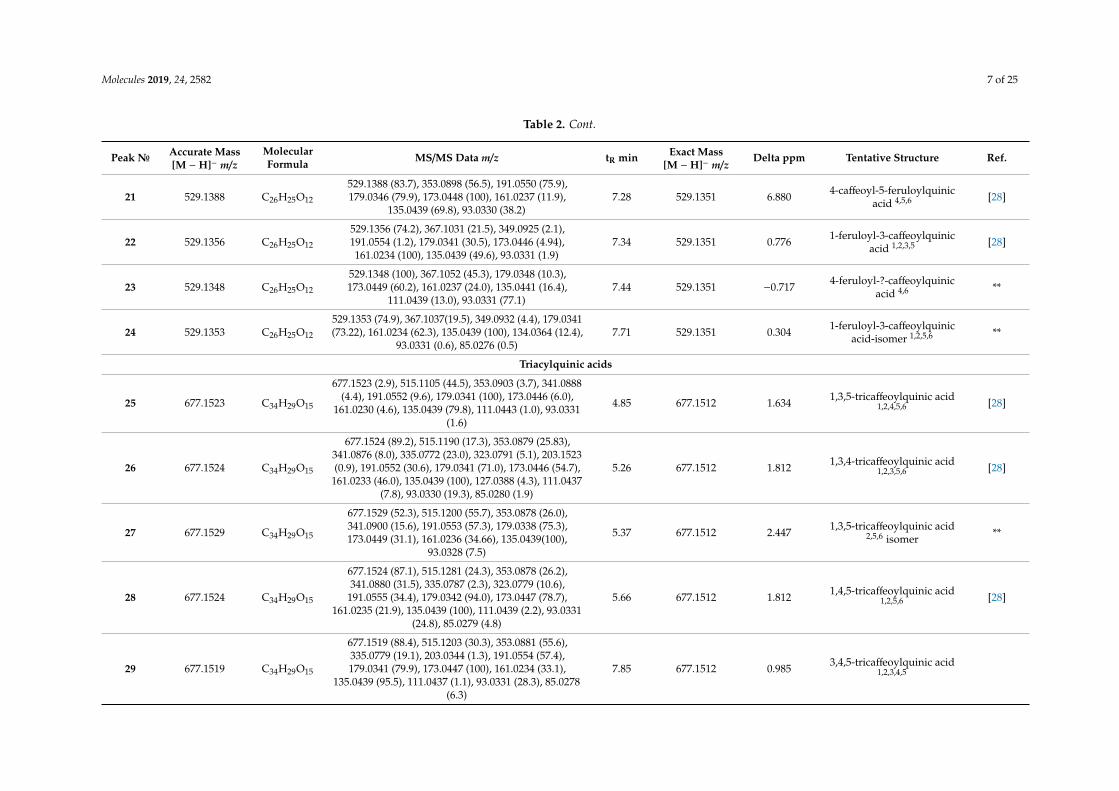
Twenty nine acylquinic acids were identified in tested Anthemis extracts (Table 2). The acylquinicacids elucidation was based on the hierarchical key developed by Clifford and colleagues [21,22]. Peaks4, 5, 6, and 7 were identified as 3-O-, 1-O-, 5-O- and 4-O-caffeoylquinic acids ([M − H]− at m/z 353.088),respectively, according to the relative abundance of the characteristic fragment ions at m/z 191.055[quinic acid − H]−, 179.034 [caffeic acid − H]−, 173.045 [quinic acid − H − H2O]−, and 135.044 [caffeicacid – H − CO2]− [21,22]. Compounds 4 and 6 were identified by comparison with neochlorogenic andchlorogenic acid, respectively. In the same manner, peaks 1, 2, and 3 were assigned as 3-O-, 5-O-, and1-O-p-coumaroylquinic acids ([M −H]− at m/z 337.093), while peaks 8, 9, 10, and 11 ([M −H]− at m/z367.103) were assigned as 3-O-, 1-O- 5-O-, and 4-O-feruloylquinic acid (Table 4). With respect to thediacylquinic acids, peaks 12–15 were related to 3,4-O-, 1,5-O, 3,5-O, and 4,5-O-dicaffeoylquinic acids([M − H]− at m/z 515.120); 12 and 13 were identified by comparison with standards. The presence of 14was evidenced by the relative abundance of the ions at m/z 191.055, 179.034, and 135.043 [21–23], whilethe ion at 173.044 was prominent for 15. Compounds 16–24, [M − H]− at m/z 529.136 were tentativelyidentified as caffeoylferuloylquinic acids [21,22]. Among the tricaffeoylquinic acids, peaks 25–29 wererelated to ([M − H]− at m/z 677.152). Compounds 26, 28, and 29 yielded indicative fragment ions at173.045 deduced 4-substituted CQA [22]. According to the presence of weak signal at m/z 203.034,the relative intensity of the fragment ion at m/z 335.078, and lipophilicity, peaks 26, 28, and 29 weretentatively assigned as 1,3,4-O-, 1,4,5-O-, and 3,4,5-O-tricaffeoylquinic acid, while 25 and 27 wererelated to 3,4,5-O-tricaffeoylquinic acid and its isomer.
2.2.2. Flavonoids
Based on literature and comparison with standards, 15 flavonoid aglycones 30–44 (most of themmethoxylated), twelve glycosides, and one caffeoyl-O-flavonoid were identified in the studied extracts(Table 4). Regarding 41–43 ([M −H]− at m/z 345.061), the fragment ion at m/z 287.020, due to consecutiveloss of 2CH3
− and CO is more intense in the product-ion spectra of 43 than 41 and 42. Probablymethoxylation of 43 in both A- and C-rings provides very stable fragments due to concurrent methylloss [24]. Fragment ion at m/z 121.028 (1,2B) (for 41 and 42) were attributed to the Retro-Diels Alder
Molecules 2019, 24, 2582 4 of 25
(RDA) cleavages of the flavonoid skeleton specific for 3′,4′-dihydroxy flavonols [25]. Thus, accordingto literature, 41–43 were tentatively identified as eupatolitin, spinatoside, and spinacetin, respectively.
The fragmentation fingerprints of 52 and 56 were associated with isorhamnetin derivatives,witnessed by the abundant fragment ion at m/z 315.051 supported by the ions at m/z 300.027 and133.028 [24]. Fragmentation patterns and monoisotopic profiles of 52 was in good agreement withthose of caffeoyl-O-isorhamnetin. The fragmentation of [M −H]− at m/z 609.1472 (56) yielded abundantion at m/z 315.0517 ([M − H − 294.095]− indicating the loss of hexose and pentose moieties.
2.2.3. Sesquiterpenes
Thirteen sesquiterpene lactones including one eudesmanolide, three germacranolides, and nineguaianolides, were tentatively identified in both ACT and ATP extracts. Concerning compound 58 ([M+ H]+ at m/z 229.122), its fragmentation pattern involved losses of 18 Da (H2O), 28 Da (CO) and 46 Da(CO2H) suggesting chamazulene carboxylic acid, a degradation product of proazulenic sesquiterpenelactones, e.g., matricarin [25]. Similar fragmentation patterns were observed in spectra of 59 and 60. Inaddition, a loss of 44 Da (CO2) and fragment ions at m/z 185.095 ([M + H – 16 − 44]+ and 95.049 (C6H7O)due to the overall fracture of lactone ring, suggested dehydroleucodin or isodehydroleucodin [26].Accordingly, 61 was assigned as leucodin ([M + H]+ at m/z 247.132), where C-13 was saturated in amethyl group. 68 and 69 were tentatively identified as matricarin and its isomer, due to the concomitantloss of (CO2 + H2O) at m/z 245.117 from the additional acetyl group [3]. Three isobaric sesquiterpenelactones 63–65 shared the same [M + H]+ at m/z 263.127 (exact mass). Peaks 63–65 demonstrateddifference of 15.995 Da, in comparison to 61, suggesting the presence of an additional hydroxyl group.Thus 63–65 were tentatively assigned to hydroxyleucodin and its isomers [27]. In the same manner,peaks 62, isobaric pair 66/67, and 70 were ascribed as parthenolide, stizolin, and ludalbine, respectively,previously identified in Anthemis species [3].
2.3. Antioxidant Activity
Oxidative stress-related diseases often arise as a result of the imbalance between the productionof free radicals and reactive oxygen/nitrogen species, and antioxidant defences. These diseases can bemanaged/prevented using natural antioxidants that represent promising therapeutic candidates [29].Different antioxidant assays are needed to obtain certain information regarding antioxidant profile ofherbal extracts. From this point, the antioxidant capacity of different extracts of ATP and ACT wereevaluated using multiple assays based on different mechanisms and the results were presented inTable 3.

Molecules 2019, 24, 2582 5 of 25
Table 2. Peak assessment of compounds in Anthemis extracts.
Peak№ Accurate Mass[M − H]− m/z
MolecularFormula MS/MS Data m/z tR min Exact Mass
[M − H]− m/zDelta ppm Tentative Structure Ref.
Acylquinnic acids
Monoacylquinic acids
1 337.0946 C16H17O8337.0946 (3.1), 191.0557 (7.7), 173.0453 (2.5), 163.0390
(100), 119.0488 (22.8) 3.05 337.0929 5.071 3-p-coumaroyl-quinicacid 1,2,6 [19]
2 337.0932 C16H17O8337.0932 (7.2), 191.0554 (100), 173.0445 (6.7), 163.0390
(5.8), 119.0489 (5.5), 93.0329 (17.6) 4.07 337.0929 0.829 5-p-coumaroyl-quinicacid 1,2,3,5,6 [19]
3 337.0930 C16H17O8
337.0930 (5.9), 191.0554 (100), 173.0446 (1.9), 163.0391(2.4), 127.0391 (1.5), 119.0487 (1.4), 111.0439 (1.3),
93.0331 (5.3), 85.0280 (7.1)4.74 337.0929 0.473 1-p-coumaroyl-quinic
acid 1,2,5,6 [19]
4 353.0879 C16H17O9
353.0879 (32.4), 191.0553 (100), 179.0341 (60.7),173.0443 (4.1),161.0233 (3.9), 135.0439 (50.7), 111.0438
(0.9), 93.0333 (4.5), 85.0279 (8.7)2.47 353.0867 0.325
neochlorogenic(3-caffeoylquinic) acid
1,2,3,4,6*
5 353.0879 C16H17O9
353.0879 (29.9), 191.0554 (47.7), 179.0341 (70.1),173.0447 (100), 135.0439 (57.7), 111.0436 (3.3), 93.331
(22.6), 85.0280 (8.6)2.48 353.0867 0.240 1-caffeoylquinic acid
1,2,3,4,6 [19]
6 353.0880 C16H17O9
353.0880 (3.8), 191.0554 (100), 179.0338 (1.4), 173.0446(0.9), 161.0234 (2.1), 135.0441 (1.1), 111.0435 (1.2),
93.0331 (2.7), 85.0279 (0.4)3.30 353.0867 0.495
chlorogenic(5-caffeoylquinic) acid
1,2,3,4,5,6*
7 353.0880 C16H17O9
353.0880 (28.1), 203.6057 (0.1), 191.0554 (100), 179.0341(58.3), 173.0446 (4.6), 161.0233 (3.9), 135.0438 (51.8),
111.0437 (2.2), 93.0330 (4.82), 85.0279 (10.2)5.92 353.0867 0.665 4-caffeoylquinic acid
1,2,3,4,6 [19]
8 367.1043 C17H19O9
367.1043 (12.5), 193.0500 (100), 173.0453 (3.9), 149.0598(2.9), 134.0361 (65.7), 127.0395 (1.0), 111.0439 (1.4),
93.0331 (2.9)3.54 367.1034 2.410 3-feruloylquinic acid
1,2,5,6 [19]
9 367.1039 C17H19O9367.1039 (49.3), 161.0234 (100), 127.0390 (1.7), 85.0281
(13.4) 3.97 367.1034 1.238 1-feruloylquinic acid1,2,5,6 [19]
10 367.1035 C17H19O9
367.1035 (14.1), 193.0499 (6.7), 191.0555 (100), 173.0447(18.2), 134.0447 (10.9), 111.0437 (3.9), 93.0331 (26.2),
85.0280 (5.3)4.52 367.1034 −0.015 5-feruloylquinic acid
1,2,3,4,5,6
11 367.1030 C17H19O9367.1030 (11.8), 193.0499 (17.1), 173.0446 (100),
111.0435 (3.1), 93.0331 (24.0) 4.78 367.1034 -1.322 4-feruloylquinic acid1,2,5,6 [19]

Molecules 2019, 24, 2582 6 of 25
Table 2. Cont.
Peak№ Accurate Mass[M − H]− m/z
MolecularFormula MS/MS Data m/z tR min Exact Mass
[M − H]− m/zDelta ppm Tentative Structure Ref.
Diacylquinic acids
12 515.1196 C25H23O12
515.1196 (100), 353.0880 (18.5), 335.0788 (6.4), 203.0331(1.3), 191.0554 (29.5), 179.0341 (58.1), 173.0446 (65.7),
161.0235 (19.8), 135.0439 (57.5), 127.0389 (3.6),111.0436 (4.7), 93.0330 (18.7), 85.0277 (4.3)
5.79 515.1184 0.137 3,4-dicaffeoylquinic acid1,2,3,4,5,6 *
13 515.1199 C25H23O12
515.1199 (21.8), 353.0881 (83.1), 335.0782 (2.6),191.0554 (100), 179.0341 (48.6), 173.0445 (10.6),
161.0233 (12.4), 135.0439 (57.5), 127.0387 (2.8), 93.0332(6.4), 85.0280 (9.0)
5.96 515.1184 1,5-dicaffeoylquinic acid1,2,3,4,5,6 *
14 515.1204 C25H23O12
515.1204 (22.6), 353.0880 (87.4), 191.0555 (100),179.0342 (52.0), 173.0455 (9.8), 161.0235 (8.9), 135.0438
(51.7), 93.0333 (2.8), 85.0281 (8.6),6.14 515.1184 1.787 3,5-dicaffeoylquinic acid 3,4 [28]
15 515.1199 C25H23O12
515.1199 (75.6), 353.0880 (60.6), 203.0343 (3.1),191.0555 (34.7), 179.0342 (72.3), 173.0447 (100),
135.0440 (48.1), 127.0384 (1.2), 111.0439 (3.7), 93.0330(17.2), 85.0280 (4.2)
6.34 515.1184 0.720 4,5-dicaffeoylquinic acid1,2,3,4,5,6 [28]
16 529.1356 C26H25O12
529.1356 (100), 367.1037 (8.3), 353.0889 (7.4), 349.0935(5.2), 335.0774 (11.8), 193.0499 (60.5), 191.0555 (9.2),
179.0342 (42.1), 173.0446 (48.1), 161.0235 (24.6),149.0596 (1.1), 134.0361 (56.0), 111.0437 (8.9), 93.0331
(14.7), 85.0276 (2.7)
6.00 529.1351 0.889 3-feruloyl-5-caffeoylquinicacid 1,2,4,5,6 [28]
17 529.1357 C26H25O12
529.1357 (55.4), 367.1038 (25.6), 193.0499 (2.6),179.0342 (3.0), 173.0449 (1.9), 161.0234 (100), 135.0441
(12.7), 134.0367 (4.3), 127.0380 (0.5), 93.0331 (1.1),85.0279 (3.2)
6.92 529.1351 1.003 1-feruloyl-5-caffeoylquinicacid 1,2,3,5 [28]
18 529.1354 C26H25O12
529.1354 (38.3), 367.1040 (39.6), 353.0883 (44.99),193.0506 (15.6), 191.0555 (100), 179.0343 (41.61),173.0454 (14.8), 161.0239 (13.0), 135.0440 (52.3),
134.0363 (21.5), 93.0332 (14.2), 85.0281 (7.4)
7.00 529.1351 0.436 3-caffeoyl-5-feruloylquinicacid 1,2,3,4 [28]
19 529.1352 C26H25O12
529.1352 (66.9), 367.1044 (100), 193.0504 (12.1),179.0333 (57.2), 173.0447 (76.9), 161.0236 (15.6),135.0439 (73.7), 134.0365 (49.6), 93.0331 (76.7)
7.12 529.1351 0.077 4-feruloyl-5-caffeoylquinicacid 4,5,6 [28]
20 529.1361 C26H25O12
529.1361 (14.2), 367.1036 (60.3), 193.0499 (14.5),173.0447 (100), 161.0239 (1.1), 134.0362 (17.5), 127.0392
(1.0), 111.0436 (3.4), 93.0330 (24.9),7.23 529.1351 1.816 3-caffeoyl-4-feruloylquinic
acid 1,2,3,6 [28]
Molecules 2019, 24, 2582 7 of 25
Table 2. Cont.
Peak№ Accurate Mass[M − H]− m/z
MolecularFormula MS/MS Data m/z tR min Exact Mass
[M − H]− m/zDelta ppm Tentative Structure Ref.
21 529.1388 C26H25O12
529.1388 (83.7), 353.0898 (56.5), 191.0550 (75.9),179.0346 (79.9), 173.0448 (100), 161.0237 (11.9),
135.0439 (69.8), 93.0330 (38.2)7.28 529.1351 6.880 4-caffeoyl-5-feruloylquinic
acid 4,5,6 [28]
22 529.1356 C26H25O12
529.1356 (74.2), 367.1031 (21.5), 349.0925 (2.1),191.0554 (1.2), 179.0341 (30.5), 173.0446 (4.94),161.0234 (100), 135.0439 (49.6), 93.0331 (1.9)
7.34 529.1351 0.776 1-feruloyl-3-caffeoylquinicacid 1,2,3,5 [28]
23 529.1348 C26H25O12
529.1348 (100), 367.1052 (45.3), 179.0348 (10.3),173.0449 (60.2), 161.0237 (24.0), 135.0441 (16.4),
111.0439 (13.0), 93.0331 (77.1)7.44 529.1351 −0.717 4-feruloyl-?-caffeoylquinic
acid 4,6 **
24 529.1353 C26H25O12
529.1353 (74.9), 367.1037(19.5), 349.0932 (4.4), 179.0341(73.22), 161.0234 (62.3), 135.0439 (100), 134.0364 (12.4),
93.0331 (0.6), 85.0276 (0.5)7.71 529.1351 0.304 1-feruloyl-3-caffeoylquinic
acid-isomer 1,2,5,6 **
Triacylquinic acids
25 677.1523 C34H29O15
677.1523 (2.9), 515.1105 (44.5), 353.0903 (3.7), 341.0888(4.4), 191.0552 (9.6), 179.0341 (100), 173.0446 (6.0),
161.0230 (4.6), 135.0439 (79.8), 111.0443 (1.0), 93.0331(1.6)
4.85 677.1512 1.634 1,3,5-tricaffeoylquinic acid1,2,4,5,6 [28]
26 677.1524 C34H29O15
677.1524 (89.2), 515.1190 (17.3), 353.0879 (25.83),341.0876 (8.0), 335.0772 (23.0), 323.0791 (5.1), 203.1523(0.9), 191.0552 (30.6), 179.0341 (71.0), 173.0446 (54.7),
161.0233 (46.0), 135.0439 (100), 127.0388 (4.3), 111.0437(7.8), 93.0330 (19.3), 85.0280 (1.9)
5.26 677.1512 1.812 1,3,4-tricaffeoylquinic acid1,2,3,5,6 [28]
27 677.1529 C34H29O15
677.1529 (52.3), 515.1200 (55.7), 353.0878 (26.0),341.0900 (15.6), 191.0553 (57.3), 179.0338 (75.3),173.0449 (31.1), 161.0236 (34.66), 135.0439(100),
93.0328 (7.5)
5.37 677.1512 2.447 1,3,5-tricaffeoylquinic acid2,5,6 isomer **
28 677.1524 C34H29O15
677.1524 (87.1), 515.1281 (24.3), 353.0878 (26.2),341.0880 (31.5), 335.0787 (2.3), 323.0779 (10.6),
191.0555 (34.4), 179.0342 (94.0), 173.0447 (78.7),161.0235 (21.9), 135.0439 (100), 111.0439 (2.2), 93.0331
(24.8), 85.0279 (4.8)
5.66 677.1512 1.812 1,4,5-tricaffeoylquinic acid1,2,5,6 [28]
29 677.1519 C34H29O15
677.1519 (88.4), 515.1203 (30.3), 353.0881 (55.6),335.0779 (19.1), 203.0344 (1.3), 191.0554 (57.4),179.0341 (79.9), 173.0447 (100), 161.0234 (33.1),
135.0439 (95.5), 111.0437 (1.1), 93.0331 (28.3), 85.0278(6.3)
7.85 677.1512 0.985 3,4,5-tricaffeoylquinic acid1,2,3,4,5

Molecules 2019, 24, 2582 8 of 25
Table 2. Cont.
Peak№ Accurate Mass[M − H]− m/z
MolecularFormula MS/MS Data m/z tR min Exact Mass
[M − H]− m/zDelta ppm Tentative Structure Ref.
Flavonoids
30 269.0457 C15H9O5269.0457 (100), 151.0027 (6.39), 149.0233 (5.74),
117.0332 (22.24), 107.0124 (5.35) 8.73 269.0444 0.644 apigenin 1,2,3,4,5,6 *
31 285.0406 C15H9O6285.0406 (100), 151.0028 (5.92), 133.0282 (25.08),
107.0126 (3.32) 7.82 285.0393 0.452 luteolin 1,2,3,4,5,6 *
32 287.0566 C15H11O6287.0566 (14.91), 151.0025 (100), 135.0435 (89.99),
125.0231 (5.03), 107.0124 (13.58) 7.53 287.0550 1.842 eriodictyol 1,2,3,4,5,6 [24]
33 299.0561 C16H11O6299.0561 (58.00), 284.0328 (100), 256.0382 (0.86),
227.0350 (3.45), 211.0393 (2.22) 8.91 299.0550 0.029 diosmetin 1,2,3,4,5,6 *
34 299.0562 C16H11O6299.0562 (95.99), 284.9335 (100), 227.047 (2.78),
151.0033 (3.67), 107.0123 (2.84) 9.09 299.0550 0.430 3,4′,7-trihydroxy-3′-methoxyflavone 1,2,3,4,5,6 [23]
35 301.0352 C15H9O7301.0352 (100), 300.0273 (24.24), 178.9976 (21.40),
151.0025 (49.72), 121,0282 (16.11), 107.0124 (14.15) 7.83 301.0342 −0.717 quercetin 1,2,3,4,5,6 *
36 315.0512 C16H11O7315.0512 (61.55), 300.0279 (100), 271.0252 (28.24),255.030 (11.23), 227.0349 (2.28), 136.9872 (2.51), 8.34 315.0499 0.584 nepetin 4,5,6 Mass bank
37 315.0513 C16H11O7315.0513 (86.15), 301.0315 (11.76), 300.0276 (100), 243.
0303 (0.70), 165.9890 (1.63), 136.9868 (9.78) 7.90 315.0499 0.965 rhamnetin 1,2,3,4,5,6 *
38 315.0514 C16H11O7315.0514 (100), 301.0316 (3.73), 300.0273 (41.59), 243.
0298 (1.08), 151.0025 (7.85), 107.0126 (6.32) 9.26 315.0499 1.156 isorhamnetin 1,2,3,4,5,6 *
39 329.0670 C17H13O7329.0607 (14.45), 314.0436 (100), 299.0198 (25.08),271.0250 (47.23),133.0282 (5.34), 107.2971 (0.52) 9.25 329.0655 0.954 jaceosidin 1,3,4,6 Mass bank
40 331.0463 C16H11O8331.0463 (100), 316.0226 (56.40), 287.0199 (15.97),271.0246 (5.47), 270.0176 (4.09), 165.9897 (19.03) 7.80 331.0448 1.086 patuletin 1,3,4,6 [23]
41 345.0618 C17H13O8345.0618 (91.18), 330.0384 (100), 315.0150 (50.33),
287.0201 (15.30), 121.0280 (1.86) 8.36 345.0604 0.694 eupatuletin 4,5,6 **
42 345.0618 C17H13O8345.0618 (100), 330.0385 (95.66), 315.0150 (46.41),
287.0198 (14.78), 121.0284 (7.72) 8.40 345.0604 0.694 spinatoside 1,2,3 [23]
43 345.0619 C17H13O8345.0619 (100), 330.0385 (42.35), 315.0145 (4.01),
301.0388 (6.46), 287.0199 (40.72) 9.38 345.0604 0.694 spinacetin 1,2,3 [23]
44 359.0775 C18H15O8359.0775 (100), 344.0539 (49.89), 329.0304 (52.64),
301.0359 (6.67), 287.0139 (4.46) 9.95 359.0761 0.750 jaceidin 1,2,3,4,5,6 [23]

Molecules 2019, 24, 2582 9 of 25
Table 2. Cont.
Peak№ Accurate Mass[M − H]− m/z
MolecularFormula MS/MS Data m/z tR min Exact Mass
[M − H]− m/zDelta ppm Tentative Structure Ref.
45 431.0981 C21H19O10 431.0981 (100), 269.0440 (27.72), 268.0378 (57.01) 6.17 431.0972 −0.673 apigenin-7-O-glucoside1,2,3,4,5,6 *
46 447.0934 C21H19O11447.0934 (100), 327.0507 (0.95), 285.0405 (99.96),151.0030 (7.20), 133.0280 (5.74), 107.0123 (4.57) 5.45 447.0921 0.348 luteolin-7-O-glucoside
1,2,3,4,5,6 *
47 461.0725 C21H17O12461.0725 (42.84), 285.0406 (100), 151.0025 (3.72),
133.0280 (10.72), 107.0121 (1.50) 5.46 461.0714 −0.150 luteolin-7-O-glucuronide1,2,3 *
48 461.1094 C22H21O11
461.1094 (100), 446.0858 (28.76), 299.0554 (12.48),284.0313 (9.73), 283.0250 (21.41), 269.0467 (2.08),255.0300 (73.76), 227.0345 (0.67), 151,0028 (0.86)
6.37 461.1078 2.220 diosmetin-O-glucoside 1,2,3 **
49 463.0887 C21H19O12463.0887 (100), 372.1873 (4.84), 301.0360 (93.71),
300.0282 (27.20) 4.77 463.0871 1.038 isoquercitrin 1,2,3,4,5,6 *
50 463.0903 C21H19O12 463.0903 (100), 301.0349 (47.50), 300.0276 (66.34) 5.29 463.0871 4.601 hyperoside 1,2,3,4,5,6 *
51 477.1041 C22H21O12
477.1041 (100), 315.0496 (14.04), 314.0434 (53.04),300.0274 (5.68), 285.0411 (6.84), 243.0297 (22.73),
271.0249 (27.98), 151.0032 (3.66)6.10 477.1027 0.442 isorhamnetin-7-O-glucoside
1,2,3,4,5,6 *
52 493.0777 C25H17O11
493.0777 (11.58), 315.0512 (99.41), 314.0435 (100),300.0277 (14.30), 285.0411 (13.30), 243.0293 (34.06),
227.0341 (4.04), 177.0182 (16.10), 151.0030 (4.86),133.0283 (12.50)
9.12 493.0765 0.194 caffeoyl-O-isorhamnetin 1,3 **
53 493.0974 C22H21O13493.0974 (100), 331.0463 (96.71), 316.0224 (22.61),287.0196 (26.93), 271.0253 (9.58), 165.9891 (8.24) 5.63 493.0976 −0.257 patuletin-O-hexoside 4,5,6 **
54 593.1504 C27H29O15593.1504 (97.39), 318.6214 (6.24), 285.0405 (100),
284.0327 (60.74), 227.0352 (29.58) 5.71 593.1500 −1.354 luteolin-7-O-rutinoside1,2,3,4,5,6 *
55 609.1467 C27H29O16
609.1467 (72.52), 343.0477 (1.65), 301.0354 (100),300.0278 (38.67), 178.9970 (1.59), 151.0027
(13.46),121.0279 (1.84), 107.0123 (4.76)5.17 609.1450 0.923 rutin 1,2,3,4,5,6 *
56 609.1472 C27H29O16609.1472 (96.84), 411.8945 (5.23), 315.0517 (100),
300.0275 (48.87), 133.0284 (7.86) 5.43 609.1450 2.149 isorhamnetin-O-pentosyl-hexoside 4,5,6 **
57 623.1613 C28H31O16623.1613 (5.57), 315.0514 (100), 301.0306 (1.22),
300.0279 (16.19), 151.0025 (7.85), 107.0125 (3.48) 6.08 623.1606 −0.703 isorhamnetin-3-O-rutinoside1,2,3,4,5,6 *

Molecules 2019, 24, 2582 10 of 25
Table 2. Cont.
Peak№ Accurate Mass[M − H]− m/z
MolecularFormula MS/MS Data m/z tR min Exact Mass
[M − H]− m/zDelta ppm Tentative Structure Ref.
Sesquiterpenes
58 229.1220 C15H17O2229.1220 (100), 211.1116 (12.69), 201.1272 (19.57),
183.1167 (40.99), 91.0548 (6.40), 6.10 229.1223 −3.643 chamazulene carboxylicacid 1,2,3 [25]
59 245.1170 C15H17O3
245.1170 (100), 227.1067 (44.35), 217.1222 (20.20),201.0910 (20.68), 199.1116 (75.08), 185.0959 (28.15),
95.0497 (66.93)10.48 245.1172 −0.942 dehydroleucodin/
isodehydroleucodin 1,2,3 [26]
60 245.1171 C15H17O3
245.1171 (84.77), 227.1065 (44.23), 217.1225 (17.55),201.0910 (17.75), 199.1116 (63.86), 185.0960 (18.51),
95.0497 (100)10.48 245.1172 −0.942 dehydroleucodin/
isodehydroleucodin 1,2,3 [26]
61 247.1325 C15H19O3
247.1325 (100), 229.1220 (90.72), 211.1113 (8.83),201.1271 (37.99), 187.0752 (37.99), 183.1167 (22.85),
91.0548 (9.16)9.79 247.1328 −1.663 desacetoxymatricarin
1,2,3,4,5,6 **
62 249.1481 C15H21O3
249.1481 (100), 231.1378 (30.41), 213.1277 (9.04),203.1431 (10.25), 193.0856 (5.87), 189.1272 (11.79),
159.1167 (22.04), 119.0857 (16.94), 105.0702 (18.62),95.0860 (10.61)
10.42 249.1485 −0.451 parthenolide 1,2,3,4,5,6 **
63 263.1275 C15H19O4
263.1275 (100), 245.1170 (55.39), 227.1061 (9.88),219.1021 (15.59), 217.1223 (47.56), 203.1065 (10.39),199.1115 (21.45), 191.0704 (28.72), 95.0497 (96.99)
10.52 263.1277 −0.316 hydroxyleucodin 1,2,3 **
64 263.1274 C15H19O4
263.1274 (100), 245.1171 (33.08), 227.1063 (4.95),219.1012 (14.30), 217.1224 (16.65), 203.1065 (10.12),199.1117 (15.19), 191.0702 (79.26), 95.0497 (20.99)
11.22 263.1277 −0.406 hydroxyleucodin isomer1,2,3 **
65 263.1275 C15H19O4
263.1275(100), 245.1170 (51.98), 227.1063 (11.55),219.1014 (34.60), 217.1223 (47.24), 203.1067 (12.57),199.1117 (22.59), 191.0702 (32.46), 95.0497 (81.14)
11.22 263.1277 −0.406 hydroxyleucodin isomer1,2,3 **
66 265.1432 C15H21O4
265.1432 (8.16), 247.1326 (34.45), 229.1221 (100),211.1115 (3.94), 201.1273 (20.45), 187.0752 (14.30),
183.1168 (7.67), 91.0548 (5.66)6.04 265.1434 −0.813 stizolin 1,3 **
67 265.1429 C15H21O4
265.1429 (24.21), 247.1325 (100), 229.1222 (71.63),219.1372 (36.25), 201.1272 (55.14), 187.1111 (17.50),
183.1174 (17.90), 91.0548 (13.40)9.78 265.1434 −1.982 stizolin isomer 1,3 **

Molecules 2019, 24, 2582 11 of 25
Table 2. Cont.
Peak№ Accurate Mass[M − H]− m/z
MolecularFormula MS/MS Data m/z tR min Exact Mass
[M − H]− m/zDelta ppm Tentative Structure Ref.
68 305.1360 C17H21O5
305.1360 (44.97), 287.1268 (3.27), 269.1206 (1.63),263.1274 (42.58), 245.1170 (100), 227.1065 (63.42),
217.1220 (60.59), 109.1116 (23.36), 201.0903 (12.29),185.0958 (19.24), 181.1010 (44.17), 171.1166 (56.51),
105.0703 (50.85), 95.0496 (25.16)
7.05 305.1383 −7.538 matricarin 3 **
69 305.1363 C17H21O5
305.1363 (23.50), 263.1279 (20.85), 245.1170 (100),227.1063 (29.92), 217.1216 (19.51), 201.0911 (2.62),
185.0961 (16.47), 181.1009 (28.17), 171.1167 (31.70),131.0857 (46.50), 95.0496 (3.40)
5.42 305.1383 −6.654 matricarin isomer 3 **
70 307.1544 C17H21O5
307.1544 (2.15), 289.1433 (0.75), 267.2721 (0.18),247.1325 (2.63), 229.1219 (100), 211.1115 (0.95),
183.1168 (2.88)9.67 307.1540 1.366 ludalbin 1,2,3 **
1-A. cretica MeOH extract, 2-A. cretica aqueous extract, 3-A. cretica EA extract, 4-A. palida MeOH extract, 5-A. palida aqueous extract, 6-A. palida EA extract. *-comparison with standardsubstance. **-tentatively identification.
Table 3. Antioxidant properties of Anthemis extracts *.
Plant Names Solvents Phosphomolybdenum(mmol TE/g) DPPH (mg TE/g) ABTS (mg TE/g) CUPRAC
(mgTE/g) FRAP (mgTE/g)Metal Chelating
Abilitiy (mgEDTAE/g)
A. tinctoria var.pallida
Ethyl acetate 2.59 ± 0.19 b 40.30 ± 0.78 e 45.52 ± 5.53 f 113.31 ± 2.26 d 47.63 ± 3.77 f 39.01 ± 4.42 a
Methanol 2.99 ± 0.14 a 407.07 ± 8.88 a 320.11 ± 5.67 a 691.17 ± 12.07 a 362.12 ± 2.63 a 28.28 ± 1.81 c
Aqueous 2.65 ± 0.02 b 298.40 ± 6.74 b 303.16 ± 8.57 b 584.01 ± 8.71 b 316.34 ± 4.15 b 33.59 ± .16 b
A. cretica subsp.tenuiloba
Ethyl acetate 1.69 ± 0.08 cd 45.47 ± 2.16 e 57.13 ± 3.89 e 112.87 ± 4.41 d 55.74 ± 2.27 e 21.90 ± 0.81 d
Methanol 1.77 ± 0.08 c 97.22 ± 0.22 c 112.41 ± 2.35 d 223.09 ± 6.17 c 143.21 ± 1.77 d 20.93 ± 1.70 d
Aqueous 1.56 ± 0.05 d 86.74 ± 2.46 d 127.68 ± 0.45 c 214.45 ± 1.39 c 130.86 ± 1.81 c 39.64 ± 1.34 a
* Values expressed are means ± S.D. of three parallel measurements. TE: Trolox equivalent; EDTAE: EDTA equivalent. Different letters indicate significant differences in the extracts(p < 0.05).

Molecules 2019, 24, 2582 12 of 25
Based on the experimental results (Table 3), it can be noticed that for both species, MeOH extractexhibited the highest total antioxidant activity. According to data presented in Table 3, ATP MeOHextract (DPPH: 407.07 ± 8.88 and ABTS: 320.11 ± 5.67 mg TE/g), followed by the aqueous extract(DPPH: 298.40 ± 6.74 and ABTS: 303.16 ± 8.57 mg TE/g) showed higher radical scavenging activityin both assays. Likewise, ACT MeOH (DPPH: 97.22 ± 0.22 and ABTS: 112.41 ± 2.35 mg TE/g) andaqueous extracts (DPPH: 86.74 ± 2.46 and ABTS: 127.68 ± 0.45 mg TE/g) showed potent radicalscavenging activity.
CUPRAC and FRAP assays were employed to assess the reducing capacity of different extracts.CUPRAC method evaluates the conversion of Cu (II) into Cu (I) while FRAP assay measures thereducing potential of an antioxidant reacting with the colourless TPTZ/Fe3+ complex to form a blueTPTZ/Fe2+ complex at low pH [30]. Remarkable reducing potencies were displayed by MeOH(CUPRAC: 691.17 ± 12.07 and FRAP: 362.12 ± 2.63) and aqueous (CUPRAC: 584.01 ± 8.71 and FRAP:316.34 ± 4.15 mg TE/g) extracts of APT. EA extract displayed the lowest reducing capacity. This trendwas also observed as regards ACT extracts (Table 3).
Chelation of pro-oxidant metals is recognized as one of the most important mechanisms of actionof antioxidants. Particularly, iron is the most powerful and abundant pro-oxidant and transition metalwhich causes oxidative changes of cellular components, such as, lipids and proteins [31]. Evaluation ofiron chelating activity showed that ATP and ACT extracts possessed notable chelation potential, withthe highest activity displayed by ATP EA extract (39.01 ± 4.42 mg EDTAE/g) and ACT aqueous extract(39.64 ± 1.34 mg EDTAE/g).
2.4. Enzyme Inhibitory Activity
The enzyme inhibitory effect of ATP and ACT extracts was determined against cholinesterases,α-amylase, α-glucosidase, and tyrosinase and the results were presented in Table 4. EA and MeOHextracts of ATP and ACT exhibited potent inhibitory activity against AChE, with the highest activityobserved for ATP MeOH extract (3.28 ± 0.43 mg GALAE/g) and ACT EA extract (4.68 ± 0.21 mgGALAE/g). On the other hand, ATP EA extract (3.48 ± 0.21 mg GALAE/g), ACT EA extract(2.51 ± 0.34 mg GALAE/g), and ACT MeOH extract (1.15 ± 0.05 mg GALAE/g) showed inhibitoryeffect against BChE. Both AChE and BChE hydrolyze acetylcholine and terminate the synaptictransmission [31]. While enhanced AChE activity is associated with early stages of AD, BChE activityis found to increase with the progression of the disease. Therefore, both enzymes are considered aslegitimate therapeutic targets for managing AD [32,33].
Table 4. Enzyme inhibitory activity of Anthemis extracts *.
PlantNames Solvents
AChEInhibition
(mgGALAE/g)
BChEInhibition
(mgGALAE/g)
TyrosinaseInhibition
(mg KAE/g)
AmylaseInhibition
(mmolACAE/g)
GlucosidaseInhibition
(mmolACAE/g)
A. tinctoriavar. pallida
Ethyl acetate 1.33 ± 0.03 c 3.48 ± 0.21 a 124.60 ± 0.15 b 0.78 ± 0.05 a 21.94 ± 1.91 b
Methanol 3.28 ± 0.43 b na 124.48 ± 1.23 b 0.54 ± 0.02 c 9.15 ± 2.26 c
Aqueous na na 72.10 ± 1.64 d 0.11 ± 0.01 d 4.95 ± 0.25 d
A. creticasubsp.
tenuiloba
Ethyl acetate 4.68 ± 0.21 a 2.51 ± 0.34 b 128.73 ± 0.71 a 0.65 ± 0.01 b 24.16 ± 0.12 a
Methanol 3.45 ± 0.26 b 1.15 ± 0.05 c 128.85 ± 1.41 a 0.52 ± 0.06 c 4.49 ± 0.93 d
Aqueous na na 88.95 ± 0.49 c 0.09 ± 0.01 d 2.49 ± 0.17 e
* Values expressed are means ± S.D. of three parallel measurements. GALAE: Galatamine equivalent; KAE: Kojicacid equivalent; ACAE: Acarbose equivalent; na: not active. Different letters indicate significant differences in theextracts (p < 0.05).
Based on the results (Table 4), it can be observed that the extracts of both species showed remarkableinhibitory activity against tyrosinase with values ranging from 124.60 ± 0.15 to 72.10 ± 1.64 mg KAE/gand from 128.85 ± 1.41 to 88.95 ± 0.49 mg KAE/g for ATP and ACT, respectively. The lowest inhibitory
Molecules 2019, 24, 2582 13 of 25
activity against tyrosinase was detected for the aqueous extracts of both species. Tyrosinase is a keyenzyme in melanin biosynthesis which is responsible for skin pigmentation. However, excessivemelanin production could lead to various skin disorders such as melasma, lentigines, age spots, andpost-inflammatory hyperpigmentation. Thus, tyrosinase inhibitors, used as hypopigmenting agents,became increasingly important for medicinal and cosmetic products [34].
Type II diabetes is a growing pandemic and poses an enormous public health challenge for almostevery country worldwide. α-Amylase and α-glucosidase are considered as key therapeutic targetsfor the management of type II diabetes. α-Amylase and α-glucosidase are carbohydrate hydrolysingenzymes, responsible for the breakdown of carbohydrates into glucose [35,36]. The present studyshowed that all ATP and ACT extracts displayed weak α-amylase inhibitory effects, despite beingactive α-glucosidase inhibitors. The highest inhibitory effect against α-amylase (0.78 and 0.65 ACAEs/gextract, for ATP and ACT, respectively) and α-glucosidase (21.94 and 24.16 ACAEs/g extract, for ATPand ACT, respectively) was displayed by EA extracts.
2.5. Multivariate Analysis
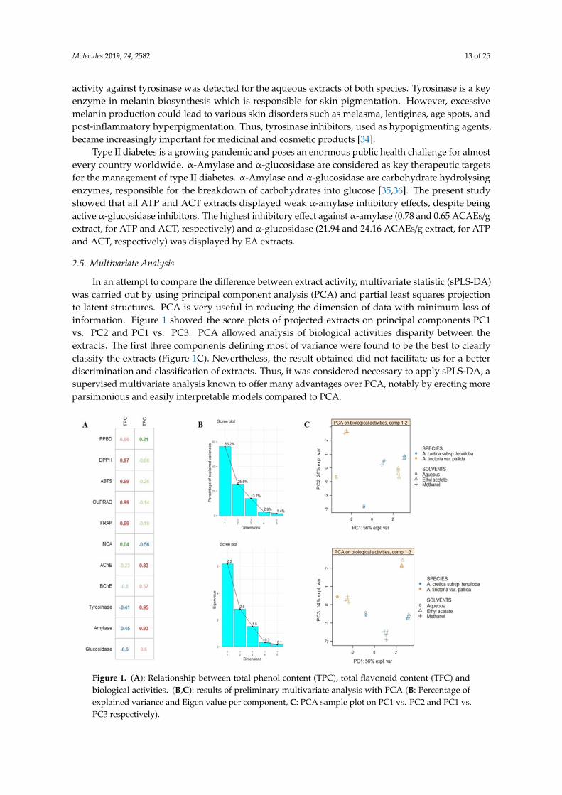
In an attempt to compare the difference between extract activity, multivariate statistic (sPLS-DA)was carried out by using principal component analysis (PCA) and partial least squares projectionto latent structures. PCA is very useful in reducing the dimension of data with minimum loss ofinformation. Figure 1 showed the score plots of projected extracts on principal components PC1vs. PC2 and PC1 vs. PC3. PCA allowed analysis of biological activities disparity between theextracts. The first three components defining most of variance were found to be the best to clearlyclassify the extracts (Figure 1C). Nevertheless, the result obtained did not facilitate us for a betterdiscrimination and classification of extracts. Thus, it was considered necessary to apply sPLS-DA, asupervised multivariate analysis known to offer many advantages over PCA, notably by erecting moreparsimonious and easily interpretable models compared to PCA.Molecules 2019, 24, x 12 of 24
Figure 1. (A): Relationship between total phenol content (TPC), total flavonoid content (TFC) and biological activities. (B&C): results of preliminary multivariate analysis with PCA (B: Percentage of explained variance and Eigen value per component, C: PCA sample plot on PC1 vs. PC2 and PC1 vs. PC3 respectively).
Firstly, analysed species were used as class membership criteria to assess whether they were characterized by distinctive biological activities. sPLS-DA samples plot was reported in Figure 2; as shown, a clear separation between A. cretica subsp. tenuiloba and A. tinctoria var. pallida was achieved, thus suggesting distinctive biological activities. Afterwards, with the aim to identify the most discriminant biological activities providing the differences overviewed in the sPLS-DA samples plot, VIP (variable importance in projection) plot was generated (Figure 2). Five biological activities including PPBD, DPPH, ABTS, CUPRAC and FRAP possessed a VIP score upper 1, which suggested them as discriminants for the two species.
Figure 1. (A): Relationship between total phenol content (TPC), total flavonoid content (TFC) andbiological activities. (B,C): results of preliminary multivariate analysis with PCA (B: Percentage ofexplained variance and Eigen value per component, C: PCA sample plot on PC1 vs. PC2 and PC1 vs.PC3 respectively).

Molecules 2019, 24, 2582 14 of 25
Firstly, analysed species were used as class membership criteria to assess whether they werecharacterized by distinctive biological activities. sPLS-DA samples plot was reported in Figure 2;as shown, a clear separation between A. cretica subsp. tenuiloba and A. tinctoria var. pallida wasachieved, thus suggesting distinctive biological activities. Afterwards, with the aim to identify themost discriminant biological activities providing the differences overviewed in the sPLS-DA samplesplot, VIP (variable importance in projection) plot was generated (Figure 2). Five biological activitiesincluding PPBD, DPPH, ABTS, CUPRAC and FRAP possessed a VIP score upper 1, which suggestedthem as discriminants for the two species.Molecules 2019, 24, x 13 of 24
Figure 2. Supervised analysis with sPLS-DA. A: sPLS-DA samples plot with confidence ellipse plots considering the species as class membership criteria. B: Performance of the model (BER) for three prediction distances using 10 × 5-fold cross-validation. C: VIP score plot displaying the biological activities having highly contributed to the discrimination of both studied species. D: Factorial plan 1-2 of the sPLS-DA with confidence ellipse plots according to the extraction conditions as class membership criteria. E: The model performance per component for the three prediction distances using 5-fold cross-validation repeated 10 times. F: VIP score plot showing the biological activities outlining the difference between the three extraction conditions
Secondly, sPLS-DA was performed considering three different extraction conditions, in order to evaluate the effect of extraction solvents on biological activities. As shown in samples plot (Figure 2) the subspace formed by the first two components showed that methanol, water and ethyl acetate extracts were well separated. Next, the prediction performance and the number of components necessary for the final model were evaluated according to BER (Balanced Error Rate). The performance of our model reached its best for two components, which suggested ncomp = 2 for a final sPLS-DA model (Figure 2). Subsequently, the biological activities having highly contributed to the separation of used solvents were identified. As it could be seen in Figure 2, AChE, BChE, tyrosinase, α-amylase and α-glucosidase were the most contributing biological activities (Figure 2).
2.6. Multidirectional Biological Evaluation
The biological activity of Anthemis extracts was formerly evaluated through allelopathy assay, a validated pharmacognostic test for discriminating herbal extract phytotoxicity. Particularly, we investigated the effects of scalar extract concentrations (100 µg/mL–10 mg/mL) on seedling germination of three commercial lettuce varieties, namely Canasta (C), Romana verde (RV), and Romana bionda (RB). After challenging the seeds with Anthemis water and EA extracts, we observed that germination process was unaffected in the tested concentration range (Figures 3A–E). Conversely, ATP MeOH extract displayed concentration-dependent inhibition of seedling germination, in the range 1–10 mg/mL (Figure 3F). The root elongation rate test revealed evident inhibitory effect, in the range 1–10 mg/mL. On the other hand, extracts resulted biocompatible at the lowest tested concentration (100 µg/mL), with percentage elongation rate ≥70% compared to vehicle
Figure 2. Supervised analysis with sPLS-DA. A: sPLS-DA samples plot with confidence ellipse plotsconsidering the species as class membership criteria. B: Performance of the model (BER) for threeprediction distances using 10 × 5-fold cross-validation. C: VIP score plot displaying the biologicalactivities having highly contributed to the discrimination of both studied species. D: Factorial plan 1-2of the sPLS-DA with confidence ellipse plots according to the extraction conditions as class membershipcriteria. E: The model performance per component for the three prediction distances using 5-foldcross-validation repeated 10 times. F: VIP score plot showing the biological activities outlining thedifference between the three extraction conditions.
Secondly, sPLS-DA was performed considering three different extraction conditions, in order toevaluate the effect of extraction solvents on biological activities. As shown in samples plot (Figure 2) thesubspace formed by the first two components showed that methanol, water and ethyl acetate extractswere well separated. Next, the prediction performance and the number of components necessary forthe final model were evaluated according to BER (Balanced Error Rate). The performance of our modelreached its best for two components, which suggested ncomp = 2 for a final sPLS-DA model (Figure 2).Subsequently, the biological activities having highly contributed to the separation of used solventswere identified. As it could be seen in Figure 2, AChE, BChE, tyrosinase, α-amylase and α-glucosidasewere the most contributing biological activities (Figure 2).
2.6. Multidirectional Biological Evaluation
The biological activity of Anthemis extracts was formerly evaluated through allelopathy assay,a validated pharmacognostic test for discriminating herbal extract phytotoxicity. Particularly, we

Molecules 2019, 24, 2582 15 of 25
investigated the effects of scalar extract concentrations (100 µg/mL–10 mg/mL) on seedling germinationof three commercial lettuce varieties, namely Canasta (C), Romana verde (RV), and Romana bionda(RB). After challenging the seeds with Anthemis water and EA extracts, we observed that germinationprocess was unaffected in the tested concentration range (Figure 3A–E). Conversely, ATP MeOH extractdisplayed concentration-dependent inhibition of seedling germination, in the range 1–10 mg/mL(Figure 3F). The root elongation rate test revealed evident inhibitory effect, in the range 1–10 mg/mL.On the other hand, extracts resulted biocompatible at the lowest tested concentration (100 µg/mL),with percentage elongation rate ≥70% compared to vehicle untreated group. The results of elongationrate test suggest a further toxicological investigation, with independent methods in order to confirmthe biocompatibility limit, as described below.
Molecules 2019, 24, x 14 of 24
untreated group. The results of elongation rate test suggest a further toxicological investigation, with independent methods in order to confirm the biocompatibility limit, as described below.
Figure 3. Seedling germination and growth of Canasta (C), Romana verde (RV) and Romana bionda (RB) seeds challenged with A. tinctoria and A. Cretica extracts. Results are expressed as root and hypocotyl (seedling) length ± SD at different concentrations and mean of GP after the fourth day since the sowing. (A): Effect of A. cretica water extract on seedling germination. (B): Effect of A. cretica ethyl acetate (EA) extract on seedling germination. (C): Effect of A. cretica water methanol (MeOH) on seedling germination. (D): Effect of A. tinctoria water extract on seedling germination. (E): Effect of A. tinctoria ethyl acetate (EA) extract on seedling germination. (F): Effect of A. tinctoria water methanol (MeOH) on seedling germination.
The potential toxicity of water, MeOH and EA extracts of Anthemis species (0.1–20 mg/mL) was also investigated through brine shrimp (Artemia salina Leach) lethality assay. Evaluation of lethality induced on brine shrimp, Artemia salina Leach, is considered predictive of cytotoxicity [37]. The results of this test revealed LC50 values < 10 mg/mL, for all tested extracts.
Additionally, we evaluated the activity of Anthemis extracts on HypoE22 cell line viability. According to brine shrimp and allelopathy assays, we tested the extracts at 100 µg/mL. MTT test revealed that Anthemis extracts were well tolerated by HypoE22 cells, with a resulting cell viability ≥70% (Figure 4). This concentration was used for subsequent ex vivo investigations aimed to elucidate extract neuroprotective effects, as following reported.
Figure 3. Seedling germination and growth of Canasta (C), Romana verde (RV) and Romana bionda(RB) seeds challenged with A. tinctoria and A. Cretica extracts. Results are expressed as root andhypocotyl (seedling) length ± SD at different concentrations and mean of GP after the fourth day sincethe sowing. (A): Effect of A. cretica water extract on seedling germination. (B): Effect of A. creticaethyl acetate (EA) extract on seedling germination. (C): Effect of A. cretica water methanol (MeOH) onseedling germination. (D): Effect of A. tinctoria water extract on seedling germination. (E): Effect ofA. tinctoria ethyl acetate (EA) extract on seedling germination. (F): Effect of A. tinctoria water methanol(MeOH) on seedling germination.
The potential toxicity of water, MeOH and EA extracts of Anthemis species (0.1–20 mg/mL) wasalso investigated through brine shrimp (Artemia salina Leach) lethality assay. Evaluation of lethality

Molecules 2019, 24, 2582 16 of 25
induced on brine shrimp, Artemia salina Leach, is considered predictive of cytotoxicity [37]. The resultsof this test revealed LC50 values < 10 mg/mL, for all tested extracts.
Additionally, we evaluated the activity of Anthemis extracts on HypoE22 cell line viability.According to brine shrimp and allelopathy assays, we tested the extracts at 100 µg/mL. MTT testrevealed that Anthemis extracts were well tolerated by HypoE22 cells, with a resulting cell viability≥70% (Figure 4). This concentration was used for subsequent ex vivo investigations aimed to elucidateextract neuroprotective effects, as following reported.
Molecules 2019, 24, x 15 of 24
Figure 4. Effect of A. tinctoria (A. T.) and A. cretica (A. C.) extracts (100 µg/mL) on HypoE22 cell line viability (MTT test). Data are means ± SD of three experiments performed in triplicate.
Cortical spreading depression (CSD) is a pathophysiological and mass depolarization of neurons and glial cells which is characterized by a change in ion and water distribution across neuron membrane associated with cytotoxic effects, including neuron death [38]. In physiological conditions, neurotransmitter release is elicited by depolarizing-stimuli (K+ 9–15 mM) which, through the increased passage of Ca2+ ions across nerve terminals via voltage-sensitive calcium channels (VSCCs), stimulates classical neurotransmitter exocytosis. On the other hand, in CSD, the neurotransmitter release increases possibly through additional mechanisms, including membrane transporter reversal [39]. Particularly, excitotoxicity depolarizing-stimuli (K+ ≥ 50 mM) were reported to increase significantly 5-HT overflow [40] which could stimulate neurotransmitter turnover, thus explaining the cortical 5-HT depletion induced by CSD, in vivo [41]. CSD has been recently described as a potential triggering mechanism in migraine with aura, via the activation of trigeminal nociceptive system, both peripherally and centrally [42]. While low 5-HT state could play a pivotal role in migraine attack, through multiple effects, including the reduction of pain perception threshold, the increased tendency of having headache and the interference with the control of cerebrovascular nociception [41]. A chronic reduction of 5-HT is also seen in migraineurs and in depressed patients, while amitriptyline and venlafaxine are first-choice drugs for treating patients suffering from migraine with comorbid depression [43]. Considering the role played by 5-HT in anxiety and migraine [41,43], and the traditional use of Anthemis species in anxiety [18], we tested ATP and ACT extracts (100 µg/mL) in isolated cortex specimens challenged with an excitotoxicity stimulus constituted by K+ (60 mM) Krebs-Ringer buffer. The results indicated that ATP and ACT EA extracts and ACT water extracts were able to completely blunt K+ (60 mM)-induced 5HIIA/5-HT ratio (Figure 5), which has long been considered as a valuable index of 5-HT degradation, in the brain [44,45]. On the other hand, MeOH extracts were ineffective in modifying K+ (60 mM)-induced 5-HT degradation (Figure 5). Conversely, MeOH extracts revealed a more selective enzyme inhibition on AChE (Table 4). Recently, pilocarpine, a muscarinic receptor agonist, was able to antagonize CSD effects, after sub-convulsing dose administration [46], thus suggesting a role played by acetylcholine signaling stimulation, in CSD. Actually, extract capacity to improve 5-HT and acetylcholine pathways could be related to their antiradical activity (Table 3) [47]. Previously, antioxidant herbal extracts were shown to blunt oxidative stress-induced reduction of neurotransmitter level, in the brain [48,49]. Specifically, water Harpagophytum procumbens extract was
Figure 4. Effect of A. tinctoria (A. T.) and A. cretica (A. C.) extracts (100 µg/mL) on HypoE22 cell lineviability (MTT test). Data are means ± SD of three experiments performed in triplicate.
Cortical spreading depression (CSD) is a pathophysiological and mass depolarization of neuronsand glial cells which is characterized by a change in ion and water distribution across neuronmembrane associated with cytotoxic effects, including neuron death [38]. In physiological conditions,neurotransmitter release is elicited by depolarizing-stimuli (K+ 9–15 mM) which, through the increasedpassage of Ca2+ ions across nerve terminals via voltage-sensitive calcium channels (VSCCs), stimulatesclassical neurotransmitter exocytosis. On the other hand, in CSD, the neurotransmitter releaseincreases possibly through additional mechanisms, including membrane transporter reversal [39].Particularly, excitotoxicity depolarizing-stimuli (K+
≥ 50 mM) were reported to increase significantly5-HT overflow [40] which could stimulate neurotransmitter turnover, thus explaining the cortical 5-HTdepletion induced by CSD, in vivo [41]. CSD has been recently described as a potential triggeringmechanism in migraine with aura, via the activation of trigeminal nociceptive system, both peripherallyand centrally [42]. While low 5-HT state could play a pivotal role in migraine attack, through multipleeffects, including the reduction of pain perception threshold, the increased tendency of having headacheand the interference with the control of cerebrovascular nociception [41]. A chronic reduction of 5-HT isalso seen in migraineurs and in depressed patients, while amitriptyline and venlafaxine are first-choicedrugs for treating patients suffering from migraine with comorbid depression [43]. Considering therole played by 5-HT in anxiety and migraine [41,43], and the traditional use of Anthemis species inanxiety [18], we tested ATP and ACT extracts (100 µg/mL) in isolated cortex specimens challenged withan excitotoxicity stimulus constituted by K+ (60 mM) Krebs-Ringer buffer. The results indicated thatATP and ACT EA extracts and ACT water extracts were able to completely blunt K+ (60 mM)-induced5HIIA/5-HT ratio (Figure 5), which has long been considered as a valuable index of 5-HT degradation, inthe brain [44,45]. On the other hand, MeOH extracts were ineffective in modifying K+ (60 mM)-induced

Molecules 2019, 24, 2582 17 of 25
5-HT degradation (Figure 5). Conversely, MeOH extracts revealed a more selective enzyme inhibitionon AChE (Table 4). Recently, pilocarpine, a muscarinic receptor agonist, was able to antagonize CSDeffects, after sub-convulsing dose administration [46], thus suggesting a role played by acetylcholinesignaling stimulation, in CSD. Actually, extract capacity to improve 5-HT and acetylcholine pathwayscould be related to their antiradical activity (Table 3) [47]. Previously, antioxidant herbal extractswere shown to blunt oxidative stress-induced reduction of neurotransmitter level, in the brain [48,49].Specifically, water Harpagophytum procumbens extract was able to prevent cortex 5-HT depletion inducedby amyloid β-peptide [48], possibly through concomitant antioxidant mechanisms, that have been,at least partially, displayed by Anthemis extracts, as well. Whereas multiple studies also pointed outthe efficacy of isolated secondary metabolites, including polyphenols and tocopherols, in bluntingoxidative stress-induced monoamine depletion, thus further suggesting a putative role in managingclinical symptoms related to neurodegenerative diseases [50,51].
Molecules 2019, 24, x 16 of 24
able to prevent cortex 5-HT depletion induced by amyloid β-peptide [48], possibly through concomitant antioxidant mechanisms, that have been, at least partially, displayed by Anthemis extracts, as well. Whereas multiple studies also pointed out the efficacy of isolated secondary metabolites, including polyphenols and tocopherols, in blunting oxidative stress-induced monoamine depletion, thus further suggesting a putative role in managing clinical symptoms related to neurodegenerative diseases [50,51].
Figure 5. Effect of A. tinctoria (A. T.) and A. cretica (A. C.) extracts (100 µg/mL) on serotonin (5-HT) turnover, expressed as 5HIIA/5-HT ratio. Turnover was evaluated on isolated rat cortex challenged with basal (K+ 3mM) and depolarizing stimuli (K+ 15 mM; K+ 60 mM). Data are means ± SD of three experiments performed in triplicate. ANOVA, p < 0.0001; post-hoc, *p < 0.05, ***p < 0.001 vs. K+ 60 mM control group.
On the other hand, after evaluating the effects of Anthemis extracts (100 µg/mL) on LDH, a well-recognized marker of tissue damage [52], we observed that ACT EA and MeOH extracts, alongside with ATP water and EA extracts, revealed effective in blunting K+ (60 mM)-induced LDH level (Figures 6). Considering the results of qualitative fingerprint analysis, we could hypothesize that the observed effects might be related to the presence of flavonoids and terpenes such as apigenin, patuletin, jaceosidin, quercetin, luteolin, and parthenolide.
In agreement with the antiradical activity (Table 2) and blunting effect on 5-HT turnover (Figure 5), ACT water extract samples has been subjected to a further proteomic study, in order to deepen our knowledge about the putative mechanism of action related to neuroprotective effects. The deepening about ACT water extract was performed in comparison with the corresponding ATP extract that, despite showing a null effect on 5-HT turnover (Figure 5), displayed a significant inhibitory effect on K+ (60 mM)-induced LDH level (Figure 6).
Figure 5. Effect of A. tinctoria (A. T.) and A. cretica (A. C.) extracts (100 µg/mL) on serotonin (5-HT)turnover, expressed as 5HIIA/5-HT ratio. Turnover was evaluated on isolated rat cortex challengedwith basal (K+ 3mM) and depolarizing stimuli (K+ 15 mM; K+ 60 mM). Data are means ± SD of threeexperiments performed in triplicate. ANOVA, p < 0.0001; post-hoc, * p < 0.05, *** p < 0.001 vs. K+
60 mM control group.
On the other hand, after evaluating the effects of Anthemis extracts (100 µg/mL) on LDH, awell-recognized marker of tissue damage [52], we observed that ACT EA and MeOH extracts, alongsidewith ATP water and EA extracts, revealed effective in blunting K+ (60 mM)-induced LDH level(Figure 6). Considering the results of qualitative fingerprint analysis, we could hypothesize that theobserved effects might be related to the presence of flavonoids and terpenes such as apigenin, patuletin,jaceosidin, quercetin, luteolin, and parthenolide.

Molecules 2019, 24, 2582 18 of 25Molecules 2019, 24, x 17 of 24
Figure 6. Effect of A. tinctoria (A. T.) and A. cretica (A. C.) extracts (100 µg/mL) on lactate dehydrogenase (LDH) level, measured on isolated rat cortex challenged with basal (K+ 3mM) and depolarizing stimuli (K+ 15 mM; K+ 60 mM). Data are means ± SD of three experiments performed in triplicate. ANOVA, p < 0.0001; post-hoc, * p <0.05 vs. K+ 60 mM control group.
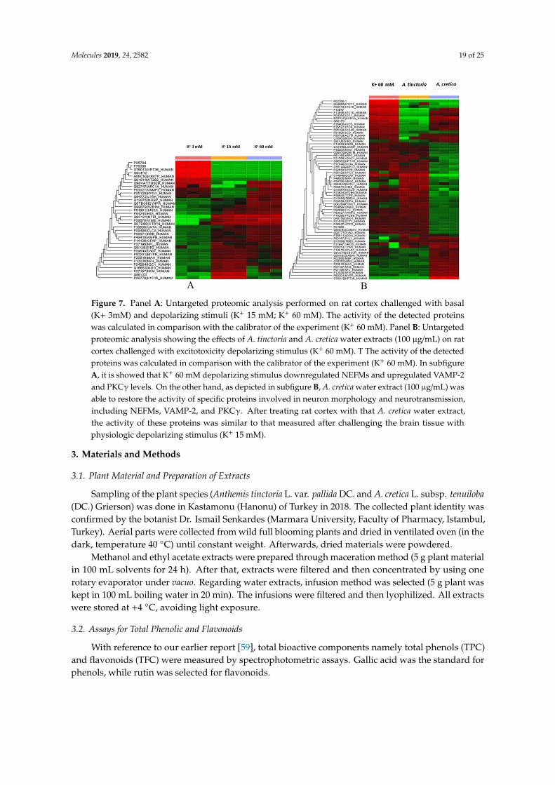
Particularly, untargeted proteomic analysis showed that K+ 60 mM was able to significantly downregulate neurofilament (NFEM) proteins (Figure 7A/supplementary data1), expressed along the axons and involved in axonal diameter regulation. Reduced NFEM levels have long been related to neurodegeneration [53]. The treatment of isolated rat cortex with ACT water extract was able to prevent NFEM downregulation, restoring the activity of NEFM proteins during K+ 15 mM physiologic depolarizing stimulus. While ATP water extract did not exert any relevant effect on NFEM level, in isolated rat cortex challenged with K+ 60 mM (Figure 7B/supplementary data 2). Conversely, K+ 60 mM stimulus led to significant upregulation of protein C kinase γ (PKCγ) and vesicle-associated membrane protein-2 (VAMP-2) (Figure 7A/supplementary data 2), compared to physiologic depolarizing stimulus (K+ 15 mM). VAMP-2 is placed on the membranes of neuronal endings’synaptic vesicles, playing a key role in synaptic vesicle fusion to the presynaptic neuronal ending membrane [54]. Multiple studies suggested upregulation of VAMP-2 level during hypoxia [55,56], which is strictly related to high K+ concentration-induced CNS injury [39]. PKCγ plays multiple roles in neuronal cells and eye tissues, such as regulation of the neuronal receptors GRIA4/GLUR4 and GRIN1/NMDAR1, modulation of receptors and neuronal functions related to sensitivity to opiates, pain and alcohol, mediation of synaptic function and cell survival after ischemia, and inhibition of gap junction activity after oxidative stress. Its level is positively related to migraine pathogenesis [57]. Additionally, PKCγ gene expression was observed in histidine triad nucleotide-binding protein 1 (Hint1) KO mice, that also showed increased anxiety-related behavior, compared to wild type control mice [58]. Also in this case, treatment of isolated rat cortex with ACT water extract was able to restore the activity of both VAMP-2 and PKCγ during K+ 15 mM-depolarizing stimulus (Figure 7A/supplementary data 2), further supporting the neuroprotective effects of this extract against the burden of oxidative stress and inflammation occurring in CSD.
Figure 6. Effect of A. tinctoria (A. T.) and A. cretica (A. C.) extracts (100 µg/mL) on lactate dehydrogenase(LDH) level, measured on isolated rat cortex challenged with basal (K+ 3mM) and depolarizing stimuli(K+ 15 mM; K+ 60 mM). Data are means ± SD of three experiments performed in triplicate. ANOVA,p < 0.0001; post-hoc, * p < 0.05 vs. K+ 60 mM control group.
In agreement with the antiradical activity (Table 2) and blunting effect on 5-HT turnover (Figure 5),ACT water extract samples has been subjected to a further proteomic study, in order to deepen ourknowledge about the putative mechanism of action related to neuroprotective effects. The deepeningabout ACT water extract was performed in comparison with the corresponding ATP extract that,despite showing a null effect on 5-HT turnover (Figure 5), displayed a significant inhibitory effect onK+ (60 mM)-induced LDH level (Figure 6).
Particularly, untargeted proteomic analysis showed that K+ 60 mM was able to significantlydownregulate neurofilament (NFEM) proteins (Figure 7A/Supplementary Data 1), expressed alongthe axons and involved in axonal diameter regulation. Reduced NFEM levels have long been relatedto neurodegeneration [53]. The treatment of isolated rat cortex with ACT water extract was able toprevent NFEM downregulation, restoring the activity of NEFM proteins during K+ 15 mM physiologicdepolarizing stimulus. While ATP water extract did not exert any relevant effect on NFEM level, inisolated rat cortex challenged with K+ 60 mM (Figure 7B/Supplementary Data 2). Conversely, K+ 60 mMstimulus led to significant upregulation of protein C kinase γ (PKCγ) and vesicle-associated membraneprotein-2 (VAMP-2) (Figure 7A/Supplementary Data 2), compared to physiologic depolarizing stimulus(K+ 15 mM). VAMP-2 is placed on the membranes of neuronal endings’synaptic vesicles, playinga key role in synaptic vesicle fusion to the presynaptic neuronal ending membrane [54]. Multiplestudies suggested upregulation of VAMP-2 level during hypoxia [55,56], which is strictly related tohigh K+ concentration-induced CNS injury [39]. PKCγ plays multiple roles in neuronal cells and eyetissues, such as regulation of the neuronal receptors GRIA4/GLUR4 and GRIN1/NMDAR1, modulationof receptors and neuronal functions related to sensitivity to opiates, pain and alcohol, mediationof synaptic function and cell survival after ischemia, and inhibition of gap junction activity afteroxidative stress. Its level is positively related to migraine pathogenesis [57]. Additionally, PKCγ
gene expression was observed in histidine triad nucleotide-binding protein 1 (Hint1) KO mice, thatalso showed increased anxiety-related behavior, compared to wild type control mice [58]. Also inthis case, treatment of isolated rat cortex with ACT water extract was able to restore the activity ofboth VAMP-2 and PKCγ during K+ 15 mM-depolarizing stimulus (Figure 7A/Supplementary Data 2),further supporting the neuroprotective effects of this extract against the burden of oxidative stress andinflammation occurring in CSD.
Molecules 2019, 24, 2582 19 of 25Molecules 2019, 24, x 18 of 24
Figure 7. Panel A: Untargeted proteomic analysis performed on rat cortex challenged with basal (K+ 3mM) and depolarizing stimuli (K+ 15 mM; K+ 60 mM). The activity of the detected proteins was calculated in comparison with the calibrator of the experiment (K+ 60 mM). Panel B: Untargeted proteomic analysis showing the effects of A. tinctoria and A. cretica water extracts (100 µg/mL) on rat cortex challenged with excitotoxicity depolarizing stimulus (K+ 60 mM). T The activity of the detected proteins was calculated in comparison with the calibrator of the experiment (K+ 60 mM). In subfigure A, it is showed that K+ 60 mM depolarizing stimulus downregulated NEFMs and upregulated VAMP-2 and PKCγ levels. On the other hand, as depicted in subfigure B, A. cretica water extract (100 µg/mL) was able to restore the activity of specific proteins involved in neuron morphology and neurotransmission, including NEFMs, VAMP-2, and PKCγ. After treating rat cortex with that A. cretica water extract, the activity of these proteins was similar to that measured after challenging the brain tissue with physiologic depolarizing stimulus (K+ 15 mM).
3. Materials and Methods
3.1. Plant Material and Preparation of Extracts
Sampling of the plant species (Anthemis tinctoria L. var. pallida DC. and A. cretica L. subsp. tenuiloba (DC.) Grierson) was done in Kastamonu (Hanonu) of Turkey in 2018. The collected plant identity was confirmed by the botanist Dr. Ismail Senkardes (Marmara University, Faculty of Pharmacy, Istambul, Turkey). Aerial parts were collected from wild full blooming plants and dried in ventilated oven (in the dark, temperature 40 °C) until constant weight. Afterwards, dried materials were powdered.
Methanol and ethyl acetate extracts were prepared through maceration method (5 g plant material in 100 mL solvents for 24 h). After that, extracts were filtered and then concentrated by using one rotary evaporator under vacuo. Regarding water extracts, infusion method was selected (5 g plant was kept in 100 mL boiling water in 20 min). The infusions were filtered and then lyophilized. All extracts were stored at +4 °C, avoiding light exposure.
3.2. Assays for Total Phenolic and Flavonoids
A B Figure 7. Panel A: Untargeted proteomic analysis performed on rat cortex challenged with basal(K+ 3mM) and depolarizing stimuli (K+ 15 mM; K+ 60 mM). The activity of the detected proteinswas calculated in comparison with the calibrator of the experiment (K+ 60 mM). Panel B: Untargetedproteomic analysis showing the effects of A. tinctoria and A. cretica water extracts (100 µg/mL) on ratcortex challenged with excitotoxicity depolarizing stimulus (K+ 60 mM). T The activity of the detectedproteins was calculated in comparison with the calibrator of the experiment (K+ 60 mM). In subfigureA, it is showed that K+ 60 mM depolarizing stimulus downregulated NEFMs and upregulated VAMP-2and PKCγ levels. On the other hand, as depicted in subfigure B, A. cretica water extract (100 µg/mL) wasable to restore the activity of specific proteins involved in neuron morphology and neurotransmission,including NEFMs, VAMP-2, and PKCγ. After treating rat cortex with that A. cretica water extract,the activity of these proteins was similar to that measured after challenging the brain tissue withphysiologic depolarizing stimulus (K+ 15 mM).
3. Materials and Methods
3.1. Plant Material and Preparation of Extracts
Sampling of the plant species (Anthemis tinctoria L. var. pallida DC. and A. cretica L. subsp. tenuiloba(DC.) Grierson) was done in Kastamonu (Hanonu) of Turkey in 2018. The collected plant identity wasconfirmed by the botanist Dr. Ismail Senkardes (Marmara University, Faculty of Pharmacy, Istambul,Turkey). Aerial parts were collected from wild full blooming plants and dried in ventilated oven (in thedark, temperature 40 ◦C) until constant weight. Afterwards, dried materials were powdered.
Methanol and ethyl acetate extracts were prepared through maceration method (5 g plant materialin 100 mL solvents for 24 h). After that, extracts were filtered and then concentrated by using onerotary evaporator under vacuo. Regarding water extracts, infusion method was selected (5 g plant waskept in 100 mL boiling water in 20 min). The infusions were filtered and then lyophilized. All extractswere stored at +4 ◦C, avoiding light exposure.
3.2. Assays for Total Phenolic and Flavonoids
With reference to our earlier report [59], total bioactive components namely total phenols (TPC)and flavonoids (TFC) were measured by spectrophotometric assays. Gallic acid was the standard forphenols, while rutin was selected for flavonoids.

Molecules 2019, 24, 2582 20 of 25
3.3. Antioxidant and Enzyme Inhibition Assays
Antioxidant properties of Anthemis extracts were determined by different in vitro assays namelyFRAP, CUPRAC, DPPH, ABTS, chelating and phospomolybdenum assays. Regarding enzymeinhibitory properties, some enzymes including tyrosinase, cholinesterase, α-amylase and α-glucosidasewere selected. All experimental procedures were given in our earlier report [59].
3.4. UHPLC-ESI/HRMS Analysis
Neochlorogenic acid (3-CQA) (4), chlorogenic acid (5-CQA) (6), apigenin (30), luteolin (31),quercetin (35), isoquercitrin (49), hyperoside (50), luteolin-7-O-rutinoside (54), and rutin (55) wereobtained from Extrasynthese (Genay, France). 3,4-O-diCQA (12), 3,4-O-diCQA (13), diosmetin (33),rhamnetin (37), isorhamnetin (38), luteolin-7-O-glucuronide (47), isorhamnetin-7-O-glucoside (51), andisorhamnetin-3-O-rutinoside (57) were purchased from PhytoLab (Vestenbergsgreuth, Germany).
The UHPLC-ESI/HRMS analyses were carried out on a Q Exactive Plus heated electrosprayionization (HESI-II) – high resolution mass spectrometer (HRMS) (ThermoFisher Scientific, Inc.,Bremen, Germany) equipped with an ultra-high-performance liquid chromatography (UHPLC) systemDionex Ultimate 3000RSLC (ThermoFisher Scientific, Inc.) [60].
3.5. Statistical Analysis for Antioxidant and Enzyme Inhibitory Assays
To interpret data gathered, R version 3.5.1 software (The R Foundation, St. Louis, MO, USA)with corrplot and mixOmics packages was used to perform univariate and multivariate statisticalanalyses. One way analysis of variance (ANOVA) and Tukey’s post hoc test were employed tocompare bioactive compounds and biological activities between the samples. Also, relationshipsbetween bioactive compounds and biological activities were evaluated by the estimation of Pearson’scorrelation. For multivariate analysis, biological activities of samples were firstly analyses by PCAto pinpoint similarities or differences between samples. Then sPLS-DA was applied by using thespecies and different extraction condition as class memberships respectively. This allowed bettercomparison between the two studied species and gauged the effect of the different extraction solventson biological activities.
3.6. Pharmacological Assays
3.6.1. Allelopathy Bioassay
Allelopathy bioassay was carried on the seeds of three commercial lettuces [Canasta (C), Romanaverde (RV) and Romana bionda (RB)], because of their fast germination rate and high sensitivity. Thedetailed procedure has been extensively reported in our recent paper [61]. Seeds were treated withscalar Anthemis extract concentrations (0.1–10 mg/mL) and considered germinated for observed rootlength ≥ 1 mm, after the third day of treatment.
3.6.2. Artemia salina Lethality Bioassay
Artemia salina lethality bioassay was performed as previously reported [61]. Brielfy, brine shrimplarvae were bred at 25–28 ◦C for 24 h in presence of Anthemis extracts (0.1–20 mg/mL) dissolved inincubation medium (artificial sea water). After incubation period (24 h) with extracts, the numberof surviving shrimps was evaluated and their vitality was compared to untreated control group.Experiments were carried out in triplicate, and percentage mortality was calculated with the followingequation: ((T − S)/T) × 100, where T and S are the total number of incubated larvae and survivalnapulii, respectively.

Molecules 2019, 24, 2582 21 of 25
3.6.3. In Vitro Studies
Rat hypothalamic Hypo-E22 cells were cultured in DMEM (Euroclone), as previously reported [48].The effects of Anthemis extracts (100 µg/mL) on Hypo-E22 cell line viability was evaluated through3-(4,5-dimethylthiazol-2-yl)-2,5-diphenyltetrazolium bromide (MTT) test.
3.6.4. Ex Vivo Cortical Spreading Depression Paradigm
Male adult Sprague-Dawley rats (200–250 g) were sacrificed by CO2 inhalation (100% CO2 at aflow rate of 20% of the chamber volume per min) and cortex specimens were immediately collectedand maintained in thermostatic shaking bath at 37 ◦C for 1 h (incubation period), in Krebs-Ringerbuffer at different K+ concentrations, as described below:
K+ 3 mM: corresponding to basal condition;K+ 15 mM: corresponding to physiologic depolarizing-stimulus;K+ 60 mM: corresponding to excitotoxicity depolarizing-stimulus.
The present experimental paradigm reproduced the neural pathophysiological condition namedcortical spreading depression (CSD), and was designed according to previous ex vivo and in vivostudies, describing the use of elevated K+ concentrations (up to 50–60 mM) to induce central nervoussystem (CNS) injury [38–40]. During incubation, cortex specimens were challenged with water, MeOHand EA A. tinctoria and A. cretica extracts (100 µg/mL). Afterwards, individual cortex slices werehomogenized in perchloric acid solution (0.05 M) in order to extract and quantify serotonin (5-HT)and its main metabolite (5-hydroxyindoleacetic acid, 5HIIA) via HPLC coupled to electrochemicaldetection, as previously reported [61,62]. The results were expressed as ng/mg wet tissue. Additionally,we carried out colorimetric evaluation of LDH level [52]. Finally, an untargeted proteomic profile wasperformed on rat cortex homogenate, as described below, in order to further elucidate the putativemechanism of action of Anthemis extracts.
3.7. Protein Extraction and Filter-aided Sample Preparation
After protein quantification, a volume corresponding to 50 ug of proteins was loaded onto aNanosep 10-kDa-cutoff filter (Pall Corporation, Michigan city, MI, USA) and digested according to theprotocol we routinely use in our laboratory. Briefly, the sample was washed twice with 200 µL ureabuffer (8 M urea, 100 mM Tris pH 8.5 in milliQ water) to remove the detergents present in the lysisbuffer. The proteins on the filter where subsequently reduced and alkylated by adding 100 µL of DTTsolution (8 mM dithiothreitol in urea buffer) and 100 µL of IAA solution (50 mM iodoacetamide inUrea buffer). For protein digestion, the buffer was exchanged with 50 mM ammonium bicarbonate,before adding trypsin to a ratio of 1:50 (enzyme:substrate). The reaction was incubated for 16 h at37 ◦C, and the mixture of peptides was collected by centrifugation, acidified with 10% trifluoroaceticacid and stored at −20 ◦C until analysis. The detailed description of mass spectrometric analysis isreported as “Supplementary Proteomic Analysis”.
3.8. Statistical Analysis for Pharmacological Assays
Statistical analysis was performed using GraphPad Prism version 5.01 for Windows (GraphPadSoftware, San Diego, CA, USA). Means ± S.E.M. were determined for each experimental groupand analyzed by one-way analysis of variance (ANOVA), followed by Newman-Keuls comparisonmultiple test. Statistical significance was set at p < 0.05. As regards the animals randomized for eachexperimental group, the number was calculated on the basis of the “Resource Equation” N = (E + T)/T(10 ≤ E ≤ 20; https://www.nc3rs.org.uk/experimental-designstatistics).

Molecules 2019, 24, 2582 22 of 25
4. Conclusions
Results collected in the present study indicated the promising biological effects of ATP and ACTextracts. As summarized in Figure 8, tested extracts showed significant antioxidant activity andpotent inhibitory effects against key enzymes, involved in Alzheimer’s disease, type II diabetes, andhyperpigmentation conditions. Particularly, EA and methanol extracts of both species showed higherenzyme inhibitory activity (at least 1.5 fold: Table 4) compared to water extracts. Conversely, ACTwater extract revealed more significant protective effects, as evidenced by reduced (−74%) cortex 5-HTturnover and restored activity of key proteins (i.e., NFEMs and PKCγ) involved in neuron morphologyand neurotransmission, in the selected model of neurotoxicity. In this context A. cretica water extractappears to be a good candidate for future investigations aimed to confirm and characterize the observedpharmacological effects, possibly through the use of independent experimental methods.
Molecules 2019, 24, x 21 of 24
higher enzyme inhibitory activity (at least 1.5 fold: Table 4) compared to water extracts. Conversely, ACT water extract revealed more significant protective effects, as evidenced by reduced (−74%) cortex 5-HT turnover and restored activity of key proteins (i.e., NFEMs and PKCγ) involved in neuron morphology and neurotransmission, in the selected model of neurotoxicity. In this context A. cretica water extract appears to be a good candidate for future investigations aimed to confirm and characterize the observed pharmacological effects, possibly through the use of independent experimental methods.
Figure 8. Protective effects induced by A. cretica and A. tinctoria extracts, as evidenced by the present pharmacological investigation.
Supplementary Materials: The following are available online. Supplementary Data 1; Supplementary Data 2; Supplementary Proteomic Analysis.
Author Contributions: Conceptualization, G.Z., L.M., G.O. and C.F.; methodology, M.R., D.Z-D., R.G.; software, L.M.; validation, C.F., M.R., R.G. and D.Z-D.; formal analysis, C.F., L.R.; investigation, A.C., S.L., A.M., G.M., M.P.D., S.D.S, I.S., C.M.N. P-A., K.I.S.; resources, G.O., L.M., C.F.; data curation, C.F., G.Z.; writing—original draft preparation, C.F., G.Z.; writing—review and editing, L.M., M.F.M., G.O.; visualization, L.B.; supervision, L.B.; project administration, L.R., G.O., C.F.; funding acquisition, G.O., C.F.
Funding: This research was supported by Italian Ministry of University Grant (FAR 2017 granted to Claudio Ferrante; FAR 2017 granted to Giustino Orlando).
Conflicts of Interest: The authors declare no conflict of interest
References
1. Davis, P. Flora of Turkey and the Aegean Islands; Edinburgh University Press: Edinburgh, UK, 1975; Volume 5.
2. Doğan, G.; Demirpolat, A.; Bağcı, E. Composition of the Volatile Oils of Anthemis coelopoda var. coelopoda from Turkey. 2015, 4, 259–265, doi:10.15671/hjbc.20154315983
3. Staneva, J.D.; Todorova, M.N.; Evstatieva, L.N. Sesquiterpene lactones as chemotaxonomic markers in genus Anthemis. Phytochemistry 2008, 69, 607–618.
4. Gonenc, T.; Argyropoulou, C.; Erdogan, T.; Gousiadou, C.; Juergenliemk, G.; Kıvçak, B.; Skaltsa, H. Chemical constituents from Anthemis wiedemanniana Fisch. & Mey. Biochem. Syst. Ecol. 2011, 39, 51–55.
5. Kilic, O.; Kocak, A.; Bagci, E. Composition of the volatile oils of two Anthemis L. taxa from Turkey. Z. Für Nat. C 2011, 66, 535–540.
6. Vaverkova, S.; Habán, M.; Eerna, K. Qualitative Properties of Anthemis Tinctoria and Anthemis nobilis,(Chamaemelum nobile) under Different Environmental Conditions. Ecophysiology of Plant Production Processes in Stress Conditions; In Proceedings of the fourth international conference, Rackova dolina, Slovakia, 12–14 September, 2001; pp. 1–2.
Figure 8. Protective effects induced by A. cretica and A. tinctoria extracts, as evidenced by the presentpharmacological investigation.
Supplementary Materials: The following are available online. Supplementary Data 1; Supplementary Data 2;Supplementary Proteomic Analysis.
Author Contributions: Conceptualization, G.Z., L.M., G.O. and C.F.; methodology, M.R., D.Z.-D., R.G.; software,L.M.; validation, C.F., M.R., R.G. and D.Z.-D.; formal analysis, C.F., L.R.; investigation, A.C., S.L., A.M., G.M.,M.P.D., S.D.S, I.S., C.M.N.P.-A., K.I.S.; resources, G.O., L.M., C.F.; data curation, C.F., G.Z.; writing—original draftpreparation, C.F., G.Z.; writing—review and editing, L.M., M.F.M., G.O.; visualization, L.B.; supervision, L.B.;project administration, L.R., G.O., C.F.; funding acquisition, G.O., C.F.
Funding: This research was supported by Italian Ministry of University Grant (FAR 2017 granted to ClaudioFerrante; FAR 2017 granted to Giustino Orlando).
Conflicts of Interest: The authors declare no conflict of interest
References
1. Davis, P. Flora of Turkey and the Aegean Islands; Edinburgh University Press: Edinburgh, UK, 1975; Volume 5.2. Dogan, G.; Demirpolat, A.; Bagcı, E. Composition of the Volatile Oils of Anthemis coelopoda var. coelopoda
from Turkey. Hacettepe J. Biol. Chem. 2015, 4, 259–265. [CrossRef]3. Staneva, J.D.; Todorova, M.N.; Evstatieva, L.N. Sesquiterpene lactones as chemotaxonomic markers in genus
Anthemis. Phytochemistry 2008, 69, 607–618. [CrossRef] [PubMed]4. Gonenc, T.; Argyropoulou, C.; Erdogan, T.; Gousiadou, C.; Juergenliemk, G.; Kıvçak, B.; Skaltsa, H. Chemical
constituents from Anthemis wiedemanniana Fisch. & Mey. Biochem. Syst. Ecol. 2011, 39, 51–55.5. Kilic, O.; Kocak, A.; Bagci, E. Composition of the volatile oils of two Anthemis L. taxa from Turkey. Z. Für
Nat. C 2011, 66, 535–540. [CrossRef]

Molecules 2019, 24, 2582 23 of 25
6. Vaverkova, S.; Habán, M.; Eerna, K. Qualitative Properties of Anthemis Tinctoria and Anthemis nobilis,(Chamaemelum nobile) under Different Environmental Conditions. Ecophysiology of Plant ProductionProcesses in Stress Conditions. In Proceedings of the Fourth International Conference, Rackova Dolina,Slovakia, 12–14 September 2001; pp. 1–2.
7. Aboee-Mehrizi, F.; Rustaiyan, A.; Zandi, H.; Ashkezari, M.D.; Zare, M. Chemical Composition andAntimicrobial Activity of the Essential Oil of Anthemis gayana Growing in Iran. J. Essent. Oil Bear. Plants2016, 19, 1557–1560. [CrossRef]
8. Bardaweel, S.K.; Tawaha, K.A.; Hudaib, M.M. Antioxidant, antimicrobial and antiproliferative activities ofAnthemis palestina essential oil. Bmc Complementary Altern. Med. 2014, 14, 297. [CrossRef] [PubMed]
9. Kurtulmus, A.; Fafal, T.; Mert, T.; Saglam, H.; Kivcak, B.; Ozturk, T.; Demirci, B.; Baser, K. Chemicalcomposition and antimicrobial activity of the essential oils of three Anthemis species from Turkey. Chem.Nat. Compd. 2009, 45, 900–904. [CrossRef]
10. Samadi, N.; Manayi, A.; Vazirian, M.; Samadi, M.; Zeinalzadeh, Z.; Saghari, Z.; Abadian, N.;Mozaffarian, V.-O.-A.; Khanavi, M. Chemical composition and antimicrobial activity of the essentialoil of Anthemis altissima L. var. altissima. Nat. Prod. Res. 2012, 26, 1931–1934. [CrossRef] [PubMed]
11. Eser, F.; Sahin Yaglioglu, A.; Dolarslan, M.; Aktas, E.; Onal, A. Dyeing, fastness, and cytotoxic properties, andphenolic constituents of Anthemis tinctoria var. tinctoria (Asteraceae). J. Text. Inst. 2017, 108, 1489–1495. [CrossRef]
12. Kizil, S.; Kayabasi, N.; Arslan, N. Determination of some agronomical and dyeing properties of dyer’schamomile (Anthemis Tinctoria L.). J. Cent. Eur. Agric. 2006, 6, 403–408.
13. Kültür, S. Medicinal plants used in Kırklareli province (Turkey). J. Ethnopharmacol. 2007, 111, 341–364.[CrossRef] [PubMed]
14. Zhang, S.; Won, Y.-K.; Ong, C.-N.; Shen, H.-M. Anti-cancer potential of sesquiterpene lactones: bioactivityand molecular mechanisms. Curr. Med. Chem. -Anti-Cancer Agents 2005, 5, 239–249. [CrossRef] [PubMed]
15. Gonenc, T.M.; Erdogan, T.F.; Demirci, B.; Baser, K.; Kivcak, B. Chemical composition of the essential oils of Anthemiscoelopoda var. bourgaei and A. aciphylla var. aciphylla. Chem. Nat. Compd. 2012, 48, 332–334. [CrossRef]
16. Pavlovic, M.; Kovacevic, N.; Tzakou, O.; Couladis, M. Essential oil composition of Anthemis triumfetti (L.)DC. Flavour Fragr. J. 2006, 21, 297–299.
17. Uysal, I.; Celik, S.; Oldacay, M. Antimicrobial activity of Anthemis coelopoda Var. bourgaei Boiss. andAnthemis tinctoria Var. pallida DC. species having ethnobotanical features. J. Appl. Sci. 2005, 5, 639–642.
18. Özüdogru, B.; Akaydın, G.; Erik, S.; Yesilada, E. Inferences from an ethnobotanical field expedition in theselected locations of Sivas and Yozgat provinces (Turkey). J. Ethnopharmacol. 2011, 137, 85–98. [CrossRef][PubMed]
19. Banagozar Mohammadi, A.; Torbati, M.; Farajdokht, F.; Sadigh-Eteghad, S.; Fazljou, S.M.B.; Vatandoust, S.M.;Golzari, S.E.J.; Mahmoudi, J. Sericin alleviates restraint stress induced depressive- and anxiety-likebehaviors via modulation of oxidative stress, neuroinflammation and apoptosis in the prefrontal cortex andhippocampus. Brain Res. 2019, 1715, 47–56. [CrossRef]
20. Prasanth, M.I.; Sivamaruthi, B.S.; Chaiyasut, C.; Tencomnao, T. A Review of the Role of Green Tea (Camelliasinensis) in Antiphotoaging, Stress Resistance, Neuroprotection, and Autophagy. Nutrients 2019, 11, 474.[CrossRef]
21. Clifford, M.N.; Johnston, K.L.; Knight, S.; Kuhnert, N. Hierarchical scheme for LC-MS n identification ofchlorogenic acids. J. Agric. Food Chem. 2003, 51, 2900–2911. [CrossRef]
22. Clifford, M.N.; Knight, S.; Kuhnert, N. Discriminating between the six isomers of dicaffeoylquinic acid byLC-MS n. J. Agric. Food Chem. 2005, 53, 3821–3832. [CrossRef]
23. Zheleva-Dimitrova, D.; Gevrenova, R.; Zaharieva, M.M.; Najdenski, H.; Ruseva, S.; Lozanov, V.; Balabanova, V.;Yagi, S.; Momekov, G.; Mitev, V. HPLC-UV and LC–MS analyses of acylquinic acids in Geigeria alata (DC)Oliv. & Hiern. and their contribution to antioxidant and antimicrobial capacity. Phytochem. Anal. 2017, 28,176–184. [PubMed]
24. Justesen, U. Collision-induced fragmentation of deprotonated methoxylated flavonoids, obtained byelectrospray ionization mass spectrometry. J. Mass Spectrom. 2001, 36, 169–178. [CrossRef] [PubMed]
25. Smelcerovic, A.; Lamshoeft, M.; Radulovic, N.; Ilic, D.; Palic, R. LC–MS Analysis of the Essential Oils ofAchillea millefolium and Achillea crithmifolia. Chromatographia 2010, 71, 113–116. [CrossRef]
26. Priestap, H.A.; Abboud, K.A.; Velandia, A.E.; Lopez, L.A.; Barbieri, M.A. Dehydroleucodin: a guaiane-typesesquiterpene lactone. Acta Crystallogr. Sect. E Struct. Rep. Online 2011, 67, o3470. [CrossRef] [PubMed]

Molecules 2019, 24, 2582 24 of 25
27. Michalska, K.; Zylewski, M.; Kisiel, W. Structure elucidation and complete NMR spectral assignments of twonew sesquiterpene lactone xylosides from Lactuca triangulata. Magn. Reson. Chem. 2008, 46, 1185–1187.[CrossRef] [PubMed]
28. Clifford, M.N.; Wu, W.; Kirkpatrick, J.; Kuhnert, N. Profiling the chlorogenic acids and other caffeic acidderivatives of herbal Chrysanthemum by LC− MS n. J. Agric. Food Chem. 2007, 55, 929–936. [CrossRef][PubMed]
29. Lobo, V.; Patil, A.; Phatak, A.; Chandra, N. Free radicals, antioxidants and functional foods: Impact onhuman health. Pharmacogn. Rev. 2010, 4, 118. [CrossRef]
30. Zengin, G.; Lobine, D.; Mollica, A.; Locatelli, M.; Carradori, S.; Mahomoodally, M.F. Multiple pharmacologicalapproaches on Fibigia eriocarpa extracts by in vitro and computational assays. Fundam. Clin. Pharmacol.2018. [CrossRef]
31. Ma, Y.-L.; Zhu, D.-Y.; Thakur, K.; Wang, C.-H.; Wang, H.; Ren, Y.-F.; Zhang, J.-G.; Wei, Z.-J. Antioxidant andantibacterial evaluation of polysaccharides sequentially extracted from onion (Allium cepa L.). Int. J. Biol.Macromol. 2018, 111, 92–101. [CrossRef]
32. Lionetto, M.G.; Caricato, R.; Calisi, A.; Giordano, M.E.; Schettino, T. Acetylcholinesterase as a biomarker inenvironmental and occupational medicine: new insights and future perspectives. Biomed Res. Int. 2013, 2013.[CrossRef]
33. Greig, N.H.; Lahiri, D.K.; Sambamurti, K. Butyrylcholinesterase: an important new target in Alzheimer’sdisease therapy. Int. Psychogeriatr. 2002, 14, 77–91. [CrossRef] [PubMed]
34. Pintus, F.; Sabatucci, A.; Maccarrone, M.; Dainese, E.; Medda, R. Amine oxidase from Euphorbia characias:Kinetic and structural characterization. Biotechnol. Appl. Biochem. 2018, 65, 81–88. [CrossRef] [PubMed]
35. Ali, H.; Houghton, P.; Soumyanath, A. α-Amylase inhibitory activity of some Malaysian plants used to treatdiabetes; with particular reference to Phyllanthus amarus. J. Ethnopharmacol. 2006, 107, 449–455. [CrossRef][PubMed]
36. Ouassou, H.; Zahidi, T.; Bouknana, S.; Bouhrim, M.; Mekhfi, H.; Ziyyat, A.; Aziz, M.; Bnouham, M. Inhibitionof α-Glucosidase, Intestinal Glucose Absorption, and Antidiabetic Properties by Caralluma europaea. Evid.-Based Complementary Altern. Med. 2018, 2018. [CrossRef] [PubMed]
37. Ohikhena, F.U.; Wintola, O.A.; Afolayan, A.J. Toxicity Assessment of Different Solvent Extracts of theMedicinal Plant, Phragmanthera capitata (Sprengel) Balle on Brine Shrimp (Artemia salina). Int. J. Pharmacol.2016, 12, 701–710.
38. Richter, F.; Eitner, A.; Leuchtweis, J.; Bauer, R.; Ebersberger, A.; Lehmenkühler, A.; Schaible, H.-G. Thepotential of substance P to initiate and perpetuate cortical spreading depression (CSD) in rat in vivo. Sci.Rep. 2018, 8, 17656. [CrossRef] [PubMed]
39. Raiteri, L.; Stigliani, S.; Zedda, L.; Raiteri, M.; Bonanno, G. Multiple mechanisms of transmitter releaseevoked by ‘pathologically’elevated extracellular [K+]: involvement of transporter reversal and mitochondrialcalcium. J. Neurochem. 2002, 80, 706–714. [CrossRef] [PubMed]
40. Sbrenna, S.; Marti, M.; Morari, M.; Calo, G.; Guerrini, R.; Beani, L.; Bianchi, C. Modulation of5-hydroxytryptamine efflux from rat cortical synaptosomes by opioids and nociceptin. Br. J. Pharmacol. 2000,130, 425–433. [CrossRef]
41. Supornsilpchai, W.; Sanguanrangsirikul, S.; Maneesri, S.; Srikiatkhachorn, A. Serotonin depletion, corticalspreading depression, and trigeminal nociception. Headache J. Head Face Pain 2006, 46, 34–39. [CrossRef]
42. Close, L.N.; Eftekhari, S.; Wang, M.; Charles, A.C.; Russo, A.F. Cortical spreading depression as a site oforigin for migraine: Role of CGRP. Cephalalgia 2018, 0333102418774299. [CrossRef]
43. Zarcone, D.; Corbetta, S. Shared mechanisms of epilepsy, migraine and affective disorders. Neurol. Sci. 2018,38, 73–76. [CrossRef] [PubMed]
44. Lee, J.; Chang, C.; Liu, I.; Chi, T.; Yu, H.; Cheng, J. Changes in endogenous monoamines in aged rats. Clin.Exp. Pharmacol. Physiol. 2001, 28, 285–289. [CrossRef] [PubMed]
45. Brunetti, L.; Orlando, G.; Ferrante, C.; Recinella, L.; Leone, S.; Chiavaroli, A.; Di Nisio, C.; Shohreh, R.;Manippa, F.; Ricciuti, A. Peripheral chemerin administration modulates hypothalamic control of feeding.Peptides 2014, 51, 115–121. [CrossRef] [PubMed]
46. Francisco, E.D.S.; Guedes, R.C. Sub-convulsing dose administration of pilocarpine reduces glycemia, increasesanxiety-like behavior and decelerates cortical spreading depression in rats suckled on various litter sizes.Front. Neurosci. 2018, 12, 897. [CrossRef] [PubMed]
Molecules 2019, 24, 2582 25 of 25
47. Mollica, A.; Stefanucci, A.; Zengin, G.; Locatelli, M.; Macedonio, G.; Orlando, G.; Ferrante, C.; Menghini, L.;Recinella, L.; Leone, S. Polyphenolic composition, enzyme inhibitory effects ex-vivo and in-vivo studies ontwo Brassicaceae of north-central Italy. Biomed. Pharmacother. 2018, 107, 129–138. [CrossRef] [PubMed]
48. Ferrante, C.; Recinella, L.; Locatelli, M.; Guglielmi, P.; Secci, D.; Leporini, L.; Chiavaroli, A.; Leone, S.;Martinotti, S.; Brunetti, L. Protective effects induced by microwave-assisted aqueous Harpagophytumextract on rat cortex synaptosomes challenged with amyloid β-peptide. Phytother. Res. 2017, 31, 1257–1264.[CrossRef] [PubMed]
49. Romano, A.; Pace, L.; Tempesta, B.; Lavecchia, A.M.; Macheda, T.; Bedse, G.; Petrella, A.; Cifani, C.;Serviddio, G.; Vendemiale, G. Depressive-like behavior is paired to monoaminergic alteration in a murinemodel of Alzheimer’s disease. Int. J. Neuropsychopharmacol. 2015, 18. [CrossRef]
50. Ramis, M.R.; Sarubbo, F.; Terrasa, J.L.; Moranta, D.; Aparicio, S.; Miralles, A.; Esteban, S. Chronicα-Tocopherol Increases Central Monoamines Synthesis and Improves Cognitive and Motor Abilities in OldRats. Rejuvenation Res. 2016, 19, 159–171. [CrossRef] [PubMed]
51. Chen, M.; Wang, T.; Yue, F.; Li, X.; Wang, P.; Li, Y.; Chan, P.; Yu, S. Tea polyphenols alleviate motorimpairments, dopaminergic neuronal injury, and cerebral α-synuclein aggregation in MPTP-intoxicatedparkinsonian monkeys. Neuroscience 2015, 286, 383–392. [CrossRef] [PubMed]
52. Menghini, L.; Leporini, L.; Vecchiotti, G.; Locatelli, M.; Carradori, S.; Ferrante, C.; Zengin, G.; Recinella, L.;Chiavaroli, A.; Leone, S. Crocus sativus L. stigmas and byproducts: qualitative fingerprint, antioxidantpotentials and enzyme inhibitory activities. Food Res. Int. 2018, 109, 91–98. [CrossRef]
53. Valdiglesias, V.; Fernández-Tajes, J.; Pásaro, E.; Méndez, J.; Laffon, B. Identification of differentially expressedgenes in SHSY5Y cells exposed to okadaic acid by suppression subtractive hybridization. Bmc Genom. 2012,13, 46. [CrossRef] [PubMed]
54. Jahn, R.; Südhof, T.C. Membrane fusion and exocytosis. Annu. Rev. Biochem. 1999, 68, 863–911. [CrossRef][PubMed]
55. Manzur, A.; Sosa, M.; Seltzer, A.M. Transient increase in rab 3A and synaptobrevin immunoreactivity aftermild hypoxia in neonatal rats. Cell. Mol. Neurobiol. 2001, 21, 39–52. [CrossRef] [PubMed]
56. Valdez, S.R.; Patterson, S.I.; Ezquer, M.E.; Torrecilla, M.; Lama, M.C.; Seltzer, A.M. Acute sublethal globalhypoxia induces transient increase of GAP-43 immunoreactivity in the striatum of neonatal rats. Synapse2007, 61, 124–137. [CrossRef] [PubMed]
57. Wu, B.; Wang, S.; Qin, G.; Xie, J.; Tan, G.; Zhou, J.; Chen, L. Protein kinase C γ contributes to centralsensitization in a rat model of chronic migraine. J. Mol. Neurosci. 2017, 63, 131–141. [CrossRef] [PubMed]
58. Varadarajulu, J.; Lebar, M.; Krishnamoorthy, G.; Habelt, S.; Lu, J.; Bernard Weinstein, I.; Li, H.; Holsboer, F.;Turck, C.W.; Touma, C. Increased anxiety-related behaviour in Hint1 knockout mice. Behav Brain Res. 2011,220, 305–311. [CrossRef] [PubMed]
59. Uysal, S.; Zengin, G.; Locatelli, M.; Bahadori, M.B.; Mocan, A.; Bellagamba, G.; De Luca, E.; Mollica, A.;Aktumsek, A. Cytotoxic and enzyme inhibitory potential of two Potentilla species (P. speciosa L. and P.reptans Willd.) and their chemical composition. Front. Pharmacol. 2017, 8, 290. [CrossRef]
60. Zengin, G.; Aktumsek, A.; Ceylan, R.; Uysal, S.; Mocan, A.; Guler, G.O.; Mahomoodally, M.F.; Glamoclija, J.;Ciric, A.; Sokovic, M. Shedding light on the biological and chemical fingerprints of three Achillea species(A. biebersteinii, A. millefolium and A. teretifolia). Food Funct. 2017, 8, 1152–1165. [CrossRef]
61. Ferrante, C.; Recinella, L.; Ronci, M.; Menghini, L.; Brunetti, L.; Chiavaroli, A.; Leone, S.; Di Iorio, L.;Carradori, S.; Tirillini, B. Multiple pharmacognostic characterization on hemp commercial cultivars: Focuson inflorescence water extract activity. Food Chem. Toxicol. 2019, 125, 452–461. [CrossRef]
62. Ferrante, C.; Orlando, G.; Recinella, L.; Leone, S.; Chiavaroli, A.; Di Nisio, C.; Shohreh, R.; Manippa, F.;Ricciuti, A.; Vacca, M. Central inhibitory effects on feeding induced by the adipo-myokine irisin. Eur. J.Pharmacol. 2016, 791, 389–394. [CrossRef]
Sample Availability: Samples of the extracts are available from the authors.
© 2019 by the authors. Licensee MDPI, Basel, Switzerland. This article is an open accessarticle distributed under the terms and conditions of the Creative Commons Attribution(CC BY) license (http://creativecommons.org/licenses/by/4.0/).