Embed Size (px)
Citation preview

Full Paper
22
Multi-Featured Macroporous Agarose–AlginateCryogel: Synthesis and Characterization forBioengineering Applications
Anuj Tripathi, Ashok Kumar*
In this study agarose–alginate scaffolds are synthesized using cryogelation technology indifferent formats like monolith, sheet, discs, and beads, and show amiable mechanicalstrength like soft tissue properties and high interconnected macroporous degradable archi-tecture. In cell–material interactions, fibroblast (NIH-3T3) cells showed good adherence andproliferation on these scaffolds presenting its potentialapplication in soft tissue engineering. The application ofcryogel beads and monoliths was also examined by theefficient immobilization of bacterial cells (BL21) on thesematrices revealing their use for recovery of product fromcontinuous fermentation systems without cell leakage.These scaffolds also showed potential as a filter forrepeated recovery of heavy metal binding, such as copperand nickel from the waste water. The cryogels preparedherein do have a number of unique features that makethem an important class of soft materials for developingmulti-featured scaffolds as a novel carrier for bioengi-neering applications.
Introduction
Hydrogels are a water insoluble, hydrophilic, three-dimen-
sional (3D)networkofcrosslinkedpolymerchains thathave
the capability of imbibing high water content or biological
fluids. Hydrogels have attracted a great deal of attention
and significant progress has been made in designing,
synthesizing, and using these materials for many bioengi-
neering applications. In addition, they have been widely
used in drug and protein delivery systems, cell cultures,
tissue engineering, medical and biological sensors, water
absorbent pads, hygiene products, breast implants, wound
A. Tripathi, A. KumarDepartment of Biological Sciences and Bioengineering, IndianInstitute of Technology Kanpur, 208016 Kanpur, IndiaFax: þ91-512-2594010; E-mail: [email protected]
Macromol. Biosci. 2011, 11, 22–35
� 2011 WILEY-VCH Verlag GmbH & Co. KGaA, Weinheim wileyonline
dressing, and for enzyme and cell immobilization, etc.[1]
Although hydrogels have shown significant potential
towardsbiomedical andbiological applications, sometimes
conventional hydrogels do not meet the set criteria for a
specific application. For example, most hydrogels show
poor mechanical strength,[2] poor mass transport, and low
swelling behavior because of small pore architecture.[3] To
overcome these limitations, different kinds of techniques
have been applied tomodify thehydrogels, such as solvent-
casting particulate-leaching methods, gas foaming meth-
ods, fiber mesh approaches, freeze-drying methods, solu-
tion castingmethods, etc[3] and thesemethods enhance the
properties of hydrogels as a scaffold.
Recently, cryogel technology has emerged as a potential
approach to produce polymeric scaffolds, so called cryo-
gels.[4] The interconnected pore morphology in a cryogel
provides a capillarynetwork throughwhich the solvent can
library.com DOI: 10.1002/mabi.201000286

Multi-Featured Macroporous Agarose–Alginate Cryogel: Synthesis and Characterization . . .
www.mbs-journal.de
flow by convective mass transport. The macroporosity
allows unhindered influx of high-molecular-weight solutes
andnutrients, aswell as the transport of cellularwaste. This
also enhances the cellular proliferation and provides
increased matrix diffusion in the gel.[5] Classical hydrogels
swell slowly because of the low diffusion or low capillary-
driven absorption, whereas macroporous cryogels show
quick swelling kinetics with substantial viscoelastic
properties, which slightly vary with the material nature
and environmental conditions. With these key advantages
over hydrogels, cryogels have been used for various
applications in biotechnology and biomedical sciences.[6]
In particular, biomaterials based on natural polymers have
been frequently used for biological and biomedical
applications because of their good biocompatibility,
biodegradability, low toxicity, and other key advantages.
Ideally, materials should be designed and engineered in
such a manner so as to address the usage in more than one
particular application area. The selection of polymers for
scaffold synthesis is a crucial step while keeping in mind
these characteristics for providing an ideal scaffold/matrix
property.
Here we have selected two natural polysaccharides, i.e.,
agarose and alginate, for the synthesis of a proper matrix/
scaffold. Both are sufficiently biodegradable, biocompati-
ble, and inert bynature andhave soft tissue likemechanical
properties.[4] Because of these properties, agarose gels have
been investigated as carriers for various biomedical
applications.[7] On the other hand, alginate is regarded as
one of the most versatile polysaccharides considering its
large number of different applications.[8] On the basis of
these properties, these two polymers can have great
potential for bioengineering applications as a composite
scaffold. On the one hand, the homopolymer scaffolds of
agarosehave showna rigid gel structurewhile alginate gels
have a weak mechanical stability. However, the combina-
tion of the above properties of the polymers has not yet
been explored as a composite macroporous scaffold.
The goal of this study is to assess the macroporous
agarose–alginate (AA) matrix/scaffold for functional (bio-
logical, biochemical, and biomechanical) properties and its
multi-disciplinary applications in the bioengineering field.
The use of cryogel technology to produce a customized
novel AA scaffold with a controlled internal micro-
architecture, and characterized with respect to physical
and chemical means, will be addressed for various
applications.
Experimental Part
Materials
Low viscosity alginic acid sodium salt (from brown algae), N-(3-
dimethylaminopropyl)-N- ethylcarbodiimide hydrochloride (EDC;
www.MaterialsViews.com
Macromol. Biosci. 20
� 2011 WILEY-VCH Verlag Gmb
FW 191.71), Dulbecco’s modifiedmedium (DMEM), 4-6-diamidino-
2-phenylindole (DAPI), andpenicillin-streptomycinantibioticwere
purchased from Sigma Chemical Co. (St. Louis, MO, USA). Agarose
(low electroendoosmosis (EEO), gelling temperature �38–40 8C)was purchased from Sisco Research Laboratories (Mumbai, India).
N-Hydroxysuccinimide (NHS) was bought from Spectrochem
(Mumbai, India). Fetal bovine serum (FBS) was purchased from
Hyclone (UT, USA). Fibroblast (NIH-3T3) was procured from NCCS
(Pune, India). Other chemicals were of analytical grade and were
used without any further purification.
Synthesis of AA Cryogel Monolith
AA cryogels were synthesized using EDC with NHS for chemical
crosslinking. Low viscosity alginate solution (3.75%) was prepared
in a plastic tube (50mL) using deionizedwater as a solvent. On the
other hand, agarose (Low EEO; gelling temperature 38–40 8C)solutions (6% and 8%)were prepared in deionizedwater by putting
the agarose-containing plastic tube (50mL) in boiling water for
about 30min or until the solution become transparent. Four
milliliters of the stock solution of the alginate (3.75%) was then
added to 5mL of completely dissolved hot agarose solution (6% or
8%) and this wasmixed by vortexing. This heterogeneous solution
was incubated at 60 8C in a water bath for 5–10min to allow good
mixing of alginate with the agarose polymer chains. The mixture
was then taken out from the water bath and cooled at room
temperature. When the temperature of heterogeneous solution
reached 45 8C, 500mL of 0.35M freshly prepared EDC solution was
added, whichwas followed by adding 500mL of 0.2M NHS solution
and was thoroughly mixed by vortexing. The total volume of the
reaction mixture was 10ml, where the final concentration of
alginate was 1.5% and the final agarose concentration was either
3%or4%dependinguponthestockconcentrationsof6%or8%used,
respectively. The final crosslinker concentration of EDC and NHS
was 17.5�10�3 and 10�10�3M, respectively. The crosslinker-
containing heterogeneous reaction mixture of AA was added to a
5mLplastic syringeand immediately incubatedat–12 8Cfor16h ina liquid cryostat (Julabo, Seelbach, Germany). After incubation of
gels for overnight, they were then thawed in deionized water and
dried at room temperature for further characterization and use.
Synthesis of AA Cryogel Beads
The aqueous solution of alginatewasmixedwith a freshly prepare
aqueous solution of agarose (as mentioned above). The ratio of
agarose toalginateconcentrationwas4 :1.5and3 : 1.5. Themixture
of AA was incubated for 5 to 10min at 60 8C and then the
heterogeneoussolutionwas left to cool to roomtemperature.When
the temperature of the polymer solution came down to 45 8C, EDCfollowed by NHS was added andmixed by vortexing. The solution
was transferred into a disposable plastic syringe and added drop-
wise into moderately frozen paraffin oil. The added polymer
droplets took a round shape in paraffin oil. The whole solution
containing beads in paraffin oil was incubated at subzero
temperature, i.e., �20 8C for 16h and the beads were then taken
out from the oil. The washing of the cryogel beads was repeated
11, 11, 22–35
H & Co. KGaA, Weinheim23

24
www.mbs-journal.de
A. Tripathi, A. Kumar
using phosphate buffered saline (PBS, pH 7.4) overnight under
gentle magnetic stirring to remove paraffin oil completely.
Characterization of the Synthesized AA Cryogel and
Morphological Analysis
The characterization of AA cryogels was done using samples 5mm
in height and 13mm in diameter prepared from the monolith
cryogelandthemorphologicalanalysisofall thedesignedformatof
cryogels were also performed as per these dimensions.
SEM and Porosity Analysis
The morphology of the synthesized AA cryogels was analyzed by
scanning electron microscopy (SEM). SEM allows direct measure-
ment of porosity, average diameter of pores, and strut thickness by
image analyzing software. The cryogel sampleswere prepared and
vacuum dried overnight using vacuum desiccators to remove any
moisture content to make an effective coating for further high
resolution microscopy analysis. Samples were then coated with
gold using a sputter coater (Vacuum Tech, Bangalore, India). SEM
examinations were made on a FEI Quanta 200 at high vacuum at
20kV with spot size of 3.5mm.
The porosity of AA cryogels was theoretically calculated
according to Archimede’s principle. By this method, the dry mass
of cryogel was recorded. The test sample was submerged under
water in a specific gravity bottle and the submerged mass of the
cryogel sample was recorded. The cryogel was then taken out and
the wet mass was recorded. Triplicate samples were used for the
study. These values were used to calculate the porosity of the
cryogel by Equation (1):
Porosity ¼ Mw�Mdð Þ= Mw�Msubð Þ (1)
where, Mw is the water saturated wet mass of the cryogel, Md is
the drymass of the cryogel, and Msub is the submergedmass of the
cryogel.
Micro-Computed Tomography (m-CT)
The micro-computed tomography (m-CT) technique was used to
analyze the 3D architecture and structural homogeneity of the
porousmaterial.Analysisof theAAcryogelwas carriedoutusingm-
CT equipment (Skyscan 1174, Belgium) with associated software
NRECON. The m-CT technique provided important information
about the shape and compactness of material without physically
sectioning them.
Permeability and Flow Rate Measurement
The hydraulic permeability was determined using Darcy’s law to
describe the relationship of the liquid flow rate versus pressure in
polymericmonoliths. The cryogel samples (13mmdiameter, 5mm
height) were prepared and placed in the permeability measure-
ment setup.[9] Under a constantwater pressureheadappliedon the
Macromol. Biosci. 2
� 2011 WILEY-VCH Verlag Gmb
porous material in the experimental setup, the water flushed out
from the outlet in 2min was collected and weighed. While in a
control experiment, a cryogelwas not put in the experiment setup.
The permeability was calculated by applying the recorded data to
Equation (2):[9]
011, 11,
H & Co
k ¼ DX
A � MB2� 2p2r4
MB1=MB2ð Þ2�1(2)
Where, K is the hydraulic permeability of the porous material, A is
the flushing area of the cryogel, DX is the thickness of the scaffold,
MB1 is the mass flow rate from the outlet of the control setup, and
MB2 is the mass flow rate from the outlet of the test setup.
The flow rate measurement was done using a peristaltic pump,
which was calibrated accordingly to Adrados et al.[10] against the
flow ratewithout connecting the test sample in between the path.
Thewaterflowrateof theAAcryogelwasdeterminedby taking the
same size (1.3 cm diameter, 2 cm height) cylindrical monolithic
cryogel in a swollen state (water saturated) and inserting it into the
same size of plastic syringe. The inlet and outlet of the syringewas
connected to the preset pumpand registering themaximumwater
flow limit (mL �min�1) of samples till the test sample was not
showing any back pressure.
Swelling Kinetics and Swelling Ratio
The swelling kinetics was carried out using a conventional
gravimetric procedure.[11] This method was used to determine
thewateruptake capacity of aporousmaterialwith respect to time
at a particular temperature. The equilibrium swelling measure-
ments were carried out in deionized water at room temperature
(�25 8C). Cryogel sections of 13mm diameter and 5mm height
were prepared from a cylindrical monolith cryogel. All sections
were dried at 60 8C in an oven. The initial dry weight of all the
cryogel sections was measured and then samples were placed in
deionized water. The samples were removed from the water and
weighed at regular time intervals. At least four samples with a
similar height and diameter were used for the study. The
percentage water uptake capacity was determined using
Equation (3):
Wu ¼ 100� Wt�Wg
� ��We
� �(3)
where Wu is thewater uptake capacity, Wt is the weight at regular
time intervals, Wg is the weight of the dry cryogel, and We is the
weight of water in the swollen gel at swelling equilibrium at a
particular temperature.
However, the swelling ratio (SR) is an important parameter
which reveals the solvent absorption capacity of sample. It is
calculated as per Equation (4):
SR¼ Ws�Wdð Þ=Wd (4)
where Ws is the weight of the swollen gel and Wd is the weight of
the dry gel.
22–35
. KGaA, Weinheim www.MaterialsViews.com

Multi-Featured Macroporous Agarose–Alginate Cryogel: Synthesis and Characterization . . .
www.mbs-journal.de
Density Measurement
The ratio of the wet weight as well as dry weight of the cryogels
with respect to their volume was used to obtain the wet and dry
densities of the test samples. The apparent density in g � cm�3 was
calculated from Equation (5):
www.M
r ¼ W=p� D=2ð Þ2 � H (5)
ere, r is the apparent density, W is the weight of the cryogel
Whsample in grams,D is the diameter of the sample in cm, andH is the
thickness of the sample in cm.
Mechanical Analysis
Unconfined Compression Analysis
For the initialmechanical characterization of the cryogels, uniform
gel samples were prepared. The height and diameter of the disc-
shaped samples were 5mm and 13mm, respectively. Cryogel
samples were saturated with PBS (pH 7.4) and were tested in
unconfined compression using a custom computer controlled
mechanical testing system (Zwick/Roell Z010, Germany). The
sampleswereuniaxially compressedbyplacingbetween twoarms
of a load frame. The test were conductedwith a 10 kN load cell at a
ramp rate of 1mmmin�1 up to 90% strain of the total length of the
samples at room temperature (�25 8C). At least four samples were
used to calculate the average unconfined compressive modulus
from the stress (kPa) and strain (%) graph shape of unconfined
mechanical test.
Cyclic Deformation Analysis (Fatigue Test)
AA cryogel samples weremade according to themethod described
earlier. The test samples of similar size (13mmdiameter and 5mm
height) were prepared from the freshly synthesized monolith.
These cryogel sampleswereaxially compressedup to40%strain for
100 000 cycles in deionized water at room temperature (�25 8C).Cyclic deformationwasmeasured for each sample across the range
of2and5HzonMTS,810MaterialTestSystem(USA).Aplasticplate
surface with circular grooves filled with water was used on the
testing apparatus to keep the samples stationary and saturated
during testing. The mass and dimensions of the samples were
measuredbeforeandafter fatiguecycling todetermine theeffect of
cyclic deformation.
In vitro Degradation Measurement
Thescaffoldwaspreparedasa5mmdisk format,withadiameterof
13mm from the cylindrical monolithic cryogel and then sterilized
by1h incubation in70%ethanol. The sampleswereplaced in50mL
tightlycappedplastic tubes,eachtubecontaining10mLof0.1MPBS
(pH 7.4). The samples were incubated at 37 8C for various time
periods up to eight weeks. At each time point, two samples were
taken out from the PBS, washed with distilled water, and air dried
overnight at room temperature. The corresponding initial dry
weight (WI) andfinal dryweight (WF)was recordedbeforeandafter
incubationof the samples and theweight losswas calculatedusing
aterialsViews.com
Macromol. Biosci. 20
� 2011 WILEY-VCH Verlag Gmb
Equation (6):
11, 11,
H & Co
Weight loss %ð Þ ¼ WI�WFð Þ=WI½ ��100 (6)
Microbial Cell Immobilization
For cell adherence studies, AA cryogel discswere prepared from the
cryogel monolith. The discs were sterilized by incubating in 70%
ethanol for 2 h. After incubation, samples were kept in vacuum
desiccators overnight to remove ethanol content completely. On
the other hand, E.coli cell (strain BL-21) culture in LBmedium in the
presence of ampicillin was setup. The bacterial culture was
incubated at 37 8C till its optical density reached 0.4 at 600nm.
Each cryogel disc pre-saturated in sterile PBS was incubated with
one milliliter of active bacterial culture broth for 15 to 20min at
room temperature inside the laminar hood. The sampleswere then
removed and gently washed with sterile PBS for 5min. For
analyzing the microbial cell adherence, the cryogel sections were
immediately fixed using 2.5% glutaraldehyde solution for over-
night incubation at room temperature. After incubation, sections
were again gently washed with sterile PBS (pH 7.4) followed by
dehydration by gradually increasing the concentration of ethanol
from20 to 100%with10% increments and the incubation timewas
10min in eachdehydration step. The sectionswere thenallowed to
dry at room temperature to evaporate ethanol content from the
samples. For entrapment, the bacterial cell mass (25mg) was
dissolved in 10mL of polymer mixture before and after bead
preparation and to fix the cells asmentioned earlier. The surface of
dried cryogel sections was analyzed by SEM after gold coating.
Mammalian Cell Culture
The cryogel sections were cut into two different thicknesses. The
5mm height and 13mm diameter sections were used for cell
viability/proliferation analysis, while thin 100mm sections were
used for fluorescent microscope observations. The thin sections
wereused for thefluorescent studybecause the ease of penetration
of light facilitates better imaging and understanding of the
growing cells. The sampleswere sterilizedwith70%ethanol for 4 h.
Samples were taken out from ethanol and kept in a 24-well tissue
culture plate inside a laminar hood. All the samples were gently
washed four times with sterile PBS (pH 7.2). Each step of washing
was for 10–15min. Before seeding, sampleswere equilibratedwith
Dulbecco’s modified eagle’s medium (DMEM) that contained 10%
fetal bovine serum and 1% streptomycin/penicillin for 4h.
Fibroblasts (NIH-3T3) were cultured and cell growth was
observed under a fluorescent microscope using 100mm sections,
while 5mm thick samples were used for cell proliferation assays.
Fibroblast cells were suspended in culture medium and were
seeded drop-wise to each AA cryogel sample (50mL on 100mm
sections and 500mL on 5mm thick sections; cell density 1�106 cellsmL�1). The cell-seeded scaffold sections were placed in a
humidified 5% CO2 atmosphere at 37 8C for up to one week. The
culture medium was exchanged with the fresh medium every
alternate day during the cell culture period.
22–35
. KGaA, Weinheim25

26
www.mbs-journal.de
A. Tripathi, A. Kumar
Cell Viability/Proliferation Analysis (MTT Assay)
The effect of the hybrid AA cryogel on themetabolic activity of the
fibroblasts (NIH-3T3)wasaccessed invitro at apredetermined time
intervals, i.e., every alternate day up to one week. The cellular
activitywasmeasured at a physiological pHof 7.4using a standard
protocol of 3-(4,5-dimethylthiazol-2-yl)-2,5-diphenyltetrazolium
bromide (MTT) assay to determine the optical density of the end
product at 570nm.[4]
Heavy Metal Binding
In the initial experiment, the potentiality of the AA cryogel was
examined for heavy metal binding and recovery by visual
observations. Copper sulfate and nickel sulfate solution (0.5M)
were used in these experiments. The AA cryogel monoliths (1.3 cm
diameter and 3 cm height) were incubated in both copper sulfate
andnickel sulfate solution for 1 h each. The cryogelmonolithswere
taken out in fresh tubes and washed with PBS to remove the non-
specificallyboundmetal solution.The repeatedlybindingofmetals
wasevaluatedby incubating themetalboundmonolith in0.1MHCl
solution to recover the bound metal and again incubated in the
same metal solutions for up to five cycles.
Results and Discussion
Synthesis of AA Cryogels
AAcryogelswere synthesized by varying the concentration
of agarose in the co-polymer and using crosslinking agents
like EDC andNHS. The synthesis is based on cryogelation of
agarose and alginate chains at subzero temperatures in the
presence of a crosslinker. The synthesized AA cryogels are
white in color and retained a three-dimensional architec-
ture even on air drying (Figure 1). On complete drying these
cryogels has shrunk approximately 1mm in size from the
original size of 13mm diameter and 5mm height. While
soaking in water they return into their original shape and
size. The crosslinking reaction between agarose and
alginatepolymer chainsusingEDChasnotbeen specifically
studied yet, but previous studies suggest that EDC may be
involved in the crosslinking within the carboxy group rich
Figure 1. The digital images of different format of AA cryogelshowing a) sheet, b) disk, c) monoliths, and d) beads.
Macromol. Biosci. 2
� 2011 WILEY-VCH Verlag Gmb
in alginate chains,[12] or in acid anhydride formation
between two carboxy groups of alginate by the action of
EDC and eventually the resultant acid anhydride may
readily react with a hydroxy group of agarose to form an
ester bond.[13] The use of NHS to improve the performance
of EDC crosslinking iswell documented in the literature.[14]
Therefore, the reaction conditions for the crosslinking of
agarose and alginate with respect to crosslinker concentra-
tionswereoptimizedbyphysicalobservationsby thenaked
eyeof thesynthesizedmatrices inorder toachieveascaffold
with good mechanical integrity. In addition, initial experi-
ments with different concentrations of agarose and
alginate polymers were attempted to synthesize the best
mechanically stable cryogel with high macroporous
architecture. We selected 3:1.5% and 4:1.5% agarose to
alginate ratios in the cryogels, respectively, for further
study, because they showed good stability and a better
macroporous structure than the other compositions (data
not shown). The characterizations of the matrix and its
screening for various applications were performed using
these two compositions.
In a separate set of control experiments in order to check
the crosslinker effect, agarose and alginate aqueous
solutions were mixed a) without using crosslinker, b) only
in thepresenceofEDC,andc)only inthepresenceofNHS.All
the combinations of polymer mixture were incubated in a
5mL plastic syringe at –12 8C for 16h. After completing the
appropriate time of incubation period, gels were thawed in
deionized water at room temperature. In all the sets of
experiments including thecontrol, itwasobserved thatgels
could be formed but did not have the properties of the
cryogels of AA synthesized in the presence of EDC and NHS
together. All the control gelswere brittle in naturewith low
elasticity andhad small pores likeahydrogel synthesizedat
room temperature, while the AA cryogels synthesized at
subzero temperatures in the presence of EDC andNHSwere
soft, spongy, elastic and had a macroporous architecture.
This particular polymer combination was used to synthe-
size cryogels in other types of formats, i.e., monoliths, disks,
sheets, and beads (Figure 1). These formats can have
significant potential for various biological applications.
Monoliths and beads can be used for direct recovery of a
product from an immobilized cell system in a continuous
system, while the disk and sheet formats of the gels can be
used as cell supports in three-dimensional architecture for
tissue engineering applications. Agarose itself can by
physically crosslinked and make a porous three-dimen-
sional scaffold while the presence of alginate in the
composite increased the softness of the cryogel as well as
the spongynature.On theotherhand, thealginatepresence
is also beneficial to cultivate different cell types for tissue
engineering applications[15] and immobilization pur-
poses.[16] Alginate has also been used to capture heavy
metals and bacteria from waste water.[17]
011, 11, 22–35
H & Co. KGaA, Weinheim www.MaterialsViews.com

Figure 2. SEM image of AA cryogel. a) AA cryogel containing 3%agarose (ratio; 3 : 1.5%) and b) AA cryogel containing 4% agarose(ratio; 4 : 1.5%). Both (a) and (b) show low magnification images intheir insets, suggesting a uniform pore distribution within themonoliths.
Multi-Featured Macroporous Agarose–Alginate Cryogel: Synthesis and Characterization . . .
www.mbs-journal.de
Morphological Analysis (SEM, Porosity, and m-CTAnalysis)
Microstructure analysis of macroporous AA cryogels was
evaluated by various microscopic observations. The SEM
observations of various formats of the AA cryogel showed
slight variation in their microstructure (Figure 2). The
monolith sections of the cryogel showed a comparatively
smaller pore diameter than thebead format. Thepore range
of cryogel sections of the monolith was 40 to 200mmwith
an average pore diameter in the approximate range of 80 to
125mm in 3:1.5% AA. On the other hand, AA (4:1.5%)
showed a pore size of 30 to 160mm and an average pore
diameter lying in the range of 60 to 85mm. The pore size
range of the cryogel sheet was more or less in the same
range as that of the monolithic cryogels. The pore range of
the internal section of the cryogel beads was 60 to 180mm
www.MaterialsViews.com
Macromol. Biosci. 20
� 2011 WILEY-VCH Verlag Gmb
and the average pore diameter was lying in the range of 95
to 127mm.
TheSEMobservationshowedauniformporedistribution
in the whole monolithic scaffold of both compositions, i.e.,
3:1.5% and 4:1.5% (Figure 2a and 2b). The pore diameter
range was higher in the 3:1.5% monolith in comparison to
4:1.5%. This might be because of the variation in agarose
polymer concentration, whereas, the formation of bigger
pores in beads compared to themonolith and sheetmay be
because of the better heat transfer abilitywithin the beads.
The size of the beads and the method used for bead
formation provide a high surface area and surroundings
within the cryogelation system, which can better circulate
and maintain the temperature. On the other hand, during
the synthesis of the monolith and sheet, the moulds used
can slightly affect theproper transport of temperature from
the cooling system to polymer system. In the cryogel
formation, a proper cooling systemhas an important role in
the formation of the desired cryogel. The rate of ice crystal
formation and crosslinking/gelation of the polymer or
polymer mixture can occur simultaneously. At subzero
temperatures most of the solvent is frozen, while that part
of solvent left unfrozen is called the unfrozen liquid
microphase. The dissolved substances in the unfrozen
phase concentrate and undergo chemical reactions, which
leads to gel formation. The ice crystals of solvent in the
frozenphase act as porogens. After thawing the ice crystals,
a system of a large interconnected porous network is
formed within the gel. So, during the cryogelation process,
variations in the temperature and other parameters can
change the structural homogeneity of a cryogel. In Figure 3,
micrographs of cryogel beads revealed that the outer
surfacewasquite different than the internalmorphologyof
the surface. The outer surface of the bead has smooth nano-
grooves (Figure 3a) although the inner part has a macro-
porous architecture (Figure 3b). This morphology may
provide a novel system to immobilize the cells or enzyme
for effective recoveryof theproduct. Thegroovespresent on
the surface of the cryogel beads help the diffusion of
product from inside the bead to the medium outside
without cell leakage. On the other hand, variation in the
agarose ratio during the cryogel synthesis has shown a
change in the property of the cryogel structure. The SEM
observation of theAAcryogel in the sheet format suggested
that it has a uniform porous architecture (Figure 3c) and
these pores were interconnected (Figure 3d, where (d) is a
magnified image of (c)). During the observations, cryogel
test samples that have a higher agarose content (4%)
showed a tight and smooth porous architecture than low
agarose-containing cryogels, i.e., 3%. The higher concentra-
tion of agarose in the composite also increased the wall
thickness as well as mechanical stability of the gel. The
cryogels show good interconnectivity revealed by inverted
microscope observations using ethidium bromide (EtBr)
11, 11, 22–35
H & Co. KGaA, Weinheim27

Figure 3. SEM images of cryogel beads and sheet. a) The outer part of the cryogel bead and b) the inner surface with correspond inset imagesat high magnification. The inset image of (a) shows nanometer-range grooves on the outer surface and the inset image of (b) suggeststhe internal macroporous bead structure. c) The side view micrograph of the sheet format of AA cryogel sheets containing 3% agarose.d) A magnified image of (c) with uniform pore distribution.
28
www.mbs-journal.de
A. Tripathi, A. Kumar
staining of 100mm sections of the 4:1.5% cryogel (Figure 4).
Porosity of the synthesized cryogels was measured using
Archimedes’s principle. Both types of cryogel monoliths
showed good porosity which was 91.47% and 84.69% in
3:1.5% and 4:1.5% cryogels, respectively (Table 1).
In the present study,m-CT is originally used as an
additional tool to accompany the SEM structural delinea-
tion (Figure 5). The three-dimensional plot of a monolith
(Figure 5d1) and sheet (Figure 5d2) form of AA cryogels
presented a percentage volume of a scaffold to determine
the structural homogeneity and also the difference in their
morphology even after a change in the synthesis format.
The micro-computed based observations showed that the
mean porosity of the cryogelmonoliths and sheets (4:1.5%;
agarose/alginate) was more than 85% and the mean pore
size was up to 122.6mm. The swelling degree of the cryogel
does notmake anynoticeable difference in themorphology
observed by the 3D plotted construct of scaffolds. The 2D
Macromol. Biosci. 2
� 2011 WILEY-VCH Verlag Gmb
histomorphometric analysis allowed for comparing the
structural integrity of the cryogel (Figure 5b and 5c).
Nevertheless, these morphological studies compliment
these novel polymeric scaffolds for fostering various
bioengineering applications, where the macroporosity is
a very important parameter, such as for tissue engineering,
bioreactors, bio-separation applications, etc.
Permeability and Flow Rate Measurement
Previous studies on scaffold design and characterization
have revealed that besides porosity, other parameters such
aspore interconnectivityandpermeabilityaffectmolecular
transport.[4,5] The highly porous scaffold should also have
interconnected pores, thus increasing the diffusion effi-
ciency as well as permeability. The calculated average
hydraulic permeability of the monolith of AA cryogels
(ratio; 3:1.5% and 4:1.5%) was 1.1� 10�9 and
011, 11, 22–35
H & Co. KGaA, Weinheim www.MaterialsViews.com

Figure 4. Inverted microscope image of thin cryogel sections; a) unstained 20mm section and b) EtBr stained 100mm section, showingoverall interconnectivity within the gel.
Table 1. Characteristics of AA cryogels prepared in different concentrations.
Conc.
of AA
cryogel
Average
porosity
Average
swelling
ratio
Density Permeability Flow
rate
Average
degree of
degradation
% g � cm�3 m4 �N�1 � s�1 mL �min–1 %
Dry Wet
3 : 1.5% 91.47 21.21� 0.23 0.029� 0.001 0.887� 0.039 1.1� 10�9 Up to 10 39.68� 1.68
4 : 1.5% 84.69 17.02� 0.32 0.042� 0.007 0.949� 0.020 7.6� 10�10 Up to 10 41.00� 1.03
Multi-Featured Macroporous Agarose–Alginate Cryogel: Synthesis and Characterization . . .
www.mbs-journal.de
7.6� 10�10m4N�1 � s�1, respectively. It can be seen that the
hydraulic permeability of the scaffolds is very efficient,
which can provide good porosity during bio-transport
processes to produce neo-tissue, or in continuous bioreac-
tors for the production and recovery of active biomolecules.
The flow rate of the AA cryogels was measured by flow
rates of water across the monolithic cryogels using a
peristaltic pump. The AA cryogels have shown good
convective flow of water as the flow rates went up to
10 mLmin�1 (Table 1). It is desirable that the convective
flow rate of the scaffold should be known, which will be
useful for cell seeding in a perfusion bioreactor or for tissue
engineering applications. This underpins the understand-
ing of the biotransport processes in the scaffolds during
tissue engineering applications. This can also be important
because an excessively high seeding density or cell growth
increases the risk of cell death if the scaffold shows high
resistance against the flow of medium.
Swelling Kinetics and Swelling Ratio
Swelling characteristics are important parameters that
influence the biological functionality of a scaffold. The
www.MaterialsViews.com
Macromol. Biosci. 20
� 2011 WILEY-VCH Verlag Gmb
swelling kinetics of AA cryogels was studied by conven-
tional gravimetric method. The AA cryogels synthesized in
different formats did not show any significant differences
in their swelling kinetics as shown in Figure 6. The rate of
water uptake capacity was approximately the same in the
both types of cryogels (3:1.4% and 4:1.5%). These cryogels
swelled up to 92–95% of their capacity within 30 s at room
temperature (�25 8C). However, both the cryogel systems
attain equilibrium within 1min. Apart from that, the
swelling ratio of different ratios of cryogels showed some
minor differences which are shown in Table 1, where the
cryogels with 3:1.5% and 4:1.5% concentration showed
average swelling ratios of 21.21� 0.23 and 17.02� 0.32,
respectively.
Density Analysis
The density of the dried as well as the wet AA cryogel
samples were calculated by measuring their weight and
dimensions, which are shown in Table 1. The AA cryogels
showed a density less than the apparent density of water,
i.e., 1 g cm�3 at room temperature, and because of this AA
cryogels float in water. It was also noticed that on
11, 11, 22–35
H & Co. KGaA, Weinheim29

Figure 5. Micro X-ray computer tomographic scan of an AA cryogel. Scaffold discs were scanned at 11.5mm resolution. a) Transmissionimages, b) a reconstructed image, c) a binarised image, and d) three-dimensional (3-D) images (d1; monolith and d2; sheet) of an AA cryogel.The white area on the images shows the scaffold material and the black areas refer to the void space. In (a), arrow 2 shows the tack ontowhich the scaffold (arrow 1) was attached for scanning. Arrow 3 shows the sample holder. Scale bars are equal to 800mm.
Figure 6. Swelling kinetics of the AA cryogel showing a very highwater uptake capacity (Wu %), where both ratios, i.e., 3 : 1.5% (-&-)and 4 : 1.5% (-^-) show the same swelling rate and wereequilibrated within 1 min.
30
www.mbs-journal.de
A. Tripathi, A. Kumar
increasing the concentration of polymer the density of the
sample also increased correspondingly because density is
dependent upon mass and volume. But above an optimal
concentration of polymers, it was observed that cryogela-
tion could not occur uniformly within the gel at subzero
temperature. However, these cryogels show a high density
Macromol. Biosci. 2
� 2011 WILEY-VCH Verlag Gmb
with low porous architecture and caused immersion in
water.
Unconfined Compression Analysis
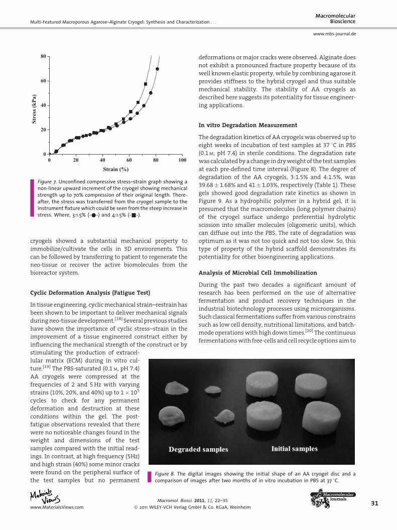
Mechanical characterizationunder unconfined compression
of theAAcryogels typically shows anon-linear stress–strain
response in a distinct upward trend (Figure 7). The
observation of dynamic strength at the strain value up to
90%showednodestruction in themorphologyof the cryogel
sections. The secant compressivemoduluswas calculated as
stress/strain at 15% compression of the total length of the
test sample. The compressive modulus of the AA cryogel of
both combinations, i.e., 3:1.5% and 4:1.5% was 67.73� 1.36
and 73.34� 2.02 kPa, respectively. The compressive moduli
of the cryogel samples containing 4% agarose showed a
higher strength than the gel which contains a low agarose
concentration. Previousstudies suggested that on increasing
the polymer concentration during scaffold synthesis it
proportionally increases its stiffness but also increases the
brittlenessdependinguponthenatureof thepolymersbeing
used. To recognize the importance of mechanical stimuli in
tissue engineering and stress reactions in a bioreactor, it
becomes important to develop a scaffold material that
maintains itsmechanical integrity duringmechanical strain
applications in the bioengineering arena. Here the AA
011, 11, 22–35
H & Co. KGaA, Weinheim www.MaterialsViews.com
0 20 40 60 80 1000
20
40
60
80
Stre
ss (k
Pa)
Strain (%)
Figure 7. Unconfined compressive stress–strain graph showing anon-linear upward increment of the cryogel showing mechanicalstrength up to 70% compression of their original length. There-after, the stress was transferred from the cryogel sample to theinstrument fixture which could be seen from the steep increase instress. Where, 3:1.5% (-*-) and 4:1.5% (-&-).
Multi-Featured Macroporous Agarose–Alginate Cryogel: Synthesis and Characterization . . .
www.mbs-journal.de
cryogels showed a substantial mechanical property to
immobilize/cultivate the cells in 3D environments. This
can be followed by transferring to patient to regenerate the
neo-tissue or recover the active biomolecules from the
bioreactor system.
Cyclic Deformation Analysis (Fatigue Test)
In tissue engineering, cyclicmechanical strain–restrain has
been shown to be important to deliver mechanical signals
duringneo-tissuedevelopment.[18] Severalpreviousstudies
have shown the importance of cyclic stress–strain in the
improvement of a tissue engineered construct either by
influencing the mechanical strength of the construct or by
Figure 8. The digital images showing the initial shape of an AA cryogel disc and acomparison of images after two months of in vitro incubation in PBS at 37 8C.
stimulating the production of extracel-
lular matrix (ECM) during in vitro cul-
ture.[19] The PBS-saturated (0.1 M, pH 7.4)
AA cryogels were compressed at the
frequencies of 2 and 5Hz with varying
strains (10%, 20%, and 40%) up to 1� 105
cycles to check for any permanent
deformation and destruction at these
conditions within the gel. The post-
fatigue observations revealed that there
were no noticeable changes found in the
weight and dimensions of the test
samples compared with the initial read-
ings. In contrast, at high frequency (5Hz)
and high strain (40%) some minor cracks
were found on the peripheral surface of
the test samples but no permanent
www.MaterialsViews.com
Macromol. Biosci. 20
� 2011 WILEY-VCH Verlag Gmb
deformations ormajor crackswere observed. Alginate does
not exhibit a pronounced fracture property because of its
well knownelastic property,while by combining agarose it
provides stiffness to the hybrid cryogel and thus suitable
mechanical stability. The stability of AA cryogels as
described here suggests its potentiality for tissue engineer-
ing applications.
In vitro Degradation Measurement
The degradation kinetics of AA cryogelswas observed up to
eight weeks of incubation of test samples at 37 8C in PBS
(0.1 M; pH 7.4) in sterile conditions. The degradation rate
wascalculatedbyachange indryweightof the test samples
at each pre-defined time interval (Figure 8). The degree of
degradation of the AA cryogels, 3:1.5% and 4:1.5%, was
39.68� 1.68% and 41� 1.03%, respectively (Table 1). These
gels showed good degradation rate kinetics as shown in
Figure 9. As a hydrophilic polymer in a hybrid gel, it is
presumed that the macromolecules (long polymer chains)
of the cryogel surface undergo preferential hydrolytic
scission into smaller molecules (oligomeric units), which
can diffuse out into the PBS. The rate of degradation was
optimum as it was not too quick and not too slow. So, this
type of property of the hybrid scaffold demonstrates its
potentiality for other bioengineering applications.
Analysis of Microbial Cell Immobilization
During the past two decades a significant amount of
research has been performed on the use of alternative
fermentation and product recovery techniques in the
industrial biotechnology processes using microorganisms.
Such classical fermentations suffer fromvarious constrains
such as low cell density, nutritional limitations, and batch-
mode operationswith high down times.[20] The continuous
fermentationswith free-cells andcell recycleoptionsaimto
11, 11, 22–35
H & Co. KGaA, Weinheim31

Figure 9. The degree of degradation of an AA cryogel (both ratio)showing approximate uniform degradation kinetics. Where (-&-)is a cryogel containing 4% agarose and (-^-) is a cryogel contain-ing 3% agarose.
32
www.mbs-journal.de
A. Tripathi, A. Kumar
enhance the cell population inside the fermentor. Immo-
bilization systems have examined the use of immobilized
cells by twomain techniques, adsorption and entrapment.
Our initial experiment showed that E.coli.-BL21 cells could
nicely adhere and were entrapped within the scaffold
surface (Figure10). Thisapproachwasaverysimplemethod
with highmass transport and cell interaction ability. These
studieshaverevealedpossibleareas for the improvementof
productivity and its recovery.
Mammalian Cell Culture
Fibroblast (NIH-3T3)-seeded cryogel samples were exam-
ined up to the 7th day of culture. Examination revealed that
NIH-3T3 could nicely adhere and proliferate on 100mm
Figure 10. SEM micrograph of microbial cell immobilization on the Aculture. a) The control, b) cell adhering on the whole surface of the ge21) adherence at high magnification (4 000�).
Macromol. Biosci. 2
� 2011 WILEY-VCH Verlag Gmb
cryogel sections (Figure 11). The cell proliferation was
examined by nuclear staining using 200 ngmL�1 working
solution of 40,6-diamidino-2-phenylindole (DAPI) prepared
in PBS. Cell-cultured cryogel sections were incubated with
DAPI solution for 5min and then gently washed with PBS.
The cryogel sections without cells were also stained with
DAPI,whichdidnot showanyfluorescence (Figure11a). The
morphology of the cell nuclei was observed after 24h and
on the 7th day of cell culture using a fluorescence
microscope at an excitation wavelength of 350nm. After
24h, cells could adhere nicely to the scaffold surface
(Figure 11b) and increased their cell number on the 7th day
of cell culture (Figure 11c).
Cell Viability/Proliferation Analysis (MTT Assay)
The AA cryogels showed an increasing cellular metabolic
activity with time, as shown in Figure 12, while the control
(2D; 24-well tissue culture plate) wells showed increasing
cellular activity up to the 3rd day of cell culture and after
that the cell viability started decreasing and declined
drastically at the 7th day of cell culture. It might be because
the growing cells reached their confluency in the control
wellswithin5days andafter that they compete for survival
and start dying, which may cause the reduction of
metabolic activity in the 2D control wells. In contrast,
cryogel samplesprovidedahighenoughsurfacearea for cell
proliferation. The effective growth of fibroblasts on the AA
cryogel showed the cellular compatibility of these scaffold
for neo-tissue development.
Heavy Metal Binding
Extensive research is being focused on the removal and
recovery of heavymetals fromwastewater.[17] In the initial
experiment, binding of heavy metals on a AA cryogel
monolithwasexaminedusingtwosolutions, i.e., CuSO4and
A cryogel. The cryogel samples were treated with active bacteriall at low magnification, and c) confirmation of bacterial cell (strain-BL
011, 11, 22–35
H & Co. KGaA, Weinheim www.MaterialsViews.com

Figure 11. Fluorescent microscope images of fibroblast (NIH-3T3)seeded on a 100mm thin cryogel section analysed by nuclearstaining using DAPI stain. a) Background staining of the cryogelscaffold without cells, b) cell adherence after 24 h of seeding, andc) high cell growth covering the whole scaffold after 7 d of cellculture.
Figure 12. The relative viability of fibroblasts (NIH-3T3) as deter-mined by MTT assay. Cells were grown up to one week in apolystyrene-coated tissue culture plate well which is used as acontrol (dotted bar), a cryogel containing 3% agarose (stripedbar), and a cryogel containing 4% agarose (blocked bar). Theabsorbance of blue formazan was measured at 570 nm at differ-ent time intervals up to one week culture.
Multi-Featured Macroporous Agarose–Alginate Cryogel: Synthesis and Characterization . . .
www.mbs-journal.de
NiSO4 (Figure 13). Visual observation showed that the color
of the cylindricalmonoliths of theAAcryogel changed from
white (Figure 13a and 13c2) to blue when treated with
CuSO4 (Figure 13c1) and green when treated with NiSO4
(Figure 13c3). This indicated the affinity binding of the
heavy metals on the cryogel monolithic column. The
system is more beneficial as it can be reused for repeated
www.MaterialsViews.com
Macromol. Biosci. 20
� 2011 WILEY-VCH Verlag Gmb
metal binding. For checking its reusable potentiality, we
tried repeated binding of CuSO4 up to five times. A HCl
solution (0.1 M) was used for the recovery of copper. The
appreciable binding of copper in each cycle was visually
observed. However, further optimizations are required to
increase and quantify the metal binding and recovery
efficiency of the gel.
Conclusion
In this study a novel hybrid AA cryogel is successfully
synthesized. The biocompatible and biodegradable AA
scaffold showed unique properties for bioengineering
applications. The study suggested that these cryogels
showed a macroporous interconnected architecture, high
swelling kinetics, and amiable mechanical properties
proving its stability and application in tissue engineering
for in vitro neo-tissue development. Experimental results
also showed that theAAcryogel canhavea role as a support
matrix for cell immobilizationand for theeffective recovery
of a product from a medium, and also as a filter to remove
heavy metals from wastewater with reusable properties.
The goal of the study was to design such a multi-featured
matrix for the various applications that have been explored
here. The materials were characterized thoroughly for
desired applications. However, further optimizations may
be required for a particular area of interest to have an in-
depth evaluation.
11, 11, 22–35
H & Co. KGaA, Weinheim33

Figure 13. Digital photographs of dry monoliths of an AA gel; a) synthesized at subzero temperature, and b) synthesized at roomtemperature. These gels were incubated with copper sulfate and nickel sulfate solutions and both the copper (c1) and nickel (c3) bindingability is shown along with its control (c2).
34
www.mbs-journal.de
A. Tripathi, A. Kumar
Acknowledgements: The authors acknowledge the financialsupport received from Department of Biotechnology (DBT),Department of Science and Technology (DST), Ministry of Scienceand Technology, Government of India, and Protista BiotechnologyAB, Lund, Sweden. A Research Fellowship to A.T. from the Councilfor Scientific and Industrial Research (CSIR), India, for DoctoralResearch work is duly acknowledged.
Received: July 9, 2010; Revised: August 20, 2010; Publishedonline: November 15, 2010; DOI: 10.1002/mabi.201000286
Keywords: agarose-alginate blend scaffolds; biomaterials; cryo-gels; heavy metal binding; immobilization; tissue engineering
[1] K. Park, Drug Delivery Technol. 2002, 2, 38.[2] H. T. Peng, L. Martineau, P. N. Shek, J. Mater. Sci. Mater. Med.
2007, 18, 975.[3] E. Sachlos, J. T. Czernuszka, Eur. Cell Mater. 2003, 5, 29.[4] A. Tripathi, N. Kathuria, A. Kumar, J. Biomed. Mater. Res.
Part A 2009, 90, 680.[5] N. Kathuria, A. Tripathi, K. K. Kar, A. Kumar, Acta Biomater.
2009, 5, 406.[6] [6a] V. Lozinsky, I. Y. Galaev, F. M. Plieva, I. N. Savina,
H. Jungvid, B. Mattiasson, Trends Biotechnol. 2003, 21, 445;[6b] S. Nilsang, K. S. Nandakumar, I. Y. Galaev, S. K. Rakshit,R. Holmdahl, B. Mattiasson, A. Kumar, Biotechnol. Prog. 2008,23, 932; [6c] P. Arvidsson, F. M. Plieva, I. N. Savina, V. I.Lozinsky, S. Fexby, L. Bulow, I. Y. Galaev, B. Mattiasson,J. Chromatogr. A 2002, 977, 27; [6d] A. Kumar, P. Fatima,I. Y. Galaev, B. Mattiasson, J. Immunol. Methods 2003, 283,185; [6e] V. I. Lozinsky, F. M. Plieva, Enzyme Microb. Technol.1998, 23, 227; [6f] L. Doretti, D. Ferrara, P. Gattolin, S. Lora,F. Schiavon, F. M. Veronese, Talanta 1998, 45, 891; [6g]K. Bloch, V. I. Lozinsky, I. Y. Galaev, K. Yavriyanz,M. Vorobeychik, D. Azarov, L. G. Damshkaln, B. Mattiasson,P. Vardi, J. Biomed. Mater. Res. Part A 2005, 75, 802; [6h]
Macromol. Biosci. 2
� 2011 WILEY-VCH Verlag Gmb
M. Dainiak, A. Kumar, I. Y. Galaev, B. Mattiasson, Proc. Natl.Acad. Sci. USA 2006, 103, 849.
[7] [7a] J. Liu, L. Li, Eur. J. Pharm. Sci. 2005, 25, 237; [7b] A. Jain, Y. T.Kim, R. J. McKeon, R. V. Bellamkonda, Biomaterials 2006, 27,497; [7c] H. Yang, H. Iwata, H. Shimizu, T. Takagi, T. Tsiji, F. Ito,Biomaterials 1994, 15, 113; [7d] S. Sakai, K. Kawabata, T. Ono,H. Ijima, K. Kawakami, Biomaterials 2005, 26, 4786; [7e]H. Gruber, G. L. Hoelscher, K. Leslie, J. A. Ingram, E. N. Hanley,Biomaterials 2006, 27, 371; [7f] H. E. Gruber, E. C. Fisher,B. Desai, A. A. Stasky, G. L. Hoelscher, E. N. Hanley, Exp. Cell.Res. 1997, 235, 13; [7g] W. Gu, H. Yao, C. Y. Huang, H. S.Cheung, J. Biomech. 2003, 36, 593; [7h] R. Mauck, M. A. Soltz,C. C. B. Wang, D. D. Wong, P.-H. G. Chao, W. B. Valhmu, C. T.Hung, G. A. Ateshian, J. Biomech. Eng. 2000, 122, 252.
[8] [8a] K. I. Draget, O. Smidsrod, G. Skjak-Braek, Alginates fromAlgae, 1st edition, Wiley-VCH, Weinheim 2005, p. 1; [8b] D. A.Lee, T. Reisler, D. L. Bader, Acta Orthop. Scand. 2003, 74, 6.
[9] J. Li, A. F. T. Mak, J. Biomater. Appl. 2005, 19, 253.[10] B. Adrados, I. Y. Galaev, K. Nilsson, B. Mattiasson,
J. Chromatogr. A 2001, 930, 73.[11] A. Srivastava, E. Jain, A. Kumar, Mater. Sci. Eng. A 2007, 464,
93.[12] S. N. Park, H. J. Lee, K. H. Lee, H. Suh, Biomaterials 2003, 24,
1631.[13] [13a] S. N. Park, J. C. Park, H. O. Kim, M. J. Song, H. Suh,
Biomaterials 2002, 24, 1205; [13b] Y. S. Choi, S. R. Hong, Y. M.Lee, K. W. Song, M. H. Park, Y. S. Nam, Biomaterials 1999, 20,409.
[14] [14a] J. M. Lee, H. H. L. Edwards, C. A. Pereira, S. I. Samii,J. Mater. Sci. Mater. Med. 1996, 7, 531; [14b] D. Sehgal, I. K.Vijay, Anal. Biochem. 1994, 218, 87.
[15] [15a] C. H. Chang, F. H. Lin, T. F. Kuo, H. C. Liu, Biomed. Eng-App. Bas. C 2005, 17, 1; [15b] S. Sakai, H.Masuhara, Y. Yamada,T. Ono, H. Ijima, K. Kawakami, J. Biosci. Bioeng. 2005, 100, 127;[15c] C. S. D. Lee, J. P. Gleghorn, N. W. Choi, M. Cabodi, A. D.Stroock, L. J. Bonassar, Biomaterials 2007, 28, 2987; [15d] S. H.Chia, M. R. Homicz, B. L. Schumacher, E. J.-M. A. Thonar, J. Am.Coll. Surg. 2005, 200, 691;. [15e] H. A. Awad, M. Q. Wickham,H. A. Leddy, J. M. Gimble, F. Guilak, Biomaterials 2004, 25,3211.
[16] [16a] C. F. Degiorgi, R. A. Pizarro, E. E. Smolko, S. Lora,M. Carenza, Radiat. Phys. Chem. 2002, 63, 109; [16b] T. A.Becker, D. R. Kipke, T. Brandon, J. Biomed. Mater. Res. 2001, 54,76; [16c] S. Fujikawa, T. Yokota, K. Koga, Appl. Microbiol.Biotechnol. 1988, 28, 440.
011, 11, 22–35
H & Co. KGaA, Weinheim www.MaterialsViews.com

Multi-Featured Macroporous Agarose–Alginate Cryogel: Synthesis and Characterization . . .
www.mbs-journal.de
[17] [17a] N. F. Y. Tam, Y. S. Wong, C. G. Simpson, Biotechnol. Tech.1998, 12, 187; [17b] K. R. Reddy, K. Rajgopal, M. L. Kantam,Catal. Lett. 2007, 114, 36.
[18] [18a] L. Niklason, J. Gao, W. Abbott, K. Hirschi, S. Houser,R. Marini, R. Langer, Science 1999, 284, 489; [18b] B. Kim,J. Nikolovski, J. Bonadia, D. Mooney, Nat. Biotechnol. 1999, 17,979.
www.MaterialsViews.com
Macromol. Biosci. 20
� 2011 WILEY-VCH Verlag Gmb
[19] [19a] M. Buschmann, Y. Gluzband, A. Grodzinsky, E. Hunziker,J. Cell. Sci. 1995, 108, 1497; [19b] C. Hunter, J. Mouw,M. Levenston, Osteoarthr. Cartil. 2004, 12, 117.
[20] [20a] T. C. Ezeji, N. Qureshi, H. P. Blaschek, J. Microbiol.Biotechnol. 2003, 19, 595; [20b] T. C. Ezeji, N. Qureshi,H. P. Blaschek, Appl. Microbiol. Biotechnol. 2004, 63,653.
11, 11, 22–35
H & Co. KGaA, Weinheim35