Embed Size (px)
Citation preview

Müller Cells in the Healthy and Diseased Retina

Andreas Reichenbach · Andreas Bringmann
Müller Cells in the Healthyand Diseased Retina
123
Andreas ReichenbachUniversitat LeipzigPaul-Flechsig-Institut
für HirnforschungAbteilung Pathophysiologie
der NeurogliaJahnallee 5904109 [email protected]
Andreas BringmannUniversitätsklinikum LeipzigKlinik und Poliklinik
für AugenheilkundeLiebigstr. 10-1404103 [email protected]
ISBN 978-1-4419-1671-6 e-ISBN 978-1-4419-1672-3DOI 10.1007/978-1-4419-1672-3Springer New York Dordrecht Heidelberg London
Library of Congress Control Number: 2009943587
© Springer Science+Business Media, LLC 2010All rights reserved. This work may not be translated or copied in whole or in part without the writtenpermission of the publisher (Springer Science+Business Media, LLC, 233 Spring Street, New York,NY 10013, USA), except for brief excerpts in connection with reviews or scholarly analysis. Use inconnection with any form of information storage and retrieval, electronic adaptation, computer software,or by similar or dissimilar methodology now known or hereafter developed is forbidden.The use in this publication of trade names, trademarks, service marks, and similar terms, even if they arenot identified as such, is not to be taken as an expression of opinion as to whether or not they are subjectto proprietary rights.While the advice and information in this book are believed to be true and accurate at the date of goingto press, neither the authors nor the editors nor the publisher can accept any legal responsibility for anyerrors or omissions that may be made. The publisher makes no warranty, express or implied, with respectto the material contained herein.
Printed on acid-free paper
Springer is part of Springer Science+Business Media (www.springer.com)
Acknowledgments
We want to thank all colleagues and PhD and MD students who contributed to theresearch in our laboratories, in particular (in alphabetical order).
Silke Agte, Anne Christiane Awißus, Bernd Biedermann, T. Ivo Chao, SladjanaDukic, Wolfgang Eberhardt, Wolfram Eichler, Ines Erdmann, Mike Francke,Kristian Franze, Janina Gentsch, Angela Germer, Iwona Goczalik, Katja Görner,Diana Grimm, Jens Grosche, Martin Gryga, Wolfgang Härtig, Petra G. Hirrlinger,Margrit Hollborn, Dominik Huster, Ian Iandiev, Folke Kalisch, Anett Karl, HannesKodal, Nicole Körber, Heidrun Kuhrt, Franziska Kutzera, Johannes Lange, NiclasLindqvist, Regina Linnertz, Stephan Lipp, Qing Liu, Yun-Bi Lu, Andreas F. Mack,Ivan Milenkovic, Vanessa Moll, Juliane Obst, Gerrit Paasche, Thomas Pannicke,Susanne Pritz-Hohmeier, Maik Raap, Matus Rehak, Winfried Reichelt, KatjaRillich, Christian Roski, Stefan Schopf, Sönke Schwarting, Jens-Uwe Stolzenburg,Steffen Syrbe, Solveig Tenckhoff, Ortrud Uckermann, Elke Ulbricht, MichaelWeick, Karl Weigand, Malte Weuste, Antje Wolf, Antje Wurm, and Yousef Yafai.
We thank also all collaborators outside of our laboratories, in particular.Jan Albrecht, Clemens Allgaier, Thomas Arendt, Hans-Gert Bernstein, Gerd
Birkenmeier, Gert Brückner, Geoffrey Burnstock, Eberhard Buse, Andrés Chávez,Pavel S. Cherkas, Thomas Claudepierre, Amin Derouiche, Nihat Dilsiz, MistyJ. Eaton, Wolfram Eichler, Volker Enzmann, Frank Faude, Christian Foja, HeikeFranke, Ulrich Gärtner, Christian Grimm, Jochen Guck, Elke Günther, GunnarHabermann, Hans-Peter Hammes, Bernd Hamprecht, Menachem Hanani, UweHeinemann, Leif Hertz, Johannes Hirrlinger, Peter Illles, Stefan Junek, JohannesKacza, Josef Käs, Helmut Kettenmann, Frank Kirchhoff, Paulo Kofuji, Leon Kohen,Konrad Kohler, G. Astrid Limb, Felix Makarov, Eric Newman, Richard W. Orkand†,Neville N. Osborne, Adrian G. Palacios, Leo Peichl, Milos Pekny, Ido Perlman,Brigitte Pfeiffer-Guglielmi, Jack Price, Martin B. Reichel, Charlotte E. Remé,Alvaro Rendon, Stephen R. Robinson, Vijay P. Sarthy, Detlev Schild, Ute E. K.Schnurrbusch-Wolf, Johannes Seeger, Mathias Seeliger, Gerald Seifert, SergueiN. Skatchkov, Tobias Stahl, Christian Steinhäuser, Eva Syková, Susann Uhlmann,Rüdiger W. Veh, Thomas H. Wheeler-Schilling, Peter Wiedemann, Frank Wohlrab,Hartwig Wolburg, Sebastian Wolf, Herbert Zimmermann, and Eberhardt Zrenner.
Research of our laboratories was supported by grants from the European UnionResearch Council, the Deutsche Forschungsgemeinschaft, the Bundesministerium
v

vi Acknowledgments
für Bildung und Forschung, the Sächsisches Staatsministerium für Wissenschaft undKunst, and the Interdisziplinäres Zentrum für Klinische Forschung (IZKF) at theFaculty of Medicine of the University of Leipzig. Special thanks are due to JensGrosche for the excellent computer graphics, and to Thomas Pannicke for tirelessproof reading.
Last but not least, we want to thank our families for their understanding andsupport. AB dedicates this book his sons Nikolai and Valentin.
The authors can be contacted at [email protected] (AR) [email protected] (AB), respectively.
Contents
1 Introduction . . . . . . . . . . . . . . . . . . . . . . . . . . . . . . . 11.1 Glial Cells – the “Second Cellular Element” of Neural Tissue . . 1
1.1.1 Definition, Origin, and Functional Role(s) of Glia . . . . 11.1.2 Basic Structural and Ultrastructural Features
of Glia in Vertebrate CNS . . . . . . . . . . . . . . . . 71.1.3 A hierarchy of Neuronal/Glial/Vascular Domains
in the CNS . . . . . . . . . . . . . . . . . . . . . . . . 111.2 The Vertebrate Retina as a Part of the CNS . . . . . . . . . . . . 15
1.2.1 Some Phylogenetic and Ontogenetic Basics . . . . . . . 151.2.2 Some Basics of Retinal Stimulus Perception
and Information Processing . . . . . . . . . . . . . . . 211.2.3 Blood Vessels and Glia: A Basis for Nutrition,
Waste Management and Inflammation . . . . . . . . . . 26
2 Müller Cells in the Healthy Retina . . . . . . . . . . . . . . . . . . 352.1 Morphology and Cellular Properties of Müller Cells
as Constituents of Retinal Tissue . . . . . . . . . . . . . . . . . 352.1.1 Basic Morphology of Müller Cells . . . . . . . . . . . . 352.1.2 Topographical Adaptations . . . . . . . . . . . . . . . . 402.1.3 Müller Cells in the Primate Fovea Centralis . . . . . . . 412.1.4 Ultrastructure of Müller Cells . . . . . . . . . . . . . . 452.1.5 Müller Cell Markers . . . . . . . . . . . . . . . . . . . 532.1.6 Intermediate Filaments . . . . . . . . . . . . . . . . . . 562.1.7 Junctional Cell Coupling . . . . . . . . . . . . . . . . . 57
2.2 Retinal Columnar Units and Domains – Role(s) of MüllerCells in Retina Organization . . . . . . . . . . . . . . . . . . . . 582.2.1 The Müller Cell Population Forms a Regular
but Locally Variable “Scaffold” . . . . . . . . . . . . . 582.2.2 Repetitive Retinal Columnar Units and Their
Diversity Among Vertebrate Retina Types . . . . . . . . 602.2.3 A Hierarchy of Retinal Domains . . . . . . . . . . . . . 662.2.4 Retina Development I: Cell Proliferation,
Progenitor Cells, and Radial Glia . . . . . . . . . . . . 66
vii
viii Contents
2.2.5 Retina Development II: Cell Differentiationand Migration – Layers and Mosaics . . . . . . . . . . . 72
2.2.6 Retina Development III: Late ShapingProcesses – Retina Expansion and Foveation . . . . . . 75
2.2.7 Functional Maturation of Efficient Glia-NeuronInteractions in the Retina . . . . . . . . . . . . . . . . . 85
2.3 Stimulus (Light) Transport to the PhotoreceptorCells – A Role for Müller Cells . . . . . . . . . . . . . . . . . . 882.3.1 Optical Properties of the Vertebrate Retina . . . . . . . 882.3.2 Individual Müller Cells Are Light-Guiding Fibers . . . . 892.3.3 The Müller Cell Population Constitutes a
Versatile “Fiberoptic Plate” . . . . . . . . . . . . . . . . 912.3.4 A Possible Contribution of Rod Cell Nuclear
“Chains” in the ONL . . . . . . . . . . . . . . . . . . . 942.3.5 And What About the Optics of the Fovea(s)? . . . . . . 96
2.4 Müller Cells Are Endowed with Tools to Controlthe Neuronal Microenvironment . . . . . . . . . . . . . . . . . . 982.4.1 Carriers, Transporters, and Enzymes:
Neurotransmitter Recycling . . . . . . . . . . . . . . . 982.4.2 Potassium Channels of Müller Cells: Retinal
Potassium Homeostasis . . . . . . . . . . . . . . . . . . 1202.4.3 Potassium and Water Channels: Retinal Water
Homeostasis . . . . . . . . . . . . . . . . . . . . . . . 1452.4.4 Regulation of Müller Cell Volume – Retinal
Volume Homeostasis . . . . . . . . . . . . . . . . . . . 1582.4.5 Contribution(s) of Other Ion Channels . . . . . . . . . . 161
2.5 Retina Metabolism: A Symbiosis Between Neuronsand Müller Cells . . . . . . . . . . . . . . . . . . . . . . . . . . 1682.5.1 Energy Metabolism . . . . . . . . . . . . . . . . . . . . 1682.5.2 Lipid Metabolism . . . . . . . . . . . . . . . . . . . . . 1722.5.3 Metabolism of Toxic Compounds . . . . . . . . . . . . 172
2.6 Other Glio-Neuronal Interactions in the Retina . . . . . . . . . . 1732.6.1 Recycling of Photopigments . . . . . . . . . . . . . . . 173
2.7 Mutual Signal Exchange Between Retinal Neuronsand Müller Cells . . . . . . . . . . . . . . . . . . . . . . . . . . 1762.7.1 Müller Cells Can Sense – And Respond to –
Retinal Neuronal Activity . . . . . . . . . . . . . . . . 1762.7.2 Müller Cells May Modulate Retinal Neuronal
Activity . . . . . . . . . . . . . . . . . . . . . . . . . . 2052.8 Physiological Müller Cell-Neuron Interactions:
A Short Summary . . . . . . . . . . . . . . . . . . . . . . . . . 213
3 Müller Cells in the Diseased Retina . . . . . . . . . . . . . . . . . . 2153.1 Reactive Müller Cells – General Properties and Roles . . . . . . 215
3.1.1 Müller Cell Gliosis . . . . . . . . . . . . . . . . . . . . 2153.1.2 Neuroprotection . . . . . . . . . . . . . . . . . . . . . 229
Contents ix
3.1.3 Müller Cell Proliferation . . . . . . . . . . . . . . . . . 2353.1.4 Müller Cells as Progenitor Cells in the Adult
Retina – Müller Stem Cells . . . . . . . . . . . . . . . . 2463.1.5 Immunomodulatory Role of Müller Cells . . . . . . . . 249
3.2 Involvement and Role(s) of Müller Cells in SpecificRetinal Disorders . . . . . . . . . . . . . . . . . . . . . . . . . 2513.2.1 Retinal Ischemia-Reperfusion Injury . . . . . . . . . . . 2513.2.2 Retinal Detachment . . . . . . . . . . . . . . . . . . . . 2563.2.3 Proliferative Retinopathies . . . . . . . . . . . . . . . . 2653.2.4 Diabetic Retinopathy . . . . . . . . . . . . . . . . . . . 2803.2.5 Macular Edema . . . . . . . . . . . . . . . . . . . . . . 2853.2.6 Neovascularization . . . . . . . . . . . . . . . . . . . . 2923.2.7 Retinal Light Damage . . . . . . . . . . . . . . . . . . 2943.2.8 Hepatic Retinopathy . . . . . . . . . . . . . . . . . . . 2963.2.9 Retinitis Pigmentosa . . . . . . . . . . . . . . . . . . . 2973.2.10 Glaucoma . . . . . . . . . . . . . . . . . . . . . . . . . 2983.2.11 Retinoschisis . . . . . . . . . . . . . . . . . . . . . . . 299
4 Conclusions and Perspectives . . . . . . . . . . . . . . . . . . . . . 303
References . . . . . . . . . . . . . . . . . . . . . . . . . . . . . . . . . . 305
Index . . . . . . . . . . . . . . . . . . . . . . . . . . . . . . . . . . . . . 395
Abbreviations
A adenosineAC amacrine cellADP adenosine 5’-diphosphateAG astrogliaAGE advanced glycation end productAMP adenosine 5’-monophosphateAMPA α-amino-3-hydroxy-5-methyl-4-isoxazolepropionic acidANP atrial natriuretic peptideAP-1 activator protein-1Apo apolipoproteinAQP aquaporinATP adenosine 5’-triphosphateBC bipolar cellBcl B cell lymphomaBDNF brain-derived neurotrophic factorbFGF basic fibroblast growth factor, FGF-2BK big conductance potassiumBL basal laminaBMP bone morphogenetic proteinBV blood vesselBzATP 2’-/3’-O-(4-benzoylbenzoyl)-ATPCA carbonic anhydrasecAMP cyclic adenosine 5’-monophosphatecGMP cyclic guanosine 5’-monophosphateCNS central nervous systemCNTF ciliary neurotrophic factorCRALBP cellular retinaldehyde-binding proteinCrx cone rod homeoboxCTGF connective tissue growth factorDHA docosahexaenoic acidDkk dickkopfDNA desoxyribonucleic acidDOPA dihydroxyphenylalanine
xi
xii Abbreviations
Dp dystrophin gene productEAAT excitatory amino acid transporterEGF epidermal growth factorEPSC excitatory postsynaptic currentER endoplasmic reticulumERK1/2 extracellular signal-regulated kinases 1 and 2, p44/p42 MAPKsFGF fibroblast growth factorflt-1 fms-like tyrosine kinase-1, VEGF receptor-1G GTP-bindingGABA γ-aminobutyric acidGAT GABA transporterGC ganglion cellGCL ganglion cell layerGDNF glial cell line-derived neurotrophic factorGFAP glial fibrillary acidic proteinGFR glial cell line-derived neurotrophic factor receptorGLAST glutamate-aspartate transporterGLT glutamate transporterGluR AMPA receptor subunitGlyT glycine transporterGSSG glutathione disulfideGTP guanosine 5’-triphosphateHB-EGF heparin-binding epidermal growth factor-like growth factorHDL high-density lipoproteinH-E hematoxylin-eosinHGF hepatocyte growth factorHVA high threshold voltage-activatedICAM intercellular adhesion moleculeIGF insulin-like growth factorIGFBP IGF binding proteinIL interleukinILM inner limiting membraneINL inner nuclear layerIP3 inositol 1,4,5-triphosphateIPL inner plexiform layerJAK Janus kinaseJNK c-Jun N-terminal kinaseKA fast transient (A-type) potassiumKA kainate receptor subunitKDR/flk-1 kinase insert domain-containing receptor/fetal liver kinase-1,
VEGF receptor-2KDR delayed rectifying potassiumKir inwardly rectifying potassiumL long-lastingLDH lactate dehydrogenase
Abbreviations xiii
LDL low-density lipoproteinLIF leukemia inhibitory factorLRP1 low-density lipoprotein-related protein, CD91LVA low threshold voltage-activatedM Müller gliaMAPK mitogen-activated protein kinaseMC Müller cellMCE Müller cell endfootMCP monocyte chemoattractant proteinMG microgliamGluR metabotropic glutamate receptorMHC major histocompatibilityMMP matrix metalloproteinasemRNA messenger ribonucleic acidNa,K-ATPase sodium and potassium-dependent ATPase, sodium pumpNAD(P)H nicotinamide adenine dinucleotide (phosphate)NFL nerve fiber layerNGF nerve growth factorNMDA N-methyl-D-aspartateNO nitric oxideNP natriuretic peptideNPY neuropeptide YNR NMDA receptor subunitNTPDase nucleoside triphosphate diphosphohydrolaseOAP orthogonal arrays of membrane particlesOLM outer limiting membraneONL outer nuclear layerOPL outer plexiform layerP postnatal dayP2X ionotropic purinergicP2Y metabotropic purinergicp75NTR low-affinity neurotrophin receptorPAP peripheral astrocytic processPax6 paired box gene 6PCNA proliferating cell nuclear antigenPDGF platelet-derived growth factorPDR proliferative diabetic retinopathyPDZ post synaptic density protein, Drosophila disc large tumor
suppressor, and zonula occludens-1 proteinPEDF pigment epithelium-derived factorPGP peripheral glial processPI3K phosphatidylinositol-3 kinasePK pyruvate kinasepO2 oxygen partial pressurPR photoreceptor

xiv Abbreviations
PRS photoreceptor segmentsPVR proliferative vitreoretinopathyRAGE AGE receptorRCS Royal College of Surgeonsrds slow retinal degenerativeRI refractory indexRPE retinal pigment epitheliumShh sonic hedgehogSrc sarcomaSTAT signal transducers and activators of transcriptionSVR surface-to-volume ratioT transientTASK TWIK-related acid-sensitive potassiumTEA tetraethylammoniumTGF transforming growth factorTNF tumor necrosis factorTrk tropomyosin receptor kinase, high-affinity neurotrophin receptorTRPC transient receptor potential canonicalUDP uridine 5’-diphosphateUTP uridine 5’-triphosphateVEGF vascular endothelial growth factorVIP vasoactive intestinal peptideWnt wingless-type MMTV integration site familyY NPY receptor
Chapter 1Introduction
At first glance one may wonder why an entire book is devoted to Müller cells, a celltype that clearly represents a minority in our central nervous system (CNS): Outof an estimated total number of about 200 billions of cells in our CNS, the 8–10millions of Müller cells in our two eyes constitute not more than some 0.005%. Tomake things even worse – Müller cells do not belong to the highly esteemed neuronsbut to the glia, a family of cells which for more than a century had been thought ofas a sort of mere filling material between the neurons. . . So one may wonder evenmore why our research group – together with an increasing number of scientistsworldwide – focuses their research on these cells now for a quarter of a century,with no end in sight.
Gentle reader! We hope to convince you that Müller cells deserve both our con-certed research effort and your effort to read the book. While everybody will agreethat the eye is a very special and versatile sense organ, it turned out over the recentyears that Müller cells are very peculiar and multipotent glial cells. In the retinaof most vertebrates and even of many mammals, Müller cells are the only type of(macro-) glial cells; thus, they are responsible for a wealth of neuron-supportivefunctions that, in the brain, rely upon a division of labour among astrocytes, oligo-dendrocytes, and ependymal cells. Even beyond such a role as “model glia” in CNS,Müller cells adapted to several exciting roles in support of vision.
Before going into detail, however, it appears appropriate to provide two intro-ductory chapters, one about glial cells – the family of neural cells Müller cellsare members of – and another one about the vertebrate retina – their habitat andworkplace.
1.1 Glial Cells – the “Second Cellular Element”of Neural Tissue
1.1.1 Definition, Origin, and Functional Role(s) of Glia
The term “neuroglia” (Greek for “nerve glue”) was introduced in the nineteenth cen-tury by the German pathologist Rudolf Virchow (Virchow, 1858). On his search fora connective tissue of the nervous system, he discovered cells which apparently were
1A. Reichenbach, A. Bringmann, Müller Cells in the Healthy and Diseased Retina,DOI 10.1007/978-1-4419-1672-3_1, C© Springer Science+Business Media, LLC 2010
2 1 Introduction
no nerve cells (i.e., neurons) and called them glia, according to what he expected tofind. Nowadays, it may be speculated that this unattractive name (reflecting the poorhypothesis behind it) was one of the reasons why these cells were neglected by mostneuroscientists for the next 100 years. It was only some neuroanatomists who werefascinated by their complex shape in Golgi-impregnated brain slices, and the neu-ropathologists who soon realized that glial cells are crucially involved in virtuallyall brain injuries and diseases. Then during the last 3 decades, the rapidly develop-ing advanced methodology in neuroscience enabled the development of innovativeparadigms and approaches which eventually revealed that there is virtually no eventduring ontogenetic development, mature functioning, and pathology of the CNSwhich would not involve a crucial contribution of glial cells.
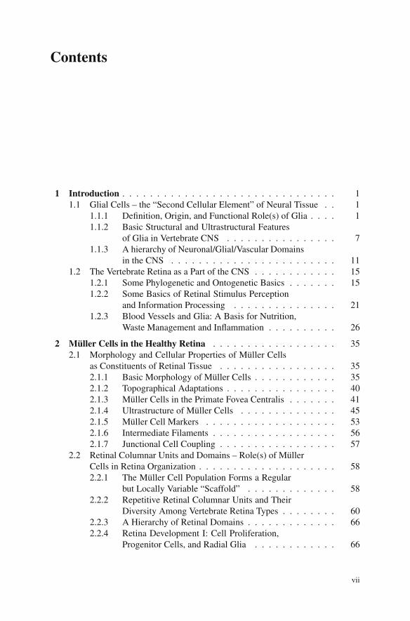
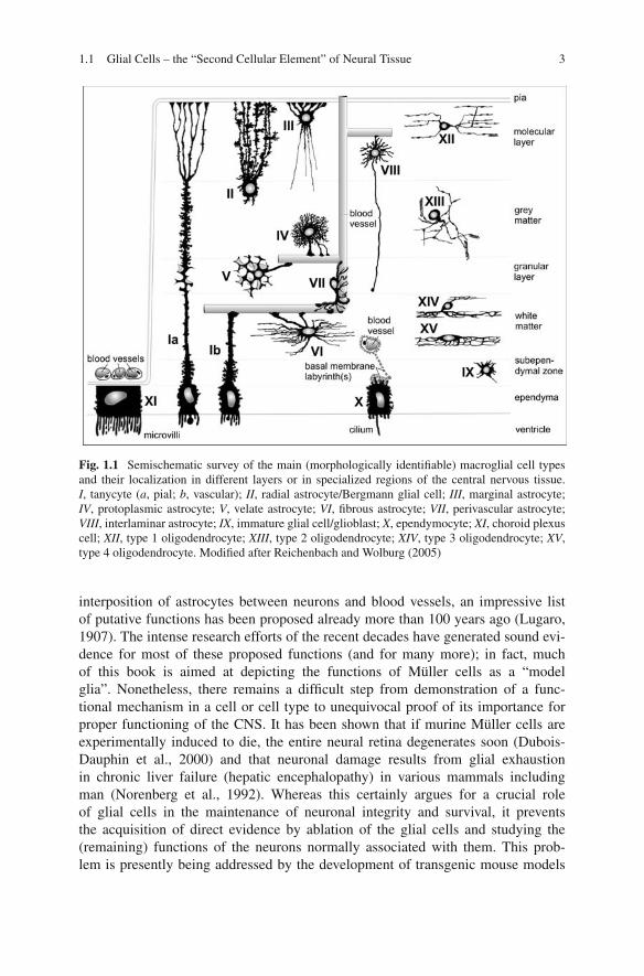
Per definition, the term “glia” applies to all cells within the CNS that (i) are notneurons and (ii) do not belong to mesenchymal structures such as the blood vesselsand the meninges. The glia proper can be divided into two “sub-families”, macro-and microglia. The term “macroglia” summarizes a wide diversity of cell typesarising from the primitive neuroepithelium together with the neurons, includingependymoglia (radial glia incl. tanycytes and Müller cells, ependymocytes, choroidplexus epithelial cells and pigment epithelial cells), several subtypes of astro-cytes, and oligodendrocytes (Fig. 1.1). By contrast, microglial cells are blood-bornemacrophages which, during late ontogenesis, invade the brain via the establishingblood vessels. As will be mentioned later, microglial cells intensely interact withMüller cells in the injured retina where they play an important role; however, in thischapter we will focus upon the macroglial cells. So if we want to define and/or toidentify a (macro-) glial cell as such, we need to make sure that it stems from pro-genitor cells of the embryonic neuroepithelium but is not a neuron. The latter pointappears as a trivial task if one has a typical neuron (e.g., a cerebellar Purkinje cell)and a typical glial cell (e.g., an astrocyte) in mind but if all types of neurons andglial cells are considered, a general discrimination becomes less easy with everynew discovery in glial cell research (see, e.g., Kimelberg, 2004). So it has beenshown that – in strong contradiction to the traditional dogma of neuroscience – glialcells do express a wealth of voltage-dependent ion channels, ligand receptors, trans-membrane transporters, second messenger pathways, and even release mechanismsfor signaling molecules which they share with the neurons. This is less of a surpriseif one considers that CNS neurons and glial cells share the same progenitors. Ininsects, “glial cells missing” (gcm) has been identified as a binary genetic switch forglia versus neurons. In the presence of gcm protein, presumptive neurons becomeglia, while in its absence, presumptive glia become neurons (Jones et al., 1995).Although a corresponding mechanism remains to be identified in vertebrates, appar-ently a similar “switch” causes quantitative shifts in the protein expression profilesof neurons and glial cells, rather than “all-or-none” decisions. What remains safe tosay is that glial cells, in contrast to neurons, are not direct parts of information pro-cessing chains – this is, they do not perceive specific environmental stimuli and/ortransmit them to specific brain centers, for example.
So if we know what glia fail to do, what do we know about their actual func-tions? Mostly prompted by morphological observations such as the “strategic”
1.1 Glial Cells – the “Second Cellular Element” of Neural Tissue 3
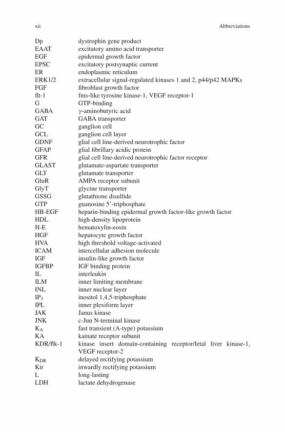
Fig. 1.1 Semischematic survey of the main (morphologically identifiable) macroglial cell typesand their localization in different layers or in specialized regions of the central nervous tissue.I, tanycyte (a, pial; b, vascular); II, radial astrocyte/Bergmann glial cell; III, marginal astrocyte;IV, protoplasmic astrocyte; V, velate astrocyte; VI, fibrous astrocyte; VII, perivascular astrocyte;VIII, interlaminar astrocyte; IX, immature glial cell/glioblast; X, ependymocyte; XI, choroid plexuscell; XII, type 1 oligodendrocyte; XIII, type 2 oligodendrocyte; XIV, type 3 oligodendrocyte; XV,type 4 oligodendrocyte. Modified after Reichenbach and Wolburg (2005)
interposition of astrocytes between neurons and blood vessels, an impressive listof putative functions has been proposed already more than 100 years ago (Lugaro,1907). The intense research efforts of the recent decades have generated sound evi-dence for most of these proposed functions (and for many more); in fact, muchof this book is aimed at depicting the functions of Müller cells as a “modelglia”. Nonetheless, there remains a difficult step from demonstration of a func-tional mechanism in a cell or cell type to unequivocal proof of its importance forproper functioning of the CNS. It has been shown that if murine Müller cells areexperimentally induced to die, the entire neural retina degenerates soon (Dubois-Dauphin et al., 2000) and that neuronal damage results from glial exhaustionin chronic liver failure (hepatic encephalopathy) in various mammals includingman (Norenberg et al., 1992). Whereas this certainly argues for a crucial roleof glial cells in the maintenance of neuronal integrity and survival, it preventsthe acquisition of direct evidence by ablation of the glial cells and studying the(remaining) functions of the neurons normally associated with them. This prob-lem is presently being addressed by the development of transgenic mouse models
4 1 Introduction
where (desirably: conditioned) deletion of certain functional components such asglia-specific enzymes, ion channels, or cytoskeletal elements helps to understandthe contribution of these glial molecules to neuronal functioning; some examples ofthis approach will be presented later.
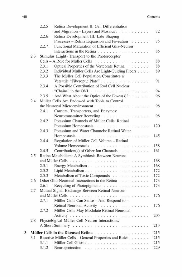
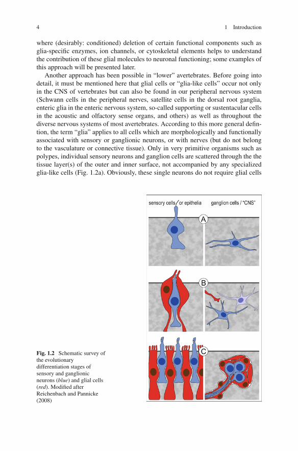
Another approach has been possible in “lower” avertebrates. Before going intodetail, it must be mentioned here that glial cells or “glia-like cells” occur not onlyin the CNS of vertebrates but can also be found in our peripheral nervous system(Schwann cells in the peripheral nerves, satellite cells in the dorsal root ganglia,enteric glia in the enteric nervous system, so-called supporting or sustentacular cellsin the acoustic and olfactory sense organs, and others) as well as throughout thediverse nervous systems of most avertebrates. According to this more general defin-tion, the term “glia” applies to all cells which are morphologically and functionallyassociated with sensory or ganglionic neurons, or with nerves (but do not belongto the vasculature or connective tissue). Only in very primitive organisms such aspolypes, individual sensory neurons and ganglion cells are scattered through the thetissue layer(s) of the outer and inner surface, not accompanied by any specializedglia-like cells (Fig. 1.2a). Obviously, these single neurons do not require glial cells
Fig. 1.2 Schematic survey ofthe evolutionarydifferentiation stages ofsensory and ganglionicneurons (blue) and glial cells(red). Modified afterReichenbach and Pannicke(2008)
1.1 Glial Cells – the “Second Cellular Element” of Neural Tissue 5
for their differentiation, function, or survival. Then the differentiation of larger/morecomplex animals resulted in the development of spezialized small sensory organs,and of small groups of loosely associated ganglion neurons (Fig. 1.2b). At thisstage, the sensory neurons (but not the ganglion neurons) usually are associatedwith glia-like cells.
Recently, Bacaj et al. (2008) demonstrated that neurons in the major sensoryorgan of Caenorhabditis elegans survive the elimination of ensheathing glia butdisplay functional deficits. In particular, the glia-deprived sensory neurons partiallyor totally lost the sensitivity for their adequate stimuli despite of an apparently nor-mal expression of their structural and functional proteins. One of the identified glialtransmembrane proteins, a KCl cotransporter, may be involved in the crucial glialcontribution to osmosensivity of the ASH neuron in C. elegans, perhaps by pro-viding a normosmotic microenvironment at (part of) the receptor neuron, as kindof a “standard” against which environmental changes can be measured. This hadlead to the view that the homeostatic functions of “sensory-associated” glia maybe required for the selection, transformation (and even transduction) of adequatestimuli by their adjacent neurons (Reichenbach and Pannicke, 2008) (in a widersense, the absorption of excess light by pigmented glial cells close to photoreceptorneurons may also be considered as a case of homeostasis). From an evolution-ary point of view, this first ancestral glia may have been separated off the neuralprogenitor line because this allowed to increase the signal-to-noise ratio of spe-cific stimulus perception in non-sessile animals with a more complex behavioralrepertoire.
Once established, the glial cells as “novel neural cell type with homeostaticcapabilities” may have occupied the large nervous centers of more advanced ani-mals where many neurons accumulate in ganglia or even brains (Fig. 1.2c). In thiscomplex environment, the perisynaptic glia becomes essential for synaptic trans-mission (Araque et al., 1999). It should be kept in mind that signal transmissionvia chemical synapses can be considered as a special case of chemosensation bythe postsynaptic neuron, with its adequate stimulus being the specific neurotrans-mitter substance released by the presynaptic terminal. Thus, an agglomerate ofchemical synapses in the ganglia may raise similar homeostatic needs as chemosen-sation in the sensory organs. In fact, at chemical synapses a special case ofdivision of labor can be observed between neurons and glial cells. The neu-ronal compartments (i.e., pre- and postsynapse) are highly specialized for rapidrelease and sensation of the neurotransmitter, respectively. For this purpose, theyare endowed with a battery of proteins involved in the synthesis, release, andperception of the neurotransmitter. The perisynaptic glial compartments, by con-trast, dominantly express transporter proteins for rapid and efficient uptake of theneurotransmitter, and specific enzymes for its conversion into a non-signaling pre-cursor molecule. This neuron-glia interaction is called “neurotransmitter recycling”(cf. Section 2.4). Again this difference is not all-or-none, however: the neuronsexpress some uptake carriers, and the glial cells express neurotransmitter receptorsas well as molecules allowing the release of neuroactive substances (see below andSection 2.7).
6 1 Introduction
Eventually, the homeostatic functions of glia appear to constitute the basisfor their currently-evaluated “more exciting roles” including direct involvementin neuronal information processing (Araque et al., 1999). On the one hand, glialhomeostatic functions may be modulated in their activity, or even “switched off”in order to increase the effectiveness of neuronal transmitter release by increas-ing the concentration and duration of presence in the synaptic cleft (Oliet et al.,2001). On the other hand, the molecular machinery required for neurotransmitterrecycling may even be elaborated into active neuron-controling mechanisms suchas gliotransmitter release (Araque et al., 1999) (cf. also Section 2.7).
In addition to an enhanced turnover of signaling molecules, the dense crowd-ing of neurons in large sensory epithelia or ganglia (or brains) of complex metazoacauses other problems. Both types of tissues are typically encapsulated against theirnon-neural environment, usually involving a blood-brain barrier to which glial cellscontribute (Wolburg et al., 2009). The insulated, highly active neurons depend onefficient nutrient delivery and clearance of waste products. This raises the needfor extracellular homeostasis in an extended sense, involving supply of nutrients(glucose or lactate/pyruvate) and removal of CO2 and water (cf. Section 2.4 and2.5). As neuronal excitation is accompanied by Na+ influx from and K+ efflux intothe extracellular space, and as elevated extracellular [K+] modulates the excitabil-ity of neurons, extracellular K+ homeostasis is an important task of glial cells (seeSection 2.4.2). Together, these problems may have been the driving force for theubiquitous appearance and further multiplication (Reichenbach, 1989c) of glia in“higher” and/or bigger animals.
To summarize from this excurse what one needs to understand the functions ofMüller cells, the essentials are that
• glial cells associated with sensory neurons increase the signal-to-noise ratioof perception, by assisting their adjacent neurons in the selection, processing,and even transduction of adequate stimuli; much of this involves homeostaticfunctions;
• glial cells associated with ganglion neurons increase the signal-to-noise ratio ofsignal transmission, mainly by a homeostatis of neurotransmitter molecules;
• glial cells in large sensory organs and in brain constitute a neural waste man-agement system and play an important role in the coupled ion and waterhomeostasis.
Noteworthy, the retina is both a large sensory epithelium and a part of the brain(see Section 1.2) such that all of the above-mentioned functions fully apply toMüller cells. It should also be mentioned here that, after the glia-cellular system hadbeen established in phylogenesis, it became available for a variety of other functionsincluding guidance of neuron migration and axon pathfinding during embryogene-sis, interactions with the immune systems, and many others. Most of these functionsrely upon the very special morphology of glial cells which is introduced in thefollowing chapter.
1.1 Glial Cells – the “Second Cellular Element” of Neural Tissue 7
1.1.2 Basic Structural and Ultrastructural Features of Gliain Vertebrate CNS
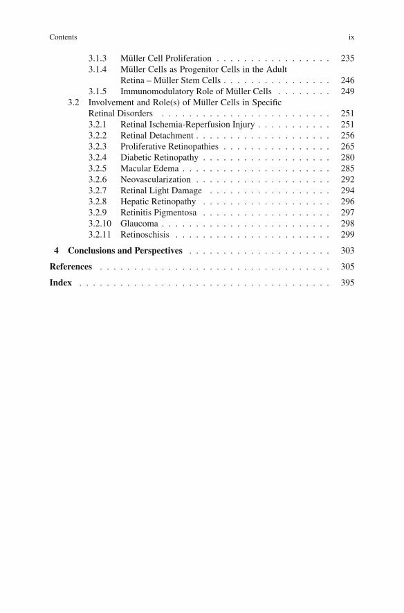
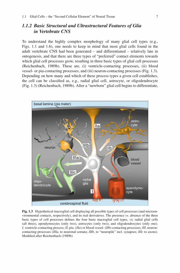
To understand the highly complex morphology of many glial cell types (e.g.,Figs. 1.1 and 1.6), one needs to keep in mind that most glial cells found in theadult vertebrate CNS had been generated – and differentiated – relatively late inontogenesis, and that there are three types of “preferred” contact elements towardswhich glial cell processes grow, resulting in three basic types of glial cell processes(Reichenbach, 1989b). These are, (i) ventricle-contacting processes, (ii) bloodvessel- or pia-contacting processes, and (iii) neuron-contacting processes (Fig. 1.3).Depending on how many and which of these process types a given cell establishes,the cell can be classified as, e.g., radial glial cell, astrocyte, or oligodendrocyte(Fig. 1.3) (Reichenbach, 1989b). After a “newborn” glial cell begins to differentiate,
Fig. 1.3 Hypothetical macroglial cell displaying all possible types of cell processes (and microen-vironmental contacts, respectively), and its real derivatives. The presence vs. absence of the threebasic types of cell processes defines the four basic macroglial cell types, viz radial glial cells(all three), ependymocytes (only two), astrocytes (only two), and oligodendrocytes (only one).I, ventricle-contacting process; II, pia- (IIa) or blood vessel- (IIb) contacting processes; III, neuron-contacting processes (IIIa, to neuronal somata; IIIb, to “neuropile” incl. synapses; IIIc to axons).Modified after Reichenbach (1989b)
8 1 Introduction
it must (try to) establish its pre-programmed contacts, a procedure which involvescell process growth (and even migration, in some instances). As mentioned above,most of the “target structures” (the pia mater, blood vessels, and neurons includingtheir elaborate neurites, including axon bundles) are already formed at this stage,and the glial cells and their processes must fit into this preexisting environment. Inaccordance with this scenario, it has been recently shown that glial cells are softerthan neurons (Lu et al., 2006). Thus, one might get some idea about the ingrowthof glial cell processes into the neuronal (and vascular) network if one imagines thebuildup of a concrete construction, when the fluid concrete mass is poured into theinterspaces between the steel scaffolding. Once (one to three of) the glia-typicalcontacts were established and the glial cell processes are differentiated, they displaytypical morphologies.
(A) The ventricle-contacting process generally ends with microvilli (in some cases,also cilia) extending into the fluid compartment (Figs. 1.3, 2.9 and 2.10).The apical microvilli provide a large membrane surface area at the inter-face between fluid and tissue, and thus facilitate the transport of moleculesbetween the two compartments. Additionally the processes form, togetherwith those of adjacent glial cells or neurons, adherent junctions that appearto be important for the maintenance of the biomechanical stability of the tis-sue and for other functions including the correct ontogenetic development ofthe tissue. In the retina, this network of zonulae adherentes is visible evenin H/E-stained histological sections, and is called “outer limiting membrane(OLM)” (Fig. 2.10). In some specialized brain regions (the circumventric-ular organs), the ventricle-contacting processes are even connected by tightjunctions such that a brain-cerebrospinal fluid-barrier is constituted (Wolburget al., 2009).
(B) The pia-contacting and the perivascular glial processes abut a basal lam-ina by so-called endfeet (Figs. 1.1, 1.3 and 2.11). These glial endfeet form,together with the basal lamina and with the mesenchymal cells at its oppo-site surface (e.g, endothelial cells or pericytes, or meningeal fibroblasts), theother interface between the neural tissue and a fluid-filled compartment (notethat the vertebrate CNS is epithelial and thus displays two opposite surfaces;cf. Section 1.2.1). The endfeet usually contain large amounts of smooth endo-plasmic reticulum (Fig. 2.11b). Their basal lamina-abutting surface is endowedwith a wealth of transport and receptor proteins as well as �–shaped mem-brane indentions indicative of active exo- and/or endocytosis. Apparently, thereoccurs a lively exchange of ions and even larger molecules between the endfeetand the fluid compartments behind the basal lamina. A particular characteristicfeature of many glial endfeet is their dense packing with so-called orthog-onal arrays of membrane particles (OAP) visible by freeze-fracture electronmicroscopy (Wolburg and Berg, 1987, 1988; Wolburg, 1995). A water chan-nel, aquaporin 4, has been identified as one of the transport molecules whichare accumulated in these OAPs (Yang et al., 1996; Verbavatz et al., 1997)(cf. Section 2.4.3).
1.1 Glial Cells – the “Second Cellular Element” of Neural Tissue 9
(C) The neuron-contacting glial processes abut at – or rather, ensheath – neuronalcell somata and/or processes (Fig. 1.3). According to the complex shape ofthe ensheathed structures this type of glial processes shows the most complexstructure (Fig. 1.6; cf. Grosche et al., 1999). In the grey matter of the CNSwhich contains a high density of synapses, the dominant glial cells are so-calledprotoplasmic astrocytes (Fig. 1.1). The many irregularly shaped processes ofthese cells give rise to numerous very thin, convoluted cytoplasmic tongues,also called lamellar processes (Wolff, 1968), lamellipodia and filopodia (Chaoet al., 2002), or peripheral astrocytic/glial processes, PAPs (Derouiche andFrotscher, 2001) or PGPs (Reichenbach et al., 2004), respectively.
The PGPs contain only a minor portion of the glial cytoplasm volume but amajority of the cell surface area. This large membrane area in small volume com-partments appears to be required to give space for a wealth of ion channels, ligandreceptors, and uptake carrier proteins necessary to maintain the variety of glia-neuron interactions. In many instances, a number of such PGPs together belong to acomplex subcellular structure called a “microdomain” (Grosche et al., 1999, 2002).Such a glial microdomain consists of a thin stalk and a small garbagge-like headfrom which latter the PGPs arise; it may contain one or a few mitochondria and isthought to be capable of a more or less autonomous interaction with the ensheathedgroup of a few synapses (cf. Section 1.1.3).
The white matter of the CNS is constituted by large numbers of axons oraxon bundles. Usually if the thickness of an axon exceeds a threshold of about0.2–0.4 μm, its internodes (i.e., the sections between two consecutive nodes ofRanvier) are myelinated by highly specialized cytoplasmic tongues arising from theprocesses of oligodendrocytes (Waxman and Black, 1995). Bundles of thin, non-myelinated axons are loosely ensheathed by cytoplasmic tongues arising from theprocesses of the so-called fibrous astrocytes (Fig. 1.1). The processes of these cellsappear less complex than those of the protoplasmic astrocytes (because there arealmost no synapses in the white matter) but are preferentially aligned in parallel tothe axon bundles. Besides ensheathing the unmyelinated axons, these astrocytic pro-cesses extend finger-like tiny processes into the perinodal spaces of the myelinatedaxons (Hildebrand et al., 1993; Butt et al.,1994; Sims et al., 1985). It has been pro-posed that these “glial fingers” monitor neuronal activity, and trigger glial responsesto it (Chao et al., 1994b).
Finally there are some CNS regions which contain densely packed small neurons,nuclear layers, such as the granule cell layer in cerebellum. Groups of such neuronalcell bodies are there ensheathed by the honey comb-like cytoplasmic tongues of so-called velate astrocytes (Fig. 1.1). Analyzing this short compilation of basic data itbecomes evident that the shape as well as the ultrastructure of glial cell processesare largely determined by the local environment and, specifically, by the type ofthe contacted/ensheathed element (cerbrospinal fluid/basal lamina/synapses, axons,or somata of neurons). For a recent comprehensive review of the various types ofglial cell processes and their specific structural and ultrastructural adaptations, seeReichenbach and Wolburg (2009).
10 1 Introduction
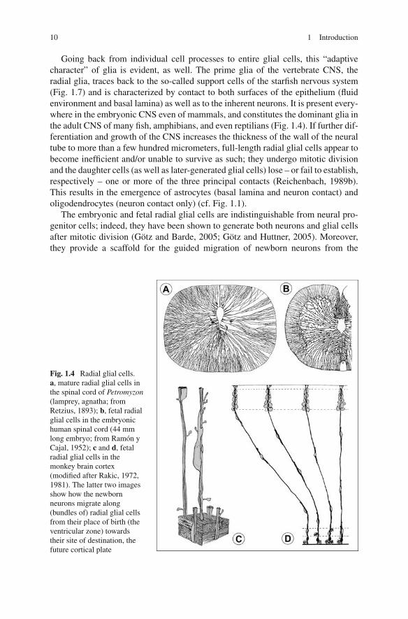
Going back from individual cell processes to entire glial cells, this “adaptivecharacter” of glia is evident, as well. The prime glia of the vertebrate CNS, theradial glia, traces back to the so-called support cells of the starfish nervous system(Fig. 1.7) and is characterized by contact to both surfaces of the epithelium (fluidenvironment and basal lamina) as well as to the inherent neurons. It is present every-where in the embryonic CNS even of mammals, and constitutes the dominant glia inthe adult CNS of many fish, amphibians, and even reptilians (Fig. 1.4). If further dif-ferentiation and growth of the CNS increases the thickness of the wall of the neuraltube to more than a few hundred micrometers, full-length radial glial cells appear tobecome inefficient and/or unable to survive as such; they undergo mitotic divisionand the daughter cells (as well as later-generated glial cells) lose – or fail to establish,respectively – one or more of the three principal contacts (Reichenbach, 1989b).This results in the emergence of astrocytes (basal lamina and neuron contact) andoligodendrocytes (neuron contact only) (cf. Fig. 1.1).
The embryonic and fetal radial glial cells are indistinguishable from neural pro-genitor cells; indeed, they have been shown to generate both neurons and glial cellsafter mitotic division (Götz and Barde, 2005; Götz and Huttner, 2005). Moreover,they provide a scaffold for the guided migration of newborn neurons from the
Fig. 1.4 Radial glial cells.a, mature radial glial cells inthe spinal cord of Petromyzon(lamprey, agnatha; fromRetzius, 1893); b, fetal radialglial cells in the embryonichuman spinal cord (44 mmlong embryo; from Ramón yCajal, 1952); c and d, fetalradial glial cells in themonkey brain cortex(modified after Rakic, 1972,1981). The latter two imagesshow how the newbornneurons migrate along(bundles of) radial glial cellsfrom their place of birth (theventricular zone) towardstheir site of destination, thefuture cortical plate
1.1 Glial Cells – the “Second Cellular Element” of Neural Tissue 11
ventricular surface of the neuroepithelium (where cell multiplication occurs) to thedistant sites of their destination (Rakic, 1988) (Fig. 1.4). Likewise, the processes offetal radial glial cells are used guidelines by the neurites of the young postmitoticneurons growing towards their targets (Silver et al., 1982; Norris and Kalil, 1991). Ithas been suggested that the cohort of neurons migrating along the same radial glial“climbing pole” later maintains and elaborates much of the contacts inevitably aris-ing between the leading and trailing processes of cells migrating together, and thatthe cells maintain a particular relationship or even “symbiosis” with the glial cell(s)along which they migrated (Rakic, 1978; Reichenbach et al., 1993a; Reichenbachand Robinson, 1995). This may be the ontogenetic basis of functional columnarunits or domains (see Sections 1.1.3 and 2.2.2) in the adult CNS, exemplified by theorientation columns in visual cortex (Mountcastle, 1957).
In relatively thin-walled brains or CNS regions (Reichenbach, 1990) the fetalradial glial cells eventually become postmitotic and differentiate into adult radialglia. On their course through the entire thickness of the CNS tissue the processes ofthese cells may pass through different local tissue specializations such as synapse-rich grey matter and axon-rich white matter in the frog spinal cord, for instance.Accordingly, such processes adopt a complex shape like protoplasmic astrocyteswithin the grey matter but assume a rather smooth shape like fibrous astrocyteswhen they enter the white matter (Fig. 1.1). The Müller cells, as radial glial cellsof the mature CNS, display all three principal types of glial processes, as well asall three types of specialized neuron ensheathment. A specific description of theirstructure and ultrastructure is given in Section 2.1.
1.1.3 A hierarchy of Neuronal/Glial/Vascular Domainsin the CNS
As already mentioned, a typical piece of CNS tissue consists of neurons, glial cells,and blood vessels (and extracellular spaces). It has been estimated that astrocytesmake up some 30% of the brain volume (Nicholson and Sykova, 1998). There areother estimates (differing in dependence on the methods used, and on the brainareas/animal species studied) but it appears to be reasonable to assume that roughly1/3 of the brain volume is occupied by glial cells and their processes, a little morethan that by neurons and their processes, and a little less than 1/3 by extracellu-lar clefts and blood vessels. Considering the huge size of the human brain, forexample, and the very complex shapes of both neurons and glial cells, it appearsto be highly improbable that the glial cells could fulfil their role as mediatorsbetween neurons and blood vessels (cf. Section 1.1.1) if the various compartmentswere randomly arranged. Indeed, it can be shown that the CNS is structurally andfunctionally compartmentalized into so-called domains at many hierarchical lev-els (Reichenbach and Wolburg, 2009). Per definition, such a domain is constitutedby neuronal and glial elements; it (i) can be structurally distinguished from otheradjacent compartments, and (ii) may function autonomously (i.e., independent onhierarchically higher structures) at least under some conditions; (iii) the range of
12 1 Introduction
elements interacting within or across the limits of a hierarchical level is variableaccording to the present and previous activity of information processing (e.g., of thestrength and/or frequency of stimulation) as well as to the metabolic conditions ofthe tissue.
To illustrate these rather theoretical considerations by some more vivid exam-ples, let’s climb the hierarchical levels of domains in cerebellum and brain cortex.At the lower end of the scale, small sub-regions of presynaptic terminals have beenshown to contain specific subtypes of neurotransmitter receptors and uptake carriers(Dorostkar and Boehm, 2008). Although compelling evidence remains to be pro-vided, it appears reasonable to assume that such neuronal “nanodomains” are facedto adjacent glial nanodomains which specifically interact with them. One step ahead,individual synapses or small groups of them are long-identified “smallest units” ofinformation processing. It has been shown that these neuronal microdomains areaccompanied by ensheathing glial structures called glial microdomains, with whichthey appear to interact specifically (Grosche et al., 1999, 2002). Then further on,an individual Purkinje neuron can be considered as a cellular neuronal domain;it interacts with its surrounding Bergmann glial cells which, thereby, constitutea (oligo-) cellular glial domain (by the way, the numerical relation between neu-rons and glial cells may vary at this level; one “velate” astrocyte in the cerebellumensheathes – and probably interacts with – several granule neurons). At the nextlevel(s) of integration, columnar arrays of hundreds or thousands of neurons mayform functional units (“mesodomains”) such as the direction-sensitivity columns(Mountcastle, 1957) and the ocular dominance columns (Müller and Best, 1989)in the visual cortex, and the barrel fields (Rice and Van der Loos, 1977) in thesomatosensory cortex of rodents. The pendants of these neuronal mesodomains arenetworks of gap junction-coupled astrocytes; it can be shown that neuronal exci-tation within these functional units is accompanied by glial responses such as, e.g.,Ca2+ rises (Aquado et al., 2002; Schummers et al., 2008). Finally at the upper end ofthe scale, so-called neuronal macrodomains involve one entire cortical area or evenseveral of them which are activated together during cognitive tasks (Horwitz, 2004),or even a whole hemisphere or the entire cortex during arousal/sleep or in pathologi-cal instances such as spreading depression, epileptiform activity or migraine. Again,this widespread neuronal activity is accompanied by glial responses within the sametissue compartments (Schipke and Kettenmann, 2004; Amzica, 2002).
Noteworthy, as soon as the hierarchically growing domains involve more than afew neuronal and glial cells (i.e., of their size exceeds the maximum distance foreasy diffusion of oxygen and other molecules), they are accompanied by a “thirdelement”, viz by blood vessels. The vascular bed perfectly fits to the size and shapeof “its” corresponding domain (e.g., Fig. 1.5c). The state of neuronal activity withinsuch domains is continuously “measured” by their glial inhabitants (Schummerset al., 2008) which then, according to the current metabolic needs, control the localblood flow by eliciting vasoconstriction or vasodilatation of the local arterioles(Gordon et al., 2008).
Inherent to this concept of hierarchical domains is an apparent paradox, as onthe one hand even small domains can function in an autonomous manner, and on
1.1 Glial Cells – the “Second Cellular Element” of Neural Tissue 13
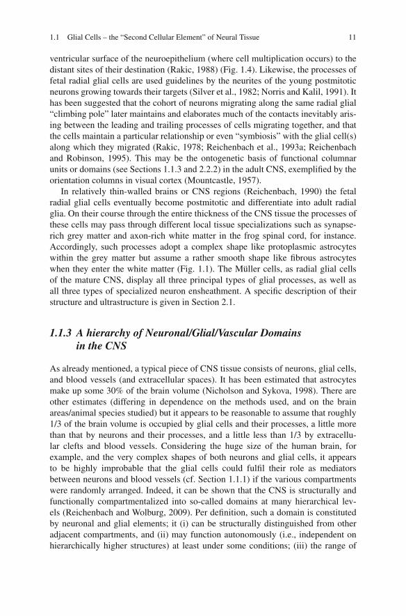
Fig. 1.5 The congruence of neuronal, glial, and vascular domains, exemplified for the olfactorybulb. a, the so-called glomerula, constituted by afferent and efferent neuronal processes and theirsynapses (newborn kitten, from Ramón y Cajal, 1952); b, the glial “envelope”of these glomerula,visualized by GFAP immunohistochemistry (adult frog; modified after Bailey et al., 1999); c, theblood supply of a glomerulum (ink injection; cat; from Kölliker (1896); the nerve fibers are alsodrawn)
the other hand they may work as interdependent cogwheels in the machinery oflarger domains. Obviously the concept requires the presence of controlled gatesbetween neighbouring domains of the same rank as well as towards higher-rankingdomains. The relatively long, thin stalk of the microdomain shown in Fig. 1.6may serve as an example for such a “gate”. Its cytoplasmic longitudinal resistanceconstitutes an obstacle against the spread of Ca2+ rises, triggered in the head byneuronal activity in the ensheathed synapses, towards the glial stem process or adja-cent microdomains. Furthermore, together with the shunt conductance of the stalkmembrane, it prevents the electrotonic propagation of even large depolarizations ofthe head membrane (Grosche et al., 2003). Whereas these estimates explain whyindividual microdomains may exclusively display Ca2+ responses in response tolow-frequency single-axon stimulation, it has also been shown that stronger/morefrequent and/or extensive stimulation may cause Ca2+ rises in several neighbour-ing microdomains or even in the whole Bergmann glial cell (Grosche et al., 1999,2003). This may be due to a spread of the activation within the neuronal com-partments (simply bypassing the glial gates), as well as by an “overrun of thegates” by the accumulation of high Ca2+ levels during repetitive release from thestores and/or by saturation of Ca2+ binding proteins in the glial cytoplasm, forinstance. A similar overrun of glial gates may play a causative role in patholog-ical events such as spreading depression and epileptiform discharges (De Keyseret al., 2008).
14 1 Introduction
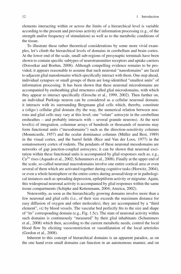
Fig. 1.6 Bergmann glial cell processes and glial microdomains. a, 3-D reconstruction of a partof a Bergmann glial cell process. The living cell was dye-injected in a perfused cerebellar slice;then, after fixation and dye-conversion, about 600 consecutive serial ultrathin sections were pho-tographed in the electron microscope, and the images of the dye-labeled profiles were reconstructedby a computer program. The inset shows a substructure labeled in blue; this part was quanti-tatively analyzed (see b, c). b, Glial microdomain as part of the 3-D reconstruction shown ina. c, Schematic drawing of such a glial microdomain and its relationships to the neuronal elements.d, 3-D reconstruction of a group of neighboring cerebellar synapses (yellow-green; synaptic clefts:orange) together with the surrounding leaflets provided by the injected Bergmann glia (blue-grey).The arrowheads point to neuronal surfaces not covered by glial sheaths from the labeled cell. Withpermission, from Reichenbach et al. (2004)
However, there are also active mechanisms of glial gate control. Glial networksare coupled via gap junctions, the conductance of which is under control of a varietyof signals including well-established intracellular second messengers (Rörig andSutor, 1996; Rouach et al., 2000).
Thus, Ca2+ waves arising in one glial cell may pass to a variable number of neigh-bour cells, depending on the current functional state of the gap junctions betweenthe cells (Enkvist and McCarthy, 1992; Venance et al., 1995). Moreover, there areextracellular “bypassing” glial signalling pathways; for instance, a stimulated glialcell may release ATP as a “gliotransmitter” which then activates ATP receptors onadjacent glial cells, which triggers intracellular Ca2+ rises in these cells and even-tually causes ATP release from them, and so far (Cotrina et al., 1998; Nedergaardet al., 2003).
Furthermore, the activity of glial homeostatic mechanisms such as uptake car-riers in their membrane can be modified by these signals and/or by the metabolicstate of the cells; this, in turn, will modify the extracellular propagation of signalmolecules released by neurons and glial cells (“volume transmission”: Syková andChvátal, 2000). Finally it should be kept in mind that large blood vessels cross theborders between different domains. In cases of stroke, for instance, the metabolismand activity of neurons and glial cells may be altered in wide areas, independent ofthe glial gates.
There are two conclusions from these considerations which appear to be impor-tant for an understanding of glia-neuron interactions in the retina (as a part of the
1.2 The Vertebrate Retina as a Part of the CNS 15
CNS: see Section 1.2), (i) the topographical relationship between neuronal and glial(plus vascular) elements reflects their intimate functional collaboration and inter-dependence, at many hierarchically scaled dimensions from sub- to multicellularlevels, and (ii) a propagation or “ascent” of activity across the limiting “gates” ofthe hierarchical levels is possible via several different mechanisms which may becarried by neuronal, glial, or even vascular elements. For the role of domains in theretina, see Section 2.2.3.
1.2 The Vertebrate Retina as a Part of the CNS
Much of the above-mentioned insights into the interplay between neurons and glialcells of the brain also applies to the retina which is a part of the CNS both by embry-ology (it arises from an evagination of the neural tube; see Section 1.2.1) and byfunction (in addition to stimulus perception it performs complex signal processing;see Section 1.2.2). In addition, the retina is a sensory organ, which causes a numberof specific requirements. So for instance, the retina must have access to its adequatestimulus, the light, which means that many parts of its surrounding ocular structuresmust be transparent and a high-quality image of the environment must be deliveredto the photoreceptor cells. These and many other tasks including the generation andrenewal of light-sensitive photopigments, the maintenance of the enormous energydemands of the specific transduction mechanism of the photoreceptors, as a fewexamples, add to the already high complexity of neuron-glia interactions in otherparts of the CNS.
1.2.1 Some Phylogenetic and Ontogenetic Basics
To understand the complex and, in some sense, even counterintuitive makeup ofthe vertebrate retina, it is essential to keep in mind that vertebrates belong to thedeuterostomian animals and that our ancestors must be searched among the relativesof recent starfish and sea urchins. If the nervous system of the starfish (Fig. 1.7) isused as a model of the origin of our CNS, two things become immediately appar-ent. First, this nervous system is not only embedded in the “skin” epithelium, itis by itself epithelial. It spans between the outer surface of the body, where itdirectly contacts the seawater as a fluid environment, to the basal lamina deliminat-ing the epidermal cells from the mesenchymal compartments. The second importantobservation is that this epithelial nervous system is polar, as well as its cells arenon-randomly oriented and polar. For instance, the so-called supporting cells –which can be considered as the ancestors of radial glial cells (Reichenbach andRobinson, 1995) – span the entire thickness of the epithelium from “watery” sur-face, into which their apical processes extend microvilli, to the inner basal laminawhere their basal processes form endfoot-like structures. The similarity of thesetwo types of cell processes to the ventricle-contacting processes and to the pia-contacting processes, respectively, of “modern” radial glial cells (Figs. 1.1 and 1.3)is apparent. The sensory cells are also polar; their sensory processes extend into the
16 1 Introduction
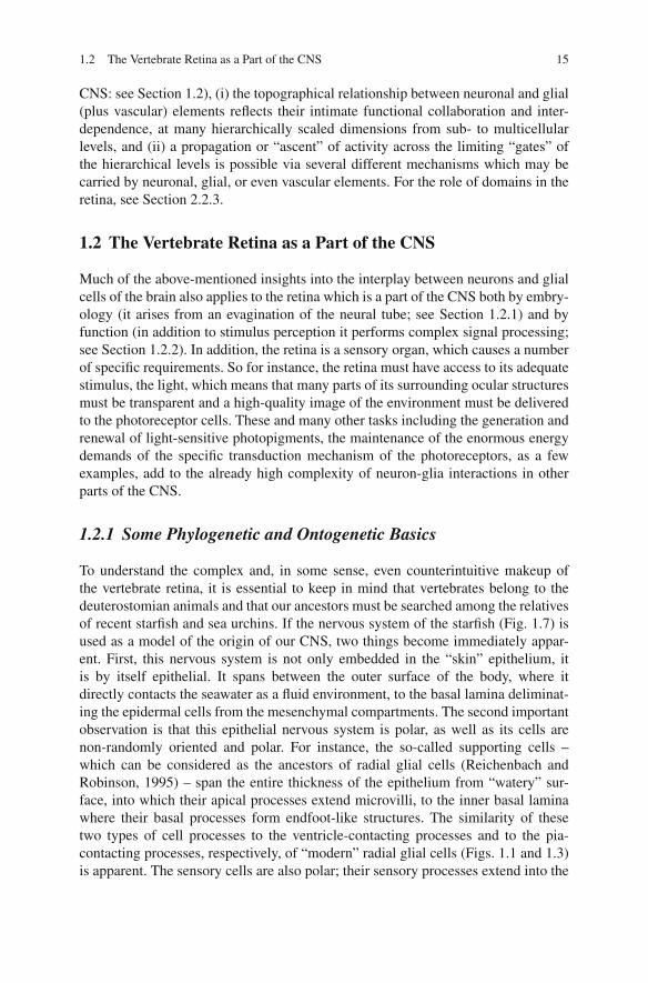
Fig. 1.7 The starfish nervous system as a (pro-)vertebrate “prototype CNS”. Cross-section throughthe radial nerve of Asterias rubens. The sensory and ganglionic neurons are surrounded by so-called supporting cells which send radial processes towards the basal lamina (delimiting theectoderma from mesoderma) which they abut with the conical endfeet of these processes. Thesecells may be considered as “ancestral radial glial cells”. Redrawn after Figs. 1.4 and 1.12 in Meyer(1906)
maritime environment of the animal as the source of the (hitherto unknown) stimulito be monitored, whereas their axons reach towards the ganglion cells as the sitesof information processing. Notably, this polarity is obviously “correct” and easilycomprehensible.
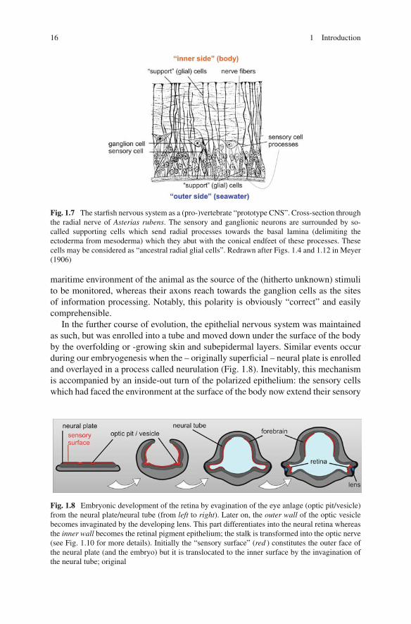
In the further course of evolution, the epithelial nervous system was maintainedas such, but was enrolled into a tube and moved down under the surface of the bodyby the overfolding or -growing skin and subepidermal layers. Similar events occurduring our embryogenesis when the – originally superficial – neural plate is enrolledand overlayed in a process called neurulation (Fig. 1.8). Inevitably, this mechanismis accompanied by an inside-out turn of the polarized epithelium: the sensory cellswhich had faced the environment at the surface of the body now extend their sensory
Fig. 1.8 Embryonic development of the retina by evagination of the eye anlage (optic pit/vesicle)from the neural plate/neural tube (from left to right). Later on, the outer wall of the optic vesiclebecomes invaginated by the developing lens. This part differentiates into the neural retina whereasthe inner wall becomes the retinal pigment epithelium; the stalk is transformed into the optic nerve(see Fig. 1.10 for more details). Initially the “sensory surface” (red ) constitutes the outer face ofthe neural plate (and the embryo) but it is translocated to the inner surface by the invagination ofthe neural tube; original
1.2 The Vertebrate Retina as a Part of the CNS 17
processes into the lumen – i.e., the inner surface – of the neural tube. Perhaps, thishad not been much of a problem in the most ancient small animals; still in therecent hemichordates this lumen is continuous with the surrounding seawater, andchemical and/or osmotic stimuli may be detected without crucial delay. However,later when the lumen was closed against the outside world, and filled by a substi-tute of the seawater – the cerebrospinal fluid – these receptors lost their originalfunction as environmental receptors, and had to be functionally replaced by “novel”receptor types and sense organs at the surface of the animals (this was perhaps theevolutionary driving force for the emergence of the peripheral nervous system).
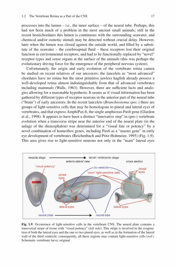
Unfortunately, the origin and early evolution of the vertebrate retina cannotbe studied on recent relatives of our ancestors; the lancelets as “most advanced”chordates have no retina but the most primitive jawless hagfish already possess awell-developed retina almost indistinguishable from that of advanced vertebratesincluding mammals (Walls, 1963). However, there are sufficient facts and analo-gies allowing for a reasonable hypothesis. It seems as if visual information has beengathered by different types of receptor neurons in the anterior part of the neural tube(“brain”) of early ancestors. In the recent lancelets (Branchiostoma spec.) there aregroups of light-sensitive cells that may be homologous to pineal and lateral eyes ofvertebrates, and that express AmphiPax-6, the single amphioxus Pax6 gene (Glardonet al., 1998). It appears to have been a distinct “innovative step” in (pre-) vertebrateevolution when a transverse stripe near the anterior end of the neural plate (in theanlage of the diencephalon) was determined for a “visual fate or potency” by anovel combination of homeobox genes, including Pax6 as a “master gene” in earlyeye development of vertebrates (Reichenbach and Pritz-Hohmeier, 1995) (Fig. 1.9).This area gives rise to light-sensitive neurons not only in the “main” lateral eyes
Fig. 1.9 Occurrence of light-sensitive cells in the vertebrate CNS. The neural plate contains atransversal stripe of tissue with “visual potency” (left side). This stripe is involved in the evagina-tion of both the lateral eyes and the one or two pineal eyes, as well as in the formation of the lateralwall of the third ventricle; consequently, all these regions may contain light-sensitive cells (red ).Schematic vertebrate larva; original
18 1 Introduction
(corresponding to our eyes) but also in one or two dorsal eyes (pineal or parietal)and in the wall of the third ventricle (as well as to visually specialized areas ofthe midbrain). Still in recent fish, amphibians and reptilians all these visual sensoryorgans can be found (Fig. 1.9). Embryonic birds have a pineal “retina” which istransformed into a neurosecretory organ during later developmental stages; such apineal retina fails to occur in mammals.
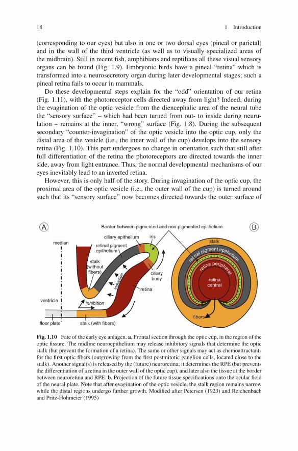
Do these developmental steps explain for the “odd” orientation of our retina(Fig. 1.11), with the photoreceptor cells directed away from light? Indeed, duringthe evagination of the optic vesicle from the diencephalic area of the neural tubethe “sensory surface” – which had been turned from out- to inside during neuru-lation – remains at the inner, “wrong” surface (Fig. 1.8). During the subsequentsecondary “counter-invagination” of the optic vesicle into the optic cup, only thedistal area of the vesicle (i.e., the inner wall of the cup) develops into the sensoryretina (Fig. 1.10). This part undergoes no change in orientation such that still afterfull differentiation of the retina the photoreceptors are directed towards the innerside, away from light entrance. Thus, the normal developmental mechanisms of oureyes inevitably lead to an inverted retina.
However, this is only half of the story. During invagination of the optic cup, theproximal area of the optic vesicle (i.e., the outer wall of the cup) is turned aroundsuch that its “sensory surface” now becomes directed towards the outer surface of
Fig. 1.10 Fate of the early eye anlagen. a, Frontal section through the optic cup, in the region of theoptic fissure. The midline neuroepithelium may release inhibitory signals that determine the opticstalk (but prevent the formation of a retina). The same or other signals may act as chemoattractantsfor the first optic fibers (outgrowing from the first postmitotic ganglion cells, located close to thestalk). Another signal(s) is released by the (future) neuroretina; it determines the RPE (but preventsthe differentiation of a retina in the outer wall of the optic cup), and later also the tissue at the borderbetween neuroretina and RPE. b, Projection of the future tissue specifications onto the ocular fieldof the neural plate. Note that after evagination of the optic vesicle, the stalk region remains narrowwhile the distal regions undergo further growth. Modified after Petersen (1923) and Reichenbachand Pritz-Hohmeier (1995)