Embed Size (px)
Citation preview

Mucormycosis in Asia:Where do we stand now?
Arunaloke ChakrabartiProfessor & Head
Center for Advanced Research in Medical Mycology
& WHO Collaborating Center
Department of Medical Microbiology
Postgraduate Institute of Medical Education & Research
Chandigarh – 160012, India

Very high incidence 0.14/1000 population
Uncontrolled diabetes
MAJOR RISK FACTOR
India & China largest
diabetics in world
Isolated renal
mucormycosisMany new flowers in the garden
Highlights of Asian epidemiology
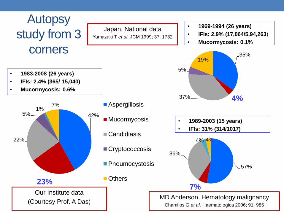
42%
23%
22%
5%1%
7% Aspergillosis
Mucormycosis
Candidiasis
Cryptococcosis
Pneumocystosis
Others
35%
4%37%
5%
19%
57%
7%
36%
4% 4%
• 1983-2008 (26 years)
• IFIs: 2.4% (365/ 15,040)
• Mucormycosis: 0.6%
Our Institute data
(Courtesy Prof. A Das)
• 1969-1994 (26 years)
• IFIs: 2.9% (17,064/5,94,263)
• Mucormycosis: 0.1%
• 1989-2003 (15 years)
• IFIs: 31% (314/1017)
MD Anderson, Hematology malignancyChamilos G et al. Haematologica 2006; 91: 986
Japan, National dataYamazaki T et al. JCM 1999; 37: 1732
Autopsy
study from 3
corners

Underlying risk factors in mucormycosisReference Count-
ries
Period Cases
No.
HM
(%)
DM
(%)
SOM/
SOT
DFO
(%)
HIV
(%)
AI/
CO
Trauma/
no
Roden, 2005 Global 1885-
2004
929 21 36 7 6 2 1 19
Bitar, 2009 France 1997-
2006
63 17 16 7 - 5 - 54
Pagano,
2009
Italy 2004-
2007
60 62 18 2 - 2 3 40
Saegeman,
2010
Belgium 2000-
2009
31 77 6 13 - 3 - 13
Ruping,
2010
Global 2006-
2009
41 63 17 10 - - - -
Skiada, 2011 Europe 2005-
2007
230 55 17 9 1 2 7 20
Chakrabarti,
2006
India 2001-
2005
178 1 74 1 - - - 19
Chakrabarti,
2009
India 2006-
2007
75 9 44 5 - 1 29 14
Lanternier,
2012
France 2005-
2007
101 50 23 3 - - - 18
HM= Hematological malignancy, DM=Diabetes mellitus, DFO= Deferroxamine therapy, HIV= human immuno-
deficiency virus, AI/CO= Autoimmune/corticosteroid therapy, SOM/SOT=Solid organ malignancy/transplant
Petrikkos et al. CID 2012; 54 (Suppl 1): S23

Mucormycosis & diabetes in India
Reference Region Population Cohort Diabetes %
Mohapatra, 2010 IJMM New Delhi All mucormycosis 29 13 44.83
Chakrabarti, 2009 PMJ Chandigarh All mucormycosis 75 48 64.00
Chakrabarti, 2006 MM Chandigarh All mucormycosis 178 131 73.60
Mohindra, 2007 MM Chandigarh All mucormycosis 27 15 55.56
Nithyanandam, 2003 IJO Bangalore All mucormycosis 34 30 88.24
Chakrabarti, 2001 JI Chandigarh All mucormycosis 96 23 23.96
Sundaram, 2006 H Hyderabad Mucormycosis (CNS) 40 30 75.00
Sundaram, 2005 M Hyderabad Mucormycosis (CNS) 56 32 57.14
Sundaram, 2011 LI Hyderabad Mucormycosis (Pulmonary) 6 5 83.33
Chander, 2010 IJMR Chandigarh Mucormycosis (Nec fascitis) 9 1 11.11
Chakrabarti Chandigarh Mucormycosis (Nec fascitis) 18 1 5.56

Countries with high diabetes burden
IDF Diabetes Atlas, Sixth Ed, 2013

• 35 cases of consecutive 22,316 diabetics (1.6
cases/1000 diabetics); 48.6% diabetic ketoacidosis
• The mean informed duration of diabetes was 6.7±4.6 y
before acquiring mucormycosis
• All patients treated with amphotericin B & 74% had
extensive surgical debridement
• 68% survived
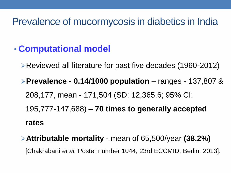
Prevalence of mucormycosis in diabetics in India
• Computational model
Reviewed all literature for past five decades (1960-2012)
Prevalence - 0.14/1000 population – ranges - 137,807 &
208,177, mean - 171,504 (SD: 12,365.6; 95% CI:
195,777-147,688) – 70 times to generally accepted
rates
Attributable mortality - mean of 65,500/year (38.2%)
[Chakrabarti et al. Poster number 1044, 23rd ECCMID, Berlin, 2013].

Chakrabarti et al. Postgrad Med J 2009; 85: 573
Uncontrolled diabetes & diabetic ketoacidosis

Diabetes & mucormycosis
• Formidable risk factor in Asia
• More common in ketoacidotic state (Roden MM et al. CID 2005; 41: 634-653;
Chakrabarti A et al. Postgrad Med J 2009; 85: 573-581)
• Mucormycosis - a diabetes-defining illness in 16-23% patients
(Roden MM et al. CID 2005; 41: 634; Chakrabarti A et al. Postgrad Med J 2009; 85: 573)
• Steroid induced diabetes in HM & transplants play role
• Not only rhino-cerebral type, but also in other types (except
cutaneous & renal mucormycosis), diabetes significantly associated
(Chakrabarti A et al. Postgrad Med J 2009; 85: 573)
• ?The incidence mucormycosis in diabetics is coming down in west
(Kontoyiannis DP. CID 2007: 44: 1089)


Obese
Non-hyperglycemic
Obese
Non-hyperglycemic
Metformin effect
Obese
Hyperglycemic
Metformin & diet
modification effect
Obese
Hyperglycemic
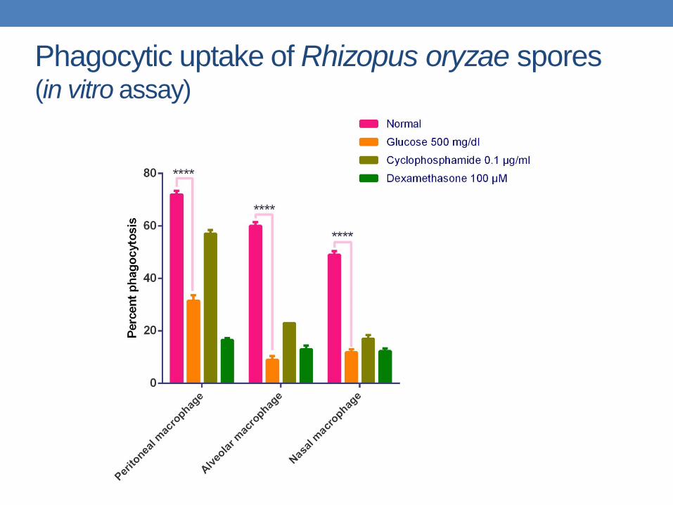
Phagocytic uptake of Rhizopus oryzae spores(in vitro assay)

• Not only nasal macrophages, but also peritoneal & alveolar
macrophages are altered in diabetes
• Increased incidence of mucormycosis in the diabetic individuals
may be due to a poor recognition of the fungi, reduced uptake
and low cytokine response in diabetic patients towards the
pathogen
• Ketoacidosis increases expression of GRP 78 (glucose
regulated protein on endothelial cells) & Cot H3 protein (spore
coat protein homologs on Mucorales) → helps in invasion (Ibrahim
et al. Mol Microbiol 2010; 77: 587; Liu et al. J Clin Invest 2010; 120: 1914; Gebremanriam et al.
J Clin Invest 2014; 124: 237)
• Further defects
Neutrophil dysfunction
Low serum pH decreases phagocytosis
Modifies transferrin system
Decreases serum inhibitory activity

Anatomical sites of involvement

ROC 48-58%
Pulmonary – 6-17%
Renal – 5-14%GI – 5-13%
Cutaneous-13-15%
Disseminated 5-12 %
Anatomic
distribution of
mucormycosis in
India
J Infect 2001; 42: 261
Med Mycol 2006; 44: 335
Mycoses 2007; 50: 271
Postgrad Med J 2009; 85: 57

E C M M
6 2 R O C
6 9 P u lm o n a ry
6 0 C u ta n e o u s
3 5 D is s e m in a te d
7 O th e rs
F u n g is c o p e
8 R O C
2 4 P u lm o n a ry
5 G a s tro in te s t in a l
8 C u ta n e o u s
6 D is s e m in a te d
3 R e n a l
1 O th e rs
R o d e n e t a l
3 6 2 R O C
2 2 3 P u lm o n a ry
6 5 G a s tro in te s t in a l
1 7 7 C u ta n e o u s
2 8 D is s e m in a te d
1 9 R e n a l
1 2 1 O th e rs
In d ia
5 8 R O C
6 P u lm o n a ry
7 G a s tro in te s t in a l
1 4 C u ta n e o u s
7 D is s e m in a te d
7 R e n a l
1 2 O th e rs
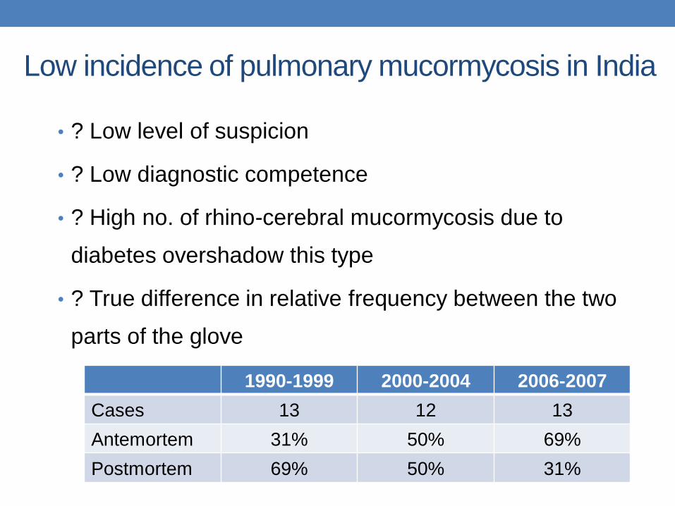
Low incidence of pulmonary mucormycosis in India
• ? Low level of suspicion
• ? Low diagnostic competence
• ? High no. of rhino-cerebral mucormycosis due to
diabetes overshadow this type
• ? True difference in relative frequency between the two
parts of the glove
1990-1999 2000-2004 2006-2007
Cases 13 12 13
Antemortem 31% 50% 69%
Postmortem 69% 50% 31%
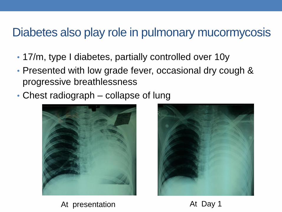
Diabetes also play role in pulmonary mucormycosis
• 17/m, type I diabetes, partially controlled over 10y
• Presented with low grade fever, occasional dry cough &
progressive breathlessness
• Chest radiograph – collapse of lung
At presentation At Day 1

• 17/m, type I diabetes, partially controlled over 10y
• Presented with low grade fever, occasional dry cough &
progressive breathlessness
• Chest radiograph – collapse of lung
• Bronchoscopy – mucosal plug
• Fexible bronchoscopy could not remove the plug
• Plug removed by rigid bronchoscopy under GA
• PCR of the sample – Rhizopus oryzae
• Lip AmB (3mg/Kg/d), surgery not possible as Carina involved
• Patient on follow up
Diabetes also play role in pulmonary mucormycosis

Another case
• 57/F, uncontrolled type II diabetes
• Cough, mucopurulent expectoration, progressive dyspnoea
• Bronchoscopy, bronchial biopsy – aseptate hyphae, PCR &
sequencing – Rhizopus oryzae
• Patient died due to surgical complication

Gastrointestinal Mucormycosis - prematurity
Presentation No. of cases
(%)
Bowel gangrene 7 (70)
Prematurity 7 (70)
Septic shock 9 (90)
Anemia 2 (20)
Renal failure 1 (10)
Hematological
malignancy
1 (10)
Steroid & cytotoxic
drug
1 (10)
Alcoholism 1 (10)
Chakrabarti et al. Postgrad Med J 2009; 85: 573
•Prematurity – GI mucormycosis in 20% of enterocolitis cases
•Suspect mucormycosis in neonate having intestinal perforation
[Patra et al. J Indian Assoc Pediatr Surg 2012, 17:153].

Health-care related mucormycosis
• Cases of necrotizing fasciitis due to Mucorales is not
unusual
• This is due to contaminated i.m. injection [(Chakrabarti & Singh.
Mycoses 2014; 57 (Suppl. 3) 1-6]
• Nosocomial mucormycosis in 9% (Chakrabarti et al. Postgrad
Med J 2009; 85: 57)
• Infection at the site of ECG leads or adhesive tapes or
from contaminated i.m. injection or from air in hospital

Renal mucormycosis
Marak RS et al. Med Mycol 2010; 48: 1088-95; Chakrabarti et al. Postgrad Med J 2009; 85: 573-81
Pathology:
Infarction
Hilar vessel thrombosis
Vasculitis
Cortical & medullary necrosis
Microabscess & granuloma

Renal mucormycosis
•50 cases in last 2 decades
at out center
•42 cases reviewed
•34 diagnosed antemortem
•75% no risk factor reported
•50% mortality
•Route of entry not known
Marak RS et al. Med Mycol 2010; 48: 1088

Medical Mycology Aug 2006; 44: 461
Primary renal zygomycosis due to Rhizopus oryzaeJin YU & Ruo Yu Li
Peking University First Hospital, Beijing, PR China
• 46 cases reviewed
• Male : Female = 10:1
• Age – 3m-77y (5 children)
• Clinical presentation – similar to India (fever, flank pain,
hematuria, anuria)
• Underlying illness• No – 30% (all children)
• Yes – 70% (i.v. drug abuser, diabetes, kidney transplant, steroid therapy etc.)

Chakrabarti & Singh. Mycoses 2014; 57 (Suppl. 3) 1-6
Human pathogenic Mucorales in different series
India India Europe France Italy

Recent study from Patel Chest, Delhi
Organism Rhino-
cerebral
Pulmonary Disseminated Cutaneous
Rhizopus arrhizus var.
delemar
2 14 2 -
R. Arrhizus var. arrhizus - 15 - -
Rhizopus microsporus 7 6 - -
Rhizopus stolonifer 1 - 2 2
Syncephalastrum
racemosum
5 - - 6
Lictheimia ramosa - 2 - 1
Mucor circinelloides - 2 - -
Apophysomyces variabilis - - - 2
Apophysomyces elegans - - - 2
Total cases (n=61) 15 39 4 13
Chowdhary et al. Mycoses 2014; 57 (Suppl. 3) Epub

Apophysomyces elegans
Rhizopus homothallicus
Saksenaea vasiformis
Thamnostylum lucknowense
Indian garden has many new flowers!

Apophysomyces elegans infection
• Of ~100 cases published in literature, a major portion
(~60%) of cases was reported from India
• Majority of patients had no underlying disease
• It usually casuses cutaneous & subcutaneous infection
• Local wound contamination with soil after accident
represents the single most common host risk factor
• The fungus has also been reported from rhinocerebral,
pulmonary, renal, and disseminated mucormycosis
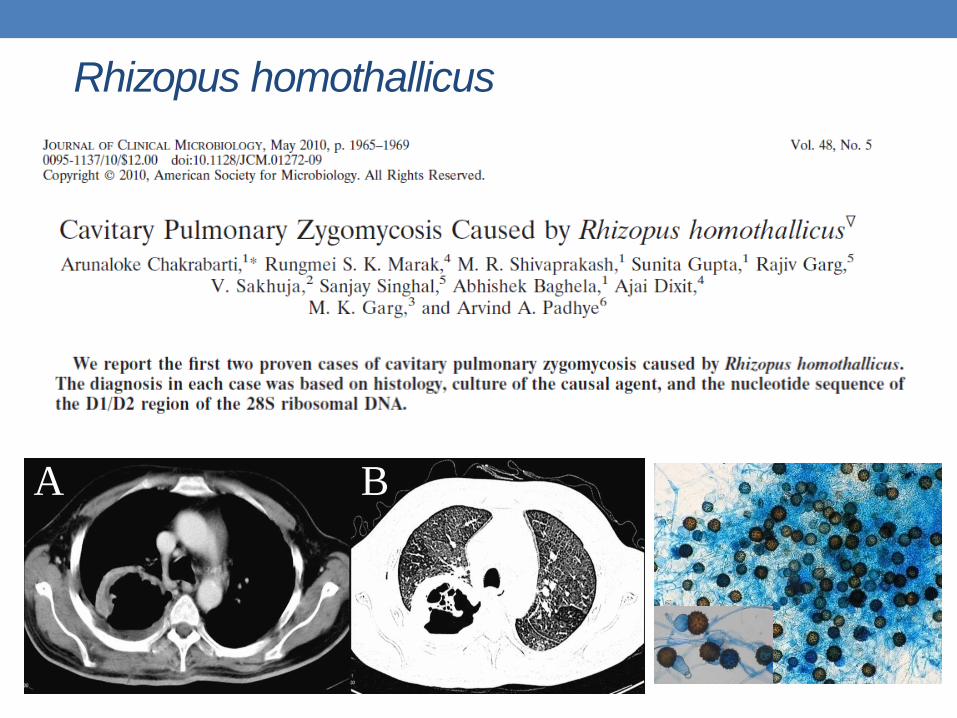
Rhizopus homothallicus
A B
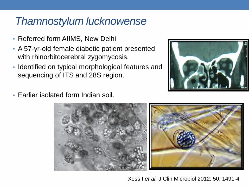
Thamnostylum lucknowense
• Referred form AIIMS, New Delhi
• A 57-yr-old female diabetic patient presented
with rhinorbitocerebral zygomycosis.
• Identified on typical morphological features and
sequencing of ITS and 28S region.
• Earlier isolated form Indian soil.
Xess I et al. J Clin Microbiol 2012; 50: 1491-4

Ecology of Mucorales
Types of soils:-
- Haryana (Calcireous sierozenic, pH>8, strongly alkaline)
- Punjab (Clay loamy, pH 7-7.5, neutral to slightly alkaline)
- Tamilnadu (Red Loamy, pH 6.8 -7.2, neutral)
- Himachal Pradesh (Slity loamy, pH< 6, acidic ).

Punjab Haryana
Tamilnadu Himachal Pradesh
Spectrum of Mucorales in Indian soils

Soil properties and Apophysomyces spp. isolation
• Alkaline soils - higher Apophysomyces isolation
• No seasonal variation in isolation of Apophysomyces spp.
• The mean temperature – 34.5±7.8
• Bivariate analysis - nitrogen, phosphorous, zinc, copper
was significantly associated with Apophysomyces isolation.
• Multivariate analysis - low nitrogen content, alkaline pH of
soil were significantly associated.

Diagnosis

Diagnostic techniques for mucormycosis
• Direct microscopy
Wet mount, Calcofluor White,
PAS, GMS
Fluorescent in situ
hybridization (FISH)
Immunohistochemistry
• Culture
• Serology
ELIspot
• Molecular techniques
Conventional PCR , RFLP
DNA sequencing
Realtime PCR
• Fungal Identification
Culture: Macro-and
Micro-morphology
DNA sequencing
Conventional PCR,
RFLP
Realtime PCR
MALDI-TOF

Molecular method for identification of Mucorales in
paraffin-embedded or frozen tissues
Reference Tissue Method Target
Dannaoui et al, 2010 Paraffin-embedded PCR+ sequencing ITS1
Hata et al, 2008 Frozen/fresh/paraffin-
embedded tissue
Real-time PCR Cyt b
Schwarz et al, 2006 Frozen/fresh tissue PCR+sequencing ITS
Machouart et al, 2006 Frozen/fresh tissue PCR+RFLP 18S
Bialek et al, 2005 Paraffin-embedded Semi-nested PCR 18S
Kobayashi et al, 2004 Frozen/fresh tissue PCR+sequencing 28S
Hayden et al, 2002 Paraffin-embedded In situ hybridization 18S
•Fresh or frozen tissue much better success
•Paraffin-embedded tissue – 70-90% success

Even closely related varieties
could be differentiated

Serological tests
Study Target
structure
Method Specimen Sensitivity/
specificity
Wyson DR, et
al., 1987
R. oryzae Ag
homogenate
Western blotting Sera of 5
patients
n.a.
Kaufman L, et
al., 1989
R. oryzae & R.
pusilus Ab
ELISA Sera of 43
patients
Minimal positivity
(1:400)
Jensen HE, et
al., 1996, 1997
R. oryzae water
soluble somatic
Ag, Hyphae
ELISA, western
blot, immuno-
histochemistry
Tissue of 40
patients
n.a.
Potenza L, et al.,
2011
Mucorales
specific IFN-
producing T cells
ELISA (ELiSpot)
or immuno-cyto-
fluorimetric
assay
Blood from 80
patients
Picked up all 3
cases with
mucormycosis

•3 of 28 patients developed mucormycosis
•17 had other infections
•Mucorales specific T cells could be
detected in 3 IM patients only

• 3 quantitative PCR assays using hydrolysis probes targeting
Rhizopus/Mucor, Lichtheimia, Rhizomucor
• 10 patients with proven mucormycosis (2-9 serum samples)
• No cross reaction with opportunistic fungi
• Detection limit 3.7-15 fentogram/10µl
• 9/10 patients – DNA detection positive
• DNA detected 69 & 3 days before mucormycosis diagnosis
Summary
• Mucormycosis is a serious problem – alarming rise
• ?disease burden, ?risk factors
• Isolated renal mucormycosis – how it occurs?
• Spectrum of agents broad – many new flowers
• Antifungal susceptibility
• Real challenge is prompt diagnosis
• Solution - ?bio-markers ?serology ?nucleic acid detection
• Therapy? – Dr. Kontoyiannis

Acknowledgement
• Manpreet Dhaliwal
• M R Shivaprakash
• Hari Prasath
• Anup Ghosh
• Ritesh Agarwal
Thank you