Embed Size (px)
Citation preview
385
[Frontiers in Bioscience, Landmark, 22, 385-406, January 1, 2017]
1. ABSTRACT
Mucopolysaccharidosis VI (MPS VI), or Maroteaux-Lamy syndrome, is an autosomal recessive lysosomal storage disorder caused by deficient activity of the enzyme arylsulfatase B (ASB). Progressive accumulation of glycosaminoglycans (GAGs) in organs and tissues leads to the development of multisystem clinical manifestations. The presentation of MPS VI is genotypically and phenotypically diverse, with a large number of potential disease-causing mutations and a phenotypic spectrum ranging from very slowly to very rapidly progressing disease. Diagnosis of MPS VI relies on presence of clinical features, increased GAG levels in urine or low ASB activity in dried blood spots, and measurement of enzyme activity levels in leukocytes or fibroblasts. The management of MPS VI involves enzyme replacement therapy and medical and surgical treatment of disease manifestations. Liquid chromatography/tandem mass spectrometry of GAG-derived disaccharides in blood or urine is emerging as a valuable method in the diagnosis, prognosis and assessment of therapeutic efficacy in MPS VI.
2. INTRODUCTION
Mucopolysaccharidosis VI (MPS VI), also known as Maroteaux-Lamy syndrome, is a progressive
Mucopolysaccharidosis VI: Pathophysiology, diagnosis and treatment
Paul R. Harmatz1, Renee Shediac2
1UCSF Benioff Children’s Hospital Oakland, Department of Gastroenterology, Oakland, CA, USA, 2BioMarin Pharmaceutical Inc., Novato, CA, USA
TABLE OF CONTENTS
1. Abstract2. Introduction3. Clinical features of MPS VI4. Biochemistry and genetics5. Primary and secondary involvement of glycosaminoglycans6. Glycosaminoglycans as biomarker of disease severity7. Diagnosis
7.1. Diagnostic algorithm7.2. Factors confounding diagnosis7.3. Future directions
8. Treatment8.1. Therapies replacing or repairing the deficient enzyme
8.1.1. Enzyme replacement therapy8.1.2. Hematopoietic stem cell transplantation8.1.3. Future directions
8.2. Management of disease manifestations8.3. Glycosaminoglycans as biomarker of therapeutic efficacy
9. Conflicts of interest10. Acknowledgements11. References
autosomal recessive lysosomal storage disorder with multisystem involvement which was first described by Dr. Pierre Maroteaux and Dr. Maurice Lamy in 1963 (1). The underlying cause of the disease is deficient activity of the enzyme N-acetylgalactosamine 4-sulfatase (arylsulfatase B (ASB)), which is involved in the degradation of the glycosaminoglycans (GAGs) dermatan sulfate and chondroitin 4-sulfate. This enzyme deficiency leads to the accumulation of partially degraded GAGs in tissues and organs, which in turn causes an array of clinical manifestations that progressively worsen with age.
MPS VI is very rare, with incidence estimates ranging from 0.36 to 1.30 per 100,000 live births, depending on the country or ethnic population examined (2). The highest incidence has been reported for the Turkish population in Germany, which may be due to a high degree of consanguinity in this population; the lowest incidence has been reported for Sweden (3,4). No worldwide estimates are currently available. Due to the lack of newborn screening programs for MPS VI and the difficulties in diagnosing the disease, the reported incidence rates are probably underestimations.
GAGs and MPS VI
386 © 1996-2017
3. CLINICAL FEATURES OF MPS VI
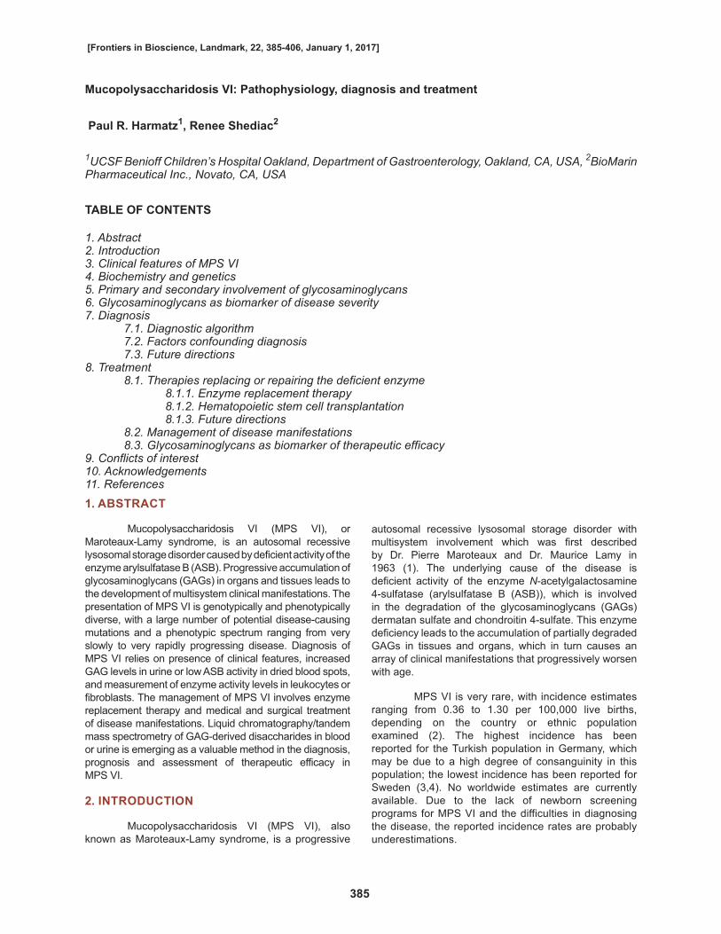
Disease manifestations, age of symptom onset and rate of disease progression vary widely among MPS VI patients (Figure 1). Although a distinction is generally made between slowly and rapidly progressing phenotypes, it is important to recognize that the phenotypic spectrum of the disease is a continuum. Table 1 gives an overview of the clinical manifestations that have been described for MPS VI.
The rapidly progressing phenotype of MPS VI is associated with a rapid onset of clinical manifestations, usually before the age of 2 years. The skeleton is generally one of the most severely affected organs in these patients. A typical finding is dysostosis multiplex, a specific radiologic expression consisting of abnormally shaped ribs and vertebrae, enlarged skull, hypoplastic epiphyses, thickened diaphyses and thickened, short metacarpal bones (5,6). These patients also frequently show scoliosis or kyphosis, hip dysplasia, genu valgum and claw hand deformities, as well as joint stiffness, joint pain and flexion contractures (6,7). Musculoskeletal disease can result in short stature (adult height below 120 cm in rapidly progressing patients), low body weight, nerve entrapment syndromes such as spinal cord compression, nerve root compression and carpal tunnel syndrome, and poor mobility (8,9). Patients with rapidly progressing MPS VI generally show normal to accelerated growth velocity during the first year of life, which slows down in the second year of life to below the fifth
percentile of Centre for Disease Control (CDC) reference curves after the age of 3 years (10-12). Other common findings in these patients are coarse facial features with frontal bossing, depressed nasal bridge, enlarged tongue, gingival hypertrophy, teeth abnormalities, hirsutism, compromised pulmonary function, impaired vision (due to corneal clouding, high hyperopia, optic nerve injury, or retinopathy), impaired hearing, cardiac valve abnormalities, hepatosplenomegaly, and umbilical and inguinal hernias (6,7). Adolescent patients can show delayed pubertal development (13). Without proper treatment, patients with rapidly progressing disease have a life expectancy below 20 years (7). These patients generally die from cardiopulmonary disease, infection, or surgical complications.
The slowly progressing phenotype of MPS VI is associated with a slower clinical course or with clinically significant symptoms occurring in fewer systems. Adult patients with this phenotype tend to have heights above 140 cm (6,7,12). They generally show normal or only mildly coarsened facial features, a slightly reduced to normal body height, and less prominent skeletal dysplasia compared to the rapidly progressing phenotype. Although disease manifestations tend to progress more slowly in these patients, clinically significant symptoms develop in an advanced stage of the disease, often requiring surgery and ultimately resulting in a reduced lifespan (14-17). In a German study of nine adult patients with slowly progressing MPS VI (19-29 years old), all patients showed reduced lung function, seven had reduced endurance, seven had corneal clouding, seven had hip dysplasia, and three had cardiac valve disease (17). Most of them required surgical interventions in the past, including ear-nose-throat interventions, spinal cord decompression, valve replacement or carpal tunnel surgery. In a cross-sectional Survey study by Swiedler et al. of 121 patients with MPS VI, only one patient was over 50 years of age (7).
Although several studies have shown abnormalities in the central nervous system of patients with MPS VI, including abnormalities in the ventricular system, white matter and perivascular spaces, communicating hydrocephalus, and cerebral atrophy (18-20), MPS VI is generally considered not to affect intellectual development. Nevertheless, in a recent study of 25 MPS VI patients, approximately 30 percent had IQ scores below the normal range (69 or lower) (20). As brain abnormalities on MRI did not correlate with IQ scores, the investigators suggested that cultural deprivation and physical limitations associated with the disease (fatigue, and visual or hearing deficits) may have an impact on the outcome of IQ testing in these patients (20).
Because of the high morbidity, frequent surgeries, and constant monitoring required to manage the common infections and other clinical manifestations,
Figure 1. Photograph of patients with MPS VI illustrating the phenotypic spectrum of the disease.
GAGs and MPS VI
387 © 1996-2017
most MPS VI patients have a poor quality of life (21). Activities of daily living can also be considerably limited due to impairments in vision, hearing, mobility, and functional capacity (7).
4. BIOCHEMISTRY AND GENETICS
MPS VI develops due to mutations in the ARSB gene, encoding for the enzyme ASB, located on chromosome 5q13-q14. Functional ASB enzyme catalyzes the hydrolysis of the C4-sulfate ester linkage in N-acetylgalactosamine 4-sulfate residues at the non-reducing ends of the GAGs dermatan sulfate and chondroitin 4-sulfate (22). These GAGs are long unbranched sulfated polysaccharides comprised of repeating disaccharides consisting of an amino sugar (N-acetylgalactosamine) and an uronic sugar. Absent or reduced ASB enzyme activity results in the progressive accumulation in tissues and organs of dermatan sulfate and sulfated oligosaccharides derived from both dermatan sulfate and chondroitin 4-sulfate (6). The accumulation of dermatan sulfate in particular is believed to give rise to the clinical manifestations associated with MPS VI (6). Disease manifestations are generally present only in patients with enzymatic activity below 10% of the lower limit of normal (6). Carriers who still have one normal allele do not exhibit clinical manifestations of MPS VI.
The ARSB gene contains 8 exons and spans about 206 kb. Like other lysosomal enzymes, the ASB enzyme is first synthesized as a large molecular mass precursor (66 kD) and then proteolytically processed in acid compartments to a mature form of lower molecular mass (43 kD) (23). Mutations in the ARSB gene may affect enzyme synthesis, stability or maturation and possibly intracellular transport time or a combination of these factors. A recent study using site-directed mutagenesis suggested that most disease-causing mutations in the ARSB gene hamper the maturation of the ASB enzyme, rather than affecting the synthesis of the 66 kD ASB precursor (24). Different mutations may lead to different levels of residual ASB activity, and thus differences in GAG accumulation and clinical presentation.
In 2007, Karageorgos and colleagues published data from a large mutational analysis in MPS VI patients. They identified 83 different mutations in 105 patients, including at least 38 missense mutations, five nonsense mutations, 11 deletions, seven splice-site mutations, one insertion, and four polymorphisms (25). Thirty three patients (31.4 percent) were homozygous. In addition to the mutations described by Karageorgos et al., there have been several publications reporting on previously described and new disease-causing mutations in the ARSB gene (24,26-30). As of April 2015, over
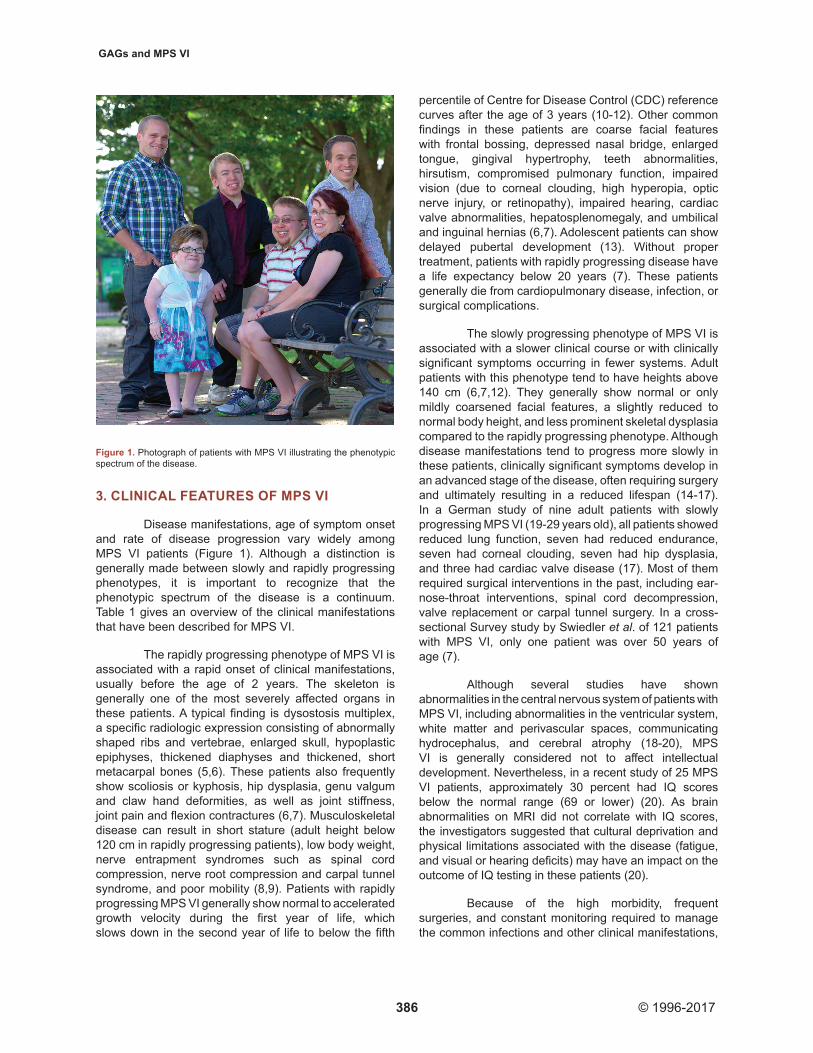
Table 1. Overview of clinical manifestations associated with mucopolysaccharidosis VI (8,19,35,40,42,52)
Musculoskeletal• Dysostosis multiplex• Genu valgum• Hip dysplasia• Kyphoscoliosis• Enlarged head• Joint stiffness• Joint contractures• Short stature
Ear, nose and throat• Gingival hyperplasia• Macroglossia• Thick lips• High‑arched palate• Distension of adenoids and/or tonsils• Impaired opening of the mouth• Conductive/neurosensory hearing loss
Respiratory• Sinusitis, recurrent bronchitis or pneumonia• Upper and lower airway obstruction• Restrictive pulmonary disease• Sleep disordered breathing (obstructive sleep apnea, nocturnal
hypoventilation)
Ocular• Corneal clouding• Refractive error problems• Glaucoma• Retinopathy• Optic disc swelling/optic atrophy
Neurological• Spinal cord/nerve root compression/myelopathy• Carpal tunnel syndrome• Abnormalities in ventricular system, white matter and perivascular spaces
Abdominal• Hepatosplenomegaly• Umbilical/inguinal hernias
Cardiac• Aortic and mitral valve insufficiency/stenosis• Pulmonary hypertension• Cardiomyopathy• Fibroelastosis• Cardiac conduction system disorders• Heart failure
Other• Coarse facial features• Teeth abnormalities (hypoplastic mandibular condyles, malposition of
unerupted teeth, large dental follicles, anterior open bite)• Hirsutism• Delayed pubertal development
GAGs and MPS VI
388 © 1996-2017
130 disease-causing mutations in the ARSB gene have been reported in the Human Gene Mutation Database (http://www.hgmd.cf.ac.uk/ac/index.php), of which 100 are missense mutations, 20 are deletions, nine are splice-site mutations, and three are insertions.
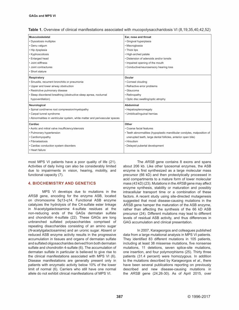
Because of the large number of mutations identified, many of which are unique or only occur in a few individuals, it has been very challenging to find genotype-phenotype correlations for MPS VI. Analysis of genotype-phenotype correlations is particularly problematic for compound heterozygous patients who have two different disease-causing alleles or when polymorphisms or the presence of two disease-causing mutations on the same allele may have an impact on disease phenotype (6). Nevertheless, several studies have suggested correlations between mutations and disease phenotypes (6). The large study by Karageorgos et al. showed a clear correlation between some genotypes and urinary GAG (uGAG) levels (25). In addition, the study showed that mutations in the active site of ASB, nonsense mutations and mutations resulting in a truncated protein product due to deletions or insertions, and some missense mutations (p.D54N, p.G144R, p.I223V, and p.I296N) appeared
to cause rapidly progressing disease. Other missense mutations were associated with a more slowly progressing phenotype. Figure 2 gives an overview of the mutations identified by Karageorgos et al. and the predicted clinical severity associated with these mutations. The genotype-phenotype correlations in this study were generated from clinical data described for these patients in other studies (7,31,32). More recently, Jurecka et al. showed a link between the relatively common p.Y210C mutation and an essentially osteoarticular phenotype (33), while patients homozygous for the p.R152W mutation were found to present with a cardiac phenotype that, although apparently slowly progressing, may lead to abrupt death (34). Despite these findings, it remains very difficult to predict a patient’s phenotype from his or her genotype.
5. PRIMARY AND SECONDARY INVOLVEMENT OF GLYCOSAMINOGLYCANS
The clinical manifestations of MPS VI are believed to be directly or indirectly caused by accumulation of dermatan sulfate in particular in tissues and organs. Dermatan sulfate is an important component
Figure 2. Location of all the mucopolysaccharidosis VI (MPS VI) mutations in the human arylsulfatase B (ARSB) gene identified in a mutational analysis study by Karageorgos et al., 2007 (25) and predicted clinical severity associated with the mutations (represented by different colors) categorized on the basis of clinical criteria defined in the natural history study by Swiedler et al. (7). The boxes with Roman numerals represent exons of the gene. Reprinted with permission from Wiley and Sons.

GAGs and MPS VI
389 © 1996-2017
of bones, cartilage and connective tissues, but also has a role as cofactor or receptor for growth factors, cytokines, adhesion molecules and chemokines and serves as a regulator of enzyme activity and signaling molecules in response to cellular damage (6,35). The accumulation of dermatan sulfate can cause engorgement, cell enlargement and cell dysfunction, but may also indirectly lead to clinical manifestations through a cascade of secondary events.
The growth plate of long bones has been shown to be very sensitive to excessive GAG storage, which interferes with the control of chondrocyte development (36). Animal studies have shown that inflammation secondary to GAG accumulation plays an important role in joint and bone disease in MPS through the activation of the Toll-like receptor-4 (TLR-4) (37). Activation of the TLR-4 stimulates the release of inflammatory cytokines such as tumor necrosis factor alpha (TNF-alpha) and interleukin-1 beta (IL-1beta) from chondrocytes, leading to apoptosis. The resulting proliferation of immature chondrocytes leads to abnormal and poorly organized matrix formation. Release of matrix metalloproteinases in synovial membranes also contributes to the bone and joint pathology. In addition, abnormal GAG metabolism has been shown to interfere with major signaling pathways involved in cartilage and bone development (35,36). The defective endochondral and membranous growth leads to the typical radiologic skeletal deformities (dysostosis multiplex; Figure 3) and short stature seen in MPS VI patients (38). Anterior hypoplasia of vertebral bodies at the thoracolumbar junction can lead to gibbus deformity or dorsolumbar kyphosis very early in the disease course (39). Hip dysplasia can develop due to a poorly formed pelvis, dysplasia of the femoral heads, and coxa valga (38). Progressive flexion contractures of the fingers can lead to claw-hand deformity and a decline in fine motor skills. Most patients show joint stiffness, pain, and flexion contractures of the elbows, shoulders, hips, and knees, leading to decreased range of motion and gait abnormalities (39).
Respiratory function in patients with MPS VI can be impaired due to airway abnormalities, which may be caused directly by GAG deposition in the mouth, nose, throat, and upper and lower airways, or indirectly by cranial and spinal abnormalities (e.g., flattened nasal bridge, short neck, high epiglottis, mandibular abnormalities, abnormal cervical vertebrae) (40). Many patients also show reduced ventilatory capacity due to altered chest wall shape and structure, diaphragmatic weakness, or compromised diaphragm excursion (due to e.g., enlarged liver and spleen). As a result, patients may develop frequent sinusitis or otitis media, recurrent bronchitis or pneumonia, sleep disordered breathing, and even respiratory failure. Airway obstruction and restrictive pulmonary disease (often in combination with cardiovascular manifestations) pose a serious anesthetic risk to MPS VI patients (41).
Abnormalities in cardiac structure and function are common in patients with MPS VI and are prominent causes of morbidity and mortality (42-45). Cardiac valve disease, particularly in the mitral and aortic valves, is among the most common findings in these patients, occurring in 96 percent of 121 patients included in the Survey study by Swiedler et al. (7,43). Cardiomyopathy, fibroelastosis and pulmonary hypertension may
Figure 3. Radiographs of (A) the hands of a patient (16 years old, male) with MPS VI showing short, broad, and irregular metacarpals and phalanges and moderately severe Madelung deformity; and (B) hips from a patient with MPS VI (19 years old, male) showing bilateral hip dysplasia with flaring of iliac wings and flattening of the acetabulum bilaterally. There is also flattening and flaring of proximal femur with incomplete containment of the femoral heads.
GAGs and MPS VI
390 © 1996-2017
also contribute to the development of heart failure in these patients (42). Dysfunction of heart muscle and morphological and biochemical changes in the connective tissues of the heart are primarily caused by accumulation of GAGs (42). Pathology studies have described excessive GAG accumulation in cytoplasmic vacuoles in endothelial cells, myocytes and fibroblasts of the endocardium, myocardium, valves, conduction system and blood vessels (42). The fact that dermatan sulfate is the dominant GAG in cardiac valve connective tissue and blood vessels in healthy subjects might explain why cardiac disease tends to be more severe in types of MPS associated with accumulation of dermatan sulfate (MPS I, II, VI and VII) than in other types (42,46). GAG accumulation can result in progressive thickening and deformation of the valves, leading to valvular regurgitation, stenosis, or both (42,44). Brands et al. recently postulated that GAG accumulation in the lysosomes of activated valvular interstitial cells (“clear” cells) deprive these cells of the capacity to maintain the extracellular matrix of the cardiac valve, leading to tissue damage. The tissue damage attracts macrophages, most likely through the TLR-4 pathway, which cause even more damage and eventually leads to valve regurgitation. In turn, valvular stenosis or regurgitation may lead to left atrial and/or left ventricular volume overload, left ventricular dilatation, left ventricular hypertrophy, and even to systolic and diastolic dysfunction (42,43). In addition, the stiff and narrow thoracic cage of MPS VI patients may limit the space available for the heart and heart beat movements (42).
Spinal cord compression in MPS VI can develop due to atlantoaxial subluxation, craniovertebral stenosis, posterior longitudinal ligament hypertrophy, dural thickening, disc bulging, kyphoscoliosis, or a combination of these factors and can cause compressive myelopathy, leading to loss of strength in the legs, pyramidal signs, paresis and, ultimately, tetraparesis (47-50). Compression occurs mostly at the upper cervical or thoracolumbar regions, but multilevel myelopathy may occur as well (50). Thickening of the dura mater, posterior elements and ligaments may be due to deposition of GAGs in these structures (19,50). A potential cause of atlantoaxial instability in MPS VI patients is odontoid process hypoplasia (19). The development of carpal tunnel syndrome, which is a relatively common finding in MPS VI, is considered to be due to bone deformity in combination with excessive GAG storage in tissues surrounding the carpal tunnel (16,51). Carpal tunnel syndrome may be associated with burning pain, tingling and numbness, finger weakness, clumsiness and, in a late stage, loss of thumb abduction and opposition strength (51) or may develop gradually without typical symptoms. Ventricular enlargement and communicating hydrocephalus in MPS VI are believed to be due to overflow impairment through bony dysostosis of the skull base in combination with deposition of GAGs in the arachnoid villi (19).
Most patients with MPS VI develop ocular manifestations, which can result in reduced visual acuity or even blindness (52). Corneal clouding is most commonly reported and develops when GAG storage in the cornea affects keratocyte size and disrupts the regular network of collagen fibrils in the stroma (53). GAGs in the anterior segments of the eye may also cause increased intraocular pressure, which in turn may lead to open-angle or narrow-angle glaucoma (53). Retinopathy can arise due to accumulation of GAGs in the retinal pigment epithelium and the interphotoreceptor matrix. Optic disc swelling and subsequent optic atrophy can be due to thickening of the sclera and impingement of the optic nerve, or may result from mismatch of the optic nerve sheath and optic canal diameter, GAG deposits within ganglion cells or the meningeal covering of the optic nerve, or from chronically increased intracranial pressure (53). Increased rigidity in the cornea and thickening of the sclera due to deposition of GAGs may cause flattening of the corneal curvature or shortening of the axial length, leading to hyperopia.
Hearing loss is a frequent finding in patients with MPS VI and is mostly due to recurrent otitis media, although it may also develop directly due to GAG deposition in the inner ear or the central nervous system (54). Young children tend to have conductive hearing loss, while adolescent and adult patients generally develop mixed hearing loss or pure sensorineural hearing loss (55). The exact mechanisms underlying this evolution are still unclear.
Very common findings in patients with MPS VI and other types of MPS are an enlarged liver and spleen (hepatosplenomegaly). Light and electron microscopic studies have revealed membrane-bound GAG inclusions within hepatocytes and Kupffer cells of MPS patients and hepatocellular accumulation of lipid droplets (56).
The frequently observed malpositioned and unerupted teeth in MPS VI have been suggested to develop secondary to large dental follicles (35). As the dermatan sulfate content does not seem to be high in large dental follicles, it remains unclear what causes this enlargement. The anterior open bite in some MPS VI patients might be due to hypoplastic mandibular condyles (35).
6. GLYCOSAMINOGLYCANS AS BIOMARKER OF DISEASE SEVERITY
In MPS VI and other types of MPS, GAGs are stored in lysosomes and the extracellular matrix of several tissues, secreted into the blood, and excreted in urine (57). The cross-sectional Survey study of 121 MPS VI patients by Swiedler et al. revealed an association between uGAG levels and disability progression rate, with higher uGAG levels being associated with a more rapidly progressing disease course (7). Elevated uGAG
GAGs and MPS VI
391 © 1996-2017
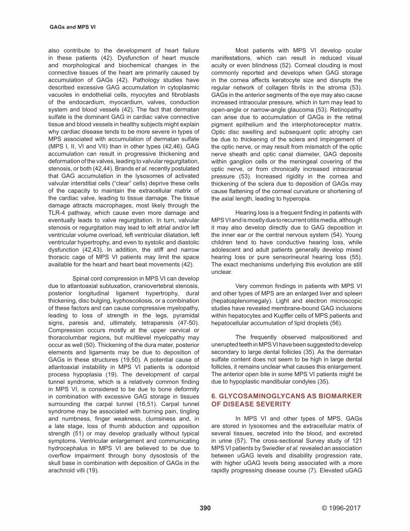
levels (higher than 200 µg/mg creatinine) in this study tended to correspond with a rapidly progressing clinical course, characterized by an age-adjusted short stature (height range 80 to 120 cm), a low body weight, impaired endurance in a walk test, and impaired pulmonary function and joint range of motion (Figure 4) (7). Patients with slowly progressing disease tended to have uGAG levels below 100 μg/mg creatinine. Low uGAG levels also seemed to be associated with a longer survival, as suggested by the finding that most patients older than 20 years had levels below 100 µg/mg creatinine (7). In line with these findings, a 10-year Resurvey study of patients who had participated in the Survey study suggested better survival in patients with low baseline uGAG levels (200 µg/mg creatinine or lower) than those with higher baseline levels (58). In addition, all patients with the most prevalent mutation (p.Y210C), associated with a slowly progressing phenotype, had uGAG levels below 100 µg/mg creatinine (7,25). Lower uGAG levels were also correlated with a higher height for age, which has been confirmed in other studies (7,13,25). The 10-year Resurvey study showed an inverse correlation between baseline uGAG levels and final heights after 10 years (58).
Taken together, these findings indicate that uGAG levels may, at least to some degree, predict disease
progression in patients with MPS VI. Based on the results of the Survey study, MPS VI patients can be classified into a rapidly progressing patient group with uGAG levels above 200 µg/mg creatinine and a slowly progressing patient group with uGAG levels below this threshold. It should be noted that this does not apply to uGAG levels in infants, because infants have been shown to have naturally high uGAG levels (59). The utility of uGAGs for classifying MPS VI is limited after onset of therapy that lowers uGAG levels, such as enzyme replacement therapy (ERT).
7. DIAGNOSIS
7.1. Diagnostic algorithmThe diagnosis of MPS VI is generally triggered
by the presence of clinical features indicative of a metabolic disorder and referral to a metabolic specialist or clinical geneticist or by the diagnosis of a close relative (sibling) with the disease. Definitive diagnosis is based on a combination of disease-specific clinical findings, including short stature, dysostosis multiplex, corneal clouding, hepatosplenomegaly and cardiac valve disease, and laboratory test results.
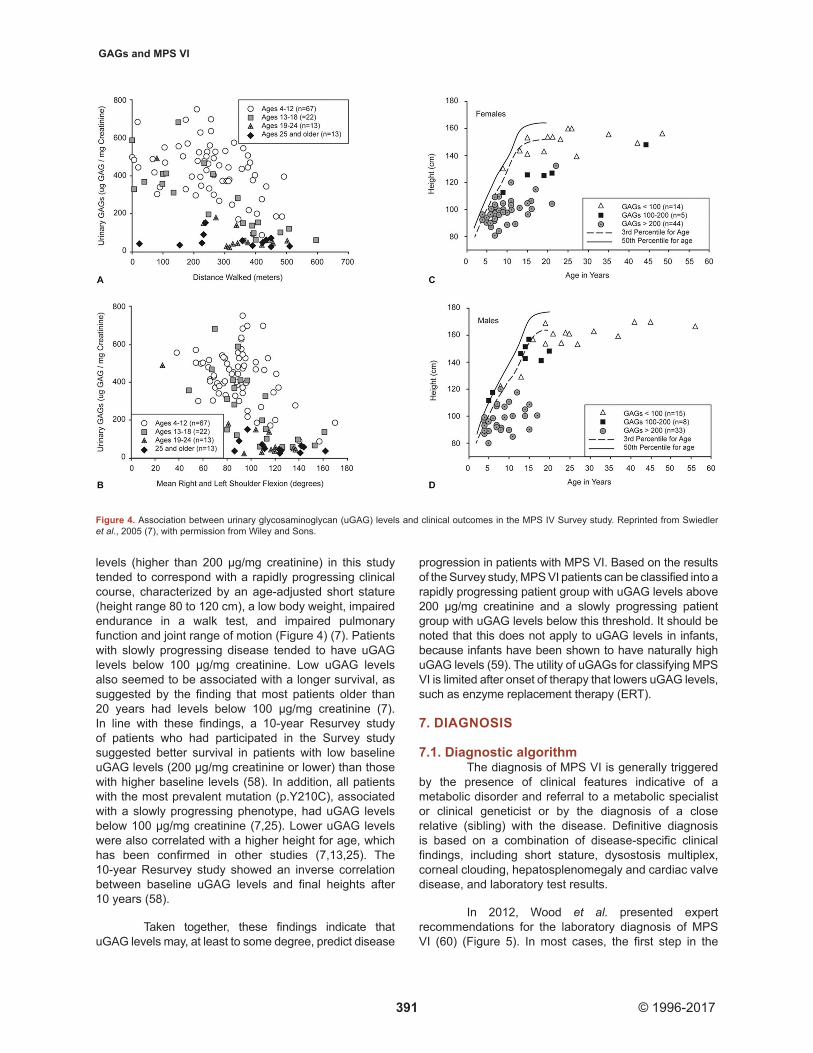
In 2012, Wood et al. presented expert recommendations for the laboratory diagnosis of MPS VI (60) (Figure 5). In most cases, the first step in the
Figure 4. Association between urinary glycosaminoglycan (uGAG) levels and clinical outcomes in the MPS IV Survey study. Reprinted from Swiedler et al., 2005 (7), with permission from Wiley and Sons.
GAGs and MPS VI
392 © 1996-2017
Figure 5. Algorithm for the laboratory diagnosis of mucopolysaccharidosis VI (MPS VI). Reprinted from Wood et al., 2012 with permission from Elsevier (60).
GAGs and MPS VI
393 © 1996-2017
diagnostic work-up is a screening test, i.e. quantitative and qualitative analyses of GAGs in urine or measurement of ASB activity in dried blood spots. If one of these tests is positive or clinical suspicion of MPS is high, the clinician should proceed to the next step in the algorithm, i.e. a diagnostic test for ASB or other MPS enzyme activity. A urine test is considered positive if it shows elevated total uGAG levels and detects dermatan sulfate. Clinicians should be aware that a negative outcome in this test does not rule out MPS VI. The screening test in dried blood spots is considered positive if ASB activity is low and if multiple sulfatase deficiency is ruled out (if activities of other enzymes tested in the same blood spot are within normal ranges). If initial screening is not positive, the test can be repeated on a new sample (when sample degradation is suspected because other enzymes were below normal ranges). If clinical suspicion is high, the clinician can proceed directly to a diagnostic test for ASB activity. When family history indicates a high risk that the patient has MPS VI, the diagnostic test is generally performed directly, without initial screening.
The diagnostic test for ASB activity is usually performed on leukocytes or fibroblasts, but may also be done on dried blood spots collected separately from the initial blood spots used in the screening assay. Multiple sulfatase deficiency should be ruled out by measuring a second sulfatase. Sample integrity can be assessed by measuring activity of a reference enzyme. If the diagnostic test is positive, the combined outcomes of all clinical and laboratory findings should be considered in the definitive diagnosis. Confirmation of ASB activity results in a different sample or tissue, the identification of two disease-causing mutations, or both are encouraged to confirm the diagnosis.
7.2. Factors confounding diagnosisDiagnosis of MPS VI can be challenging due
to the phenotypic heterogeneity resulting from the large number of disease-causing mutations and other genetic, metabolic and environmental factors (60). Patients with slowly progressive MPS VI may be particularly difficult to diagnose because they often first present as adolescents or adults to specialists not familiar with the disease such as rheumatologists or ophthalmologists. Also, most of these patients do not show the skeletal features of classical dysostosis multiplex (61). Clinicians should be aware that the radiographic findings in these patients may be very similar to those of other disorders such multiple epiphyseal dysplasia, some forms of spondylo-epiphyseal dysplasia and bilateral Perthes-like disease (61). An additional problem is that slowly progressing patients may have uGAG levels within the normal range and thus may be missed if only a quantitative uGAG analysis is performed.
7.3. Future directionsTo overcome some limitations of the currently
available screening methods, alternative screening
methods have been developed in more recent years. Liquid chromatography/tandem mass spectrometry (LC-MS/MS) has emerged as a powerful qualitative and quantitative method for the simultaneous analysis of disaccharides derived from different GAGs in urine, blood and/or dried blood spots (57,62-65). This method has shown good sensitivity and specificity for detecting all subtypes of MPS, and may also be useful in defining clinical severity and monitoring therapeutic efficacy (57). However, LC‑MS/MS is time‑consuming, which limits its utility for screening large numbers of samples. An alternative GAG assay by RapidFire high-throughput mass spectrometry was recently developed to overcome these limitations (66). Although this method is faster and more efficient than conventional LC-MS/MS methods and has shown sensitivity and specificity equivalent to LC-MS/MS, its use is limited by the fact that it cannot distinguish between disaccharides with identical molecular weights (57). In the future, LC-MS/MS may become a useful test for newborn screening of MPS (67).
8. TREATMENT
8.1. Therapies replacing or repairing the deficient enzyme
For a long time, the treatment of MPS VI was limited to symptom management and palliative care. More recently, therapies that directly target the underlying cause of the disease, i.e. the deficiency of the ASB enzyme, have been developed. The therapies currently available for MPS VI are ERT and hematopoietic stem cell transplantation (HSCT).
8.1.1. Enzyme replacement therapyERT is currently the recommended first‑line
treatment for MPS VI (39). The therapy involves weekly intravenous infusions with recombinant human ASB or galsulfase (Naglazyme®, BioMarin Pharmaceutical Inc., Novato, CA, USA) to replace the defective enzyme. The recombinant enzyme is produced in genetically engineered Chinese Hamster Ovary cells (31).
Preclinical studies in MPS VI cats established the safety of galsulfase and its efficacy in reversing GAG storage and limiting bone disease when started early in life (68). These studies were followed by phase 1/2, 2 and 3 studies in humans with MPS VI (31,32,69), which led to approval of the therapy in the United States in May 2005 (70) and in the European Union in January 2006. These clinical studies evaluated the efficacy and safety of intravenous ERT (0.1 or 1 mg/kg/week) in a total of 56 MPS VI patients ranging from 5 to 29 years of age at study initiation (Table 2). Supportive efficacy and safety data in a larger and more heterogeneous MPS VI population than in the clinical trials and over a longer period of time are being collected in the MPS VI Clinical Surveillance Program (MPS VI CSP), which was established to collect observational data from routine
GAGs and MPS VI
394 © 1996-2017
clinical and laboratory assessments of patients with MPS VI over a period of at least 15 years (8). In March 2010, the MPS VI CSP included data from 132 MPS VI patients with ages ranging from 1 to 59 years collected at 39 sites in Europe and the US (8). A phase 4 clinical open-label study in four infants (aged 3.3-12.7 months at inclusion) who received galsulfase 1.0 or 2.0 mg/kg/week for at least 52 weeks has recently been completed as well (71). In addition, the impact of early initiation of ERT on disease progression has been examined in three (ongoing) sibling control studies in children with MPS VI (72-74). In the Resurvey study which examined 10-year follow-up data of 59 patients who had previously participated in the Survey study by Swiedler et al., 55 patients received ERT (58). The mean duration of ERT treatment was 6.8 years in the 52 patients with known ERT start date (58). The outcomes of these studies were supported by a number of retrospective studies and case reports (11,24,75-82).
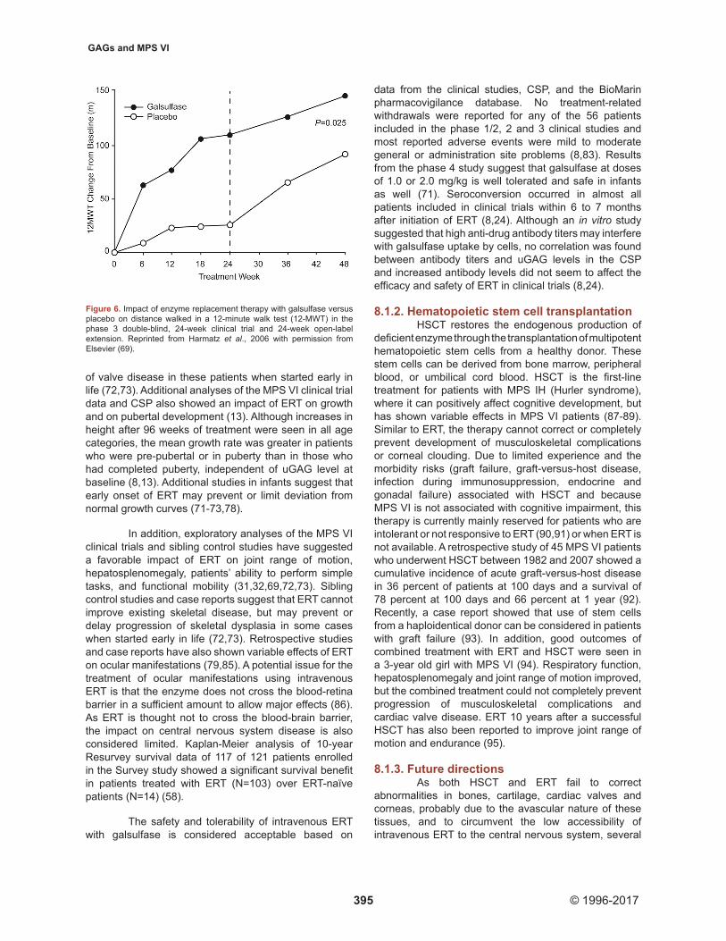
All clinical studies showed a significant reduction in uGAG after initiation of ERT (31,32,69). This reduction was sustained through 240 weeks of ERT (83). In the MPS VI CSP, uGAG data before and after initiation of ERT were available for 59 patients (8). uGAG decreased after initiation of ERT by a mean of at least 65 percent in patients with a baseline uGAG level higher than 200 μg/mg and by approximately 50 percent in those with a baseline uGAG below 200 μg/mg for the duration of the study. Improvements in endurance in a 6- or 12 minute walk test (MWT) and 3-minute stair climb test (3MSCT) were also seen in all clinical studies after onset of ERT (31,32,69). After the 24‑week double‑blind part of the phase 3 study, patients in the ERT group walked on average 92 meters more in the 12MWT (P = 0.025)
(Figure 6) and climbed 5.7 stairs per minute more in the 3MSCT (P = 0.053) than patients in the placebo group (69). Continued improvement in endurance was observed during the open-label extension (83). In the MPS VI CSP, distance walked in timed walk tests increased after initiation of ERT in 76 percent of patients (N = 25) (increases of at least 15 meters in the 6MWT and/or at least 30 meters in the 12MWT) (8). The increase in walking distance was observed through 4 years of ERT (N = 9 at 4 years follow-up). The 10-year Resurvey study also showed improvements in mean 6MWT distance in the ERT group, with patients with low baseline uGAG levels (below 200 µg/mg creatinine) showing greater increases after 10 years than those with high baseline uGAG levels (58).
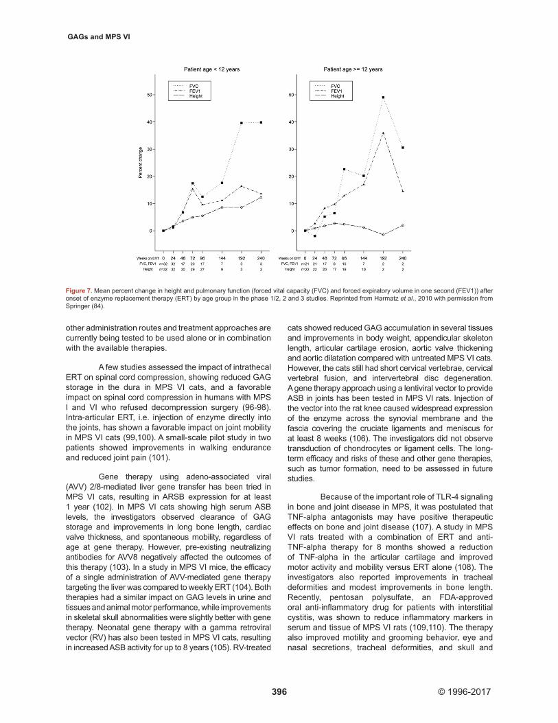
A post-hoc analysis of the pooled long-term data of the MPS VI clinical trials and data from the MPS VI CSP and 10-year Resurvey study also showed significant improvements in pulmonary function, i.e. forced vital capacity, forced expiratory volume in 1 second and maximum voluntary ventilation, after onset of ERT, which was sustained in the long term (8,58,84). Improvements in pulmonary function occurred in both patients aged younger than 12 years and patients aged 12 years or older, despite a smaller percentage increase in height in the latter group (Figure 7). Post-hoc analysis of cardiac ultrasound data from the clinical trials and additional studies suggested that ERT preserves cardiac dimensions and function, decreases ventricular wall thickness, and does not prevent progression of valve disease (44,58,75,76). Although the clinical studies showed no impact of ERT on cardiac valve disease in MPS VI patients, there are indications from sibling control studies that ERT might prevent or delay the progression
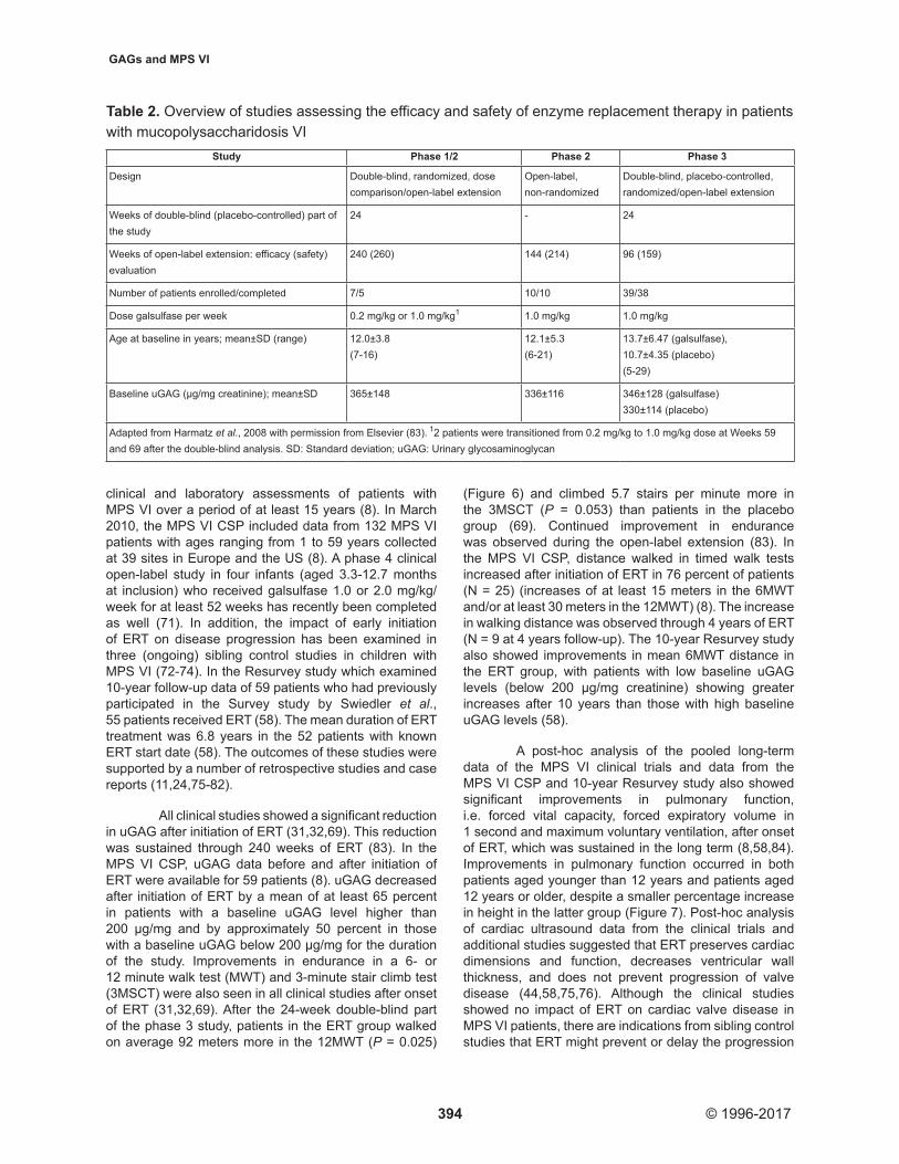
Table 2. Overview of studies assessing the efficacy and safety of enzyme replacement therapy in patients with mucopolysaccharidosis VI
Study Phase 1/2 Phase 2 Phase 3
Design Double-blind, randomized, dose comparison/open-label extension
Open-label, non-randomized
Double-blind, placebo-controlled, randomized/open-label extension
Weeks of double-blind (placebo-controlled) part of the study
24 - 24
Weeks of open-label extension: efficacy (safety) evaluation
240 (260) 144 (214) 96 (159)
Number of patients enrolled/completed 7/5 10/10 39/38
Dose galsulfase per week 0.2 mg/kg or 1.0 mg/kg1 1.0 mg/kg 1.0 mg/kg
Age at baseline in years; mean±SD (range) 12.0±3.8 (7-16)
12.1±5.3 (6-21)
13.7±6.47 (galsulfase), 10.7±4.35 (placebo) (5-29)
Baseline uGAG (µg/mg creatinine); mean±SD 365±148 336±116 346±128 (galsulfase)330±114 (placebo)
Adapted from Harmatz et al., 2008 with permission from Elsevier (83). 12 patients were transitioned from 0.2 mg/kg to 1.0 mg/kg dose at Weeks 59 and 69 after the double‑blind analysis. SD: Standard deviation; uGAG: Urinary glycosaminoglycan
GAGs and MPS VI
395 © 1996-2017
of valve disease in these patients when started early in life (72,73). Additional analyses of the MPS VI clinical trial data and CSP also showed an impact of ERT on growth and on pubertal development (13). Although increases in height after 96 weeks of treatment were seen in all age categories, the mean growth rate was greater in patients who were pre-pubertal or in puberty than in those who had completed puberty, independent of uGAG level at baseline (8,13). Additional studies in infants suggest that early onset of ERT may prevent or limit deviation from normal growth curves (71-73,78).
In addition, exploratory analyses of the MPS VI clinical trials and sibling control studies have suggested a favorable impact of ERT on joint range of motion, hepatosplenomegaly, patients’ ability to perform simple tasks, and functional mobility (31,32,69,72,73). Sibling control studies and case reports suggest that ERT cannot improve existing skeletal disease, but may prevent or delay progression of skeletal dysplasia in some cases when started early in life (72,73). Retrospective studies and case reports have also shown variable effects of ERT on ocular manifestations (79,85). A potential issue for the treatment of ocular manifestations using intravenous ERT is that the enzyme does not cross the blood‑retina barrier in a sufficient amount to allow major effects (86). As ERT is thought not to cross the blood‑brain barrier, the impact on central nervous system disease is also considered limited. Kaplan-Meier analysis of 10-year Resurvey survival data of 117 of 121 patients enrolled in the Survey study showed a significant survival benefit in patients treated with ERT (N=103) over ERT‑naïve patients (N=14) (58).
The safety and tolerability of intravenous ERT with galsulfase is considered acceptable based on
data from the clinical studies, CSP, and the BioMarin pharmacovigilance database. No treatment-related withdrawals were reported for any of the 56 patients included in the phase 1/2, 2 and 3 clinical studies and most reported adverse events were mild to moderate general or administration site problems (8,83). Results from the phase 4 study suggest that galsulfase at doses of 1.0 or 2.0 mg/kg is well tolerated and safe in infants as well (71). Seroconversion occurred in almost all patients included in clinical trials within 6 to 7 months after initiation of ERT (8,24). Although an in vitro study suggested that high anti-drug antibody titers may interfere with galsulfase uptake by cells, no correlation was found between antibody titers and uGAG levels in the CSP and increased antibody levels did not seem to affect the efficacy and safety of ERT in clinical trials (8,24).
8.1.2. Hematopoietic stem cell transplantationHSCT restores the endogenous production of
deficient enzyme through the transplantation of multipotent hematopoietic stem cells from a healthy donor. These stem cells can be derived from bone marrow, peripheral blood, or umbilical cord blood. HSCT is the first‑line treatment for patients with MPS IH (Hurler syndrome), where it can positively affect cognitive development, but has shown variable effects in MPS VI patients (87-89). Similar to ERT, the therapy cannot correct or completely prevent development of musculoskeletal complications or corneal clouding. Due to limited experience and the morbidity risks (graft failure, graft-versus-host disease, infection during immunosuppression, endocrine and gonadal failure) associated with HSCT and because MPS VI is not associated with cognitive impairment, this therapy is currently mainly reserved for patients who are intolerant or not responsive to ERT (90,91) or when ERT is not available. A retrospective study of 45 MPS VI patients who underwent HSCT between 1982 and 2007 showed a cumulative incidence of acute graft-versus-host disease in 36 percent of patients at 100 days and a survival of 78 percent at 100 days and 66 percent at 1 year (92). Recently, a case report showed that use of stem cells from a haploidentical donor can be considered in patients with graft failure (93). In addition, good outcomes of combined treatment with ERT and HSCT were seen in a 3-year old girl with MPS VI (94). Respiratory function, hepatosplenomegaly and joint range of motion improved, but the combined treatment could not completely prevent progression of musculoskeletal complications and cardiac valve disease. ERT 10 years after a successful HSCT has also been reported to improve joint range of motion and endurance (95).
8.1.3. Future directionsAs both HSCT and ERT fail to correct
abnormalities in bones, cartilage, cardiac valves and corneas, probably due to the avascular nature of these tissues, and to circumvent the low accessibility of intravenous ERT to the central nervous system, several
Figure 6. Impact of enzyme replacement therapy with galsulfase versus placebo on distance walked in a 12-minute walk test (12-MWT) in the phase 3 double-blind, 24-week clinical trial and 24-week open-label extension. Reprinted from Harmatz et al., 2006 with permission from Elsevier (69).
GAGs and MPS VI
396 © 1996-2017
other administration routes and treatment approaches are currently being tested to be used alone or in combination with the available therapies.
A few studies assessed the impact of intrathecal ERT on spinal cord compression, showing reduced GAG storage in the dura in MPS VI cats, and a favorable impact on spinal cord compression in humans with MPS I and VI who refused decompression surgery (96-98). Intra‑articular ERT, i.e. injection of enzyme directly into the joints, has shown a favorable impact on joint mobility in MPS VI cats (99,100). A small-scale pilot study in two patients showed improvements in walking endurance and reduced joint pain (101).
Gene therapy using adeno-associated viral (AVV) 2/8-mediated liver gene transfer has been tried in MPS VI cats, resulting in ARSB expression for at least 1 year (102). In MPS VI cats showing high serum ASB levels, the investigators observed clearance of GAG storage and improvements in long bone length, cardiac valve thickness, and spontaneous mobility, regardless of age at gene therapy. However, pre‑existing neutralizing antibodies for AVV8 negatively affected the outcomes of this therapy (103). In a study in MPS VI mice, the efficacy of a single administration of AVV-mediated gene therapy targeting the liver was compared to weekly ERT (104). Both therapies had a similar impact on GAG levels in urine and tissues and animal motor performance, while improvements in skeletal skull abnormalities were slightly better with gene therapy. Neonatal gene therapy with a gamma retroviral vector (RV) has also been tested in MPS VI cats, resulting in increased ASB activity for up to 8 years (105). RV-treated
cats showed reduced GAG accumulation in several tissues and improvements in body weight, appendicular skeleton length, articular cartilage erosion, aortic valve thickening and aortic dilatation compared with untreated MPS VI cats. However, the cats still had short cervical vertebrae, cervical vertebral fusion, and intervertebral disc degeneration. A gene therapy approach using a lentiviral vector to provide ASB in joints has been tested in MPS VI rats. Injection of the vector into the rat knee caused widespread expression of the enzyme across the synovial membrane and the fascia covering the cruciate ligaments and meniscus for at least 8 weeks (106). The investigators did not observe transduction of chondrocytes or ligament cells. The long-term efficacy and risks of these and other gene therapies, such as tumor formation, need to be assessed in future studies.
Because of the important role of TLR-4 signaling in bone and joint disease in MPS, it was postulated that TNF-alpha antagonists may have positive therapeutic effects on bone and joint disease (107). A study in MPS VI rats treated with a combination of ERT and anti‑TNF-alpha therapy for 8 months showed a reduction of TNF-alpha in the articular cartilage and improved motor activity and mobility versus ERT alone (108). The investigators also reported improvements in tracheal deformities and modest improvements in bone length. Recently, pentosan polysulfate, an FDA-approved oral anti-inflammatory drug for patients with interstitial cystitis, was shown to reduce inflammatory markers in serum and tissue of MPS VI rats (109,110). The therapy also improved motility and grooming behavior, eye and nasal secretions, tracheal deformities, and skull and
Figure 7. Mean percent change in height and pulmonary function (forced vital capacity (FVC) and forced expiratory volume in one second (FEV1)) after onset of enzyme replacement therapy (ERT) by age group in the phase 1/2, 2 and 3 studies. Reprinted from Harmatz et al., 2010 with permission from Springer (84).
GAGs and MPS VI
397 © 1996-2017
teeth abnormalities. Improvements in spinal stability were suggested as well. There were no improvements in cortical bone or femur length.
A potential therapeutic approach that has been proposed for MPS VI patients with nonsense mutations is pharmacological enhancement of ribosomal read-through of nonsense mutations to restore production of the full-length ASB protein. Bartolomeo et al. showed that PTC124 could increase the level of ASB activity in fibroblast cell lines to less than 2.5 percent of normal activity and reduce lysosomal size (111). The clinical significance of these findings needs to be established.
8.2. Management of disease manifestationsIn addition to the above-described systemic
therapies, patients with MPS VI need continuous management of disease manifestations, including use of adaptive or supportive devices, physiotherapy, occupational therapy, symptom-based medications, and surgical interventions. Because most patients show impairments in multiple organ systems, a multi-disciplinary management approach is required. General guidelines for the diagnosis and management of MPS VI have been developed by an international team of experts and were published in 2007 (39). These guidelines include a recommended multi-disciplinary schedule of assessments to detect and monitor disease manifestations. Since the publication of these general guidelines, several guidelines for the management of specific disease manifestations, including orthopedic problems, respiratory disease, spinal cord compression, cardiac disease, carpal tunnel syndrome, and anesthesia risk, in patients with MPS VI or patients with MPS in general have been published (40,41,43,50,51,53,86,112-114).
Even with ERT or HSCT, patients with MPS VI often require orthopedic surgery for correction of deformities of the spine, hips and extremities or carpal tunnel release to optimize long-term function and quality of life (51,112,113). Orthopedic surgery in these patients can be difficult due to abnormally shaped bones and the patients’ small stature (112). Moreover, the effectiveness of these interventions can be limited by the progressive nature of the disease. Physical therapy and anti-inflammatory drugs can also help to alleviate joint pain in patients with skeletal dysplasia.
Patients with upper airway obstruction may benefit from the use of nasal decongestants to control excessive mucus production or nasal steroids to reduce inflammation and swelling (40). In severe cases, tonsillectomy and/or adenoidectomy may be required (40). These procedures can be difficult due to the patients’ small airways and often thick and hard oropharyngeal tissues. Patients with sleep disordered breathing can considerably benefit from application of continuous positive airways pressure (for obstructive sleep apnea) or non-invasive ventilator support such as bilevel positive airway pressure (for nocturnal hypoventilation). Tracheostomy
may be required when assisted ventilation is ineffective or when airway obstruction is present at daytime (40). Other supportive therapies that can help maintain optimal functional status include regular pneumococcus and influenza vaccinations, aggressive treatment of respiratory tract infections, inhaled bronchodilators and manual or mechanic airway clearance techniques to improve secretion clearance.
Patients who develop cardiac valve stenosis or regurgitation may benefit from medical therapy (e.g., diuretics, aldosterone antagonists, ACE inhibitors, beta blockers, anticoagulants), or may require cardiac valve replacement in severe cases (43,114). There is currently very limited literature on the impact of medical treatments, implantable cardiac devices or surgical interventions in MPS VI patients with heart failure, pulmonary hypertension, coronary artery disease or other cardiac issues. Recommendations from general guidelines should be considered to manage these manifestations (114). Clinicians should be aware that cardiac insufficiency may be masked by limited maximal exercise levels due to joint stiffness, skeletal malformations or respiratory dysfunction (42).
Increased intracranial pressure or hydrocephalus can be decompressed using ventriculoperitoneal shunts (39). Detailed guidelines for the diagnosis, monitoring and management of spinal cord compression in patients with MPS VI have been published in 2012 (50). Early signs of myelopathy can be detected by functional assessments and clinical neurological examinations. Magnetic resonance imaging (MRI) is the gold standard for detecting cervical cord compression. Spinal cord compression can be treated surgically by cervical decompression with or without stabilization.
In patients with corneal clouding, corneal transplantation can be used to restore vision (53,86). However, the effectiveness of corneal transplantation can be limited by the concomitant presence of retinopathy, glaucoma or optic atrophy or by the recurrence of GAG deposits in the transplanted cornea (53). Glaucoma can be very difficult to diagnose due to corneal clouding, hampering the observation of chamber angles or optic disc cupping, or falsely high intraocular pressure measurements. The outcome of medical or surgical glaucoma therapy in MPS patients is uncertain (53,86).
Interventions requiring general anesthesia can be very dangerous in patients with MPS VI who are at high risk of perioperative morbidity and mortality. Problems that may arise include airway obstruction after induction or extubation, intubation difficulties or failure (sometimes requiring emergency tracheostomy), and cardiovascular and cervical spine issues (41). Because of the high anesthetic risk, the advantages of surgery should
GAGs and MPS VI
398 © 1996-2017
always be balanced against the associated risks for each individual patient and the procedures should be performed by experienced personnel (41). Neurophysiological monitoring is recommended during spine or head and neck surgery but may also be considered for other procedures requiring general anesthesia (50).
8.3. Glycosaminoglycans as biomarker of therapeutic efficacy
More than a decade ago, studies in MPS VI cats showed that reductions in uGAG levels after initiation of ERT are associated with reductions in GAG levels in a variety of well-vascularized tissues (68,115). As ERT demonstrates a clear impact on uGAG levels, these findings suggested that uGAG may be useful as a biomarker of therapeutic efficacy. Total uGAG determined with dimethylmethylene blue (DMB) staining and normalized with creatinine has been used as a surrogate for the extent of GAG clearance from lysosomal storage in all MPS VI clinical trials (31,32,69). The phase 1/2 clinical study showed a greater percentage reduction in uGAG with the higher dose of galsulfase (1 mg/kg) than with the lower dose (0.2 mg/kg).
In 2004, Crawley et al. evaluated GAG-derived oligosaccharides by tandem mass spectrometry in the urine of MPS VI cats receiving high and low doses of ERT and demonstrated a clear dose response during the first 4 weeks of ERT (115). Both doses also removed or prevented accumulation of GAGs in lysosomes of several soft tissue, with the highest dose showing a greater effect. The study also showed an advantage of tandem mass spectrometry determination of GAG-derived oligosaccharides for monitoring therapeutic efficacy over total uGAG determination by DMB staining (115). Several other methods for screening and monitoring of GAGs have been developed for MPS VI and other types of MPS, including antibody-based assays, ligand-binding assays and total GAG analysis by mass spectrometry (116). More recently, assays using LC-MS/MS to measure GAG-derived disaccharides in blood or urine have emerged as valuable methods, not only for diagnosis and screening, but also for prognosis of clinical severity and assessment of therapeutic efficacy in MPS patients (57,65, 66,117,118).
9. CONFLICTS OF INTEREST
P.R. Harmatz has provided consulting services, received research grants, participated in advisory board meetings and received speaker honoraria and travel support from BioMarin Pharmaceutical Inc. R. Shediac is an employee and shareholder of BioMarin.
10. ACKNOWLEDGEMENTS
The authors are grateful to Ismar Healthcare NV for their assistance in the writing of the manuscript, which was funded by BioMarin.
11. REFERENCES
1. P. Maroteaux, B. Levêque, J. Marie, M. Lamy: A new dysostosis with urinary elimination of chondroitin sulfate B. Presse Med 71, 1849-1852 (1963)
2. J. Muenzer: Overview of the mucopolysaccharidoses. Rheumatology (Oxford) 50(Suppl 5), v4-v12 (2011)
3. F. Baehner, C. Schmiedeskamp, F. Krummenauer, E. Miebach, M. Bajbouj, C. Whybra, A. Kohlschutter, C. Kampmann, M. Beck: Cumulative incidence rates of the mucopolysaccharidoses in Germany. J Inherit Metab Dis 28, 1011-1017 (2005)DOI: 10.1007/s10545-005-0112-z
4. G. Malm, A.M. Lund, J.E. Månsson, A. Heiberg: Mucopolysaccharidoses in the Scandinavian countries: incidence and prevalence. Acta Paediatr 97, 1577-1581 (2008)DOI: 10.1111/j.1651-2227.2008.00965.x
5. R. Lachman, K.W. Martin, S. Castro, M.A. Basto, A. Adams, E.L. Teles: Radiologic and neuroradiologic findings in the mucopolysaccharidoses. J Pediatr Rehabil Med 3, 109-118 (2010)DOI: 10.3233/PRM-2010-0115
6. V. Valayannopoulos, H. Nicely, P. Harmatz, S. Turbeville: Mucopolysaccharidosis VI. Orphanet J Rare Dis 5, 5 (2010)DOI: 10.1186/1750-1172-5-5
7. S.J. Swiedler, M. Beck, M. Bajbouj, R. Giugliani, I. Schwartz, P. Harmatz, J.E. Wraith, J. Roberts, D. Ketteridge, J.J. Hopwood, N. Guffon, M.C. Sá Miranda, E.L. Teles, K.I. Berger, C. Piscia-Nichols: Threshold effect of urinary glycosaminoglycans and the walk test as indicators of disease progression in a survey of subjects with mucopolysaccharidosis VI (Maroteaux-Lamy syndrome). Am J Med Genet 134A, 144-150 (2005)DOI: 10.1002/ajmg.a.30579
8. C.J. Hendriksz, R. Giugliani, P. Harmatz, C. Lampe, A.M. Martins, G.M. Pastores, R.D. Steiner, E. Leão Teles, V. Valayannopoulos: Design, baseline characteristics, and early findings of the MPS VI (mucopolysaccharidosis VI) Clinical Surveillance Program (CSP). J Inherit Metab Dis 36, 373-384 (2011)DOI: 10.1007/s10545-011-9410-9
GAGs and MPS VI
399 © 1996-2017
9. F.S. Haddad, D.H.A. Jones, A. Vellodi, N. Kane, M.C. Pitt: Carpal tunnel syndrome in the mucopolysaccharidoses and mucolipidoses. J Bone Joint Surg Br 79, 576-582 (1997)DOI: 10.1302/0301-620x.79b4.7547
10. D. Heron, C. Baumann, J.J. Benichou, J.P. Harpey, M. Le Merrer: Early diagnosis of Maroteaux-Lamy syndrome in two patients with accelerated growth and advanced bone maturation. Eur J Pediatr 163, 323-326 (2004)DOI: 10.1007/s00431-004-1428-7
11. M. Scarpa, R. Barone, A. Fiumara, L. Astarita, G. Parenti, A. Rampazzo, S. Sala, G. Sorge, R. Parini: Mucopolysaccharidosis VI: the Italian experience. Eur J Pediatr 168, 1203-1206 (2009)DOI: 10.1007/s00431-008-0910-z
12. A. Quartel, C.J. Hendriksz, R. Parini, S. Graham, P. Lin, P. Harmatz: Growth charts for individuals with mucopolysaccharidosis VI (Maroteaux-Lamy Syndrome). JIMD Rep 18, 1-11 (2015)DOI: 10.1007/8904_2014_333
13. C. Decker, Z. Yu, R. Giugliani, I.V.D. Schwartz, N. Guffon, E.L. Teles, M.C. Sá Miranda, J.E. Wraith, M. Beck, L. Arash, M. Scarpa, D. Ketteridge, J.J. Hopwood, B. Plecko, R. Steiner, C.B. Whitley, P. Kaplan, S.J. Swiedler, S. Conrad, P. Harmatz, MPS VI Study Group: Enzyme replacement therapy for mucopolysaccharidosis VI: growth and pubertal development in patients treated with recombinant human N-acetylgalactosamine 4-sulfatase. J Pediatr Rehabil Med 3, 89-100 (2010)DOI: 10.3233/PRM-2010-0113
14. H. Pilz, K. von Figura, H.H. Goebel: Deficiency of arylsulfatase B in 2 brothers aged 40 and 38 years (Maroteaux-Lamy syndrome, type B). Ann Neurol 6, 315-325 (1979)DOI: 10.1002/ana.410060405
15. D.A. Brooks, G.J. Gibson, L. Karageorgos, L.K. Hein, E.F. Robertson, J.J. Hopwood: An index case for the attenuated end of the mucopolysaccharidosis type VI clinical spectrum. Mol Genet Metab 85, 236-238 (2005)DOI: 10.1016/j.ymgme.2005.02.008
16. M. Scarpa, E. Buffone, P. La Marca, M. Campello, A. Rampazzo: Difficulties in diagnosing slowly progressive
mucopolysaccharidosis VI: a case series. J Pediatr Rehabil Med 3, 71-75 (2010)DOI: 10.3233/PRM-2010-0104
17. A. Thümler, E. Miebach, C. Lampe, S. Pitz, W. Kamin, C. Kampmann, B. Link, E. Mengel: Clinical characteristics of adults with slowly progressing mucopolysaccharidosis VI: a case series. J Inherit Metab Dis 35, 1071-1079 (2012)DOI: 10.1007/s10545-012-9474-1
18. L. Vedolin, I.V.D. Schwartz, M. Komlos, A. Schuch, A.C. Azevedo, T. Vieira, F.K. Maeda, A.M. Marques da Silva, R. Giugliani: Brain MRI in mucopolysaccharidosis: effect of aging and correlation with biochemical findings. Neurology 69, 917-924 (2007)DOI: 10.1212/01.wnl.0000269782.80107.fe
19. F. Borlot, P.R. Arantes, C.R. Quaio, J.F.D.S. Franco, C.M. Lourenco, D.R. Bertola, C.A. Kim: New insights in mucopolysaccharidosis type VI: neurological perspective. Brain Dev 36, 585-592 (2014)DOI: 10.1016/j.braindev.2013.07.016
20. A.C.M. Azevedo, O. Artigalás, L. Vedolin, M. Komlós, A. Pires, R. Giugliani, I.V.D. Schwartz: Brain magnetic resonance imaging findings in patients with mucopolysaccharidosis VI. J Inherit Metab Dis 36, 357-362 (2013)DOI: 10.1007/s10545-012-9559-x
21. M.M.G. Brands, D. Güngör, J.M.P. van den Hout, F.P.J. Karstens, E. Oussoren, I. Plug, J.J. Boelens, P.M. van Hasselt, C.E.M. Hollak, M.F. Mulder, E.R. Gozalbo, J.A. Smeitink, G.P.A. Smit, F.A. Wijburg, H. Meutgeert, A.T. Van der Ploeg: Pain: a prevalent feature in patients with mucopolysaccharidosis. Results of a cross-sectional national survey. J Inherit Metab Dis (2014)DOI: 10.1007/s10545-014-9737-0
22. Neufeld EF and Muenzer J: The mucopolysaccharidoses, in Scriver CR, Beaudet AL, Sly WS et al. (Eds): The metabolic and molecular bases of inherited disease, 8 ed. New York, McGraw‑Hill Medical Publishing Division, 2001, pp 3421-3452.
23. J.A. Taylor, G.J. Gibson, D.A. Brooks, J.J. Hopwood: Human N-acetylgalactosamine-4-sulphatase biosynthesis and maturation in normal, Maroteaux-Lamy and multiple-sulphatase-deficient fibroblasts. Biochem J 268, 379-386 (1990)
GAGs and MPS VI
400 © 1996-2017
24. M.M. Brands, M. Hoogeveen‑Westerveld, M.A. Kroos, W. Nobel, G.J. Ruijter, L. Özkan, I. Plug, D. Grinberg, L. Vilageliu, D.J. Halley, A.T. Van der Ploeg, A.J. Reuser: Mucopolysaccharidosis type VI phenotypes-genotypes and antibody response to galsulfase. Orphanet J Rare Dis 8, 51 (2013)DOI: 10.1186/1750-1172-8-51
25. L. Karageorgos, D.A. Brooks, A. Pollard, E.L. Melville, L.K. Hein, P.R. Clements, D. Ketteridge, S.J. Swiedler, M. Beck, R. Giugliani, P. Harmatz, J.E. Wraith, N. Guffon, T.E. Leao, M.C. Sa Miranda, J.J. Hopwood: Mutational analysis of 105 mucopolysaccharidosis type VI patients. Hum Mutat 28, 897-903 (2007)DOI: 10.1016/j.ymgme.2006.10.008
26. E. Garrido, B. Cormand, J.J. Hopwood, A. Chabás, D. Grinberg, L. Vilageliu: Maroteaux-Lamy syndrome: functional characterization of pathogenic mutations and polymorphisms in the arylsulfatase B gene. Mol Genet Metab 94, 305-312 (2008)DOI: 10.1016/j.ymgme.2008.02.012
27. G.R.D. Villani, M. Grosso, G. Pontarelli, A. Chierchia, R. Sessa, M. Sibilio, G. Parenti, P. Di Natale: Large deletion involving exon 5 of the arylsulfatase B gene caused apparent homozygosity in a mucopolysaccharidosis type VI patient. Genet Test Mol Biomarkers 14, 113-120 (2010)DOI: 10.1089/gtmb.2009.0138
28. N. Nouri, N. Nouri, O. Aryani, B. Kamalidehghan, M. Houshmand: Identification of a novel arylsulfatase B gene mutation in three unrelated Iranian mucopolysaccharidosis type-VI patients with different phenotype severity. Iran Biomed J 16, 169-171 (2012)
29. P.N. Kantaputra, H. Kayserili, Y. Guven, W. Kantaputra, M.C. Balci, P. Tanpaiboon, N. Tananuvat, A. Uttarilli, A. Dalal: Clinical manifestations of 17 patients affected with mucopolysaccharidosis type VI and eight novel ARSB mutations. Am J Med Genet A 164A, 1443-1453 (2014)DOI: 10.1002/ajmg.a.36489
30. A. Jurecka, E. Piotrowska, L. Cimbalistiene, N. Gusina, A. Sobczynska, B. Czartoryska, K. Czerska, K. Ounap, G. Wegrzyn, A. Tylki-Szymanska: Molecular analysis of mucopolysaccharidosis type VI in Poland,
Belarus, Lithuania and Estonia. Mol Genet Metab 105, 237-243 (2012)DOI: 10.1016/j.ymgme.2011.11.003
31. P. Harmatz, C.B. Whitley, L. Waber, R. Pais, R. Steiner, B. Plecko, P. Kaplan, J. Simon, E. Butensky, J.J. Hopwood: Enzyme replacement therapy in mucopolysaccharidosis VI (Maroteaux-Lamy syndrome). J Pediatr 144, 574-580 (2004)DOI: 10.1016/j.jpeds.2004.03.018
32. P. Harmatz, D. Ketteridge, R. Giugliani, N. Guffon, E.L. Teles, M.C. Miranda, Z.F. Yu, S.J. Swiedler, J.J. Hopwood: Direct comparison of measures of endurance, mobility, and joint function during enzyme-replacement therapy of mucopolysaccharidosis VI (Maroteaux-Lamy syndrome): results after 48 weeks in a phase 2 open-label clinical study of recombinant human N-acetylgalactosamine 4-sulfatase. Pediatrics 115, e681-e689 (2005)DOI: 10.1542/peds.2004-1023
33. A. Jurecka, E. Zakharova, V. Malinova, E. Voskoboeva, A. Tylki-Szymanska: Attenuated osteoarticular phenotype of type VI mucopolysaccharidosis: a report of four patients and a review of the literature. Clin Rheumatol 33, 725-731 (2014)DOI: 10.1007/s10067-013-2423-z
34. A. Jurecka, A. Golda, V. Opoka‑Winiarska, E. Piotrowska, A. Tylki‑Szymanska: Mucopolysaccharidosis type VI (Maroteaux-Lamy syndrome) with a predominantly cardiac phenotype. Mol Genet Metab 104, 695-699 (2011)DOI: 10.1016/j.ymgme.2011.11.089
35. P.N. Kantaputra, H. Kayserili, Y. Güven, W. Kantaputra, M.C. Balci, P. Tanpaiboon, A. Uttarilli, A. Dalal: Oral manifestations of 17 patients affected with mucopolysaccharidosis type VI. J Inherit Metab Dis 37, 263-268 (2014)DOI: 10.1007/s10545-013-9645-8
36. T. Alliston: Chondroitin sulfate and growth factor signaling in the skeleton: possible links to MPS VI. J Pediatr Rehabil Med 3, 129-138 (2010)DOI: 10.3233/PRM-2010-0117
37. C.M. Simonaro: Cartilage and chondrocyte pathology in the mucopolysaccharidoses: the role of glycosaminoglycan-mediated inflammation. J Pediatr Rehabil Med 3,
GAGs and MPS VI
401 © 1996-2017
85-88 (2010)DOI: 10.3233/PRM-2010-0120
38. K.K. White: Orthopaedic aspects of mucopolysaccharidoses. Rheumatology (Oxford) 50(Suppl 5), v26-v33 (2011)DOI: 10.1093/rheumatology/ker393
39. R. Giugliani, P. Harmatz, J.E. Wraith: Management guidelines for mucopolysaccharidosis VI. Pediatrics 120, 405-418 (2007)DOI: 10.1542/peds.2006-2184
40. K.I. Berger, S.C. Fagondes, R. Giugliani, K.A. Hardy, K.S. Lee, C. McArdle, M. Scarpa, M.J. Tobin, S.A. Ward, D.M. Rapoport: Respiratory and sleep disorders in mucopolysaccharidosis. J Inherit Metab Dis 36, 201-210 (2013)DOI: 10.1007/s10545-012-9555-1
41. R. Walker, K.G. Belani, E.A. Braunlin, I.A. Bruce, H. Hack, P.R. Harmatz, S. Jones, R. Rowe, G.A. Solanki, B. Valdemarsson: Anaesthesia and airway management in mucopolysaccharidosis. J Inherit Metab Dis 36, 211-219 (2013)DOI: 10.1007/s10545-012-9563-1
42. A. Golda, A. Jurecka, A. Tylki‑Szymanska: Cardiovascular manifestations of mucopolysaccharidosis type VI (Maroteaux-Lamy syndrome). Int J Cardiol 158, 6-11 (2012)DOI: 10.1016/j.ijcard.2011.06.097
43. E.A. Braunlin, P.R. Harmatz, M. Scarpa, B. Furlanetto, C. Kampmann, J.P. Loehr, K.P. Ponder, W.C. Roberts, H.M. Rosenfeld, R. Giugliani: Cardiac disease in patients with mucopolysaccharidosis: presentation, diagnosis and management. J Inherit Metab Dis 34, 1183-1197 (2011)DOI: 10.1007/s10545-011-9359-8
44. E. Braunlin, H. Rosenfeld, C. Kampmann, J. Johnson, M. Beck, R. Giugliani, N. Guffon, D. Ketteridge, C.M. Sá Miranda, M. Scarpa, I.V. Schwartz, E. Leão Teles, J.E. Wraith, P. Barrios, E. Dias da Silva, G. Kurio, M. Richardson, G. Gildengorin, J.J. Hopwood, M. Imperiale, A. Schatz, C. Decker, P. Harmatz, MPS VI Study Group: Enzyme replacement therapy for mucopolysaccharidosis VI: long-term cardiac effects of galsulfase (Naglazyme®) therapy. J Inherit Metab Dis 36, 385-394 (2013)DOI: 10.1007/s10545-012-9481-2
45. C.F. Wippermann, M. Beck, D. Schranz, R. Huth, I. Michel‑Behnke, B.K. Jüngst: Mitral
and aortic regurgitation in 84 patients with mucopolysaccharidoses. Eur J Pediatr 154, 98-101 (1995)DOI: 10.1007/bf01991908
46. M. Brands, J. Roelants, R. de Krijger, A. Bogers, A. Reuser, A. van der Ploeg, W. Helbing: Macrophage involvement in mitral valve pathology in mucopolysaccharidosis type VI (Maroteaux-Lamy syndrome). Am J Med Genet A 161A, 2550-2553 (2013)DOI: 10.1002/ajmg.a.36105
47. E. Kachur, R. Del Maestro: Mucopolysaccharidoses and spinal cord compression: case report and review of the literature with implications of bone marrow transplantation. Neurosurgery 47, 223-228 (2000)DOI: 10.1227/00006123-200007000-00046
48. J.A. Thorne, M. Javadpour, D.G. Hughes, E. Wraith, R.A. Cowie: Craniovertebral abnormalities in type VI mucopolysaccharidosis (Maroteaux-Lamy syndrome). Neurosurgery 48, 849-853 (2001)DOI: 10.1097/00006123-200104000-00031
49. A. Jurecka, V. Opoka‑Winiarska, E. Jurkiewicz, J. Marucha, A. Tylki‑Szymanska: Spinal cord compression in Maroteaux-Lamy syndrome: case report and review of the literature with effects of enzyme replacement therapy. Pediatr Neurosurg 48, 191-198 (2012)DOI: 10.1159/000345635
50. G.A. Solanki, T.D. Alden, B.K. Burton, R. Giugliani, D.D.G. Horovitz, S.A. Jones, C. Lampe, K.W. Martin, M.E. Ryan, M.K. Schaefer, A. Siddiqui, K.K. White, P. Harmatz: A multinational, multidisciplinary consensus for the diagnosis and management of spinal cord compression among patients with mucopolysaccharidosis VI. Mol Genet Metab 107, 15-24 (2012)DOI: 10.1016/j.ymgme.2012.07.018
51. K. White, T. Kim, J.A. Neufeld: Clinical assessment and treatment of carpal tunnel syndrome in the mucopolysaccharidoses. J Pediatr Rehabil Med 3, 57-62 (2010)DOI: 10.3233/PRM-2010-0103
52. J.L. Ashworth, F.E. Kruse, B. Bachmann, A.P. Tormene, A. Suppiej, R. Parini, N. Guffon: Ocular manifestations in the mucopolysaccharidoses - a review. Clin Experiment Ophthalmol 28, 12-22 (2010)
GAGs and MPS VI
402 © 1996-2017
DOI: 10.1111/j.1442-9071.2010.02364.x53. K.T. Fahnehjelm, J.L. Ashworth, S. Pitz,
M. Olsson, A.L. Törnquist, P. Lindahl, C.G. Summers: Clinical guidelines for diagnosing and managing ocular manifestations in children with mucopolysaccharidosis. Acta Ophthalmol 90, 595-602 (2012)DOI: 10.1111/j.1755-3768.2011.02280.x
54. M.A. Simmons, I.A. Bruce, S. Penney, E. Wraith, M.P. Rothera: Otorhinolaryngological manifestations of the mucopolysaccharidoses. Int J Pediatr Otorhinolaryngol 69, 589-595 (2005)DOI: 10.1016/j.ijporl.2005.01.017
55. H.Y. Lin, S.C. Shih, C.K. Chuang, K.S. Lee, M.R. Chen, H.C. Lin, P.C. Chiu, D.M. Niu, S.P. Lin: Assessment of hearing loss by pure-tone audiometry in patients with mucopolysaccharidoses. Mol Genet Metab 111, 533-538 (2014)DOI: 10.1016/j.ymgme.2014.02.003
56. J.M. Resnick, C.B. Whitley, A.S. Leonard, W. Krivit, D.C. Snover. Light and electron microscopic features of the liver in mucopolysaccharidosis. Hum Pathol 25, 276-86 (1994)DOI: 10.1016/0046-8177(94)90200-3
57. S. Tomatsu, T. Shimada, R.W. Mason, A.M. Montano, J. Kelly, W.A. LaMarr, F. Kubaski, R. Giugliani, A. Guha, E. Yasuda, W. Mackenzie, S. Yamaguchi, Y. Suzuki, T. Orii: Establishment of glycosaminoglycan assays for mucopolysaccharidoses. Metabolites 4, 655-679 (2014)DOI: 10.1016/j.ymgme.2014.12.263
58. R. Giugliani, C. Lampe, N. Guffon, D. Ketteridge, E. Leão‑Teles, J.E. Wraith, S.A. Jones, C. Piscia‑Nichols, P. Lin, A. Quartel, P. Harmatz: Natural history and galsulfase treatment in mucopolysaccharidosis VI (MPS VI, Maroteaux-Lamy syndrome)-10-year follow-up of patients who previously participated in an MPS VI survey study. Am J Med Genet A 164A, 1953-1964 (2014)DOI: 10.1002/ajmg.a.36584
59. M.P. Gallegos-Arreola, M.V. Machorro-Lazo, S.E. Flores‑Martínez, G.M. Zúñiga‑González, L.E. Figuera, A. González‑Noriega, J. Sánchez‑Corona: Urinary glycosaminoglycan excretion in healthy subjects and in patients with mucopolysaccharidoses. Arch Med Res
31, 505-510 (2000)DOI: 10.1016/s0188-4409(00)00104-1
60. T. Wood, O.A. Bodamer, M.G. Burin, V. D’Almeida, M. Fietz, R. Giugliani, S.M. Hawley, C.J. Hendriksz, W.L. Hwu, D. Ketteridge, Z. Lukacs, N.J. Mendelsohn, N. Miller, M. Pasquali, A. Schenone, K. Schoonderwoerd, B. Winchester, P. Harmatz: Expert recommendations for the laboratory diagnosis of MPS VI. Mol Genet Metab 106, 73-82 (2012)DOI: 10.1016/j.ymgme.2012.02.005
61. R.S. Lachman, B.K. Burton, L.A. Clarke, S. Hoffinger, S. Ikegawa, D.K. Jin, H. Kano, O.H. Kim, C. Lampe, N.J. Mendelsohn, R. Shediac, P. Tanpaiboon, K.K. White: Mucopolysaccharidosis IVA (Morquio A syndrome) and VI (Maroteaux-Lamy syndrome): under-recognized and challenging to diagnose. Skeletal Radiol 43, 359-369 (2014)DOI: 10.1007/s00256-013-1797-y
62. T. Oguma, S. Tomatsu, A.M. Montano, O. Okazaki: Analytical method for the determination of disaccharides derived from keratan, heparan, and dermatan sulfates in human serum and plasma by high-performance liquid chromatography/turbo ionspray ionization tandem mass spectrometry. Anal Biochem 368, 79-86 (2007)DOI: 10.1016/j.ab.2007.05.016
63. T.A. Duffey, M. Sadilek, C.R. Scott, F. Turecek, M.H. Gelb: Tandem mass spectrometry for the direct assay of lysosomal enzymes in dried blood spots: application to screening newborns for mucopolysaccharidosis VI (Maroteaux-Lamy syndrome). Anal Chem 82, 9587-9591 (2010)DOI: 10.1021/ac102090v
64. C. Auray‑Blais, P. Lavoie, H. Zhang, R. Gagnon, J.T.R. Clarke, B. Maranda, S.P. Young, Y. An, D.S. Millington: An improved method for glycosaminoglycan analysis by LC-MS/MS of urine samples collected on filter paper. Clin Chim Acta 413, 771-778 (2012)DOI: 10.1016/j.cca.2012.01.012
65. C.K. Chuang, H.Y. Lin, T.J. Wang, C.C. Tsai, H.L. Liu, S.P. Lin: A modified liquid chromatography/tandem mass spectrometry method for predominant disaccharide units of urinary glycosaminoglycans in patients with
GAGs and MPS VI
403 © 1996-2017
mucopolysaccharidoses. Orphanet J Rare Dis 9, 135 (2014)DOI: 10.1186/s13023-014-0135-3
66. S. Tomatsu, T. Shimada, R.W. Mason, J. Kelly, W.A. LaMarr, E. Yasuda, Y. Shibata, H. Futatsumori, A.M. Montano, S. Yamaguchi, Y. Suzuki, T. Orii: Assay for glycosaminoglycans by tandem mass spectrometry and its applications. J Anal Bioanal Tech 2014(Suppl 2), 006 (2014)DOI: 10.4172/2155-9872.S2-006
67. S. Tomatsu, T. Fujii, M. Fukushi, T. Oguma, T. Shimada, M. Maeda, K. Kida, Y. Shibata, H. Futatsumori, A.M. Montano, R.W. Mason, S. Yamaguchi, Y. Suzuki, T. Orii: Newborn screening and diagnosis of mucopolysaccharidoses. Mol Genet Metab 110, 42-53 (2013)DOI: 10.1016/j.ymgme.2013.06.007
68. D. Auclair, J.J. Hopwood, D.A. Brooks, J.F. Lemontt, A.C. Crawley: Replacement therapy in mucopolysaccharidosis type VI: advantages of early onset of therapy. Mol Genet Metab 78, 163-174 (2003)DOI: 10.1016/S1096-7192(03)00007-6
69. P. Harmatz, R. Giugliani, I. Schwartz, N. Guffon, E.L. Teles, M.C. Sá Miranda, J.E. Wraith, M. Beck, L. Arash, M. Scarpa, Z.F. Yu, J. Wittes, K.I. Berger, M.S. Newman, A.M. Lowe, E. Kakkis, S.J. Swiedler: Enzyme replacement therapy for mucopolysaccharidosis VI: a phase 3, randomized, double-blind, placebo-controlled, multinational study of recombinant human N-acetylgalactosamine 4-sulfatase (recombinant human arylsulfatase B or rhASB) and follow-on, open-label extension study. J Pediatr 148, 533-539 (2006)DOI: 10.1016/j.jpeds.2005.12.014
70. Galsulfase. Arylsulfatase B, BM 102, recombinant human arylsulfatase B, recombinat human N-acetylgalactosamine-4-Sulfatase, rhASB. Drugs 6, 312-315 (2005)No DOI link available
71. P.R. Harmatz, P. Garcia, N. Guffon, L.M. Randolph, R. Shediac, E. Braunlin, R.S. Lachman, C. Decker: Galsulfase (Naglazyme®) therapy in infants with mucopolysaccharidosis VI. J Inherit Metab Dis 37, 277-287 (2014)DOI: 10.1007/s10545-013-9654-7
72. M. Furujo, T. Kubo, M. Kosuga, T. Okuyama:
Enzyme replacement therapy attenuates disease progression in two Japanese siblings with mucopolysaccharidosis type VI. Mol Genet Metab 104, 597-602 (2011)DOI: 10.1016/j.ymgme.2011.08.029
73. J.J. McGill, A.C. Inwood, D.J. Coman, M.L. Lipke, D. de Lore, S.J. Swiedler, J.J. Hopwood: Enzyme replacement therapy for mucopolysaccharidosis VI from 8 weeks of age - a sibling control study. Clin Genet 77, 492-498 (2010)DOI: 10.1111/j.1399-0004.2009.01324.x
74. E. Ribeiro, K. R. F. Bezerra, D. F. Giovannetti, C. B. Silva: Enzyme replacement therapy in mucopolysaccharidosis VI: early treatment with galsulfase in three siblings. 11th International Symposium on Mucopolysaccharide and Related Diseases(abs. P104)-2010)NoDOI link available
75. C. Kampmann, C. Lampe, C. Whybra-Trümpler, C.M. Wiethoff, E. Mengel, L. Arash, M. Beck, E. Miebach: Mucopolysaccharidosis VI: cardiac involvement and the impact of enzyme replacement therapy. J Inherit Metab Dis 37, 269-276 (2014)DOI: 10.1007/s10545-013-9649-4
76. M.M.M.G. Brands, I.M. Frohn-Mulder, M.L.C. Hagemans, W.C.J. Hop, E. Oussoren, W.A. Helbing, A.T. Van der Ploeg: Mucopolysaccharidosis: cardiologic features and effects of enzyme-replacement therapy in 24 children with MPS I, II and VI. J Inherit Metab Dis 36, 227-234 (2013)DOI: 10.1007/s10545-011-9444-z
77. P. Garcia, S.B. Sousa, T.P. Ling, M. Conceição, J. Seabra, K.K. White, L. Diogo: Skeletal complications in mucopolysaccharidosis VI patients: case reports. J Pediatr Rehabil Med 3, 63-69 (2010)DOI: 10.3233/PRM-2010-0108
78. D.D.G. Horovitz, E. M. Ribeiro, A. Acosta, L. Guiliani, M. Kerstenestzy, C. A. Kim, T. S. P. C. Magelhães, D. Palhares, J. C. Llerena, Jr.: Enzyme replacement therapy in eight mucopolysaccharidosis type VI Brazilian children under age three: preliminary data. Mol Genet Metab98, 67 (abs. 379)-2009)
79. S. Pitz, O. Ogun, L. Arash, E. Miebach, M. Beck: Does enzyme replacement therapy influence the ocular changes in type VI mucopolysaccharidosis? Graefes Arch Clin
GAGs and MPS VI
404 © 1996-2017
Exp Ophthalmol 247, 975-980 (2009)DOI: 10.1007/s00417-008-1030-1
80. R.G. Schumacher, R. Brzezinska, G. Schulze-Frenking, S. Pitz: Sonographic ocular findings in patients with mucopolysaccharidoses I, II and VI. Pediatr Radiol 38, 543-550 (2008)DOI: 10.1007/s00247-008-0788-y
81. P. Harmatz, A. Wen, H. Nicely, S. Turbeville, M. Heard, K. Hardy, C. Decker: Tracheostomy reversal in an MPS VI patient due to improved pulmonary function while on enzyme replacement therapy (ERT). 15th Annual Meeting of the American College of Medical GEnetics (ACMG)(abs. 188)-2008)
82. S. Ospina, R. Benavidez, D. Giovannetti, H. Arevalo, M. Solano: Maroteaux Lamy syndrome: enzyme replacement therapy outcome in a severe form. Mol Genet Metab98, 86-87 (abs. 469) (2009)
83. P. Harmatz, R. Giugliani, I.V.D. Schwartz, N. Guffon, E.L. Teles, M.C.S. Miranda, J.E. Wraith, M. Beck, L. Arash, M. Scarpa, D. Ketteridge, J.J. Hopwood, B. Plecko, R. Steiner, C.B. Whitley, P. Kaplan, Z.F. Yu, S.J. Swiedler, C. Decker: Long-term follow-up of endurance and safety outcomes during enzyme replacement therapy for mucopolysaccharidosis VI: final results of three clinical studies of recombinant human N-acetylgalactosamine 4-sulfatase. Mol Genet Metab 94, 469-475 (2008)DOI: 10.1016/j.ymgme.2008.04.001
84. P. Harmatz, Z.F. Yu, R. Giugliani, I.V.D. Schwartz, N. Guffon, E.L. Teles, M.C. Sá Miranda, J.E. Wraith, M. Beck, L. Arash, M. Scarpa, D. Ketteridge, J.J. Hopwood, B. Plecko, R. Steiner, C.B. Whitley, P. Kaplan, S.J. Swiedler, K. Hardy, K.I. Berger, C. Decker: Enzyme replacement therapy for mucopolysaccharidosis VI: evaluation of long-term pulmonary function in patients treated with recombinant human N-acetylgalactosamine 4-sulfatase. J Inherit Metab Dis 33, 51-60 (2010)DOI: 10.1007/s10545-009-9007-8
85. C.G. Summers, K.T. Fahnehjelm, S. Pitz, N. Guffon, S.T. Koseoglu, P. Harmatz, M. Scarpa: Systemic therapies for mucopolysaccharidosis: ocular changes following haematopoietic stem cell transplantation or enzyme replacement therapy - a review. Clin Experiment Ophthalmol 38, 34-42 (2010)DOI: 10.1111/j.1442-9071.2010.02366.x
86. S. Ferrari, D. Ponzin, J.L. Ashworth, K.T. Fahnehjelm, C.G. Summers, P.R. Harmatz, M. Scarpa: Diagnosis and management of ophthalmological features in patients with mucopolysaccharidosis. Br J Ophthalmol 95, 613-619 (2011)DOI: 10.1136/bjo.2010.179937
87. M. Beck, P. Arn, R. Giugliani, J. Muenzer, T. Okuyama, J. Taylor, S. Fallet: The natural history of MPS I: global perspectives from the MPS I Registry. Genet Med 16, 759-765 (2014)DOI: 10.1016/j.ymgme.2012.11.034
88. E. Herskhovitz, E. Young, J. Rainer, C.M. Hall, V. Lidchi, K. Chong, A. Vellodi: Bone marrow transplantation for Maroteaux-Lamy syndrome (MPS VI): long-term follow-up. J Inherit Metab Dis 22, 50-62 (1999)DOI: 10.1023/a:1005447232027
89. W. Krivit, M.E. Pierpont, K. Ayaz, M. Tsai, N.K. Ramsay, J.H. Kersey, S. Weisdorf, R. Sibley, D. Snover, M.M. McGovern.,: Bone-marrow transplantation in the Maroteaux-Lamy syndrome (mucopolysaccharidosis type VI). Biochemical and clinical status 24 months after transplantation. N Engl J Med 311, 1606-1611 (1984)DOI: 10.1056/nejm198412203112504
90. V.K. Prasad, J. Kurtzberg: Transplant outcomes in mucopolysaccharidoses. Semin Hematol 47, 59-69 (2010)DOI: 10.1053/j.seminhematol.2009.10.008
91. R. Mitchell, I. Nivison-Smith, A. Anazodo, K. Tiedemann, P.J. Shaw, L. Teague, C.J. Fraser, T.L. Carter, H. Tapp, F. Alvaro, T.A. O’Brien: Outcomes of haematopoietic stem cell transplantation for inherited metabolic disorders: a report from the australian and new zealand children’s haematology oncology group and the australasian bone marrow transplant recipient registry. Pediatr Transplant 17, 582-588 (2013)DOI: 10.1111/petr.12109
92. S. Turbeville, H. Nicely, J.D. Rizzo, T.L. Pedersen, P.J. Orchard, M.E. Horwitz, E.M. Horwitz, P. Veys, C. Bonfim, A. Al‑Seraihy: Clinical outcomes following hematopoietic stem cell transplantation for the treatment of mucopolysaccharidosis VI. Mol Genet Metab 102, 111-115 (2011)DOI: 10.1016/j.ymgme.2010.09.010
93. S. Jester, J. Larsson, E.A. Eklund, D.
GAGs and MPS VI
405 © 1996-2017
Papadopoulou, J.E. Månsson, A.N. Békássy, D. Turkiewicz, J. Toporski, I. Øra: Haploidentical stem cell transplantation in two children with mucopolysaccharidosis VI: clinical and biochemical outcome. Orphanet J Rare Dis 8, 134 (2013)DOI: 10.1186/1750-1172-8-134
94. D. Sillence, K. Waters, S. Donaldson, P.J. Shaw, C. Ellaway: Combined enzyme replacement therapy and hematopoietic stem cell transplantation in mucopolysacharidosis type VI. JIMD Rep 2, 103-106 (2012)DOI: 10.1007/8904_2011_56
95. Y.B. Sohn, S.W. Park, S.H.Kim, S.Y. Cho, S.T. Ji, E.K. Kwon, S.J. Han, S.J. Oh, Y.J. Park, A.R. Ko, K.H. Paik, J. Lee, D.H. Lee, D.K. Jin. Enzyme replacement therapy improves joint motion and outcome of the 12-min walk test in a mucopolysaccharidosis type VI patient previously treated with bone marrow transplantation. Am J Med Genet A 158A:1158-63 (2012)DOI: 10.1002/ajmg.a.35263
96. D. Auclair, J. Finnie, S.U. Walkley, J. White, T. Nielsen, M. Fuller, A. Cheng, C.A. O’Neill, J.J. Hopwood: Intrathecal recombinant human 4-sulfatase reduces accumulation of glycosaminoglycans in dura of mucopolysaccharidosis VI cats. Pediatr Res 71, 39-45 (2012)DOI: 10.1038/pr.2011.13
97. M.V. Munoz-Rojas, T. Vieira, R. Costa, S. Canani, A. John, L.B. Jardim, L.M. Vedolin, M. Raymundo, P.I. Dickson, E. Kakkis, R. Giugliani: Intrathecal enzyme replacement therapy in a patient with mucopolysaccharidosis type I and symptomatic spinal cord compression. Am J Med Genet A 146A, 2538-2544 (2008)DOI: 10.1002/ajmg.a.32294
98. M.V. Muñoz‑Rojas, D.D.G. Horovitz, L.B. Jardim, M. Raymundo, J.C. Llerena, Jr., T.S.C.P. de Magalhães, T.A. Vieira, R. Costa, E. Kakkis, R. Giugliani: Intrathecal administration of recombinant human N-acetylgalactosamine 4-sulfatase to a MPS VI patient with pachymeningitis cervicalis. Mol Genet Metab 99, 346-350 (2010)DOI: 10.1016/j.ymgme.2009.11.008
99. D. Auclair, L.K. Hein, J.J. Hopwood, S. Byers: Intra-articular enzyme administration for joint disease in feline mucopolysaccharidosis VI:
enzyme dose and interval. Pediatr Res 59, 538-543 (2006)DOI: 10.1203/01.pdr.0000203090.41012.a6
100. D. Auclair, J.J. Hopwood, J.F. Lemontt, L. Chen, S. Byers: Long-term intra-articular administration of recombinant human N-acetylgalactosamine-4-sulfatase in feline mucopolysaccharidosis VI. Mol Genet Metab 91, 352-361 (2007)DOI: 10.1016/j.ymgme.2007.04.009
101. D. Auclair, D. Ketteridge, S. Oates, J.J. Hopwood, S. Byers: An overview of intra‑articular therapy for mucopolysaccharidosis VI. J Pediatr Rehabil Med 3, 3-6 (2010)DOI: 10.3233/PRM-2010-0101
102. G. Cotugno, P. Annunziata, A. Tessitore, T. O’Malley, A. Capalbo, A. Faella, R. Bartolomeo, P. O’donnell, P. Wang, F. Russo, M.M. Sleeper, V.W. Knox, S. Fernandez, L. Levanduski, J. Hopwood, E. De Leonibus, M. Haskins, A. Auricchio: Long‑term amelioration of feline Mucopolysaccharidosis VI after AAV-mediated liver gene transfer. Mol Ther 19, 461-469 (2011)DOI: 10.1038/mt.2010.257
103. R. Ferla, T. O’Malley, R. Calcedo, P. O’donnell, P. Wang, G. Cotugno, P. Claudiani, J.M. Wilson, M. Haskins, A. Auricchio: Gene therapy for mucopolysaccharidosis type VI is effective in cats without pre-existing immunity to AAV8. Hum Gene Ther 24, 163-169 (2013)DOI: 10.1089/hum.2012.179
104. R. Ferla, P. Claudiani, G. Cotugno, P. Saccone, E. De Leonibus, A. Auricchio. Similar therapeutic efficacy between a single administration of gene therapy and multiple administrations of recombinant enzyme in a mouse model of lysosomal storage disease. Hum Gene Ther 25:609-18 (2014)DOI: 10.1089/hum.2013.213
105. K.P. Ponder, T.M. O’Malley, P. Wang, P.A. O’Donnell, A.M. Traas, V.W. Knox, G.A. Aguirre, N.M. Ellinwood, J.A. Metcalf, B. Wang, E.J. Parkinson‑Lawrence, M.M. Sleeper, D.A. Brooks, J.J. Hopwood, M.E. Haskins: Neonatal gene therapy with a gamma retroviral vector in mucopolysaccharidosis VI cats. Mol Ther 20, 898-907 (2012)DOI: 10.1038/mt.2012.9
106. S. Byers, M. Rothe, J. Lalic, R. Koldej, D.S. Anson. Lentiviral-mediated correction of MPS
GAGs and MPS VI
406 © 1996-2017
VI cells and gene transfer to joint tissues. Mol Genet Metab 97, 102-8 (2009)DOI: 10.1016/j.ymgme.2009.02.008
107. C.M. Simonaro, Y. Ge, E. Eliyahu, X. He, K.J. Jepsen, E.H. Schuchman: Involvement of the Toll-like receptor 4 pathway and use of TNF-a antagonists for treatment of the mucopolysaccharidoses. Proc Natl Acad Sci U S A 107, 222-227 (2010)DOI: 10.1073/pnas.0912937107
108. E. Eliyahu, T. Wolfson, Y. Ge, K.J. Jepsen, E.H. Schuchman, C.M. Simonaro: Anti‑TNF-alpha therapy enhances the effects of enzyme replacement therapy in rats with mucopolysaccharidosis type VI. PLoS One 6, e22447 (2011)DOI: 10.1371/journal.pone.0022447
109. E.H. Schuchman, Y. Ge, A. Lai, Y. Borisov, M. Faillace, E. Eliyahu, X. He, J. Iatridis, H. Vlassara, G. Striker, C.M. Simonaro: Pentosan polysulfate: a novel therapy for the mucopolysaccharidoses. PLoS One 8, e54459 (2013)DOI: 10.1371/journal.pone.0054459
110. M. Frohbergh, Y. Ge, F. Meng, N. Karabul, A. Solyom, A. Lai, J. Iatridis, E.H. Schuchman, C.M. Simonaro: Dose responsive effects of subcutaneous pentosan polysulfate injection in mucopolysaccharidosis type VI rats and comparison to oral treatment. PLoS One 9, e100882 (2014)DOI: 10.1371/journal.pone.0100882
111. R. Bartolomeo, E.V. Polishchuk, N. Volpi, R.S. Polishchuk, A. Auricchio: Pharmacological read-through of nonsense ARSB mutations as a potential therapeutic approach for mucopolysaccharidosis VI. J Inherit Metab Dis 36, 363-371 (2013)DOI: 10.1007/s10545-012-9521-y
112. K.K. White: Orthopaedic surgery for mucopolysaccharidosis. Curr Orthop Pract 23, 394-399 (2012)DOI: 10.1097/bco.0b013e318269c313
113. K.K. White, P. Harmatz: Orthopedic management of mucopolysaccharide disease. J Pediatr Rehabil Med 3, 47-56 (2010)DOI: 10.3233/PRM-2010-0102
114. A. Golda, A. Jurecka, V. Opoka‑Winiarska, A. Tylki-Szymanska: Mucopolysaccharidosis type VI: a cardiologist’s guide to diagnosis and treatment. Int J Cardiol 167, 1-10 (2013)
DOI: 10.1016/j.ijcard.2012.05.115115. A. Crawley, S.L. Ramsay, S. Byers, J.
Hopwood, P.J. Meikle: Monitoring dose response of enzyme replacement therapy in feline mucopolysaccharidosis type VI by tandem mass spectrometry. Pediatr Res 55, 585-591 (2004)DOI: 10.1203/01.pdr.0000113789.30640.5c
116. R. Lawrence, J.R. Brown, F. Lorey, P.I. Dickson, B.E. Crawford, J.D. Esko: Glycan‑based biomarkers for mucopolysaccharidoses. Mol Genet Metab 111, 73-83 (2014)DOI: 10.1016/j.ymgme.2013.07.016
117. H. Zhang, T. Wood, S.P.Young, D.S. Millington. A straightforward, quantitative ultra-performance liquid chromatography-tandem mass spectrometric method for heparan sulfate, dermatan sulfate and chondroitin sulfate in urine: an improved clinical screening test for the mucopolysaccharidoses. Mol Genet Metab 114:123-8 (2015)DOI: 10.1016/j.ymgme.2014.09.009
118. C. Auray-Blais, P. Bhérer, R. Gagnon, S.P. Young, H.H. Zhang, Y. An, J.T. Clarke, D.S. Millington. Efficient analysis of urinary glycosaminoglycans by LC-MS/MS in mucopolysaccharidoses type I, II and VI. Mol Genet Metab 102:49-56 (2011)DOI: 10.1016/j.ymgme.2010.09.003
Abbreviations: 3MSCT: 3-minute stair climb test; 6/12MWT: 6/12-minute walk test; ASB: arylsulfatase B; AVV: Adeno-associated virus; CSP: Clinical Surveillance Program; ERT: enzyme replacement therapy; GAGs: glycosaminoglycans; HSCT: hematopoietic stem cell transplantation; IL: interleukin; LC-MS/MS: Liquid chromatography/tandem mass spectrometry; MPS: mucopolysaccharidosis; RV: retroviral vector; TLR-4: Toll-like receptor-4; TNF: tumor necrosis factor; uGAG: urinary glycosaminoglycan
Key Words: Mucopolysaccharidosis VI, MPS VI, Maroteaux-Lamy Syndrome, Glycosominoglycans, Arylsulfatase B, Enzyme Replacement Therapy, Dermatan sulfate, Review
Send correspondence to: Paul Harmatz, UCSF Benioff Children’s Hospital Oakland, 747 52nd St., Oakland, CA 94609, United States of America, Tel: 510-428-3058, Fax: 510-450-5813, E‑mail: [email protected]