Embed Size (px)
Citation preview

8/6/2019 Msc Medical Physics Medical Imaging Exam 1
http://slidepdf.com/reader/full/msc-medical-physics-medical-imaging-exam-1 1/9
ANDREW JOHNSTONE B3459426 S809 TMA1
1 (a). The hand is relatively easy to image compared to other parts of the bodyprimarily because of the thickness of it. The X-Ray transmitter and the detector canbe placed very close to each other. The transmitted power can be kept relativelysmall because of this. Being able to move the detector closer to the source meansthat there is more leeway in finding the optimal positioning of transmitter, hand anddetector to minimise the effects of scattering.
The physiology and anatomy of the hand also make the hand relatively simple toimage. The hand can be laid relatively flat and is hugely articulate in that it can bepositioned and held still. The dimensions of the hand also mean that the hand can bebrought closer to the transmitter because a greater field of view is not needed thusincreasing beam density. The smaller the detector the lesser the chances of wider angle scattering on the quality of the image.
(b). Beam intensity.
0
0
exp ( )
:
is the intensity after the ray has passed through a distance of
is the initial intensity
is the linear attenuation coefficient
i ii I I x
where
I x
I
µ
µ
= −∑
(c). Beam intensity through a specific part of the hand containing:
Muscle thickness 15 mmCortical bone 5 mm
Compared to a specific part containing 20mm of muscle.
ANDREW PAUL JOHNSTONE B3459426S809 IMAGING IN MEDICINE
Page 1 of 9

8/6/2019 Msc Medical Physics Medical Imaging Exam 1
http://slidepdf.com/reader/full/msc-medical-physics-medical-imaging-exam-1 2/9
Muscle and bone:
0
0
1 1
2 2
1.08
exp ( )
1(constant)0.028 @ x 15 (muscle)
= 0.132 @ x = 5mm (bone)
I= exp {( 0.028x15) ( 0.132x5)}
= e
30.34
Intensity outPercentage intensity passing through medium =
Intensity i
i ii I I x
I mm
µ
µ
µ
−
= −
=
= =
∴ − + −
=
∑
∑
0.34x 100= x100=34%
n 1
Muscle only:
0
0
1 1
( 0.028x20)
0.56
exp ( )
1
0.028 @ x 20 (muscle)
0.57
Intensity out 0.57Percentage intensity passing through medium = x 100= x100=57%
Intensity in 1
i ii I I x
I
mm
I e
e
µ
µ
−
−
= −
=
= =
∴ =
=
=
∑
23% more intensity passes through the medium not containing bone this is sufficient
to distinguish between bone and flesh.
(d). Not removing the scattered radiation in an x ray would result in a blurring of theimage, this would diminish the overall spatial resolution of the image.
(e)(i) Roentgens image would be show less definition between different tissues as
the primary attenuation differences were reduced. The exposure time to the radiationwould have to be increased and would cause over exposure of the image.
(ii) Frau Roentgens hand would have to be exposed to the radiation source for longer thus increasing the radiation dosage and tissue damage.
ANDREW PAUL JOHNSTONE B3459426S809 IMAGING IN MEDICINE
Page 2 of 9
8/6/2019 Msc Medical Physics Medical Imaging Exam 1
http://slidepdf.com/reader/full/msc-medical-physics-medical-imaging-exam-1 3/9
2.A description of ultrasound as a ‘kind’ modality is appropriate, the productive pointsare:
• Ultrasound does not use ionising radiation.
•
Lightweight and portable and can be taken to the patient.• Can provide instant live feedback and reassurance for example in pregnancy
• The patient does not have to be man handled
• Relatively quiet
• Non enclosing such as in CT and MRI
• Minimally invasive
• Can be used with children with parents present and integrated into “play”
• Parents can hear their unborn child
• Live imaging used as a guide during surgery to minimise discomfort, somepatients gain relief in operative procedures if they are allowed to watch whatis going on.
•Relatively unobtrusive in the diagnosis of complaints in personal areas e.g.testicles
• Painless
• Especially useful in trauma and in conflict zones where concussion or strikecan cause internal rupture.
3(a). A constant supply of Mo-99 is important because it is the parent radionuclide toTechnetium-99m (Tc-99m). (Tc-99m) is highly sought after as it emits Gammaradiation with a photon energy of 140 keV which is easy to detect using conventionalx ray detection. It also has a short biological and physical half life so it is flushedfrom the body relatively quickly.
(b)(i)
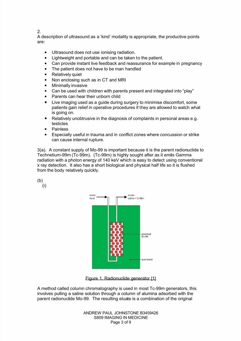
Figure 1. Radionuclide generator [1]
A method called column chromatography is used in most Tc-99m generators, thisinvolves pulling a saline solution through a column of alumina adsorbed with the
parent radionuclide Mo-99. The resulting eluate is a combination of the original
ANDREW PAUL JOHNSTONE B3459426S809 IMAGING IN MEDICINE
Page 3 of 9

8/6/2019 Msc Medical Physics Medical Imaging Exam 1
http://slidepdf.com/reader/full/msc-medical-physics-medical-imaging-exam-1 4/9
saline plus the daughter radionuclide Tc-99m. Because of the half life of the Tc-99mand the time that it takes to produce enough of the element from the decay of theparent source , the generator or “cow” is “milked” twice a day at least 6 hours apart.The useful life of one of these generators is recommended by the manufacturer [1] at3 half lives of the parent which is approx one week. The whole system is encased ina lead shield.
(ii)
99 99
42 43
99 99
43 43
m
m
Mo Tc
Tc Tc
β ν
γ
−→ + +
→ +
(c)
Web of Science [2]
Topic. Molybdenum.Author. Ballinger
Abstract:
Most nuclear medicine studies use Tc-99(m), which is the decay product of Mo-99.The world supply of Mo-99 comes from only five nuclear research reactors andavailability has been much reduced in recent times owing to problems at the largestreactors. In the short-term there are limited actions that can be taken owing tocapacity issues on alternative imaging modalities. In the long-term, stability of Mo-99supply will rely on a combination of replacing conventional reactors and developingnew technologies.
Ballinger, JR. (2010). Short- and long-term responses to molybdenum-99 shortages
in nuclear medicine. British Journal of Radiology. 83(995), 899-901.
ANDREW PAUL JOHNSTONE B3459426S809 IMAGING IN MEDICINE
Page 4 of 9

8/6/2019 Msc Medical Physics Medical Imaging Exam 1
http://slidepdf.com/reader/full/msc-medical-physics-medical-imaging-exam-1 5/9
4 (a) Frequency of precession (Larmor Frequency)
5
Hydrogen
=B
:
Larmour Frequncy Hz
B = Magnetic field strength T
Gyromagnetic Ratio
3.0T (compared to the earths magnetic field of 5x10 )
=42.58MHz x 3 = 127.74 Mhz
where
MRI T
ω γ
ω
γ
ω
−
=
=
=
∴
(b) Number of pixels = 5480Average Greyscale = 154.107
(c)
MRI Proton Density 1 Phantom, Area 5480 Pixels
Sample C1 C2 C3 C4 Water Unknown
% Water 50 60 70 75 100
AverageGreyscaleValue
154.107 165.075 176.624 183.569 219.876 178.849
Figure 2. Table of Average Greyscale Results for Various Samples
ANDREW PAUL JOHNSTONE B3459426S809 IMAGING IN MEDICINE
Page 5 of 9

8/6/2019 Msc Medical Physics Medical Imaging Exam 1
http://slidepdf.com/reader/full/msc-medical-physics-medical-imaging-exam-1 6/9
Figure 3. Average Greyscale against Percentage Water, MRI Proton Density 1Phantom
Figure 3. shows the plotted graph for samples C1 to C4 plus the water controlsample. The unknown sample is marked and annotated.
Average Greyscale Value is directly proportional to Percentage Water Content.
(d) From the graph shown at Figure 3. The estimated water content of the UnknownSample is approximately 72%
Figure 4. Image J Histogram Analysis of Unknown Sample.
Greyscale Value of Unknown Sample = 178.849 ± 6.216
5(a)
ANDREW PAUL JOHNSTONE B3459426S809 IMAGING IN MEDICINE
Page 6 of 9

8/6/2019 Msc Medical Physics Medical Imaging Exam 1
http://slidepdf.com/reader/full/msc-medical-physics-medical-imaging-exam-1 7/9
(i)
Equivalent dose:
3 3
Sieverts (Sv)
Where:
H Equivalent Dose to Tissue.
Radiation Weighting Factor
D = Absorbed dose to tissue
D 4.5mGy and W = 1 for X rays
H 1 x 4.5x10 4.5x10 (per breast)
T R T
T
R
T
T R
T
H W D
W
Since
Sv− −
=
=
=
=
= =
Effective dose:
3 6
Sv
:
W Tissue Weighting Factor
H Equivalent Dose to Tissue
Since W 0.12 for breast and H 4.5
0.12 x 4.5x10 540x10 (per breast)
T T
T
T
T T
E W H
Where
mSv
E Sv− −
=
=
=
= =
= =
ANDREW PAUL JOHNSTONE B3459426S809 IMAGING IN MEDICINE
Page 7 of 9

8/6/2019 Msc Medical Physics Medical Imaging Exam 1
http://slidepdf.com/reader/full/msc-medical-physics-medical-imaging-exam-1 8/9
(ii)
Background Equivalent Radiation Time
( )
3
3
effective dose (mSv)Uk annual background radiation level(mSv/yr)
0.54x100.2
2.7x10
assuming 1 year is 365 days BERT = 0.2 x 365 = 73 days
yrs BERT
BERT years−
−
=
∴ = =
∴
(b)
The women would be subject to indirect DNA damage due to the low radiationweighting factor of x rays and the high tissue weighting factor of breasts. Anydeterministic effects are unlikely as the amount of radiation received is well belowany threshold factor however there is always the chance of a stochastic effect.
(ii) Population odds.
(iii) Risk of death from smoke fire or flame = 1 in 1200. [4].
Risk of death from 3 mammograms in 10 year = 1 in 12346 people
The women are approximately 10 times more likely to die from smoke fire or flamethan of 3 mammograms in 10 years.
ANDREW PAUL JOHNSTONE B3459426S809 IMAGING IN MEDICINE
Page 8 of 9
3
Total effective dose = 3 x 0.54mSv=1.62mSv
Single value of risk coefficient for thewhole population(except for developingchild)=5% per Sievert. [3]
Risk of Stochastic effects(%)=1.62x10 x5=0.0081%
P
−∴
100 100opulation odds = 12345.679
Risk 0.0081
Increased lifetime risk 1 in 12346 people
= =
∴ ;

8/6/2019 Msc Medical Physics Medical Imaging Exam 1
http://slidepdf.com/reader/full/msc-medical-physics-medical-imaging-exam-1 9/9
References.
1.http://www.medcyclopaedia.com/library/topics/volume_i/r/radionuclide_generator/dradionuclide_generato_fig1.aspx (accessed 01/02/2011)
2.http://apps.isiknowledge.com/full_record.do?product=UA&search_mode=GeneralSearch&qid=1&SID=Q2gg1E4d2JiGaKogIAF&page=1&doc=1&colname=WOS (accessed 01/02/2011)
3. The 2007 Recommendations of the international commission of RadiologicalProtection ICRP publication 103, 2007. Annals of the ICRB vol 37 pp 1-332.
4. US National Safety Council, 1995.
ANDREW PAUL JOHNSTONE B3459426S809 IMAGING IN MEDICINE
Page 9 of 9