Embed Size (px)
Citation preview
MS 1 Cardiology ExamAmanda Krauss
Disclaimer
● I put this lecture together from notes I took 3 years ago. I supplemented that information with resources that you will likely use next year.
● Items in blue are attempts to plant a seed for next year or paint a bigger picture of pathophysiology. It will be lower yield for your upcoming exam.
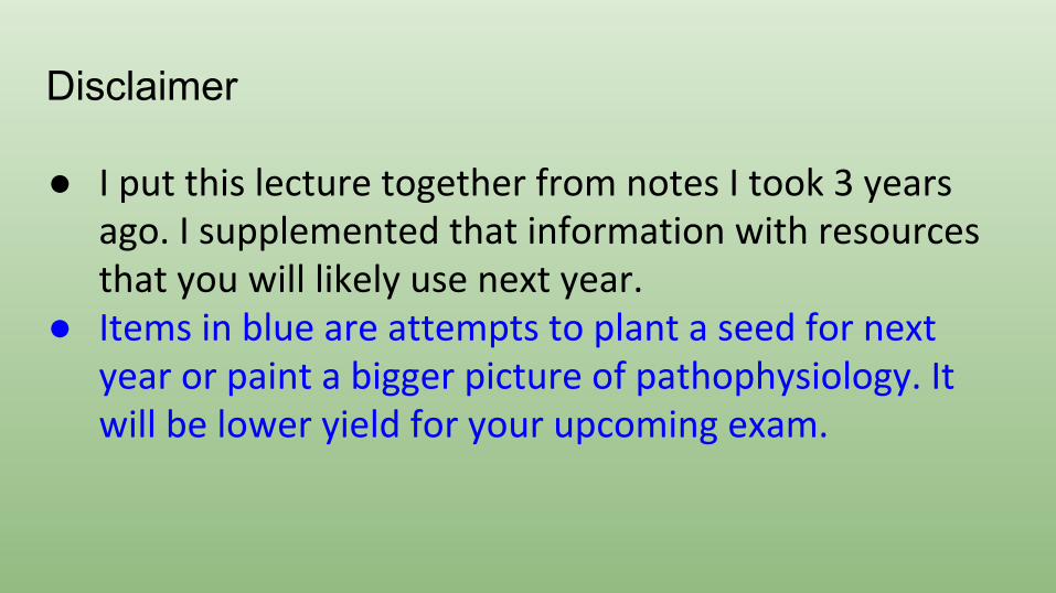
Cardiac Physiology-Electrical Conduction
● Normal conduction system goes SA to AV to Bundle of His to Bundle Branches (Right, Left Anterior and Left Posterior), Purkinje fibers
● Two different electrically conducting cells
○ SA node and AV node■ Funny current adds to
rhythmicity of these pacemakers
○ Purkinje and Bundles■ Lacks funny current
● The small differences in ion conduction play into autonomics and medications
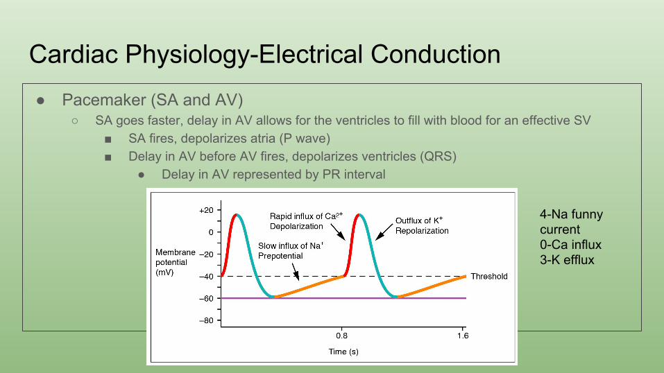
Cardiac Physiology-Electrical Conduction● Pacemaker (SA and AV)
○ SA goes faster, delay in AV allows for the ventricles to fill with blood for an effective SV■ SA fires, depolarizes atria (P wave)■ Delay in AV before AV fires, depolarizes ventricles (QRS)
● Delay in AV represented by PR interval
4-Na funny current0-Ca influx3-K efflux
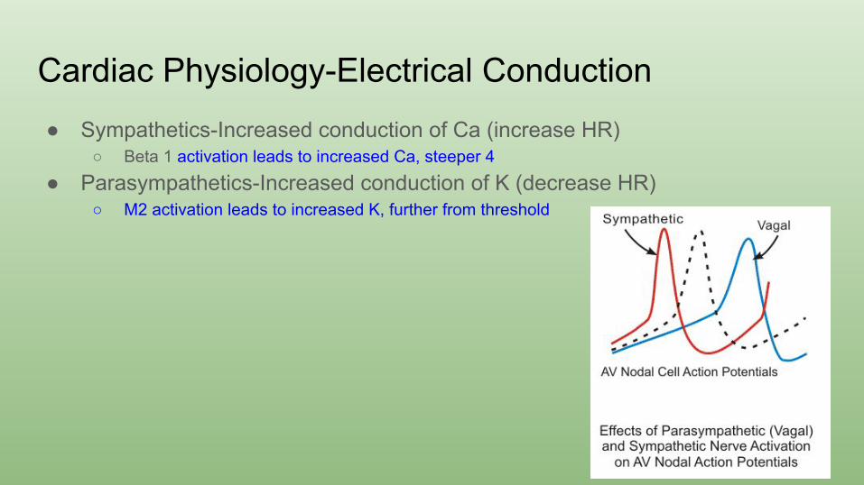
Cardiac Physiology-Electrical Conduction● Sympathetics-Increased conduction of Ca (increase HR)
○ Beta 1 activation leads to increased Ca, steeper 4
● Parasympathetics-Increased conduction of K (decrease HR)○ M2 activation leads to increased K, further from threshold
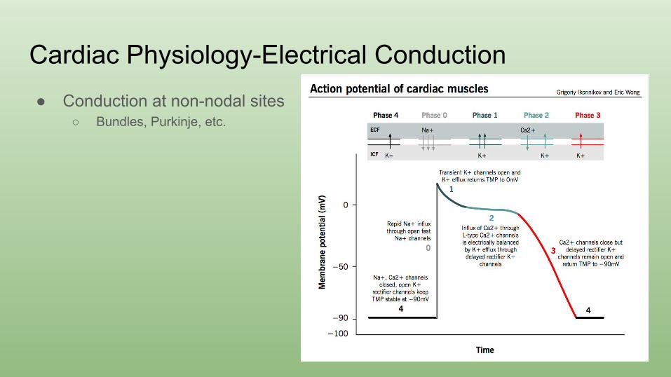
Cardiac Physiology-Electrical Conduction● Conduction at non-nodal sites
○ Bundles, Purkinje, etc.
Genetics● Channelopathies● Long QT
○ LQT1-slow K channel causes arrhythmia with exercise○ LQT2-fast K causes arrhythmia with loud noises○ LQT3-Na channel causes arrhythmia in NREM
● Short QT○ Can lead to atrial fibrillation
● Connexin 40-can lead to atrial fibrillation
EKG
• Try to understand rather than just memorize and recognize a pattern
• Dr. Petrany does a great job teaching a systematic way of going through EKGs-rate, rhythm, axis, PR, QRS, ST, QT
• Regardless of which field you go into-you WILL order an EKG on your patient at some point-so know how to read them
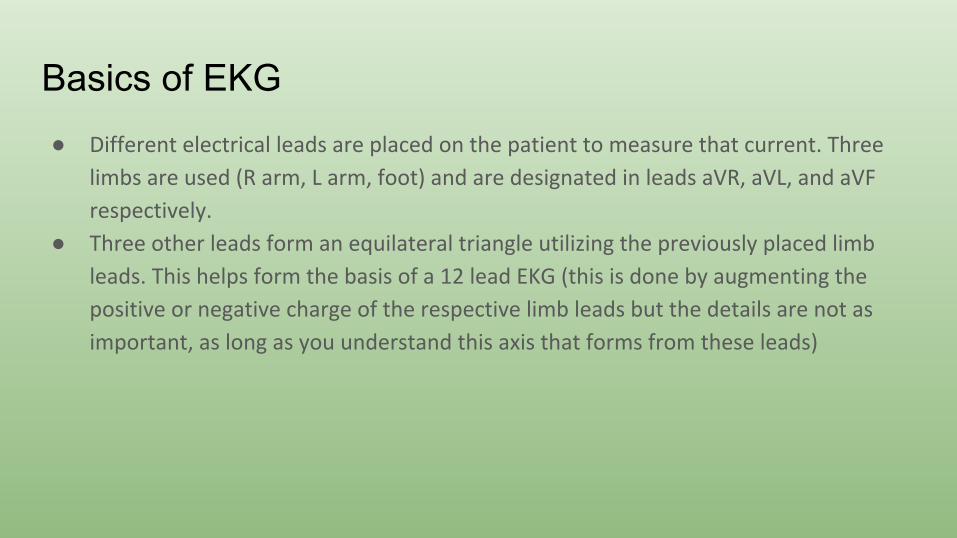
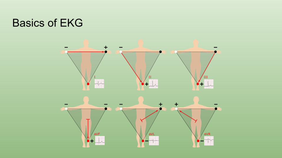
Basics of EKG● Different electrical leads are placed on the patient to measure that current. Three
limbs are used (R arm, L arm, foot) and are designated in leads aVR, aVL, and aVF
respectively.
● Three other leads form an equilateral triangle utilizing the previously placed limb
leads. This helps form the basis of a 12 lead EKG (this is done by augmenting the
positive or negative charge of the respective limb leads but the details are not as
important, as long as you understand this axis that forms from these leads)
Basics of EKG
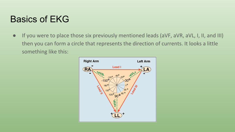
Basics of EKG● If you were to place those six previously mentioned leads (aVF, aVR, aVL, I, II, and III)
then you can form a circle that represents the direction of currents. It looks a little
something like this:
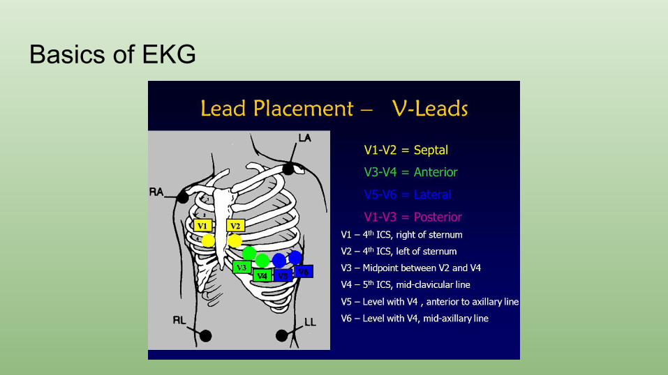
Basics of EKG● Septal leads, designated as V1 through V6
● These leads start out measuring the conduction of the right side (V1 is furthest right)
and go to the left side (V6 is furthest left)
● These leads give a better idea of conduction and provide a left vs right view of
conduction. (Particularly in pediatric patients, looking at V1-V3 can be helpful in
looking for RVH and V4-V6 for LVH as they will show huge depolarizations in those
respective lead groups)
Basics of EKG
Basics of EKG
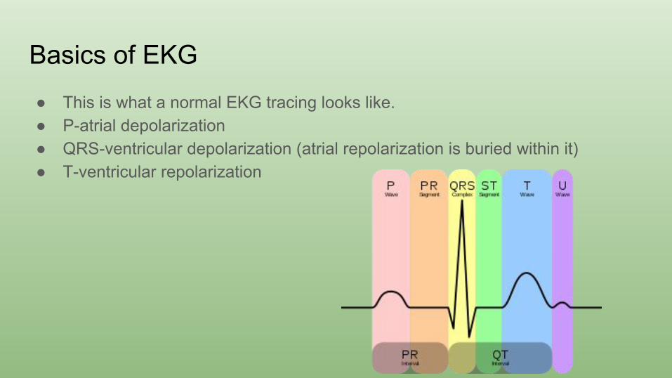
● This is what a normal EKG tracing looks like. ● P-atrial depolarization● QRS-ventricular depolarization (atrial repolarization is buried within it)● T-ventricular repolarization
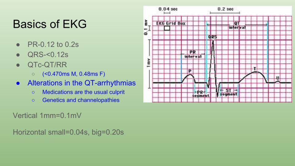
Basics of EKG● PR-0.12 to 0.2s● QRS-<0.12s● QTc-QT/RR
○ (<0.470ms M, 0.48ms F)
● Alterations in the QT-arrhythmias○ Medications are the usual culprit○ Genetics and channelopathies
Vertical 1mm=0.1mV
Horizontal small=0.04s, big=0.20s
Basics of EKG
• Remember-EKGs are measuring vectors based on particular lead placement
• Heart should depolarize from R to L and top to bottom• Net current towards something gives an upward deflection,
away gives a downward deflection
• The magnitude of these deflections reflects tissue mass or conduction delays
EKG Basics
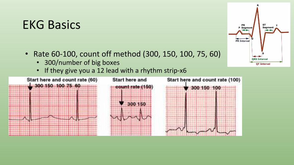
• Rate 60-100, count off method (300, 150, 100, 75, 60)• 300/number of big boxes• If they give you a 12 lead with a rhythm strip-x6
EKG Basics-Axis
Back to Basics
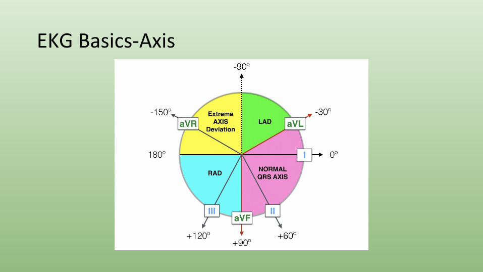
• Axis-normal from -30 to 90• Remember that positive currents mean that the net depolarization is going
towards that lead• Lead I lines up with 0 and aVF lines up with 90
• If they are both positive you are between 0 and 90• If aVF is positive and I is negative you have RAD• If I is positive and aVF is negative, you are between 0 and -90
• To determine if LAD or normal, look at II• If II is positive-NAD• If II is negative-LAD
Axis
• Normal axis is -30 to 90• Axis gives representation of tissue mass and/or electrical
conduction• Causes of LAD
• Inferior wall MI• LAHB• LVH• Endocardial cushion defects
• Causes of RAD• RVH• LPHB• Massive PE
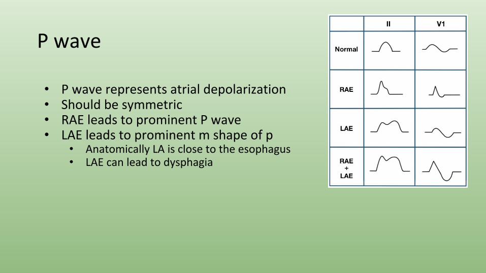
P wave
• P wave represents atrial depolarization• Should be symmetric• RAE leads to prominent P wave• LAE leads to prominent m shape of p
• Anatomically LA is close to the esophagus• LAE can lead to dysphagia
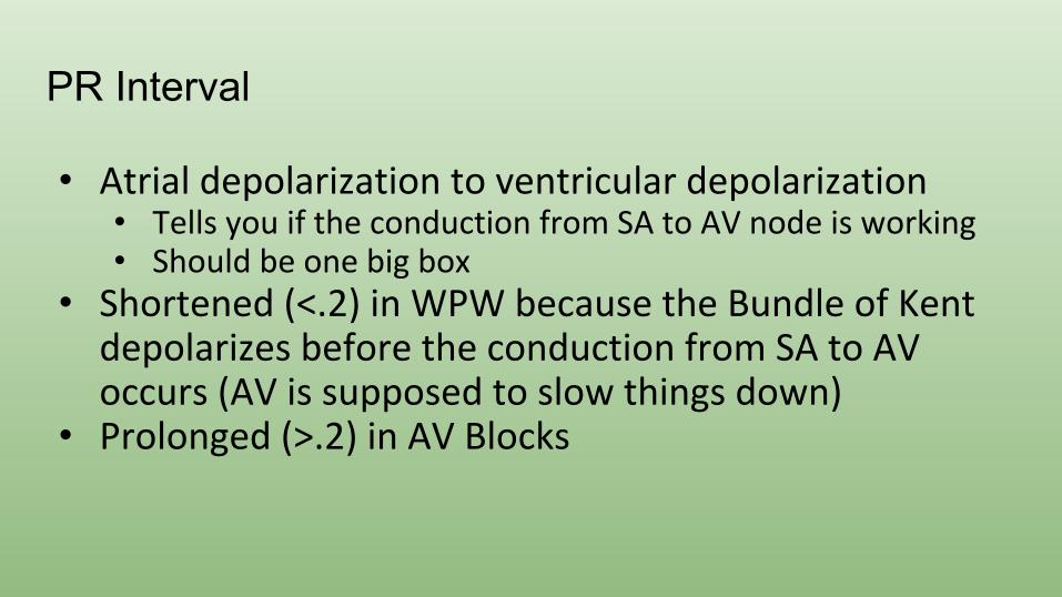
PR Interval
• Atrial depolarization to ventricular depolarization• Tells you if the conduction from SA to AV node is working• Should be one big box
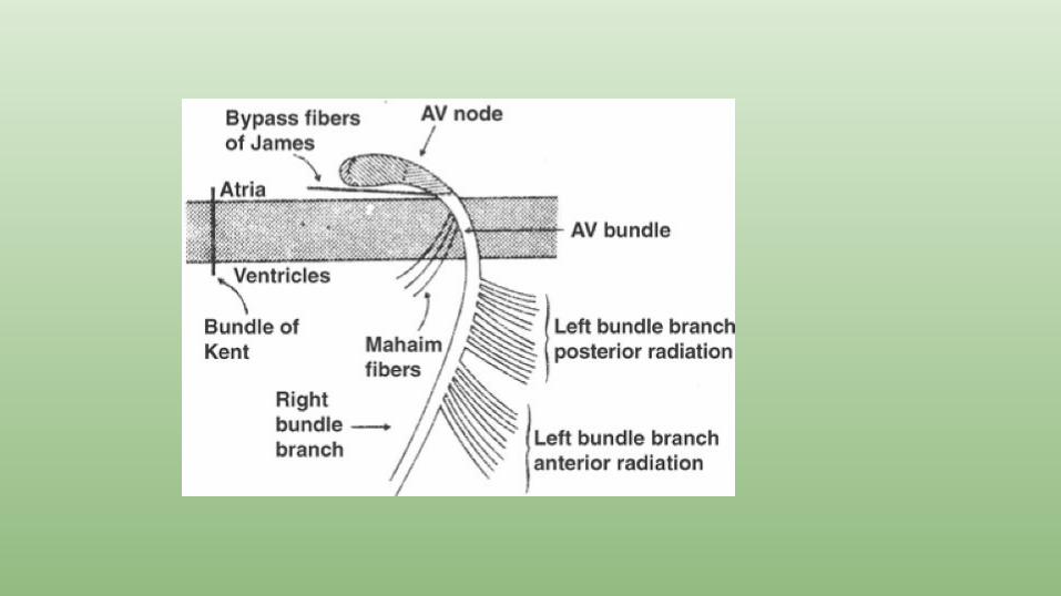
• Shortened (<.2) in WPW because the Bundle of Kent depolarizes before the conduction from SA to AV occurs (AV is supposed to slow things down)
• Prolonged (>.2) in AV Blocks
Importance of the PR interval
• Heart Block• 1st Degree is no big deal, just a long PR interval• 2nd Degree type 1-PR lengthens then drops• 2nd Degree type 2-drops a beat without lengthening • 3rd Degree-no association with P and QRS
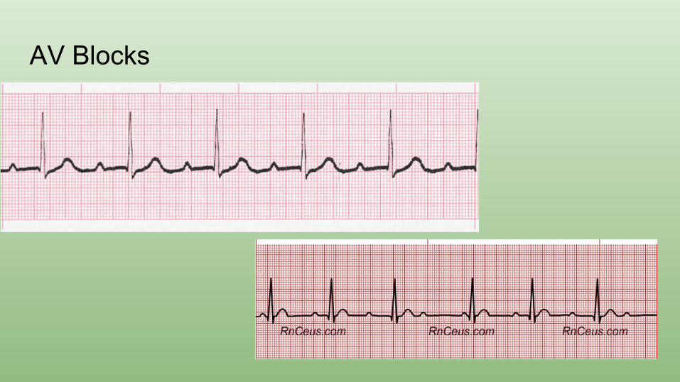
AV Blocks

AV Blocks
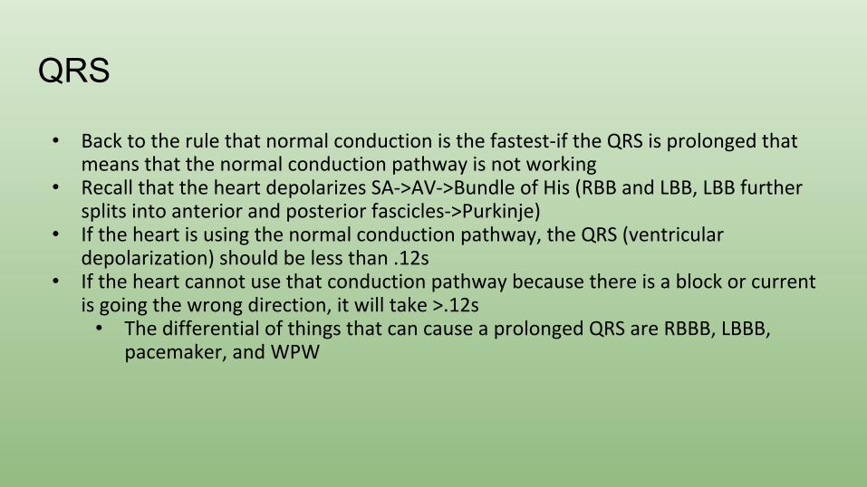
QRS
• Back to the rule that normal conduction is the fastest-if the QRS is prolonged that means that the normal conduction pathway is not working
• Recall that the heart depolarizes SA->AV->Bundle of His (RBB and LBB, LBB further splits into anterior and posterior fascicles->Purkinje)
• If the heart is using the normal conduction pathway, the QRS (ventricular depolarization) should be less than .12s
• If the heart cannot use that conduction pathway because there is a block or current is going the wrong direction, it will take >.12s
• The differential of things that can cause a prolonged QRS are RBBB, LBBB, pacemaker, and WPW
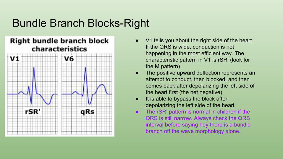
Bundle Branch Blocks-Right● V1 tells you about the right side of the heart.
If the QRS is wide, conduction is not happening in the most efficient way. The characteristic pattern in V1 is rSR’ (look for the M pattern)
● The positive upward deflection represents an attempt to conduct, then blocked, and then comes back after depolarizing the left side of the heart first (the net negative).
● It is able to bypass the block after depolarizing the left side of the heart
● The rSR’ pattern is normal in children if the QRS is still narrow. Always check the QRS interval before saying hey there is a bundle branch off the wave morphology alone.
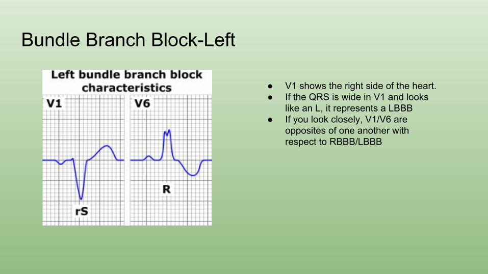
Bundle Branch Block-Left
● V1 shows the right side of the heart.● If the QRS is wide in V1 and looks
like an L, it represents a LBBB● If you look closely, V1/V6 are
opposites of one another with respect to RBBB/LBBB
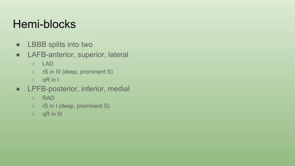
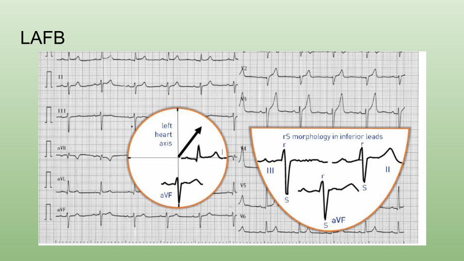
Hemi-blocks● LBBB splits into two● LAFB-anterior, superior, lateral
○ LAD○ rS in III (deep, prominent S)○ qR in I
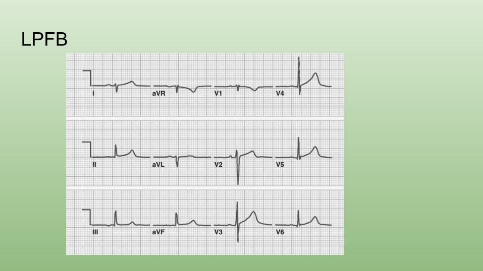
● LPFB-posterior, inferior, medial○ RAD○ rS in I (deep, prominent S)○ qR in III
LAFB
LPFB
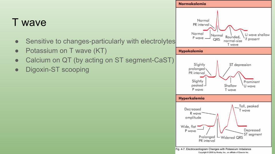
T wave● Sensitive to changes-particularly with electrolytes● Potassium on T wave (KT)● Calcium on QT (by acting on ST segment-CaST)● Digoxin-ST scooping
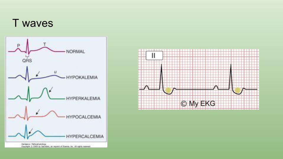
T waves
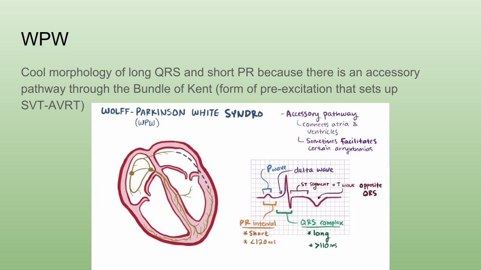
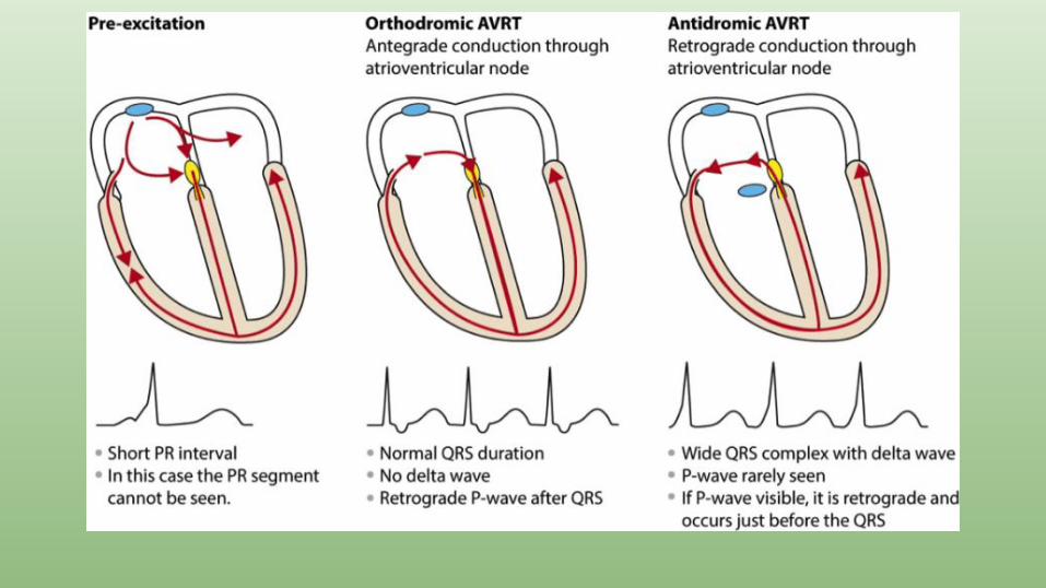
WPWCool morphology of long QRS and short PR because there is an accessory pathway through the Bundle of Kent (form of pre-excitation that sets up SVT-AVRT)

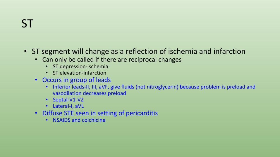
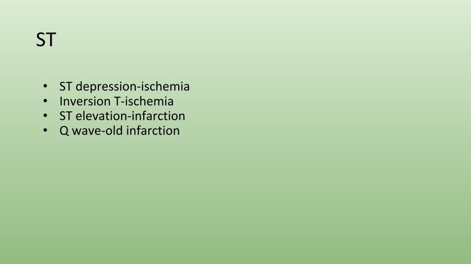
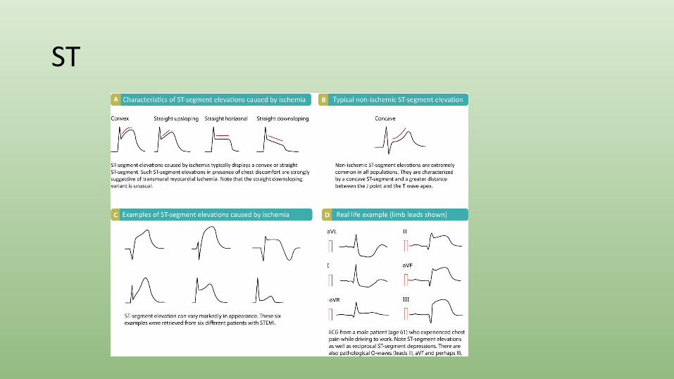
ST
• ST segment will change as a reflection of ischemia and infarction• Can only be called if there are reciprocal changes
• ST depression-ischemia • ST elevation-infarction
• Occurs in group of leads• Inferior leads-II, III, aVF, give fluids (not nitroglycerin) because problem is preload and
vasodilation decreases preload• Septal-V1-V2• Lateral-I, aVL
• Diffuse STE seen in setting of pericarditis • NSAIDS and colchicine
ST
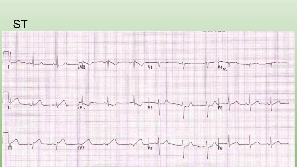
• ST depression-ischemia• Inversion T-ischemia• ST elevation-infarction• Q wave-old infarction
ST
ST
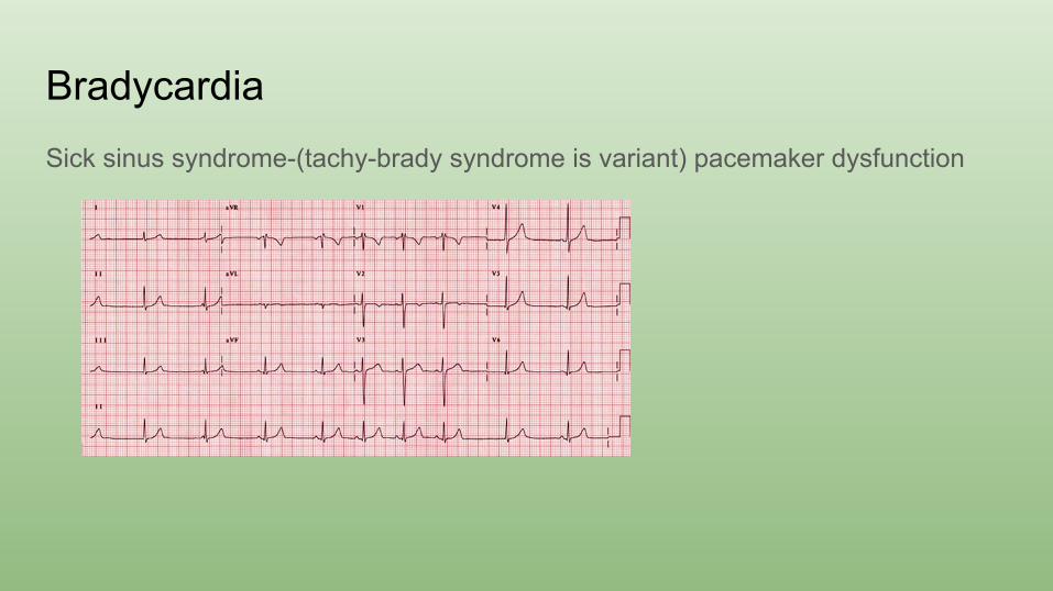
BradycardiaSick sinus syndrome-(tachy-brady syndrome is variant) pacemaker dysfunction
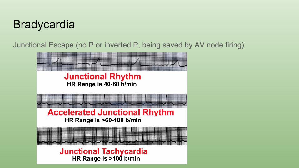
BradycardiaJunctional Escape (no P or inverted P, being saved by AV node firing)
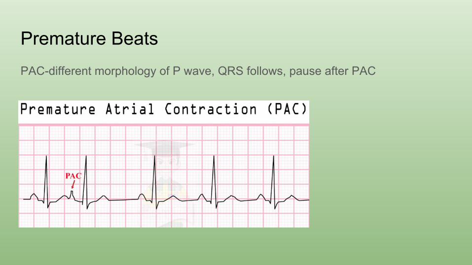
Premature BeatsPAC-different morphology of P wave, QRS follows, pause after PAC
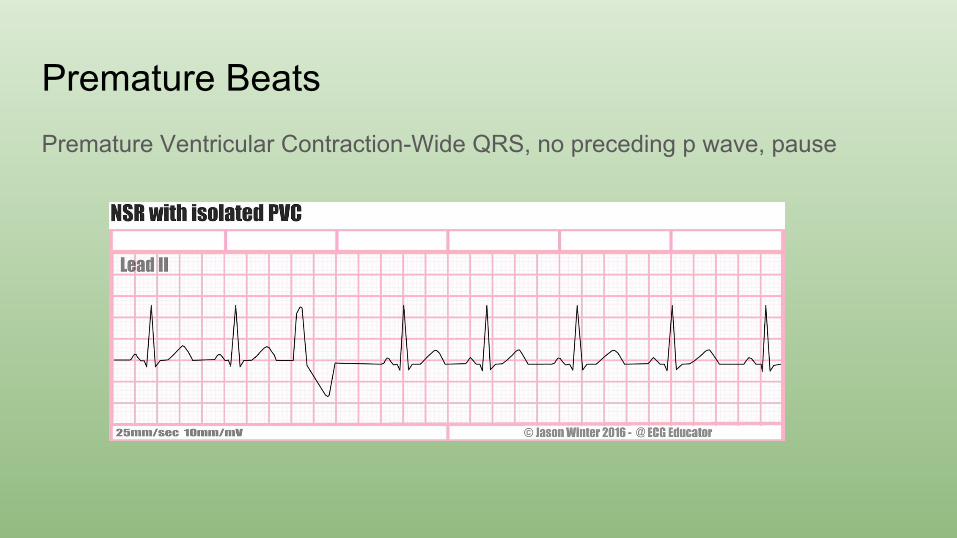
Premature BeatsPremature Ventricular Contraction-Wide QRS, no preceding p wave, pause
SVT● Generic term for tachycardia arising from above the ventricles● AVRT-WPW is a pre-excitation pattern
○ Orthodromic-narrow QRS○ Antidromic-wide QRS
● AVNRT-dependent on different characteristics of tissue around the node● Atrial fibrillation● Atrial flutter● Ectopic atrial tachycardia
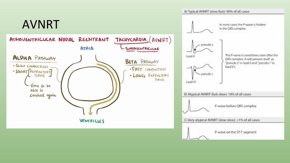
AVNRT
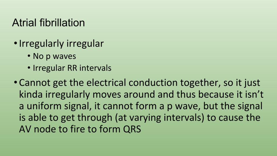
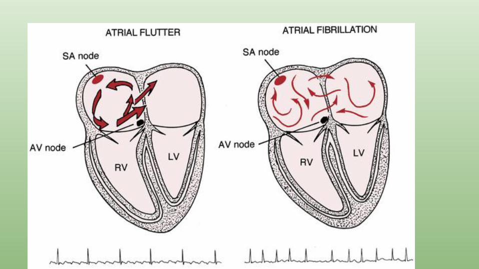
Atrial fibrillation
• Irregularly irregular• No p waves• Irregular RR intervals
• Cannot get the electrical conduction together, so it just kinda irregularly moves around and thus because it isn’t a uniform signal, it cannot form a p wave, but the signal is able to get through (at varying intervals) to cause the AV node to fire to form QRS
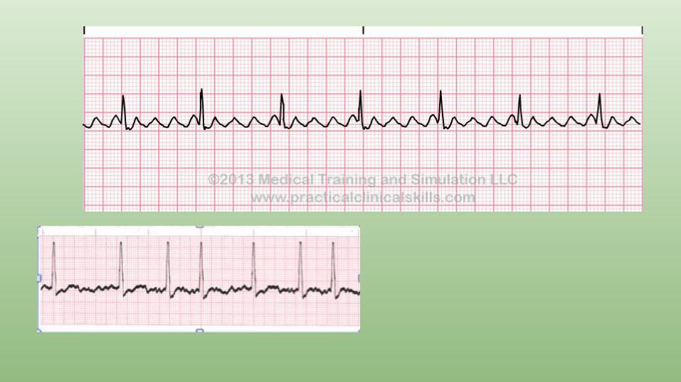
Atrial flutter
• Consistent electrical circuit-but it just isn’t the fastest
• Sawtooth appearance
• Consistent RR interval
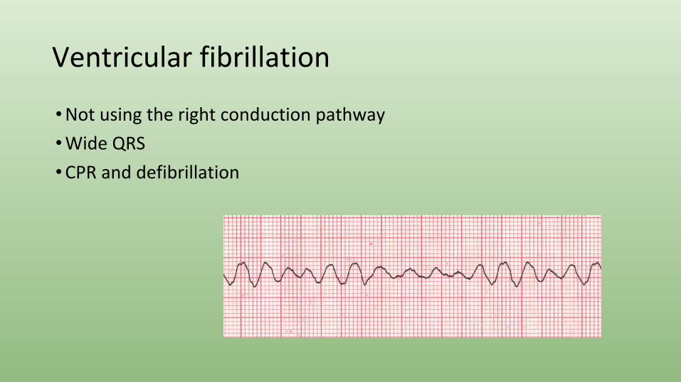
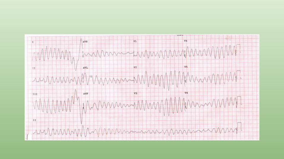
Ventricular fibrillation
• Not using the right conduction pathway
• Wide QRS
• CPR and defibrillation
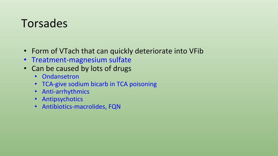
Torsades
• Form of VTach that can quickly deteriorate into VFib• Treatment-magnesium sulfate• Can be caused by lots of drugs
• Ondansetron • TCA-give sodium bicarb in TCA poisoning • Anti-arrhythmics• Antipsychotics• Antibiotics-macrolides, FQN
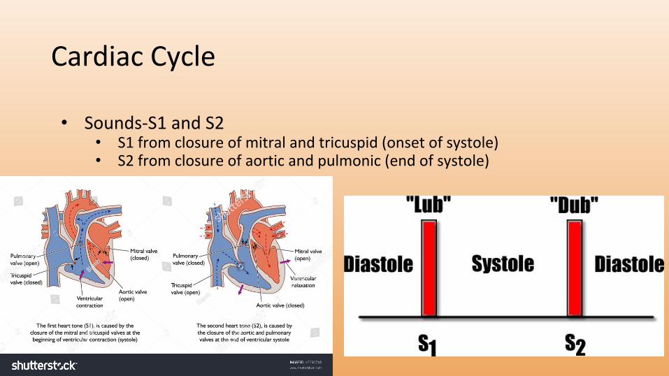
Cardiac Cycle
• Sounds-S1 and S2• S1 from closure of mitral and tricuspid (onset of systole)• S2 from closure of aortic and pulmonic (end of systole)
Cardiac Cycle
• Murmurs occur when extra sounds are heard from obstructed blood flow
• Using knowledge of opened vs. closed valves in the cardiac cycle, you can deduce what is causing the murmur
• Hearing a noise when a valve should be closed from regurgitant flow• Pulmonary valve/aortic valve-diastole• Tricuspid valve/mitral valve-systole
• Hearing a noise when a valve should be opened from stenotic obstruction
• Pulmonary valve/aortic valve-systole (ASS)• Tricuspid valve/mitral valve-diastole
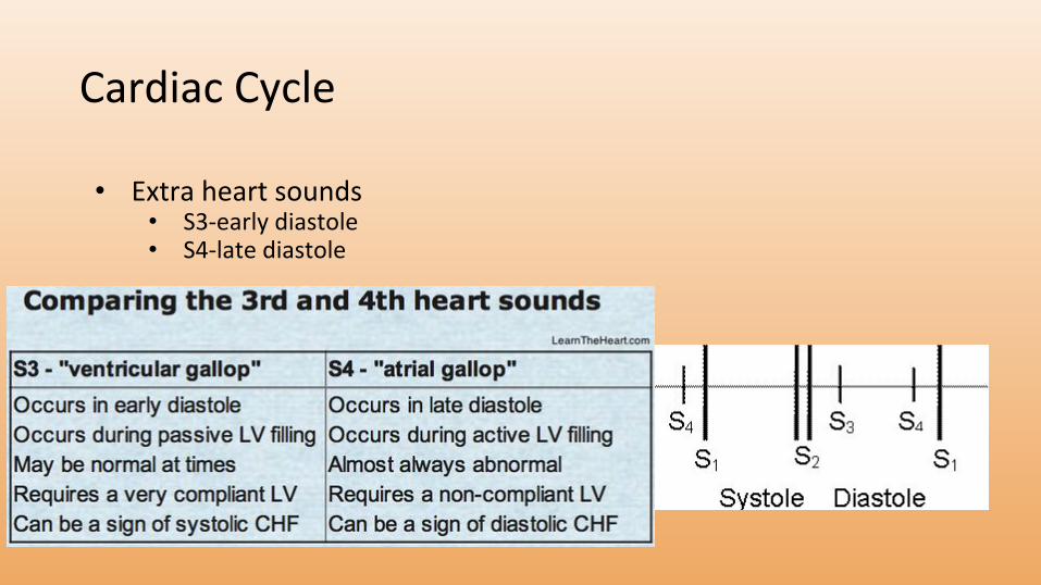
Cardiac Cycle
• Extra heart sounds• S3-early diastole• S4-late diastole
Cardiac Cycle
• Quick advice about cardiac cycle• Easy to tie in lots of graphs• If you get lost-look at the axis and tie into the very basic concepts of
cardiac physiology• Movement of blood dependent on pressure differentials• Opening of valves dependent on pressure differentials• Greater pressures on R side of heart than L side of heart
Cardiac Cycle
Cardiac Cycle
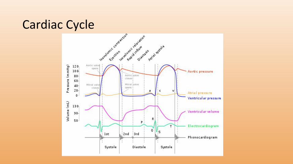
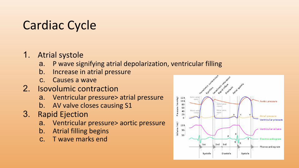
1. Atrial systolea. P wave signifying atrial depolarization, ventricular fillingb. Increase in atrial pressurec. Causes a wave
2. Isovolumic contractiona. Ventricular pressure> atrial pressureb. AV valve closes causing S1
3. Rapid Ejectiona. Ventricular pressure> aortic pressureb. Atrial filling beginsc. T wave marks end
Cardiac Cycle
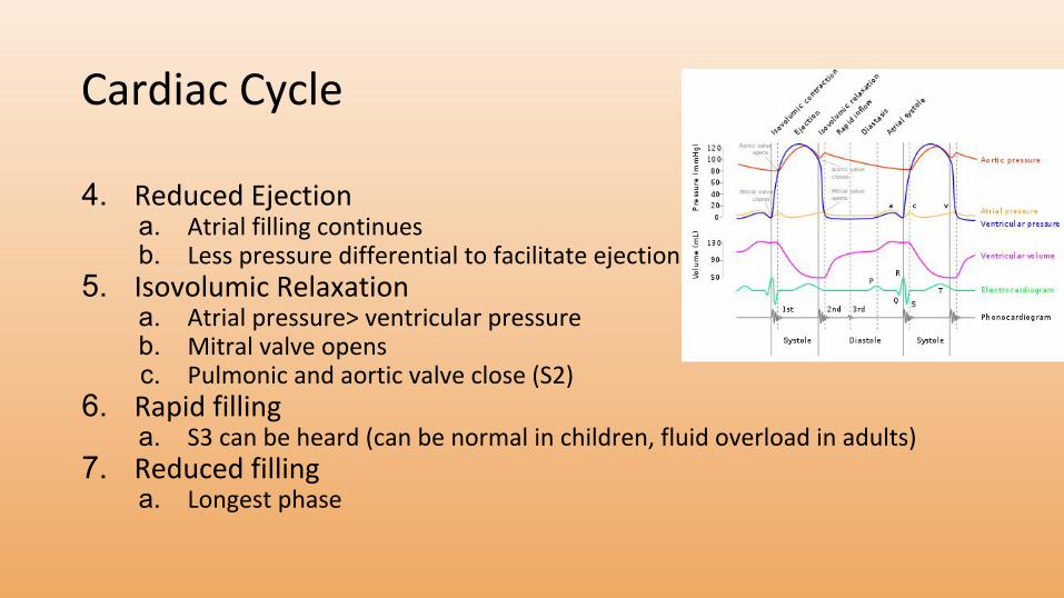
4. Reduced Ejectiona. Atrial filling continuesb. Less pressure differential to facilitate ejection
5. Isovolumic Relaxationa. Atrial pressure> ventricular pressureb. Mitral valve opensc. Pulmonic and aortic valve close (S2)
6. Rapid fillinga. S3 can be heard (can be normal in children, fluid overload in adults)
7. Reduced fillinga. Longest phase
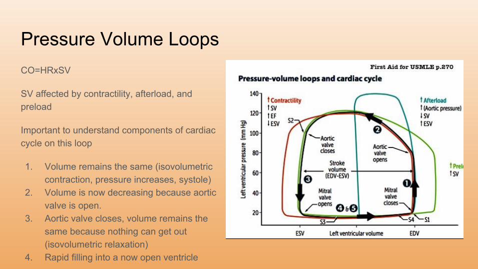
Pressure Volume LoopsCO=HRxSV
SV affected by contractility, afterload, and preload
Important to understand components of cardiac cycle on this loop
1. Volume remains the same (isovolumetric contraction, pressure increases, systole)
2. Volume is now decreasing because aortic valve is open.
3. Aortic valve closes, volume remains the same because nothing can get out (isovolumetric relaxation)
4. Rapid filling into a now open ventricle
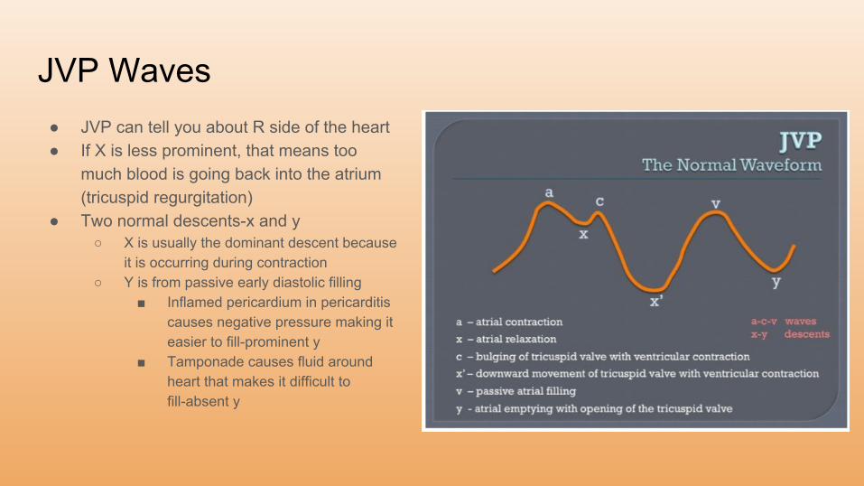
JVP Waves● JVP can tell you about R side of the heart● If X is less prominent, that means too
much blood is going back into the atrium (tricuspid regurgitation)
● Two normal descents-x and y○ X is usually the dominant descent because
it is occurring during contraction○ Y is from passive early diastolic filling
■ Inflamed pericardium in pericarditis causes negative pressure making it easier to fill-prominent y
■ Tamponade causes fluid around heart that makes it difficult to fill-absent y
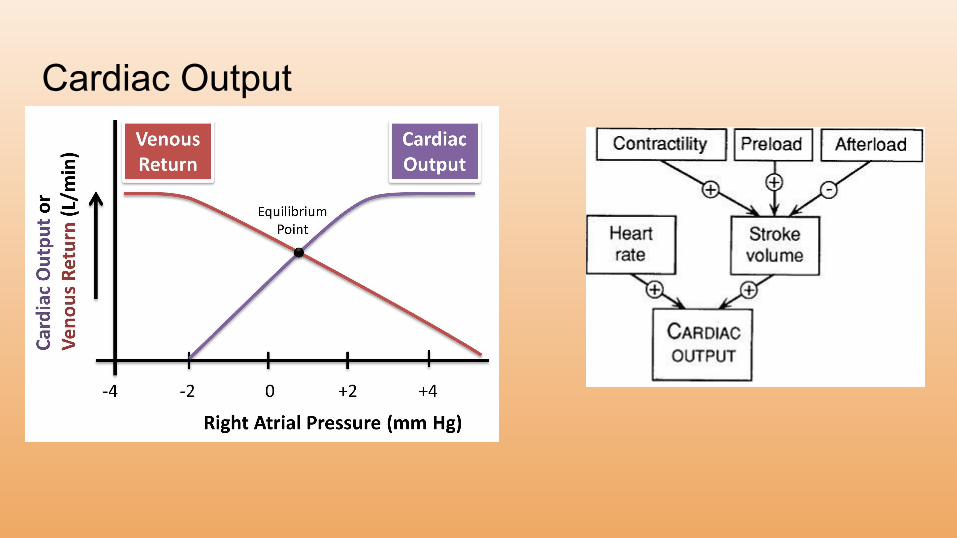
Cardiac Output
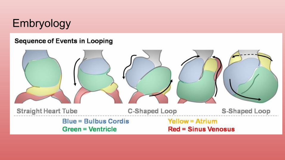
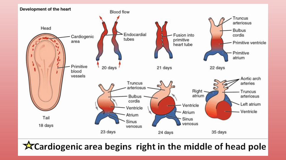
Embryology
Embryology● Truncus arteriosus-ascending aorta and pulmonary trunk
○ Failure to septate can give a cyanotic heart lesion called truncus arteriosus
● Bulbus cordis-smooth parts of left and right ventricle (LVOT, RVOT)● Endocardial cushion-atrial septum, membranous interventricular septum; AV
and semilunar valves○ Endocardial cushion defects are common CHD with Trisomy 21 (LAD at birth)
● Primitive atrium-trabeculated LA and RA● Primitive ventricle-trabeculated LV and RV
○ RV>LV, if too trrabeculated indication for ASA
● Primitive pulmonary vein-smooth part of left atrium● Left horn of sinus venosus-coronary sinus● Right horn of sinus venosus-smooth part of right atrium● Right common cardinal vein and right anterior cardinal vein-superior vena
cava
Embryology● Ductus arteriosus-ligamentum arteriosum● Ductus venosum-ligamentum venosum● Foramen ovale-fossa ovalis● Umbilical vein-ligamentum teres hepatis
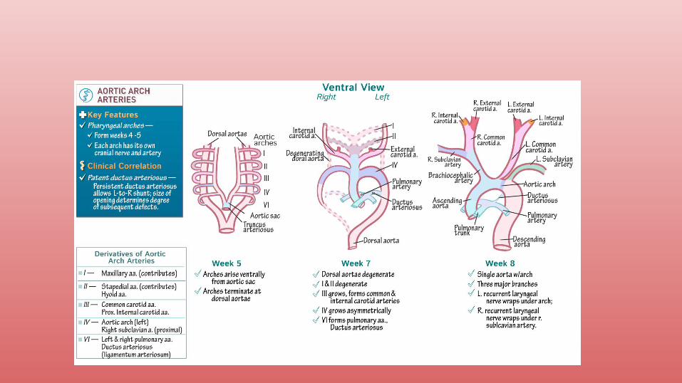
Embryology● Aortic arches
○ 1st-maxillary artery○ 2nd-stapedial artery○ 3rd-common carotid artery○ 4th-aorta○ 5th-nothing○ 6th-pulmonary trunk○ 7th intersegmental-left subclavian
Vessels and Circulation● Lymphatics
○ Junction of adjacent endothelial cells overlap forming a valve○ Muscles work to pump lymphatics (myogenic response)
● Filtration and absorption determined by permeability and pressure differentials○ Pc-Pi-(ONCc-ONCi)
■ Positive leads to filtering● Edema occurs when filtration exceeds rate of capillary reabsoprtion
■ Negative leads to reabsorption
Circulation● Heart
○ Because of origin of coronary arteries, as HR increases, less time for perfusion
● Cerebral○ Autoregulation between 60 and 130 mmHg○ Sensitive to changes in pCO2
● Autonomics○ Parasynpathetic-Ach to muscarinic vasodilates○ Sympathetic-Epi to adrenergic vasoconstricts
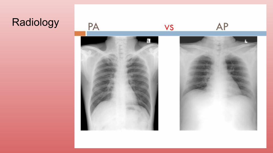
Radiology● PA CXR is standard (chest against film)● AP CXR if patient is supine (super common in hospital for admitted patients)● L higher than R due to stomach, gas bubble should be seen● Spinous processes midline, clavicle medial● Inspiration-6/7 anterior, 9/10 posterior● Penetration-how much XR goes through, over penetration is black, under
penetrated cannot see intervertebral discs
Radiology