Embed Size (px)
Citation preview
2015-09-08
1
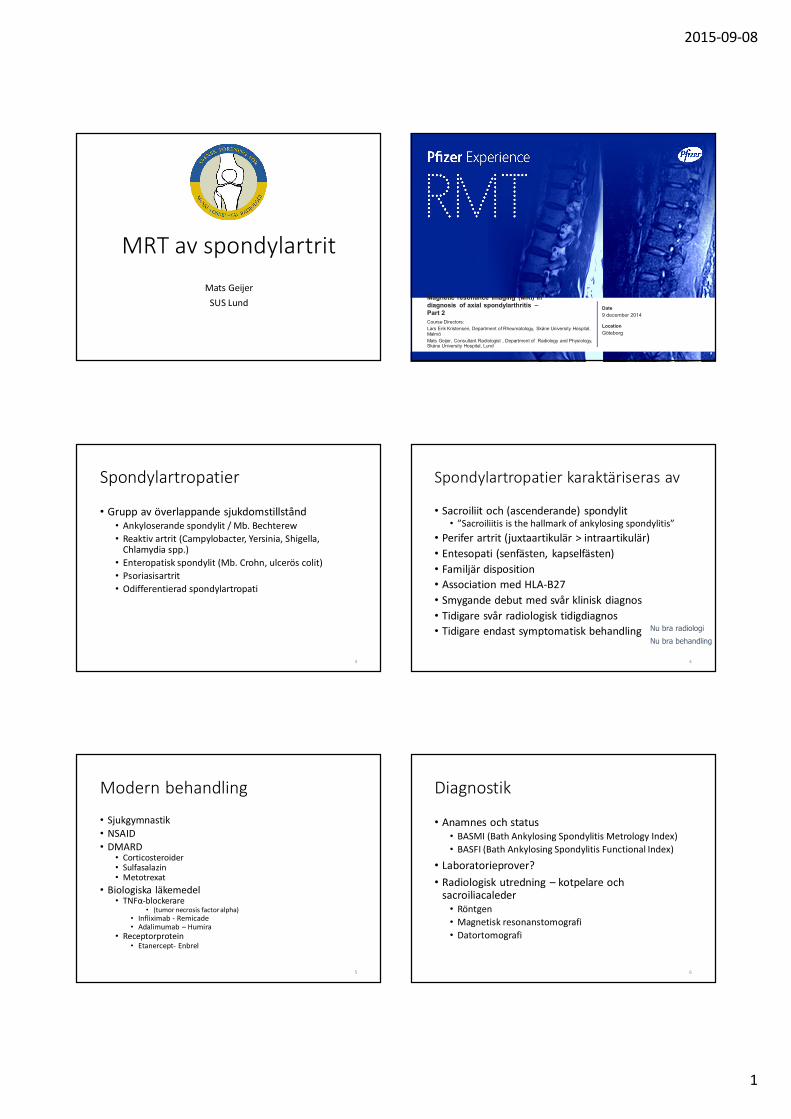
MRT av spondylartrit
Mats Geijer
SUS LundDate
Location
Magnetic resonance imaging (MRI) indiagnosis of axial spondylarthritis –Part 2
Course Directors:
Lars Erik Kristensen, Department of Rheumatology, Skåne University Hospital, Malmö
Mats Geijer, Consultant Radiologist , Department of Radiology and Physiology, Skåne University Hospital, Lund
9 december 2014
Göteborg
EN
B20140307P
SE
03
3
Spondylartropatier
• Grupp av överlappande sjukdomstillstånd• Ankyloserande spondylit / Mb. Bechterew
• Reaktiv artrit (Campylobacter, Yersinia, Shigella, Chlamydia spp.)
• Enteropatisk spondylit (Mb. Crohn, ulcerös colit)
• Psoriasisartrit
• Odifferentierad spondylartropati
4
Spondylartropatier karaktäriseras av
• Sacroiliit och (ascenderande) spondylit• ”Sacroiliitis is the hallmark of ankylosing spondylitis”
• Perifer artrit (juxtaartikulär > intraartikulär)
• Entesopati (senfästen, kapselfästen)
• Familjär disposition
• Association med HLA-B27
• Smygande debut med svår klinisk diagnos
• Tidigare svår radiologisk tidigdiagnos
• Tidigare endast symptomatisk behandlingNu bra behandling
Nu bra radiologi
5
Modern behandling
• Sjukgymnastik• NSAID• DMARD
• Corticosteroider• Sulfasalazin• Metotrexat
• Biologiska läkemedel• TNFα-blockerare
• (tumor necrosis factor alpha)• Infliximab - Remicade• Adalimumab – Humira
• Receptorprotein• Etanercept- Enbrel
6
Diagnostik
• Anamnes och status• BASMI (Bath Ankylosing Spondylitis Metrology Index)
• BASFI (Bath Ankylosing Spondylitis Functional Index)
• Laboratorieprover?
• Radiologisk utredning – kotpelare och sacroiliacaleder
• Röntgen
• Magnetisk resonanstomografi
• Datortomografi
2015-09-08
2
7 8
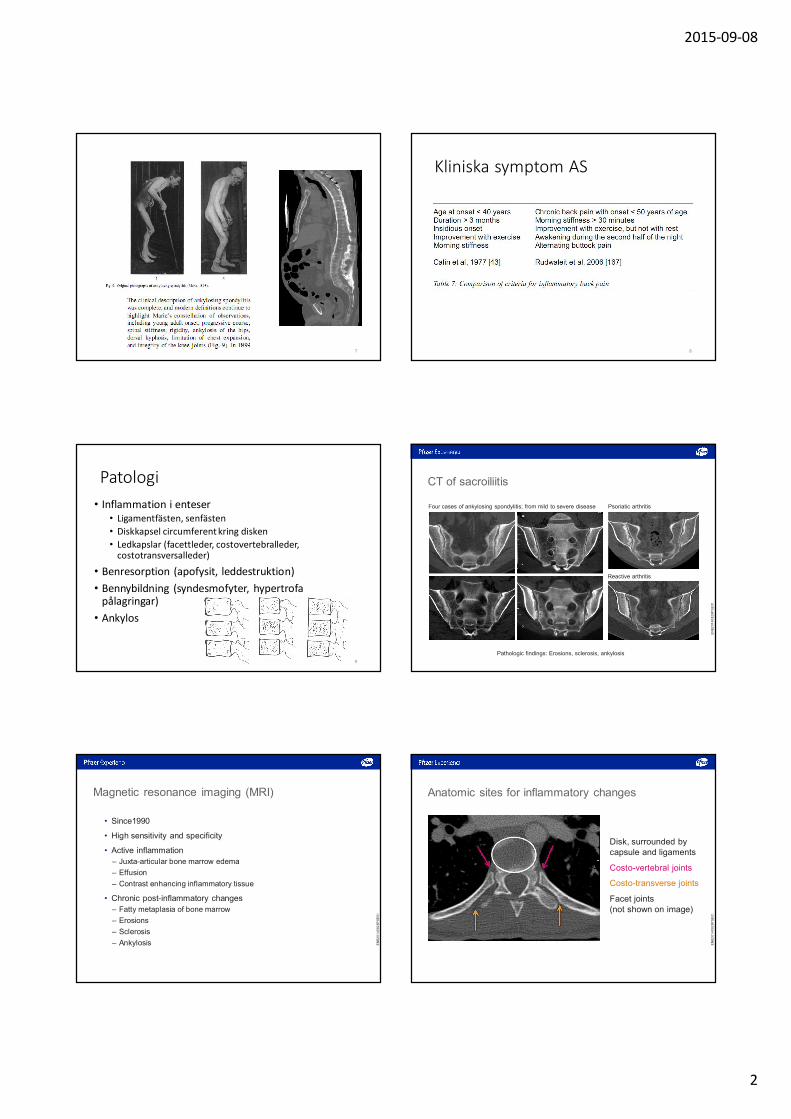
Kliniska symptom AS
Patologi
• Inflammation i enteser• Ligamentfästen, senfästen
• Diskkapsel circumferent kring disken
• Ledkapslar (facettleder, costovertebralleder, costotransversalleder)
• Benresorption (apofysit, leddestruktion)
• Bennybildning (syndesmofyter, hypertrofapålagringar)
• Ankylos
9
CT of sacroiliitis
Reactive arthritis
Psoriatic arthritisFour cases of ankylosing spondylitis; from mild to severe disease
Pathologic findings: Erosions, sclerosis, ankylosis
EN
B2
01
40
82
8P
SE
01
Magnetic resonance imaging (MRI)
• Since1990
• High sensitivity and specificity
• Active inflammation
– Juxta-articular bone marrow edema
– Effusion
– Contrast enhancing inflammatory tissue
• Chronic post-inflammatory changes
– Fatty metaplasia of bone marrow
– Erosions
– Sclerosis
– Ankylosis EN
B2
01
40
82
8P
SE
01
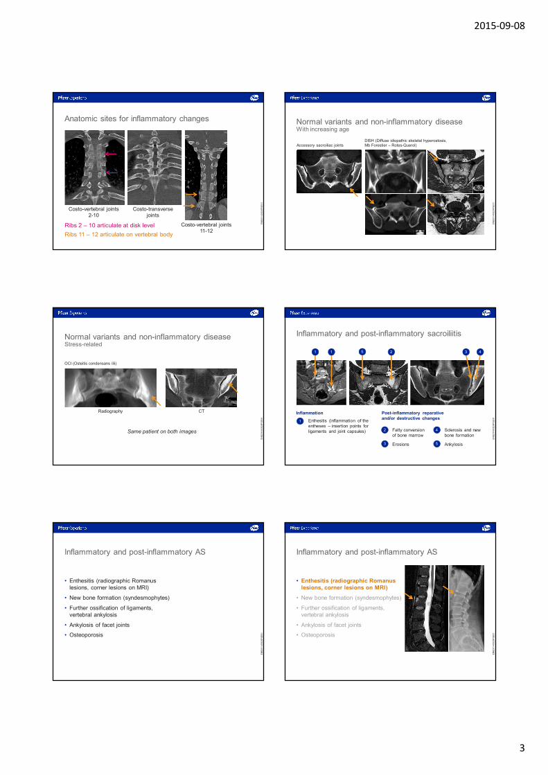
Anatomic sites for inflammatory changes
Disk, surrounded by capsule and ligaments
Costo-vertebral joints
Costo-transverse joints
Facet joints(not shown on image)
EN
B2
01
40
82
8P
SE
01
2015-09-08
3
Ribs 2 – 10 articulate at disk level
Ribs 11 – 12 articulate on vertebral body
Costo-vertebral joints 2-10
Costo-vertebral joints 11-12
Costo-transversejoints
Anatomic sites for inflammatory changes
EN
B2
01
40
82
8P
SE
01
Normal variants and non-inflammatory diseaseWith increasing age
Accessory sacroiliac jointsDISH (Diffuse idiopathic skeletal hyperostosis, Mb Forestier – Rotes-Querol)
EN
B2
01
40
82
8P
SE
01
Normal variants and non-inflammatory diseaseStress-related
OCI (Osteitis condensans ilii)
Radiography CT
Same patient on both images
EN
B2
01
40
82
8P
SE
01
• Ankylosis• Erosions
• Enthesitis (inflammation of the entheses – insertion points for ligaments and joint capsules)
Inflammatory and post-inflammatory sacroiliitis
Inflammation
1
1 1 25
Post-inflammatory reparative and/or destructive changes
• Sclerosis and new bone formation
• Fatty conversionof bone marrow
2
3
4
5
3 4
EN
B2
01
40
82
8P
SE
01
Inflammatory and post-inflammatory AS
• Enthesitis (radiographic Romanuslesions, corner lesions on MRI)
• New bone formation (syndesmophytes)
• Further ossification of ligaments, vertebral ankylosis
• Ankylosis of facet joints
• Osteoporosis
EN
B2
01
40
82
8P
SE
01
Inflammatory and post-inflammatory AS
• Enthesitis (radiographic Romanuslesions, corner lesions on MRI)
• New bone formation (syndesmophytes)
• Further ossification of ligaments, vertebral ankylosis
• Ankylosis of facet joints
• Osteoporosis
EN
B2
01
40
82
8P
SE
01

2015-09-08
4
Inflammatory and post-inflammatory AS
• Enthesitis (radiographic Romanus lesions, corner lesions on MRI)
• New bone formation (syndesmophytes)
• Further ossification of ligaments,vertebral ankylosis
• Ankylosis of facet joints
• Osteoporosis
EN
B2
01
40
82
8P
SE
01
Inflammatory and post-inflammatory AS
• Enthesitis (radiographic Romanus lesions, corner lesions on MRI)
• New bone formation (syndesmophytes)
• Further ossification of ligaments, vertebral ankylosis
• Ankylosis of facet joints
• Osteoporosis
EN
B2
01
40
82
8P
SE
01
Inflammatory and post-inflammatory AS
• Enthesitis (radiographic Romanus lesions, corner lesions on MRI)
• New bone formation (syndesmophytes)
• Further ossification of ligaments,vertebral ankylosis
• Ankylosis of facet joints
• Osteoporosis
EN
B2
01
40
82
8P
SE
01
Inflammatory and post-inflammatory AS
• Enthesitis (radiographic Romanus lesions, corner lesions on MRI)
• New bone formation (syndesmophytes)
• Further ossification of ligaments,vertebral ankylosis
• Ankylosis of facet joints
• Osteoporosis
EN
B2
01
40
82
8P
SE
01
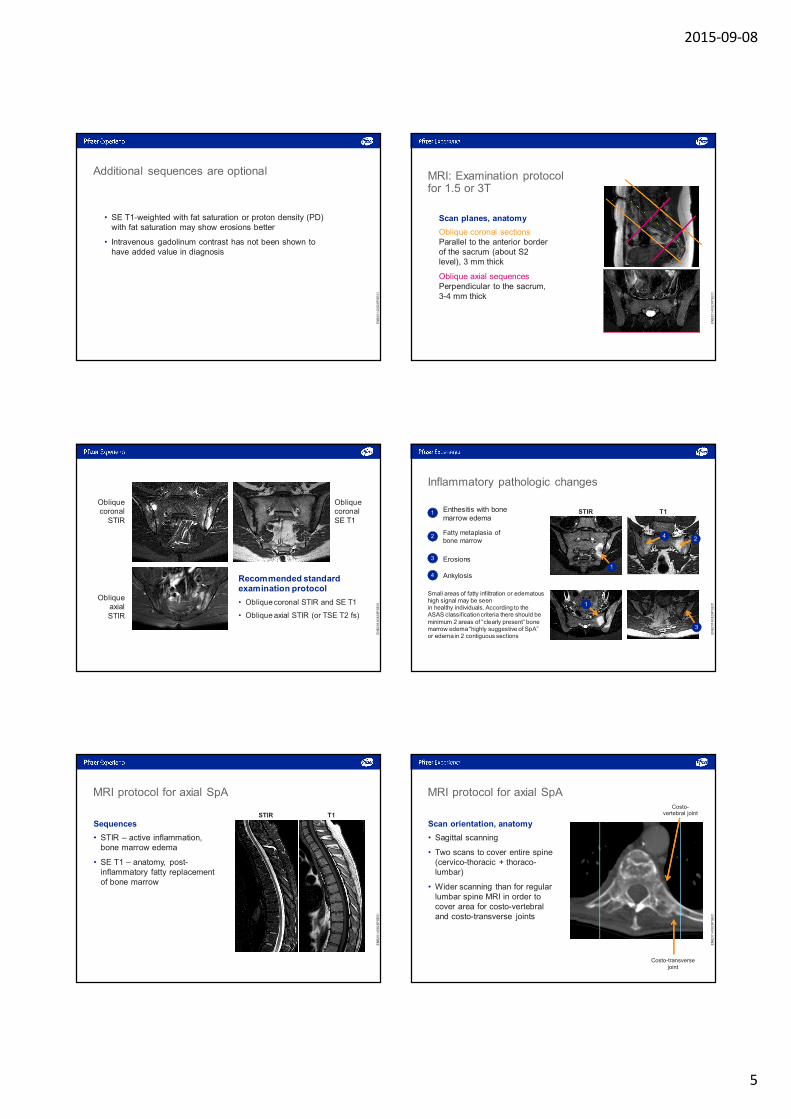
MRI: Examination protocol for 1.5 or 3T
STIR (Short tau inversion recovery) sequence.
Used to detect active inflammation with bone marrow edema. All signal from fat is extinguished, and signal is derived exclusively from water.
Bonemarrowedema
Dark sub-cutaneousfat
Bright pelvicveins
EN
B2
01
40
82
8P
SE
01
MRI: Examination protocol for 1.5 or 3T
Spin-echo (SE) T1-weighted sequence.
Used for anatomy, and for chronic changes such as fatty metaplasia of bone marrow, sclerosis, and erosions.
Signal information from fat.
Erosions
Bright sub-cutaneous fat
EN
B2
01
40
82
8P
SE
01
2015-09-08
5
Additional sequences are optional
• SE T1-weighted with fat saturation or proton density (PD) with fat saturation may show erosions better
• Intravenous gadolinum contrast has not been shown to have added value in diagnosis
EN
B2
01
40
82
8P
SE
01
MRI: Examination protocol for 1.5 or 3T
Scan planes, anatomy
Oblique coronal sectionsParallel to the anterior border of the sacrum (about S2 level), 3 mm thick
Oblique axial sequencesPerpendicular to the sacrum,3-4 mm thick
EN
B2
01
40
82
8P
SE
01
Oblique coronal SE T1
Oblique coronal
STIR
Oblique axial STIR
Recommended standard examination protocol
• Oblique coronal STIR and SE T1
• Oblique axial STIR (or TSE T2 fs)
EN
B2
01
40
82
8P
SE
01
Inflammatory pathologic changes
Enthesitis with bonemarrow edema
1
2
3
4
STIR T1
4 2
1
1
3
Fatty metaplasia ofbone marrow
Erosions
Ankylosis
Small areas of fatty infiltration or edematous high signal may be seenin healthy individuals. According to the ASAS classification criteria there should be minimum 2 areas of “clearly present” bone marrow edema “highly suggestive of SpA” or edema in 2 contiguous sections
EN
B2
01
40
82
8P
SE
01
MRI protocol for axial SpA
Sequences
• STIR – active inflammation, bone marrow edema
• SE T1 – anatomy, post-inflammatory fatty replacement of bone marrow
STIR T1
EN
B2
01
40
82
8P
SE
01
MRI protocol for axial SpA
Scan orientation, anatomy
• Sagittal scanning
• Two scans to cover entire spine (cervico-thoracic + thoraco-lumbar)
• Wider scanning than for regular lumbar spine MRI in order to cover area for costo-vertebral and costo-transverse joints
Costo-transverse joint
Costo-vertebral joint
EN
B2
01
40
82
8P
SE
01
2015-09-08
6
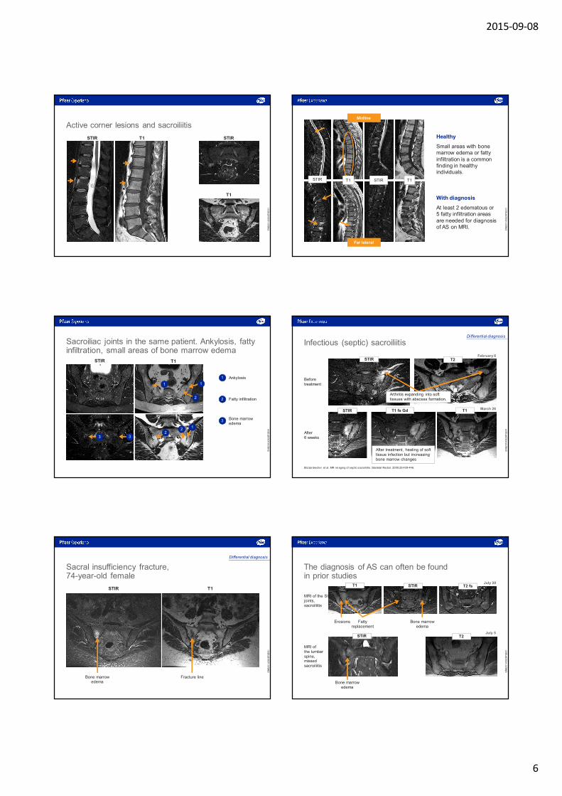
Active corner lesions and sacroiliitis
STIR T1 STIR
T1
EN
B2
01
40
82
8P
SE
01
Healthy
Small areas with bone marrow edema or fatty infiltration is a common finding in healthy individuals.
With diagnosis
At least 2 edematous or5 fatty infiltration areas are needed for diagnosis of AS on MRI.
STIR STIR T1T1
MidlineMidline
Far lateralFar lateral
EN
B2
01
40
82
8P
SE
01
Sacroiliac joints in the same patient. Ankylosis, fatty infiltration, small areas of bone marrow edema
T1STIR
Ankylosis1
Fatty infiltration2
Bone marrow edema
3
1 1
1
2
22
3 3
EN
B2
01
40
82
8P
SE
01
Infectious (septic) sacroiliitis
February 6
March 26
T2STIR
After treatment, healing of soft tissue infection but increasing bone marrow changes
Arthritis expanding into soft tissues with abscess formation.
Beforetreatment
After6 weeks
T1 fs GdSTIR
Differential diagnosis
T1
Stürzenbecher et al. MR imaging of septic sacroiliitis. Skeletal Radiol. 2000;29:439-446.
EN
B2
01
40
82
8P
SE
01
Sacral insufficiency fracture,74-year-old female
T1STIR
Bone marrow edema
Fracture line
Differential diagnosis
EN
B2
01
40
82
8P
SE
01
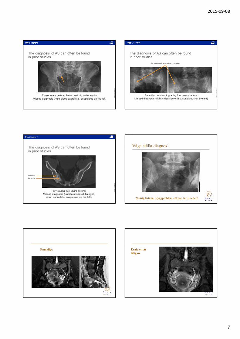
The diagnosis of AS can often be found in prior studies
Erosions Fattyreplacement
Bone marrowedema
MRI of the SI joints, sacroiliitis
July 20
MRI ofthe lumbar spine, missed sacroiliitis
T2 fsSTIR
July 5
Bone marrowedema
T2STIR
T1
EN
B2
01
40
82
8P
SE
01
2015-09-08
7
Three years before: Pelvis and hip radiography.Missed diagnosis (right-sided sacroiliitis, suspicious on the left)
The diagnosis of AS can often be found in prior studies
EN
B2
01
40
82
8P
SE
01
Sacroiliitis with sclerosis and erosions
Sacroiliac joint radiography four years before:Missed diagnosis (right-sided sacroiliitis, suspicious on the left)
The diagnosis of AS can often be found in prior studies
EN
B2
01
40
82
8P
SE
01
Erosions
Sclerosis
Polytrauma five years before:Missed diagnosis (unilateral sacroiliitis right-
sided sacroiliitis, suspicious on the left)
The diagnosis of AS can often be found in prior studies
EN
B2
01
40
82
8P
SE
01
Våga ställa diagnos!
22-årig kvinna. Ryggproblem ett par år. SI-leder?
Samtidigt: Exakt ett år tidigare
2015-09-08
8
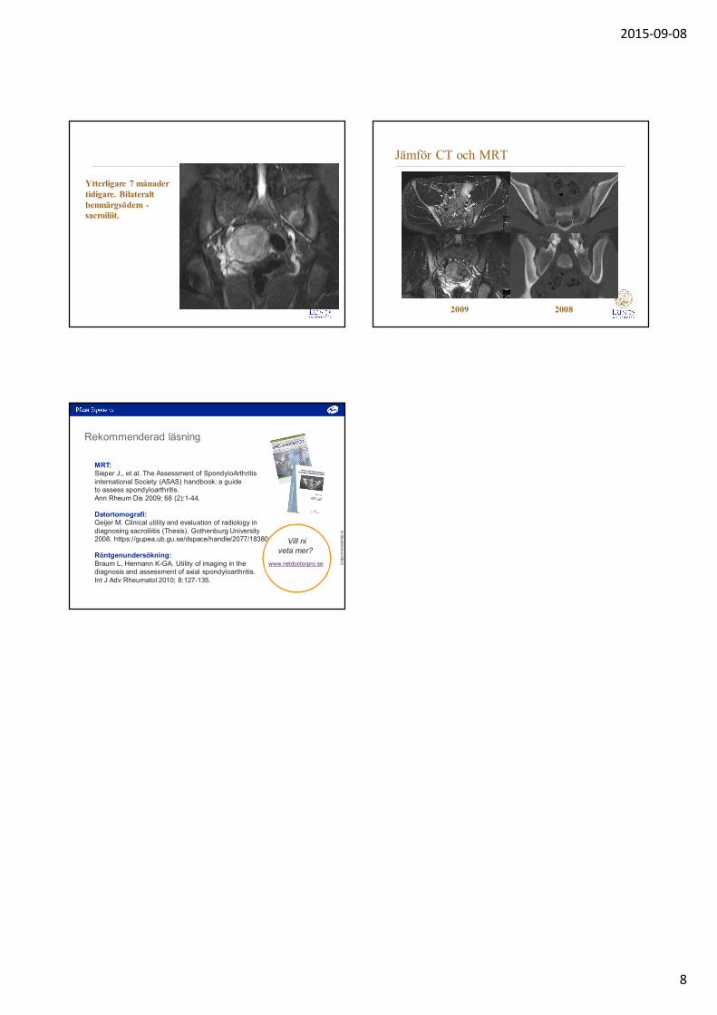
Ytterligare 7 månader tidigare. Bilateralt benmärgsödem -sacroiliit.
Jämför CT och MRT
2009 2008
Rekommenderad läsning
MRT: Sieper J., et al. The Assessment of SpondyloArthritisinternational Society (ASAS) handbook: a guideto assess spondyloarthritis. Ann Rheum Dis 2009; 68 (2):1-44.
Datortomografi: Geijer M. Clinical utility and evaluation of radiology in diagnosing sacroiliitis (Thesis). Gothenburg University 2008. https://gupea.ub.gu.se/dspace/handle/2077/18380.
Röntgenundersökning:Braum L, Hermann K-GA. Utility of imaging in thediagnosis and assessment of axial spondyloarthritis.Int J Adv Rheumatol 2010; 8:127-135.
Vill niveta mer?
www.netdoctorpro.se EN
B2
01
40
90
5P
SE
10