Embed Size (px)
Citation preview
RESEARCH Open Access
MRSA decolonization failure—are biofilmsthe missing link?Frank Günther1, Brigitte Blessing1, Evelina Tacconelli2,3 and Nico T. Mutters1,2*
Abstract
Background: Device-associated infections due to biofilm-producing methicillin-resistant Staphylococcus aureus(MRSA) have been recently associated with the failure of antibiotic treatment and decolonization measures. Thegoal of our study was to evaluate the extent to which the formation of biofilms influenced the efficacy of topicaldecolonization agents or disinfectants such as mupirocin (MUP), octenidine (OCT), chlorhexidine (CHG),polyhexanide (POL), and chloroxylenol (CLO).
Methods: Bacterial killing in biofilms by the disinfectants and MUP was determined as the reduction [%] inmetabolic activity determined by a biofilm viability assay that uses kinetic analysis of metabolic activity.The test substances were diluted in water with standardized hardness (WSH) at 25 °C at the standardconcentration as well as half the standard concentration to demonstrate the dilution effects in a practicalsetting. The tested concentrations were: CHG 1%, 2%; OCT 0.1%, 0.05%; PH 0.04%, 0.02%; and CLO 0.12%, 0.24%. A test organism suspension, 1 mL containing ~1 × 10⁹ bacterial cells/mL, and 1 mL of sterile WSHwere mixed and incubated for six different exposure times (15 s, 1, 3, 5, 10 and 20 min) after the testsubstance was added.Additionally, the bactericidal effects of all substances were tested on planktonic bacteria and measured as thelog10 reduction.
Results: The disinfectants OCT and CHG showed good efficacy in inhibiting MRSA in biofilms withreduction rates of 94 ± 1% and 91 ± 1%, respectively. POL, on the other hand, had a maximum efficacy ofonly 81 ± 7%. Compared to the tested disinfectants, MUP showed a significantly lower efficacy with <20%inhibition (p < .05). Bactericidal effects were the greatest for CHG (log10 reduction of 9.0), followed byOCT (7.7), POL (5.1), and CLO (6.8). MUP, however, showed a very low bactericidal effect of only 2.1. Evenwhen the exposure time was increased to 24 h, 2% MUP did not show sufficient bactericidal effect.
Conclusions: Our data provide evidence that OCT and CHG are effective components for disinfection ofMRSA-biofilms. On the other hand, exposure to MUP at the standard concentrations in topicalpreparations did not effectively inhibit MRSA-biofilms and also did not show adequate bactericidal effects.Combining an MUP-based decolonization regimen with a disinfectant such as OCT or CHG could decreasedecolonization failure.
Keywords: MRSA, Decolonization, Biofilm, Chlorhexidine, Octenidine, Mupirocin, Polyhexanide,Chloroxylenol, Infection control
* Correspondence: [email protected] of Infectious Diseases, Heidelberg University Hospital, ImNeuenheimer Feld 324, 69126 Heidelberg, Germany2Division of Infectious Diseases - Department of Internal Medicine I,Tübingen University Hospital, Tübingen, GermanyFull list of author information is available at the end of the article
© The Author(s). 2017 Open Access This article is distributed under the terms of the Creative Commons Attribution 4.0International License (http://creativecommons.org/licenses/by/4.0/), which permits unrestricted use, distribution, andreproduction in any medium, provided you give appropriate credit to the original author(s) and the source, provide a link tothe Creative Commons license, and indicate if changes were made. The Creative Commons Public Domain Dedication waiver(http://creativecommons.org/publicdomain/zero/1.0/) applies to the data made available in this article, unless otherwise stated.
Günther et al. Antimicrobial Resistance and Infection Control (2017) 6:32 DOI 10.1186/s13756-017-0192-1
BackgroundMethicillin-resistant and biofilm-forming Staphylococcusaureus (MRSA) isolates have become a common clinicalproblem [1]. In recent years, MRSA incidences seemedto be decreasing, and the focus of infection control spe-cialists was multidrug-resistant Gram-negative bacteria[2–4]. However, Public Health England recently reportedan alarming 26% increase in MRSA bloodstream infec-tions [5]. Although the rise in numbers coincided withthe Department of Health’s change in policy on screen-ing for MRSA, from universal to targeted screening, itunderlines the fact that MRSA cannot be considered“out of the picture”. The formation of biofilms as a reac-tion to therapeutic interventions, which can lead toincreased antimicrobial resistance and a higher chanceof treatment failure, is being increasingly recognized asan infection control problem [6, 7]. Accordingly, treat-ment and decolonization failure occur more frequentlywhen topical drugs like mupirocin are used againstbiofilm-forming microorganisms [8–12].The organization of bacteria into biofilms is the com-
mon mode of bacterial survival, since this form increasestheir ability to withstand antibiotics, disinfectants, andhost responses. Biofilm formation is a multifarious, con-trolled bacterial process that induces many additionalfunctional and phenotypic alterations, including loss ofmotility, reduced growth rate, increased surface adhe-sion, as well as an altered susceptibility to the hostresponse [13–17]. An association with biofilm formationhas been reported for many hospital-acquired infections,such as urinary tract and catheter-related bloodstreaminfections as well as infections of implanted medicaldevices including indwelling catheters, artificial heartvalves, orthopedic prostheses, or osteosynthesis mate-rials [7, 18–24]. Colonization with MRSA is associatedwith a high risk of acquiring an MRSA infection duringhospital stays [25, 26]. Decolonization may reduce therisk of MRSA infection in individual carriers and preventtransmission to other patients [25]. However, the mostcommonly used agent for decolonization, mupirocin,comes with a considerable risk of resistance if widelyemployed [26]. There have been many other attempts toeradicate carriage, mostly with topical agents, but suc-cess rates have not been consistent or applicable to allpopulations [26], and even mupirocin decolonizationsuccess rates can be low [25]. Many international guide-lines (i.e., in Germany, Ireland, Netherlands, Slovenia)already state that attempts at decolonization are unlikelyto be successful in patients with chronic skin conditions,ulcers, or in-dwelling catheters [27–30]. Some studies,however, showed that decolonization can be effective inpatients with lines and catheters [25] and that the ina-bility to decolonize was most closely associated with fail-ure to use a standardized decolonization protocol [31].
However, to our knowledge, no study has been able toidentify a consistent subgroup of patients at higher riskfor decolonization failure. Perhaps, the focus has beenon the wrong variable in the equation, and the reasonfor decolonization failure is not the patient but thebacterium and its biofilm-forming capacities. Alarm-ingly, one study on biofilm formation among MRSAnasal carriers showed that all of the isolated MRSA hadthe ability to form biofilms [7]. The goal of our study,therefore, was to evaluate the extent to which MRSAbiofilms are influenced by the use of the topicaldecolonization antibiotic MUP and the widely used top-ical disinfectants, i.e., octenidine (OCT), chlorhexidine(CHG), polyhexanide (POL), and chloroxylenol (CLO).
MethodsBacterial isolatesTo ensure practical relevance, clinical MRSA isolates, aswell as American Type Culture Collection (Manassas,VA, USA; ATCC®) control strains, were tested in thisstudy. Clinical isolates were recovered as follows: screen-ing swab samples were inoculated on Columbia 5%sheep blood agar plate (BD Diagnostics, Sparks, USA)and chromogenic plates for MRSA detection (ChromAgarMRSA II, BD) and incubated under aerobic conditions for48 h at 36 °C. If growth on chromogenic plates wasdetected, identification by matrix-assisted laser desorptionionization-time-of-flight mass spectrometry (MALDI-TOF MS) (Bruker Daltonics, Bremen, Germany) wasperformed [32]. Agglutination with Pastorex® StaphPlus(Alere, Jena, Germany) was performed to confirm S.aureus growth. Susceptibility testing was performed byVITEK2 (bioMérieux) and results were interpreted ac-cording to EUCAST breakpoints. Biofilm-forming capaci-ties of each isolate were determined by the crystal violetstaining technique (data not shown). Six representativeisolates with significant biofilm-forming capacity com-pared to the standard disinfectant efficacy test isolateATCC® 6538™, according to ATCC® product sheet, wereselected and used for further testing.
Preparation of antimicrobials and neutralizerFor standardization of experimental conditions, in eachexperiment, water of standardized hardness (WSH) wasprepared with a total hardness of 300 ppm (CaCO3) with0.119 g/l magnesium chloride (Carl Roth, Karlsruhe,Germany), 0.277 g/l calcium chloride (Carl Roth), and0.28 g/l sodium hydrogen carbonate (Carl Roth). WSH ata pH of 7.0 ± 0.2 at 25 °C was used as a diluent. Trypticsoy broth (TSB) containing lecithin, Tween 80, histidine,and sodium thiosulfate neutralizing agent (all from MerckMillipore, Darmstadt, Germany) (LTHTh) was used inboth treatment and control groups, immediately followingdisinfection according to the manufacturer’s instructions.
Günther et al. Antimicrobial Resistance and Infection Control (2017) 6:32 Page 2 of 7
The media and neutralizers used in this study wereapproved for effective neutralization of the applied disin-fectants prior to the experiments (data not shown).
Biofilm viability assayThe MRSA isolates were cultured on Columbia bloodagar plates at 37 °C for 12 h. Bacterial killing in biofilmswas determined as reduction [%] in metabolic activityusing a kinetic biofilm viability assay as previously de-scribed [33]. Briefly, the test substances were diluted inWSH at 25 °C at the standard concentration as well ashalf the standard concentration to demonstrate the dilu-tion effects in a practical setting. For testing of anti-microbial effects on the bacterial metabolic activity inbiofilms, the antimicrobial substances were diluted inWSH at standard working concentrations 0.05% and0.1% (w/v) for OCT (TCI, Eschborn, Germany), 1% and2% (w/v) for CHG (Sigma Aldrich, Taufkirchen,Germany), 0.02% and 0.04% (w/v) for POL (Fagron,Barsbuettel, Germany), 0.12% and 0.24% (w/v) for CLO(Sigma Aldrich), and 1% and 2% (w/v) for MUP(Fagron). Each preparation was applied to the preparedbiofilms for different exposure times of 15 s, 1, 3, 5, 10and 20 min at 37°Cfor the OCT, CHG, POL, and CLOsolutions. Prolonged exposure times of up to 3.5 h wereused for MUP to analyze the different mode of action ofthis substance. To take into account the different modeof action of MUP compared to disinfectants, extendedexposure times of up to 3.5 h were used for this substanceadapted to simplified testing protocols for determination ofbactericidal activity on Staphylococcus aureus isolates aspreviously described [34]. After exposure, the remainingmetabolic activity in the biofilm was measured. Biofilmsexposed to WSH alone (without supplements) served as acontrol for 100% viability or 0% inhibition. The bacterialkilling by the disinfectants in the biofilms was determinedas the reduction [%] in metabolic activity as compared tothe untreated controls.
Live/dead staining of biofilmsTo analyze the killing effects on the bacterial cells in bio-films, biofilms were cultured on glass coverslips (CarlRoth). After incubation, biofilms were washed twice in a0.9% NaCl solution. Then, 100 μL M63 minimal mediumconsisting of 0.015 M ammonium hydrogen sulfate(Carl Roth), 0.1 M potassium dihydrogen sulfate(Sigma-Aldrich, Steinheim, Germany), 1.8 μM ironsulfate heptahydrate (Carl Roth), 1 mM magnesiumsulfate heptahydrate, 2 ml/L glycerol (VWR Chemi-cals, Darmstadt, Germany) and 1 g/L casein hydrolys-ate standard (Carl Roth)containing disinfectants indifferent concentrations was added, and the biofilmswere incubated for up to 2 h at 37 °C. After incubation,biofilms were washed twice in 0.9% NaCl solution and
stained using the LIVE/DEAD BacLight Bacterial ViabilityKit (Molecular Probes, Leiden, Netherlands) according tothe manufacturer’s instructions. Stained biofilms wereanalyzed after mounting on object slides by using a BZ8100 fluorescence microscope (Keyence, Neu-Isenburg,Germany) in the green fluorescent band for Syto 9 stain-ing of dead and living bacterial cells, and in red fluores-cent band for selective propidium iodide staining of deadcells or cells with disturbed cell integrity; therefore, yellowcolor in the overlay image is indicative of dead bacteria inthe biofilm.
Determination of bactericidal activityFor determination of bactericidal effects, the antimi-crobial substances were diluted in WSH at standard
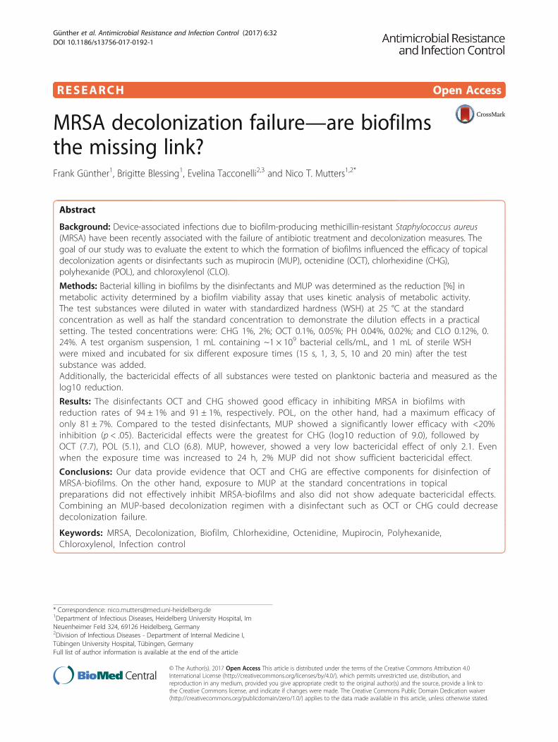
Fig. 1 Disinfectant efficacy on MRSA-biofilms Reduction of metabolicactivity (%) in bacterial biofilms ± standard deviation (SD) afterdisinfectant treatment at different concentrations and for varyingexposure times, determined by a kinetic metabolic assay
Günther et al. Antimicrobial Resistance and Infection Control (2017) 6:32 Page 3 of 7
working concentrations 0.1% (w/v) for OCT, 2% forCHG, 0.04% (w/v) for POL, 0.24% (w/v) for CLO,and 2% (w/v) for MUP, and at least 1x10⁹ bacterialcells were added for different exposure times. Eachsample was tested for the surviving bacterial countby membrane filtration of the disinfectant solutionthrough a 0.45-μm nitrocellulose membrane (Sartor-ius Stedim, Göttingen, Germany) followed by rinsingthree times using 100 ml of a NaCl-peptone solution(Becton Dickinson, Heidelberg, Germany) for removalof remaining disinfectant. Afterwards, the filters weretransferred to Caso Agar containing LTHTh as aneutralizer (Merck Millipore, Darmstadt, Germany).The neutralization and washing steps were validatedfor effective neutralization of the tested disinfectantsprior to this study (data not shown). The mediawere incubated for 48 h at 37 °C and then checkedfor microbial growth. Colony forming units (CFUs)were counted and the Log10 reduction factor (LRF10)was calculated compared to the untreated controls asa measure of the bactericidal effect.
StatisticsFor descriptive purposes, arithmetic mean value, stand-ard deviation, median, interquartile range, and cumula-tive frequencies were calculated as appropriate. P valuesof ≤ .05 were considered statistically significant. Statis-tical analysis was performed using the SPSS ver. 21.0statistical package (SPSS, Chicago, IL).
ResultsThe effects of the different disinfectants on metabolicactivity in established MRSA biofilms were tested usinga kinetic metabolic assay (Fig. 1) [33]. OCT showedmoderate efficacy in inhibiting microbial metabolic ac-tivity with 94 ± 1% inhibition after only 15 s of exposureand at a concentration of 0.05%. The overall efficacy ofOCT on MRSA biofilm inhibition was not significantlychanged due to modifications of the concentration used,ranging from 0.05% to 1%, or due to modifications ofthe exposure time, ranging from 15 s to 20 min. The CHGsolution showed a efficacy of 91 ± 1% in inhibiting micro-bial metabolic activity after 3 min of exposure to a 1%CHG solution. Furthermore, after 1 min of exposure, therewas no significant increase in efficacy of CHG on MRSAbiofilms for the tested concentration range. POL, in con-trast, yielded a lower overall medium efficacy of inhibitionof metabolic activity in biofilms of 65.4 ± 11%, while theefficacy was strongly dependent on the applied exposuretime. CLO showed the lowest efficacy of 15.8 ± 27%. Add-itionally, the efficacy of CLO was strongly dependent onthe applied exposure time and concentration.On the other hand, the antibiotic MUP showed detect-
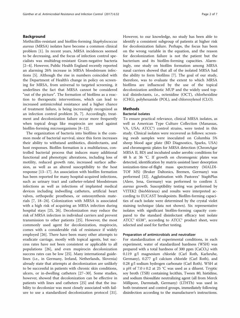
able efficacy in inhibiting metabolic activity in MRSAbiofilms after short exposure times (Fig. 2). To rule outthat the lack of efficacy is caused by the different modeof action of MUP, namely the inhibition of bacterial pro-tein synthesis, the exposure times for MUP on theMRSA biofilms were extended to up to 3.5 h. Even after
Fig. 2 MUP efficacy on MRSA-biofilms and planktonic bacteria Upper panel: Reduction of metabolic activity (%) in bacterial biofilms ± SD afterMUP treatment at different concentrations and varying exposure times, determined by a kinetic metabolic assay. Lower panel: Bactericidal activityof a 2% MUP solution on planktonic MRSA, shown as reduction of cell counts [CFU] compared to untreated controls in WSH
Günther et al. Antimicrobial Resistance and Infection Control (2017) 6:32 Page 4 of 7
3.5 h of exposure to 2% MUP solution, the level of meta-bolic inhibition of the bacteria in the MRSA biofilmsdid not exceed 20%, while significant inhibition (p <0.05) was reached after at least 2 h of. Taken to-gether, 2% MUP showed between 1 and 20% inhib-ition of the metabolic activity of MRSA biofilms afterexposure times of up to 3.5 h (Fig. 2).Exposure of biofilms to OCT and CHG solutions led
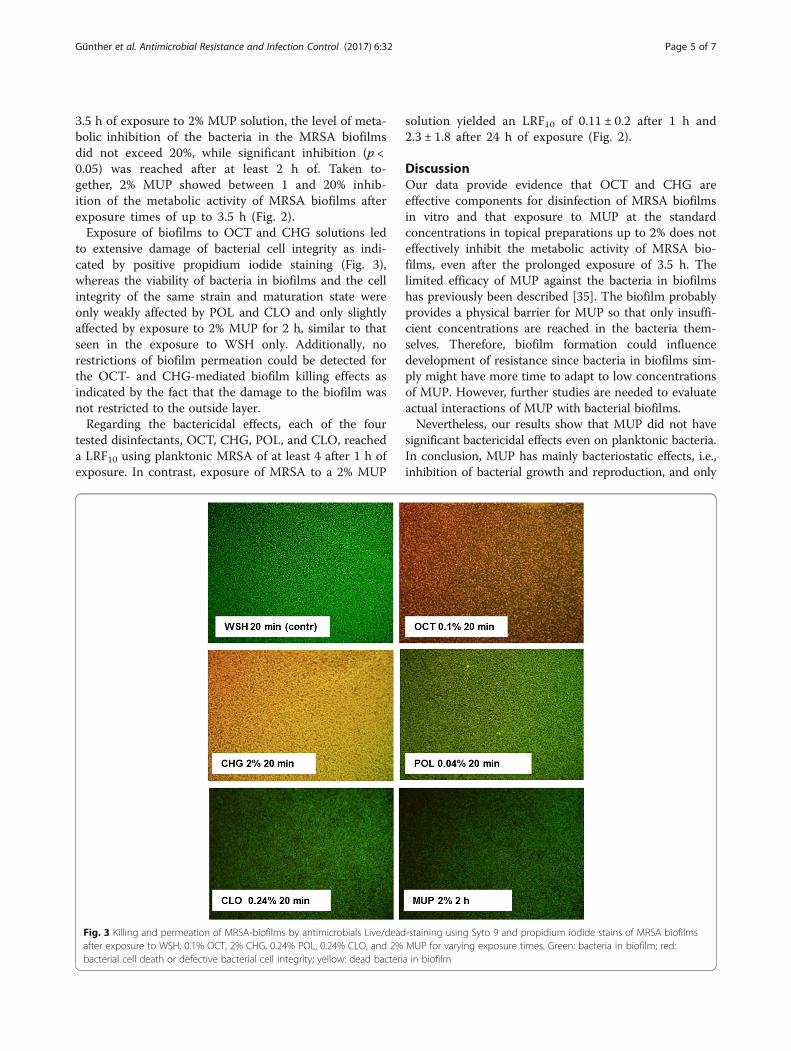
to extensive damage of bacterial cell integrity as indi-cated by positive propidium iodide staining (Fig. 3),whereas the viability of bacteria in biofilms and the cellintegrity of the same strain and maturation state wereonly weakly affected by POL and CLO and only slightlyaffected by exposure to 2% MUP for 2 h, similar to thatseen in the exposure to WSH only. Additionally, norestrictions of biofilm permeation could be detected forthe OCT- and CHG-mediated biofilm killing effects asindicated by the fact that the damage to the biofilm wasnot restricted to the outside layer.Regarding the bactericidal effects, each of the four
tested disinfectants, OCT, CHG, POL, and CLO, reacheda LRF10 using planktonic MRSA of at least 4 after 1 h ofexposure. In contrast, exposure of MRSA to a 2% MUP
solution yielded an LRF10 of 0.11 ± 0.2 after 1 h and2.3 ± 1.8 after 24 h of exposure (Fig. 2).
DiscussionOur data provide evidence that OCT and CHG areeffective components for disinfection of MRSA biofilmsin vitro and that exposure to MUP at the standardconcentrations in topical preparations up to 2% does noteffectively inhibit the metabolic activity of MRSA bio-films, even after the prolonged exposure of 3.5 h. Thelimited efficacy of MUP against the bacteria in biofilmshas previously been described [35]. The biofilm probablyprovides a physical barrier for MUP so that only insuffi-cient concentrations are reached in the bacteria them-selves. Therefore, biofilm formation could influencedevelopment of resistance since bacteria in biofilms sim-ply might have more time to adapt to low concentrationsof MUP. However, further studies are needed to evaluateactual interactions of MUP with bacterial biofilms.Nevertheless, our results show that MUP did not have
significant bactericidal effects even on planktonic bacteria.In conclusion, MUP has mainly bacteriostatic effects, i.e.,inhibition of bacterial growth and reproduction, and only
Fig. 3 Killing and permeation of MRSA-biofilms by antimicrobials Live/dead-staining using Syto 9 and propidium iodide stains of MRSA biofilmsafter exposure to WSH, 0.1% OCT, 2% CHG, 0.24% POL, 0.24% CLO, and 2% MUP for varying exposure times. Green: bacteria in biofilm; red:bacterial cell death or defective bacterial cell integrity; yellow: dead bacteria in biofilm
Günther et al. Antimicrobial Resistance and Infection Control (2017) 6:32 Page 5 of 7
minor effects when bacteria are in biofilms. Biofilms mightbe the missing link in understanding the rapid develop-ment of resistance when MUP-based regimens are usedroutinely among general inpatient populations.One limitation of our study is that we cannot estimate
the fraction of MRSA strains that are strong biofilm for-mers. However, this issue should be addressed in futurestudies. As previously mentioned, one study on 810 nasalcarriers of S. aureus and MRSA showed that all isolatedMRSA strains were biofilm formers; however, only 34.6%were medium to strong biofilm producers [7]. The largerthe fraction of strong biofilm formers among MRSAstrains, the more impact a change in the decolonizationregimen will have.
ConclusionsThe standard MRSA decolonization protocols at referral atour health care facility consist of 5 days of MUP treatmentas nasal ointment and additional antiseptic washing of bodyand daily change of bed linen. Our data suggest that com-bining an MUP-based decolonization regimen with a disin-fectant such as OCT or CHG could increase the efficacy ofdecolonization in patients colonized with biofilm-formingMRSA. In patients who failed the first decolonization at-tempt, one possible approach could be to change the 5 daysof MUP treatment of the standard decolonization regimento (1) two days of primary disinfection with either OCT orCHG as a nasal ointment followed by (2) three days oftreatment with MUP for suppression of re-colonization.Obviously, this new regimen needs to be evaluated asrandomized-controlled trial. Decolonization studies usingOCT already show good decolonization rates (67%); how-ever, to our knowledge, no long-lasting effects with regardsto re-colonization have yet been reported [36].
AbbreviationsCFU: Colony forming unit; CHG: Chlorhexidine; CLO: Chloroxylenol;LTHTh: Lecithin, Tween 80, histidine, and sodium thiosulfate neutralizingagent; MRSA: Methicillin-resistant Staphylococcus aureus; MUP: Mupirocin;OCT: Octenidine; POL: Polyhexanide; TSB: Tryptic soy broth; WSH: Water withstandardized hardness
AcknowledgementsLanguage editing was done by Elsevier Language Editing Service.
FundingWe acknowledge financial support by Deutsche Forschungsgemeinschaftand Ruprecht-Karls-Universität Heidelberg within the funding programmeOpen Access Publishing.
Availability of data and materialsThe data is not publicly available. Please contact the corresponding authorfor further information.
Authors’ contributionsFG designed the study, analyzed and interpreted the data, and drafted themanuscript. BB contributed to the design of the study and conducted theexperiments. ET contributed to the design of the study and wrote parts of themanuscript. NTM contributed to the design of the study, analyzed the data, andwrote the manuscript. All authors read and approved the final manuscript.
Competing interestsThe authors declare that they have no competing interests.
Consent for publicationNot applicable.
Ethics approval and consent to participateAt Heidelberg University Hospital, all MRSA strains are routinely collected inthe microbiology laboratory and stored at − 70 °C. The current study thus isdescriptive of a bacterial collection of those isolates. No patient-related datawere collected. Ethical approval was therefore not required. The study was alaboratory-based basic science study.
Publisher’s NoteSpringer Nature remains neutral with regard to jurisdictional claims inpublished maps and institutional affiliations.
Author details1Department of Infectious Diseases, Heidelberg University Hospital, ImNeuenheimer Feld 324, 69126 Heidelberg, Germany. 2Division of InfectiousDiseases - Department of Internal Medicine I, Tübingen University Hospital,Tübingen, Germany. 3German Centre for Infection Research (DZIF), TübingenGermany.
Received: 17 January 2017 Accepted: 21 March 2017
References1. Bhattacharya S, Bir R, Majumdar T. Evaluation of multidrug resistant
staphylococcus aureus and their association with biofilm production in aTertiary Care Hospital, Tripura, Northeast India. J Clin Diagn Res. 2015;9:DC01–04.
2. Nordmann P, Dortet L, Poirel L. Carbapenem resistance inEnterobacteriaceae: here is the storm! Trends Mol Med. 2012;18:263–72.
3. Nordmann P, Poirel L. The difficult-to-control spread of carbapenemaseproducers among Enterobacteriaceae worldwide. Clin Microbiol Infect. 2014;20:821–30.
4. Mutters NT, Gunther F, Sander A, Mischnik A, Frank U. Influx of multidrug-resistant organisms by country-to-country transfer of patients. BMC InfectDis. 2015;15:466.
5. MRSA bacteraemia: annual data [https://www.gov.uk/government/statistics/mrsa-bacteraemia-annual-data].
6. Ohadian Moghadam S, Pourmand MR, Aminharati F. Biofilm formation andantimicrobial resistance in methicillin-resistant Staphylococcus aureusisolated from burn patients, Iran. J Infect Dev Ctries. 2014;8:1511–7.
7. Rezaei M, Moniri R, Mousavi SGA, Shiade MJ. Prevalence of biofilmformation among methicillin resistance staphylococcus aureus isolated fromnasal carriers. Jundishapur J Microbiol. 2013; 6(6):e9601.
8. McNeil JC, Hulten KG, Kaplan SL, Mason EO. Mupirocin resistance inStaphylococcus aureus causing recurrent skin and soft tissue infections inchildren. Antimicrob Agents Chemother. 2011;55:2431–3.
9. Thangamani S, Younis W, Seleem MN. Repurposing ebselen for treatment ofmultidrug-resistant staphylococcal infections. Sci Rep. 2015;5:11596.
10. Thangamani S, Mohammad H, Abushahba MF, Sobreira TJ, Seleem MN.Repurposing auranofin for the treatment of cutaneous staphylococcalinfections. Int J Antimicrob Agents. 2016;47:195–201.
11. Thangamani S, Younis W, Seleem MN. Repurposing clinical moleculeebselen to combat drug resistant pathogens. PLoS One. 2015;10:e0133877.
12. Roche ED, Renick PJ, Tetens SP, Carson DL. A model for evaluating topicalantimicrobial efficacy against methicillin-resistant Staphylococcus aureusbiofilms in superficial murine wounds. Antimicrob Agents Chemother. 2012;56:4508–10.
13. Gunther F, Wabnitz GH, Stroh P, Prior B, Obst U, Samstag Y, Wagner C,Hansch GM. Host defence against Staphylococcus aureus biofilms infection:phagocytosis of biofilms by polymorphonuclear neutrophils (PMN). MolImmunol. 2009;46:1805–13.
14. Dunne Jr WM. Bacterial adhesion: seen any good biofilms lately? ClinMicrobiol Rev. 2002;15:155–66.
15. Davey ME, O'Toole GA. Microbial biofilms: from ecology to moleculargenetics. Microbiol Mol Biol Rev. 2000;64:847–67.
Günther et al. Antimicrobial Resistance and Infection Control (2017) 6:32 Page 6 of 7
16. Meyle E, Stroh P, Gunther F, Hoppy-Tichy T, Wagner C, Hansch GM.Destruction of bacterial biofilms by polymorphonuclear neutrophils: relativecontribution of phagocytosis, DNA release, and degranulation. Int J ArtifOrgans. 2010;33:608–20.
17. Watnick P, Kolter R. Biofilm, city of microbes. J Bacteriol. 2000;182:2675–9.18. Donlan RM. Biofilm formation: a clinically relevant microbiological process.
Clin Infect Dis. 2001;33:1387–92.19. Donlan RM. Biofilms and device-associated infections. Emerg Infect Dis.
2001;7:277–81.20. Donlan RM, Murga R, Bell M, Toscano CM, Carr JH, Novicki TJ, Zuckerman C,
Corey LC, Miller JM. Protocol for detection of biofilms on needleless connectorsattached to central venous catheters. J Clin Microbiol. 2001;39:750–3.
21. Gottenbos B, Busscher HJ, Van Der Mei HC, Nieuwenhuis P. Pathogenesisand prevention of biomaterial centered infections. J Mater Sci Mater Med.2002;13:717–22.
22. Zimmerli W, Trampuz A, Ochsner PE. Prosthetic-joint infections. N EnglJ Med. 2004;351:1645–54.
23. Costerton JW, Stewart PS, Greenberg EP. Bacterial biofilms: a common causeof persistent infections. Science. 1999;284:1318–22.
24. Cha JO, Yoo JI, Yoo JS, Chung HS, Park SH, Kim HS, Lee YS, Chung GT.Investigation of biofilm formation and its association with the molecularand clinical characteristics of methicillin-resistant staphylococcus aureus.Osong Public Health Res Perspect. 2013;4:225–32.
25. Sai N, Laurent C, Strale H, Denis O, Byl B. Efficacy of the decolonization ofmethicillin-resistant Staphylococcus aureus carriers in clinical practice.Antimicrob Resist Infect Control. 2015;4:56.
26. Abad CL, Pulia MS, Safdar N. Does the nose know? An update on MRSAdecolonization strategies. Curr Infect Dis Rep. 2013;15:455–64.
27. Royal-College-of-Physicians-Ireland. Prevention and Control of Methicillin-resistant Staphylococcus aureus (MRSA) National Clinical Guideline. 2013.
28. University-Clinic-of-Respiratory-and-Allergic-Diseases-Golnik-Slovenia.Guidelines to Control the Spread of MRSA. 2008.
29. Empfehlung der KRINKO beim Robert-Koch-Institut. Empfehlungen zurPrävention und Kontrolle von Methicillin-resistenten Staphylococcus aureus-Stämmen (MRSA) in medizinischen und pflegerischen Einrichtungen.Bundesgesundheitsbl. 2014; 57:696–732.
30. SWAB-Stichting-Werkgroep-Antibioticabeleid. Herziening SWAB richtlijnBehandeling MRSA dragers. 2012.
31. Dow G, Field D, Mancuso M, Allard J. Decolonization of methicillin-resistantStaphylococcus aureus during routine hospital care: efficacy and long-termfollow-up. Can J Infect Dis Med Microbiol. 2010;21:38–44.
32. Eigner U, Holfelder M, Oberdorfer K, Betz-Wild U, Bertsch D, Fahr AM.Performance of a matrix-assisted laser desorption ionization-time-of-flightmass spectrometry system for the identification of bacterial isolates in theclinical routine laboratory. Clin Lab. 2009;55:289–96.
33. Gunther F, Scherrer M, Kaiser SJ, DeRosa A, Mutters NT. Comparative testingof disinfectant efficacy on planktonic bacteria and bacterial biofilms using anew assay based on kinetic analysis of metabolic activity. J Appl Microbiol.2017;122:625–33.
34. Shanholtzer CJ, Peterson LR, Mohn ML, Moody JA, Gerding DN. MBCs forStaphylococcus aureus as determined by macrodilution and microdilutiontechniques. Antimicrob Agents Chemother. 1984;26:214–9.
35. Haisma EM, Goblyos A, Ravensbergen B, Adriaans AE, Cordfunke RA,Schrumpf J, Limpens RW, Schimmel KJ, den Hartigh J, Hiemstra PS, et al.Antimicrobial peptide P60.4Ac-containing creams and gel for eradication ofmethicillin-resistant staphylococcus aureus from cultured skin and airwayepithelial surfaces. Antimicrob Agents Chemother. 2016;60:4063–72.
36. Danilevicius M, Juzeniene A, Juzenaite-Karneckiene I, Versinina A. MRSAdecontamination using octenidine-based products. Br J Nurs. 2015;24(S36):S38–40.
• We accept pre-submission inquiries
• Our selector tool helps you to find the most relevant journal
• We provide round the clock customer support
• Convenient online submission
• Thorough peer review
• Inclusion in PubMed and all major indexing services
• Maximum visibility for your research
Submit your manuscript atwww.biomedcentral.com/submit
Submit your next manuscript to BioMed Central and we will help you at every step:
Günther et al. Antimicrobial Resistance and Infection Control (2017) 6:32 Page 7 of 7