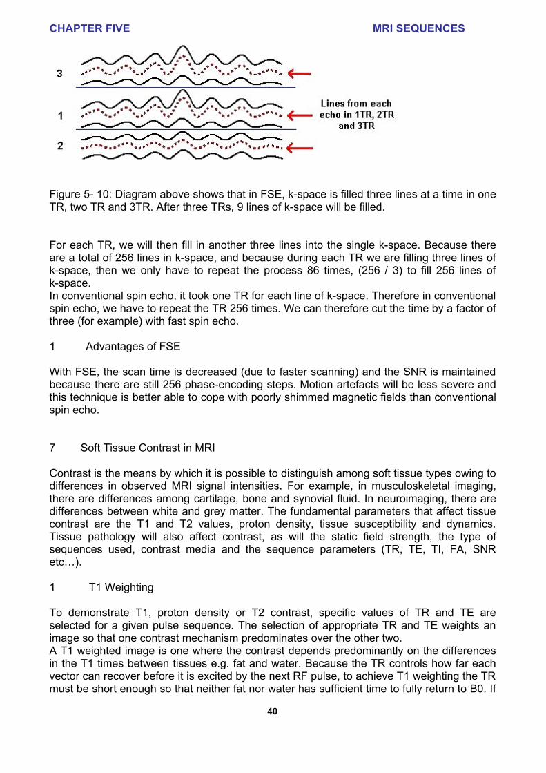
CHAPTER FIVE MRI SEQUENCES 34 5 MRI SEQUENCES An MRI sequence is an ordered combination of RF and gradient pulses designed to acquire the data to form the image. In this chapter I will describe the basic gradient echo, spin echo and inversion recovery sequences used in MRI. The data to create an MR image is obtained in a series of steps. First the tissue magnetisation is excited using an RF pulse in the presence of a slice select gradient. The other two essential elements of the sequence are phase encoding and frequency encoding/read out, which are required to spatially localise the protons in the other two dimensions. Finally, after the data has been collected, the process is repeated for a series of phase encoding steps. The MRI sequence parameters are chosen to best suit the particular clinical application. The parameters affecting soft tissue contrast are described, and advanced sequences such as STIR, FLAIR, FISP, and FLASH are briefly introduced at the end of the chapter. 1 Gradient Echo Sequence The gradient echo sequence is the simplest type of MRI sequence. It consists of a series of excitation pulses, each separated by a repetition time TR. Data is acquired at some characteristic time after the application of the excitation pulses and this is defined as the echo time TE. TE is the time between the mid-point of the excitation pulse and the mid-point of the data acquisition as shown in the sequence diagram, figure 5-1 below. The contrast in the image will vary with changes to both TR and TE (see chapter 6). In terms of k-space representation, the simultaneous application of the phase encode and read dephase gradients results in translation from the centre of k-space from A to B. This is followed by frequency encoding from B to C via the centre of k-space. Each line of data is FT to extract frequency information from the signal and the process is repeated for different phase encode gradient strengths. Figure 5-1 below shows the principle of a gradient echo sequence 1. Figure 5- 1: (L) Gradient Echo Sequence and (R) k-space representation Gradient Echo imaging does not compensate for B0 inhomogeneities. Therefore, there is an increased sensitivity to T2* decay caused by the lack of a 180º refocusing pulse. Gradient Echo sequences have advantages and disadvantages and these are highlighted in