Embed Size (px)
Citation preview
Page 1 of 17
MRI Patterns Of Shoulder Denervation: A Way To Make ItEasy
Poster No.: C-2059
Congress: ECR 2018
Type: Educational Exhibit
Authors: E. Rossetto1, P. Schvartzman2, V. N. Alarcon2, M. E. Scherer2, D.
M. Cecchi3, F. M. Olivera Plata4; 1Buenos Aires, Capital Federal/
AR, 2Buenos Aires/AR, 3Capital Federal, Buenos Aires/AR,4Ciudad Autonoma de Buenos Aires/AR
Keywords: Musculoskeletal joint, Musculoskeletal soft tissue, Neuroradiologyperipheral nerve, MR, Education, eLearning, Edema, Inflammation,Education and training
DOI: 10.1594/ecr2018/C-2059
Any information contained in this pdf file is automatically generated from digital materialsubmitted to EPOS by third parties in the form of scientific presentations. Referencesto any names, marks, products, or services of third parties or hypertext links to third-party sites or information are provided solely as a convenience to you and do not inany way constitute or imply ECR's endorsement, sponsorship or recommendation of thethird party, information, product or service. ECR is not responsible for the content ofthese pages and does not make any representations regarding the content or accuracyof material in this file.As per copyright regulations, any unauthorised use of the material or parts thereof aswell as commercial reproduction or multiple distribution by any traditional or electronicallybased reproduction/publication method ist strictly prohibited.You agree to defend, indemnify, and hold ECR harmless from and against any and allclaims, damages, costs, and expenses, including attorneys' fees, arising from or relatedto your use of these pages.Please note: Links to movies, ppt slideshows and any other multimedia files are notavailable in the pdf version of presentations.

Page 2 of 17
www.myESR.org
Page 3 of 17
Learning objectives
To describe and illustrate the most frequent MR patterns of shoulder muscle denervation.
To review of specific anatomic places commonly involved in this type of pathology.
We perform a diagnosis algorithm in order to simplify the association of denervatedmuscle and the nerve compromise
Background
We performed a retrospective analysis of patients with shoulder muscle denervationin our institution with 1.5/3T MRI between the years 2014 and 2017, correlating theradiological findings, nerve compromise and etiology.
Peripheral nerve injury associated with muscular denervation is an uncommon causeof shoulder pain and could lead to mislead to other pathologies with similar clinicalpresentation.
The knowledge of the brachial plexus anatomy and the suprascapular and axillary nervepath are critical in order to understand the correlation of the denervated muscle groupwith the compromised nerve and the possible location of its injury.
The nerves most commonly affected are the suprascapular and axillary.
• Suprascapular Nerve
Sensorimotor nerve that arises from the upper trunk of the brachial plexus, with isoriginate by C5 and C6 roots, with variable contribution from C4. Supplies motor branchesto the supraspinatus and infraspinatus, receiving sensory branches fron glenohumeraland acromioclavicaular joints, rotator cuff and posterior two thirds of the capsule.
Sites of entrapment include the suprascapular notch and the spinoglenoid notch.
• Axillary nerve
Arises from the posterior trunk of the brachial plexus, formed by C5 and C6 roots.Supplies teres minor muscle and anterior and middle parts of deltoid muscle.
Sites of entrapment include the quadrilateral space and the area anteroinferior to theglenohumeral joint.
Page 4 of 17
Images for this section:
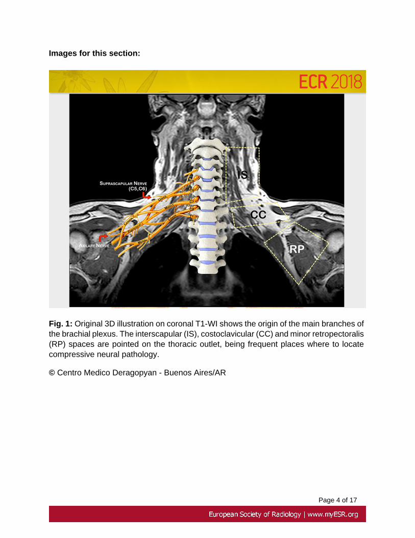
Fig. 1: Original 3D illustration on coronal T1-WI shows the origin of the main branches ofthe brachial plexus. The interscapular (IS), costoclavicular (CC) and minor retropectoralis(RP) spaces are pointed on the thoracic outlet, being frequent places where to locatecompressive neural pathology.
© Centro Medico Deragopyan - Buenos Aires/AR
Page 5 of 17
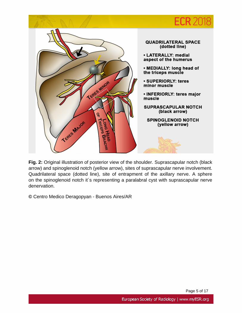
Fig. 2: Original illustration of posterior view of the shoulder. Suprascapular notch (blackarrow) and spinoglenoid notch (yellow arrow), sites of suprascapular nerve involvement.Quadrilateral space (dotted line), site of entrapment of the axillary nerve. A sphereon the spinoglenoid notch it´s representing a paralabral cyst with suprascapular nervedenervation.
© Centro Medico Deragopyan - Buenos Aires/AR
Page 6 of 17
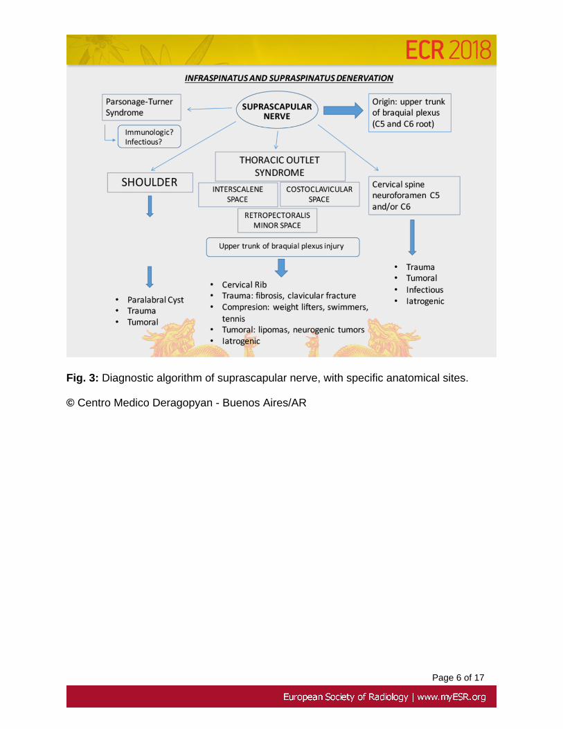
Fig. 3: Diagnostic algorithm of suprascapular nerve, with specific anatomical sites.
© Centro Medico Deragopyan - Buenos Aires/AR
Page 7 of 17
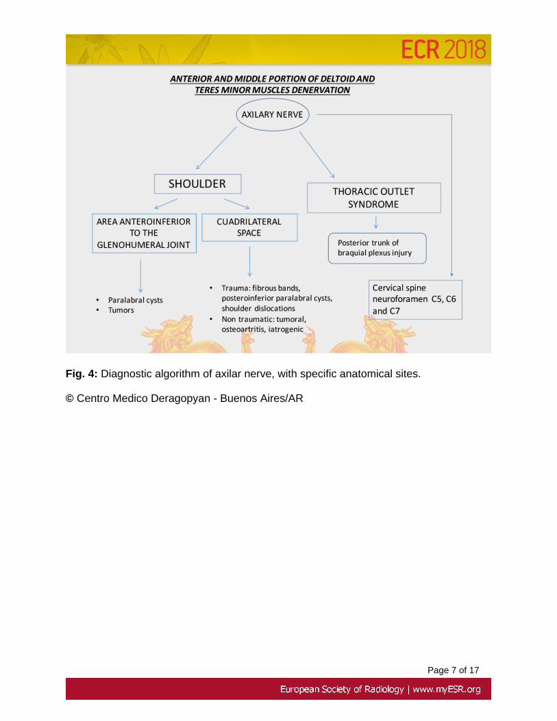
Fig. 4: Diagnostic algorithm of axilar nerve, with specific anatomical sites.
© Centro Medico Deragopyan - Buenos Aires/AR
Page 8 of 17
Findings and procedure details
The entrapment and compression neuropathies are usually diagnosed on the basis ofclinical history and electrophysiologic studies (electromyography and nerve conductionstudies). MRI has proved significantly useful since it allows to identify the precise locationof the injury depending on the affected muscle group and has a remarkable advantageover the EMG, which does not show signs of muscle denervation up to 2-3 weeks afterinjury (Linda, D. et al.¹)
The denervated muscles show us the possible nerve compromised and guide us to findthe location of the nerve injury.
The finding of high signal intensity in T2 and STIR secuences depicting muscleedema, represents an acute lesion. Fatty atrophy reflects chronic denervation, showinghiperintense signal in T1 and T2.
The muscles which usually present a denervatory pattern are: infraspinatus,supraspinatus, teres minor and deltoid.
Parsonage-Turner Syndrome (Fig. 5 and 6)
Also know as acute brachial neuritis and neuralgic amyotrophy.
Etiology: Not clear. Autoimmune mechanisms? Viral?.
Self-limited. 3rd-8th decade. Bilateral 30%.
Characteristic manifestation:
Acute and severe shoulder pain followed by weakness.
Most common muscle involved is supraspinatus (97% of cases, and 50% isolatedaccording to Gaskin et al.³).
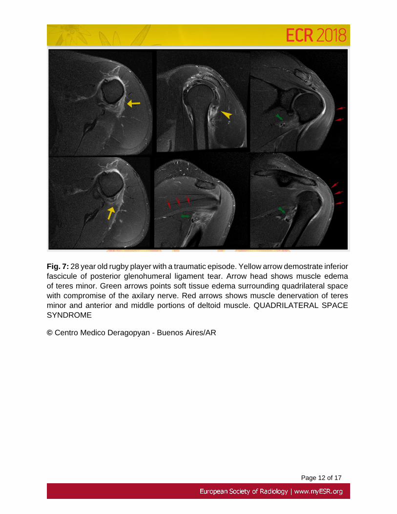
Quadrilateral Space Syndrome (Fig. 7)
Due to compression of the axillary nerve and posterior circumflex humeral artery,resulting in tener minor and anterior and medial portions of deltoid muscle denervation.
Page 9 of 17
Etiology: Traumatic and atraumatic causes
Mass lesions such as tumors, posteroinferior paralabral cysts or fibrous bands (mostfrequent), posteroinferior paralabral acute lesion with edema (fig. ), osteoartritis, etc.
Post-traumatic nerve injury may occur in almoust half of shoulder dislocations, since thenerve is stretched over the dislocated humeral head.
Most axillary nerve injuries are secondary to brachial plexus impairment.
Isolated injury may occur in open surgical intervention with a deltoid muscle-splittingapproach or reverse shoulder arthroplasty.
Suprascapular Nerve Entrapment
Sites of entrapment include
Suprascapular notch: supraspinatus and infraspinatus denervation (fig. 9).
Spinoglenoid notch: infraspinatus denervation.
2nd - 6th decades.
Etiology:
Compression from space-occupying lesions, including paralabral cysts (fig. 9), tumors,iatrogenic surgical injury, and enlarged varicosities.
Thickened transverse scapular ligament (repetitive strain on the abducted and externallyrotated shoulder), with entrapment on the suprascapular notch.
Thoracic Outlet Syndrome
Entrapment of the brachial plexus at specific anatomic places.
Sites of entrapment include the retropectoralis minor, costoclavicular, and interscalenespaces (fig. 1).
Etiology: Cervical Rib; trauma (fig. 8 and 9), fibrosis, clavicular fracture; compresion:weight lifters, swimmers, tennis; tumoral: lipomas, neurogenic tumors; Iatrogenic (Erb-Duchenne fig. 11).
Clinical presentation depends on the affected nerve, involving more frequently axillaryand supraspacular nerve.
Page 10 of 17
Images for this section:
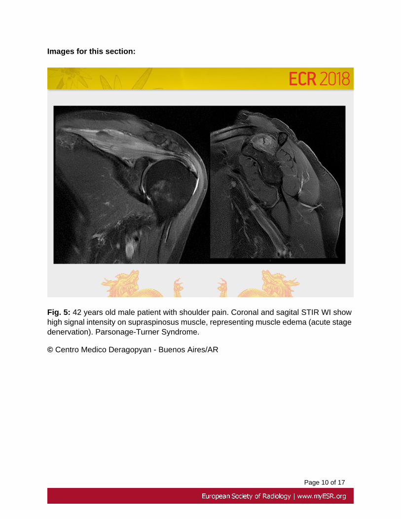
Fig. 5: 42 years old male patient with shoulder pain. Coronal and sagital STIR WI showhigh signal intensity on supraspinosus muscle, representing muscle edema (acute stagedenervation). Parsonage-Turner Syndrome.
© Centro Medico Deragopyan - Buenos Aires/AR
Page 11 of 17
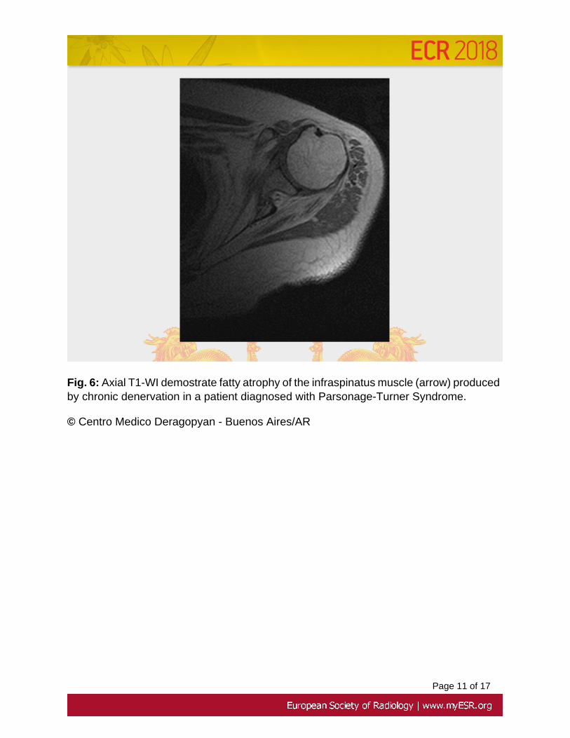
Fig. 6: Axial T1-WI demostrate fatty atrophy of the infraspinatus muscle (arrow) producedby chronic denervation in a patient diagnosed with Parsonage-Turner Syndrome.
© Centro Medico Deragopyan - Buenos Aires/AR
Page 12 of 17
Fig. 7: 28 year old rugby player with a traumatic episode. Yellow arrow demostrate inferiorfascicule of posterior glenohumeral ligament tear. Arrow head shows muscle edemaof teres minor. Green arrows points soft tissue edema surrounding quadrilateral spacewith compromise of the axilary nerve. Red arrows shows muscle denervation of teresminor and anterior and middle portions of deltoid muscle. QUADRILATERAL SPACESYNDROME
© Centro Medico Deragopyan - Buenos Aires/AR
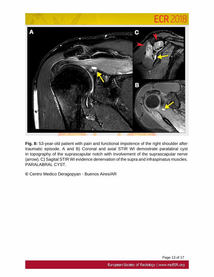
Page 13 of 17
Fig. 8: 53-year-old patient with pain and functional impotence of the right shoulder aftertraumatic episode. A and B) Coronal and axial STIR WI demostrate paralabral cystin topography of the suprascapular notch with involvement of the suprascapular nerve(arrow). C) Sagital STIR WI evidence denervation of the supra and infraspinatus muscles.PARALABRAL CYST.
© Centro Medico Deragopyan - Buenos Aires/AR
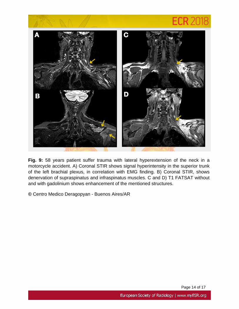
Page 14 of 17
Fig. 9: 58 years patient suffer trauma with lateral hyperextension of the neck in amotorcycle accident. A) Coronal STIR shows signal hyperintensity in the superior trunkof the left brachial plexus, in correlation with EMG finding. B) Coronal STIR, showsdenervation of supraspinatus and infraspinatus muscles. C and D) T1 FATSAT withoutand with gadolinium shows enhancement of the mentioned structures.
© Centro Medico Deragopyan - Buenos Aires/AR
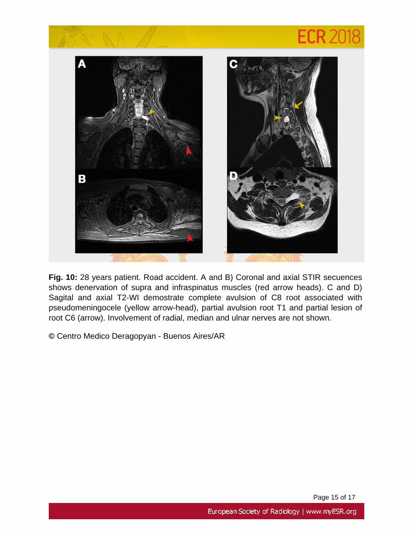
Page 15 of 17
Fig. 10: 28 years patient. Road accident. A and B) Coronal and axial STIR secuencesshows denervation of supra and infraspinatus muscles (red arrow heads). C and D)Sagital and axial T2-WI demostrate complete avulsion of C8 root associated withpseudomeningocele (yellow arrow-head), partial avulsion root T1 and partial lesion ofroot C6 (arrow). Involvement of radial, median and ulnar nerves are not shown.
© Centro Medico Deragopyan - Buenos Aires/AR
Page 16 of 17
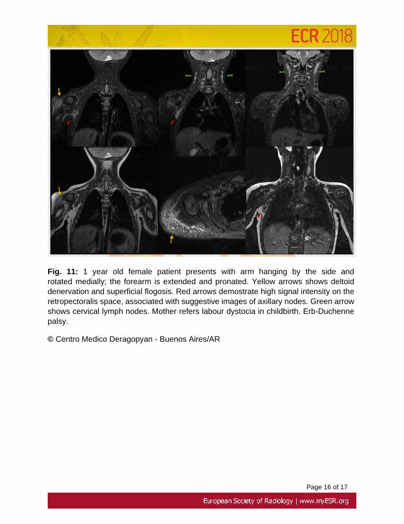
Fig. 11: 1 year old female patient presents with arm hanging by the side androtated medially; the forearm is extended and pronated. Yellow arrows shows deltoiddenervation and superficial flogosis. Red arrows demostrate high signal intensity on theretropectoralis space, associated with suggestive images of axillary nodes. Green arrowshows cervical lymph nodes. Mother refers labour dystocia in childbirth. Erb-Duchennepalsy.
© Centro Medico Deragopyan - Buenos Aires/AR
Page 17 of 17
Conclusion
The knowledge of the patterns of MRI muscle shoulder denervation in correlation with acorrect understanding of the anatomy helps to locate the topography of the nerve injuryand could determine the possible etiology cause in order to be attended.
Personal information
References
1. Linda, D., Harish, S., Stewart, B., & Finlay, K. (2010). Multimodality Imagingof Peripheral Neuropathies of the Upper Limb and Brachial Plexus 1.Radiographics, 6, 1373-1400. https://doi.org/10.1148/rg.305095169/-/DC1
2. Yanny, S., & Toms, A. P. (2010). MR patterns of denervation around theshoulder. American Journal of Roentgenology, 195(2), 157-163. https://doi.org/10.2214/AJR.09.4127
3. Gaskin, C. M., & Helms, C. A. (2006). Parsonage-Turner Syndrome: MRImaging Findings and Clinical Information of 27 Patients. Radiology, 240(2),501-507. https://doi.org/10.1148/radiol.2402050405
4. Muse R, L., & Contreras O, O. (2003). Sindrome De Parsonage-Turner O Neuritis Braquial: a Proposito De Dos Casos Clinicos.Revista Chilena de Radiología, 9(3), 137-139. https://doi.org/10.4067/S0717-93082003000300005
5. Safran, M. R. (2004). Nerve Injury about the Shoulder in Athletes, Part1: Suprascapular Nerve and Axillary Nerve. American Journal of SportsMedicine, 32(3), 803-819. https://doi.org/10.1177/0363546504264582