Embed Size (px)
Citation preview

MRI observation of the light-induced release of a contrast agent from photo-controllable
polymer micelles
This article has been downloaded from IOPscience. Please scroll down to see the full text article.
2007 Phys. Med. Biol. 52 N249
(http://iopscience.iop.org/0031-9155/52/10/N04)
Download details:
IP Address: 129.174.55.245
The article was downloaded on 31/05/2012 at 11:40
Please note that terms and conditions apply.
View the table of contents for this issue, or go to the journal homepage for more
Home Search Collections Journals About Contact us My IOPscience

IOP PUBLISHING PHYSICS IN MEDICINE AND BIOLOGY
Phys. Med. Biol. 52 (2007) N249–N255 doi:10.1088/0031-9155/52/10/N04
NOTE
MRI observation of the light-induced release of acontrast agent from photo-controllable polymermicelles
Martin Lepage1, Jinqiang Jiang2,3, Jerome Babin2, Bo Qi2,Luc Tremblay1 and Yue Zhao2
1 Departement de medecine nucleaire et de radiobiologie, Universite de Sherbrooke et Centred’imagerie moleculaire de Sherbrooke, Sherbrooke, Quebec, Canada2 Departement de chimie, Universite de Sherbrooke, Sherbrooke, Quebec, Canada
E-mail: [email protected] and [email protected]
Received 14 February 2007, in final form 10 April 2007Published 1 May 2007Online at stacks.iop.org/PMB/52/N249
AbstractThe encapsulation of molecules into nanocarriers is studied for its potential indelivering a high dose of anticancer drugs to a tumor, while minimizing sideeffects. Most systems either release their content in a non-specific manneror under specific environmental conditions such as temperature or pH. Wehave synthesized a novel class of photo-controllable polymer micelles that canstably encapsulate a hydrophilic compound and subsequently release it uponabsorption of UV light. Here, we describe an in vitro magnetic resonanceimaging assay that can evaluate the state of incorporation of a small Gd-basedcontrast agent. Our results indicate that the contrast agent alone can diffusethrough a filter, but that the same agent incorporated into micelles cannot. Afterexposure to UV light, the micelles released the contrast agent, which could thendiffuse through the filter.
1. Introduction
Using polymer nanocarriers for drug delivery has received much attention in recent years(Haag 2004). Targeting groups, such as antibodies, can be attached to the surface ofpolymer nanocarriers to deliver drugs at specific locations. The interest to use nanocarriersas vehicles for drug transport and release has been strengthened by recent reports that suchnanoscaled polymer assemblies may exploit the enhanced permeability of tumor blood vesselsto accumulate in solid tumors, and may be absorbed inside cells via endocytosis (Haag 2004).Currently, the main research effort in this field is dedicated to developing pH-responsivepolymer nanocarriers for which the drug release is triggered by changes in pH (Bae et al3 Present address: School of Chemistry and Material Engineering, Southern Yangtze University, People’s Republicof China.
0031-9155/07/100249+07$30.00 © 2007 IOP Publishing Ltd Printed in the UK N249

N250 M Lepage et al
2003, Gillies and Frechet 2003, Haag 2004). In most cases, the nanocarriers are stable atphysiological pH but break up at low pH leading to the release of either encapsulated orcovalently linked drugs (Bae et al 2003, Gillies and Frechet 2003, Haag 2004). pH-inducedphase transition was also used to disrupt polymer micelles for drug release (Taillefer et al2000). This general approach represents a step forward toward controlled drug delivery sincedrugs, in principle, are released only after the polymer nanocarrier reaches the inside of tumorcells or the tumor tissue where pH is acidic. However, most pH-sensitive micelles can bedisrupted by a range of acid pH values, which means that targeted release on a specific sitehaving a specific pH value is difficult to achieve. We recently reported on the synthesis andthe characterization of novel photo-controllable polymer micelles. We showed the extent ofdisruption of the nanocarrier can be controlled by the intensity of irradiation which, in turn,controls the period of time over which the encapsulated drug is released (Jiang et al 2006,2007).
Magnetic resonance imaging (MRI) is a well-established method able to visualize softtissues in vivo with a high spatial resolution, which can be well below 300 µm in live animalsusing a high magnetic field scanner. A contrast between different tissues (e.g., betweennormal and tumor tissues) can be enhanced by contrast agents (CAs) that often contain theparamagnetic Gd atom (Merbach et al 2001). In addition, the kinetics of CA accumulationin and elimination from tumor sites can provide unique information on the tumor micro-environment, such as the transcapillary transfer rates and the extravascular extracellular volumefraction (Tofts and Berkowitz 1993, Tofts et al 1995, 1999) both correlated with the tumorstage.
Liposomes containing hydrophobic or amphiphilic MRI CAs have been developed already(Gløgård et al 2000, Grant et al 1987, Kabalka et al 1988, Magin et al 1986, Navon et al1986). Their encapsulation in polymeric micelles has also been demonstrated (Torchilin 2002).However, normal polymer micelles comprising a hydrophobic core and a hydrophilic coronaare not suitable for encapsulating hydrophilic, commercially available, MRI CAs.
We anticipate a photo-controllable nanocarrier system able to encapsulate a drug, anda diagnostic imaging agent could be useful in the visualization of the site-specific releaseprocess. In such a case, the extent of nanocarrier disruption could be assessed in real timewhile the system is exposed to photons of the appropriate wavelength.
In this contribution, we demonstrate that a new type of photo-controllable reverse polymermicelle can encapsulate a hydrophilic MRI CA, and that this CA can be released after expositionof the micelles to UV light. More specifically, we observed a MRI signal enhancement outsidea filter tube containing the CA alone or containing micelles mixed with CA and exposed toUV, but not containing intact micelles mixed with CA.
2. Materials and methods
2.1. Synthesis, encapsulation and illumination of micelles
The basic chemical structure of the polymer forming the photo-controllable shell-crosslinkedreverse micelle (SCRM) is shown in figure 1(a). It was prepared based on an amphiphilicdiblock copolymer composed of a hydrophilic block of poly(ethylene oxide) (PEO) anda hydrophobic block being a random copolymer of coumarin methacrylate and methylmethacrylate, referred to as PEO-b-P(CMA-co-MMA). Details on the synthesis of this diblockcopolymer using atom transfer radical polymerization (ATRP) were reported elsewhere (Jianget al 2007) so only the salient features will be described here. A sample of PEO-b-P(CMA-co-MMA) containing ∼112 EO, 8 CMA and 20 MMA units was used in the present study.

Release of MRI contrast agents from photo-controllable micelles N251
Figure 1. (a) The chemical structure of the polymer forming PNBMA-grafted SCRM, and itsphotolysis upon UV illumination giving rise to PMA-grafted SCRM. (b) Schematic illustration ofthe stable encapsulation of a hydrophilic MRI contrast agent and its release upon UV illumination.
(This figure is in colour only in the electronic version)
Its reverse micelle was first obtained by dissolving the diblock copolymer in n-pentanol whilestirring (0.5 mg ml−1); the micellar solution was then exposed to UV light (λ > 320 nm,Novacure UV-Vis spot curing system) to fully dimerize the pendant coumarin groups resultingin a SCRM. The SCRM was collected through precipitation in hexane. Afterwards, driedSCRM was dissolved in chlorobenzene (50 mg in 2 ml), together with 2-nitrobenzylmethacrylate (NBMA, 1 g), CuBr (106 mg) and N,N,N′,N′,N′′-pentamethyldiethylenetriamine(PMDETA, 130 mg), and the whole was heated to 100 ◦C (2 days) for ATRP of the NBMAmonomer. After being dissolved in 200 ml of chloroform and washed with water until therewas no color in the water layer, the product was a SCRM whose shell surface was graftedwith the hydrophobic poly (2-nitrobenzyl methacrylate) (PNBMA). It is denoted as PEO-b-P(CMA-co-MMA)-graft-PNBMA hereafter. As shown in figure 1(a), upon UV illumination(λ > 320 nm), the photolysis of NBMA units converts the hydrophobic PNBMA grafts intohydrophilic poly(methacrylic acid) (PMA) grafts (Jiang et al 2006). This is at the origin of thelight-induced release described below. 1H NMR spectra suggest the grafting of 470 units ofNBMA on the SCRM, while dynamic light scattering (DLS) measurements found an averageparticle size of 183 nm for PEO-b-P(CMA-co-MMA)-graft-PNBMA and an average size of156 nm after the UV photolysis resulting in PEO-b-P(CMA-co-MMA)-graft-PMA.
Figure 1(b) illustrates our strategy of using PEO-b-P(CMA-co-MMA)-graft-PNBMA toload a hydrophilic MRI CA and to subsequently release it with an illumination. The CAused was gadolinium tetraphenylporphyrin (GdTPP). GdTPP was prepared by dissolving10 mg of sulfonated porphine (0.01 mmole) and 7.9 mg of gadolinium (III) bromide hydrate(0.02 mmole) in 1 ml of water/THF (1/1, v/v) with stirring at 60 ◦C overnight. A typical
N252 M Lepage et al
loading experiment was as follows. Ten mg of PEO-b-P(CMA-co-MMA)-graft-PNBMA wasdissolved under stirring in 2 ml of THF, in which PNBMA grafts were soluble. Then, 1 mlof the GdTPP solution (2 mg ml−1) was added to allow the hydrophilic PEO core to solubilizeGdTPP through diffusion. The solution was stirred at 60 ◦C for 48 h before 7 ml of waterwas added slowly; the solution was cooled to room temperature under stirring. THF andunloaded CA were removed by dialysis against water with stirring for 3 days (water wasfrequently refreshed). As depicted in figure 1(b), after dialysis, CA-loaded PNBMA-graftedSCRM dispersed in water was obtained. Being hydrophobic, PNBMA grafts should collapseon the surface of the crosslinked shell, which thickens the hydrophobic corona and prevents(or hinders) the release of GdTPP. Approximately, 2 ml of the micellar solution was exposedto UV light (Novacure spot curing system equipped with a 320 and 450 nm bandpass filter,total intensity 2000 mW) for 20 min to fully photolyze nitrobenzyl groups, under continuousstirring. Upon UV illumination, the PNBMA grafts become PMA grafts that, being watersoluble, allow GdTPP to diffuse across the crosslinked shell and be released into the aqueoussolution.
2.2. Magnetic resonance imaging.
MR images were acquired using a 7 T Varian animal scanner with a bore size of 210 mm.Samples were inserted in a volume coil having an inner diameter of 63 mm. Thirty-twoconsecutive sets of T1-weighted axial gradient echo images were acquired using the followingparameters: TR/TE 250/2.3 ms, flip angle 40◦, NA 24, FOV 6 × 6 cm2, matrix 1282, ten slicesand a slice thickness of 2 mm. The spin-lattice relaxation time constant T1 was determined atthe end of the experiment using an inversion-recovery spin-echo sequence with the inversiontimes (170, 350, 600, 1000, 3000 ms), TR/TE 6000/20 ms and NA 4. The time course wasextracted from the images by manually drawing a circular ROI covering 37 pixels on themiddle of each sample. The mean value is reported while the standard deviation from themean was normally below 1%.
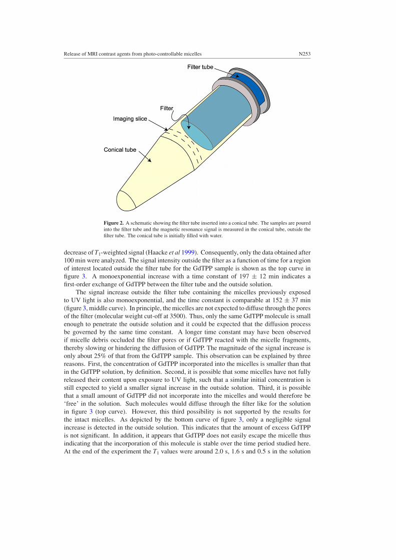
For this study, a GdTPP-loaded PNBMA-grafted SCRM aqueous solution (0.5 mg ml−1)was divided into two containers, one was exposed to the UV lamp (hereafter referred to as‘exposed micelles’) while the second remained intact (hereafter referred to as ‘intact micelles’).For comparison, a third container was filled with a solution of GdTPP, without any micelles.Samples were poured in a 0.5 ml filter tube (Slide-A-Lyzer MINI Dialysis Unit, PierceBiotechnology) with molecular weight cut-off at 3500, which was subsequently inserted intoa 1.5 ml plastic conical tube (EppendorfTM) filled with tap water at room temperature (22 ◦C)(see figure 2). The tubes were then sealed with a flexible film (ParafilmTM) and secured intothe volume coil for imaging. An extra plastic tube containing only water was imaged as acontrol. The time delay between the insertion of the filter tube into the conical tube and thebeginning of the serial imaging acquisition was 15 min. A feedback warm air system wasused to keep the air around the samples at 27 ◦C throughout the experiments.
3. Results and discussion
The signal of T1-weighted image for diluted solutions of a paramagnetic compound iscorrelated to the spin-lattice relaxation rate constant R1 (R1 = 1/T1) of the solution. Fordilute paramagnetic solutions, this rate constant is proportional to the concentration of thecontrast agent (Toth et al 2001). A signal decrease in the water tube was observed for thefirst 100 min. This is attributed to the temperature increase of the samples in the magnetbore in the presence of a flow of warm air at 27 ◦C, leading to a decrease of R1 and thus to a
Release of MRI contrast agents from photo-controllable micelles N253
Figure 2. A schematic showing the filter tube inserted into a conical tube. The samples are pouredinto the filter tube and the magnetic resonance signal is measured in the conical tube, outside thefilter tube. The conical tube is initially filled with water.
decrease of T1-weighted signal (Haacke et al 1999). Consequently, only the data obtained after100 min were analyzed. The signal intensity outside the filter as a function of time for a regionof interest located outside the filter tube for the GdTPP sample is shown as the top curve infigure 3. A monoexponential increase with a time constant of 197 ± 12 min indicates afirst-order exchange of GdTPP between the filter tube and the outside solution.
The signal increase outside the filter tube containing the micelles previously exposedto UV light is also monoexponential, and the time constant is comparable at 152 ± 37 min(figure 3, middle curve). In principle, the micelles are not expected to diffuse through the poresof the filter (molecular weight cut-off at 3500). Thus, only the same GdTPP molecule is smallenough to penetrate the outside solution and it could be expected that the diffusion processbe governed by the same time constant. A longer time constant may have been observedif micelle debris occluded the filter pores or if GdTPP reacted with the micelle fragments,thereby slowing or hindering the diffusion of GdTPP. The magnitude of the signal increase isonly about 25% of that from the GdTPP sample. This observation can be explained by threereasons. First, the concentration of GdTPP incorporated into the micelles is smaller than thatin the GdTPP solution, by definition. Second, it is possible that some micelles have not fullyreleased their content upon exposure to UV light, such that a similar initial concentration isstill expected to yield a smaller signal increase in the outside solution. Third, it is possiblethat a small amount of GdTPP did not incorporate into the micelles and would therefore be‘free’ in the solution. Such molecules would diffuse through the filter like for the solutionin figure 3 (top curve). However, this third possibility is not supported by the results forthe intact micelles. As depicted by the bottom curve of figure 3, only a negligible signalincrease is detected in the outside solution. This indicates that the amount of excess GdTPPis not significant. In addition, it appears that GdTPP does not easily escape the micelle thusindicating that the incorporation of this molecule is stable over the time period studied here.At the end of the experiment the T1 values were around 2.0 s, 1.6 s and 0.5 s in the solution

N254 M Lepage et al
100 150 200 250 300 350 400
4
5
6
7
8
9
MR
SIG
NA
L I
NT
EN
SIT
Y (
arb.
uni
ts)
INTACT
UV
GdTPP alone
TIME (minutes)
Figure 3. MR signal intensity (arbitrary units) as a function of time in the solution outside the filtertube containing either GdTPP alone (top curve), micelles containing GdTPP after disruption fromthe exposition to UV light (middle curve) and intact micelles containing GdTPP (bottom curve).Monoexponential fits (top and middle) and a linear fit (bottom) are shown as solid lines.
outside the filter tube containing the intact micelles, the exposed micelles and the GdTPPsolution, respectively.
4. Conclusions
We studied with MRI the diffusion of GdTPP through a filter under three different conditions.A comparable time constant was obtained for diffusion from a free GdTPP solution andfrom a solution with a photo-controllable micelle into which GdTPP was incorporated, afterexposition to UV light. Conversely, only a negligible signal change was observed from thesame photo-controllable micelle without exposition to UV light. The assay described is thusable to monitor the state of encapsulation of a contrast agent in photo-controllable micelles.
Acknowledgments
This study was supported by The Cancer Research Society Inc. ML is the Canada ResearchChair in magnetic resonance imaging. YZ thanks Mrs Rodica Plesu (Laval University) forDLS measurements.
References
Bae Y, Fukushima S, Harada A and Kataoka K 2003 Design of environment-sensitive supramolecular assemblies forintracellular drug delivery: polymeric micelles that are responsive to intracellular pH change Angew. Chem.,Int. Ed. Engl. 42 4640–3

Release of MRI contrast agents from photo-controllable micelles N255
Gillies E R and Frechet J M 2003 A new approach towards acid sensitive copolymer micelles for drug delivery Chem.Commun. 14 1640–1
Gløgård C, Hovland R, Fossheim S L, Aasen A J and Klaveness J 2000 Synthesis and physicochemical characterisationof new amphiphilic gadolinium DO3A complexes as contrast agents for MRI J. Chem. Soc. Perkin Trans. 21047–52
Grant C W M, Barber K R, Florio E and Karlik S 1987 A phospholipid spin label used as a liposome-associated MRIcontrast agent Magn. Reson. Med. 5 371–6
Haacke E M, Brown R B, Thompson M R and Venkatesan R 1999 Magnetic Resonance Imaging: Physical Principlesand Sequence Design (New York: Wiley)
Haag R 2004 Supramolecular drug-delivery systems based on polymeric core-shell architectures Angew. Chem., Int.Ed. Engl. 43 278–82
Jiang J, Qi B, Lepage M and Zhao Y 2007 Polymer micelles stabilization on demand through reversible photo-cross-linking Macromolecules 40 790–2
Jiang J, Tong X, Morris D and Zhao Y 2006 Toward photocontrolled release using light-dissociable block copolymermicelles Macromolecules 39 4633–40
Kabalka G W, Buonocore E, Hubner K, Davis M and Huang L 1988 Gadolinium-labeled liposomes containingparamagnetic amphipathic agents: targeted MRI contrast agents for the liver Magn. Reson. Med. 8 89–95
Magin R L, Wright S M, Niesman M R, Chan H C and Swartz H M 1986 Liposome delivery of NMR contrast agentsfor improved tissue imaging Magn. Reson. Med. 3 440–7
Merbach A E and Toth E 2001 The Chemistry of Contrast Agents in Medical Magnetic Resonance Imaginged A E Merbach (Chichester: Wiley)
Navon G, Panigel R and Valensin G 1986 Liposomes containing paramagnetic macromolecules as MRI contrastagents Magn. Reson. Med. 3 876–80
Taillefer J, Jones M, Brasseur N, van Lier J E and Leroux J 2000 Preparation and characterization of ph-responsivepolymeric micelles for the delivery of photosensitizing anticancer drugs J. Pharm. Sci. 89 52–62
Tofts P S and Berkowitz B A 1993 Rapid measurement of capillary permeability using the early part of the dynamicGd-DTPA MRI enhancement curve J. Magn. Reson. B 102 129–36
Tofts P S, Berkowitz B and Schnall M D 1995 Quantitative analysis of dynamic GD-DTPA enhancement in breasttumors using a permeability model Magn. Reson. Med. 33 564–8
Tofts P S et al 1999 Estimating kinetic parameters from dynamic contrast-enhanced T1-weighted MRI of a diffusabletracer: standardized quantities and symbols J. Magn. Reson. Imaging 10 223–32
Torchilin V P 2002 PEG-based micelles as carriers of contrast agents for different imaging modalities Adv. DrugDeliv. Rev. 54 235–52
Toth E, Helm L and Merbach A E 2001 Relaxivity of gadolinium(iii) complexes: theory and mechanism The Chemistryof Contrast Agents in Medical Magnetic Resonance Imaging ed A E Merbach and E Toth (Chichester: Wiley)pp 45–119