Embed Size (px)
DESCRIPTION
MRI- Magnetic resonance imaging. By Prashil Patel. What is MRI. Test that uses a magnetic field and pulses of radio wave energy to make pictures of organs and structures inside the body. Body is placed inside a special machine that contains a strong magnet. Cont …. - PowerPoint PPT Presentation
Citation preview
By Prashil Patel
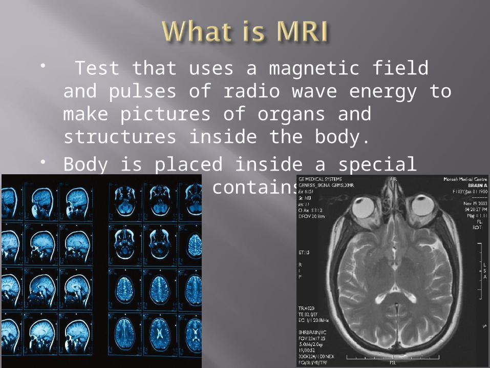
Test that uses a magnetic field and pulses of radio wave energy to make pictures of organs and structures inside the body.
Body is placed inside a special machine that contains a strong magnet.
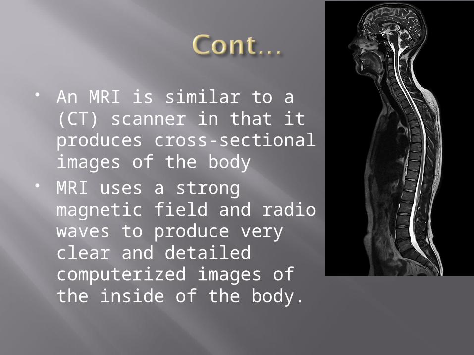
An MRI is similar to a (CT) scanner in that it produces cross-sectional images of the body
MRI uses a strong magnetic field and radio waves to produce very clear and detailed computerized images of the inside of the body.
Felix Bloch of Stanford University and Edward Purcell of Harvard University made the first successful nuclear magnetic resonance, NMR, experiment to study chemical compounds in 1946
NMR creates magnetic fields and radio waves cause atoms to give off tiny radio signals.
In the early 1980s, the first "human" magnetic resonance imaging (MRI) scanners became available, producing images of the inside of the body.
Used to find problems such as TumorsBleeding InjuryBlood vessel diseasesInfection
MRI scan can be done forHeadChestBlood vesselsAbdomen and pelvisBones and joinsspine
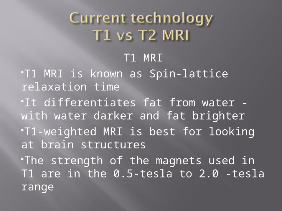
T1 MRIT1 MRI is known as Spin-lattice relaxation timeIt differentiates fat from water - with water darker and fat brighterT1-weighted MRI is best for looking at brain structuresThe strength of the magnets used in T1 are in the 0.5-tesla to 2.0 -tesla range
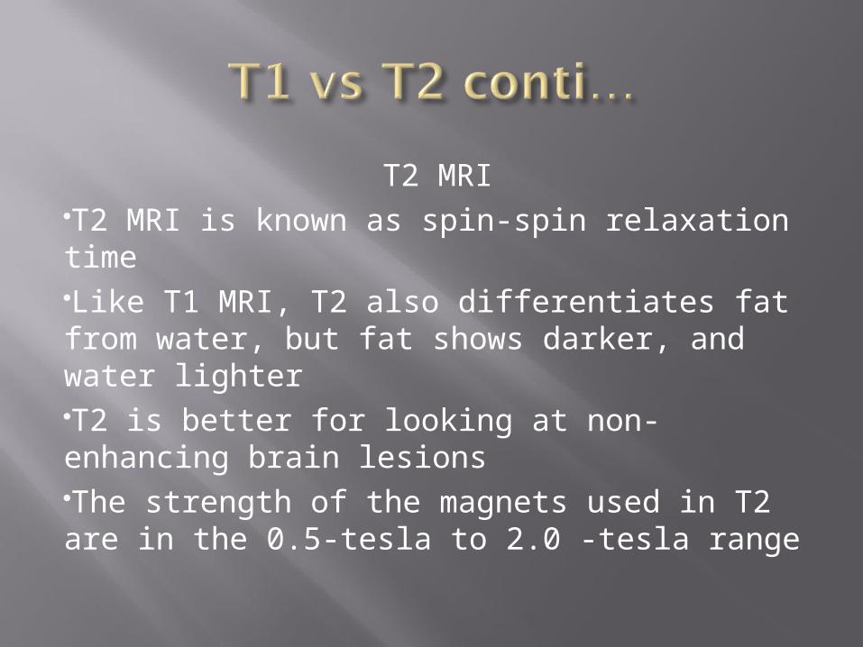
T2 MRIT2 MRI is known as spin-spin relaxation timeLike T1 MRI, T2 also differentiates fat from water, but fat shows darker, and water lighterT2 is better for looking at non-enhancing brain lesionsThe strength of the magnets used in T2 are in the 0.5-tesla to 2.0 -tesla range
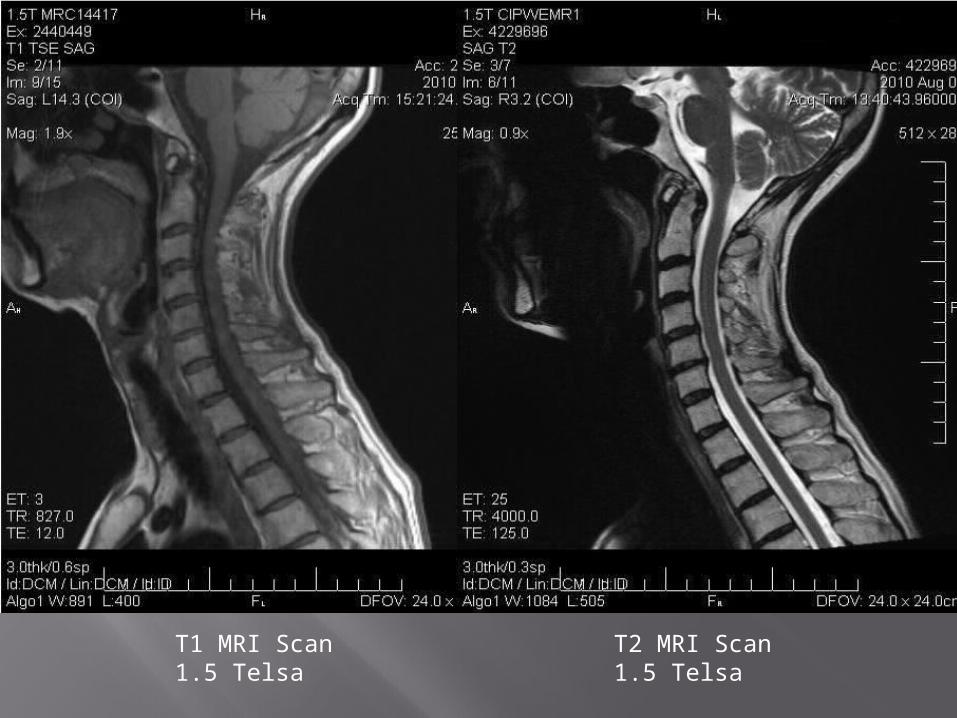
T1 Image T2ImageT1 MRI Scan1.5 Telsa
T2 MRI Scan1.5 Telsa
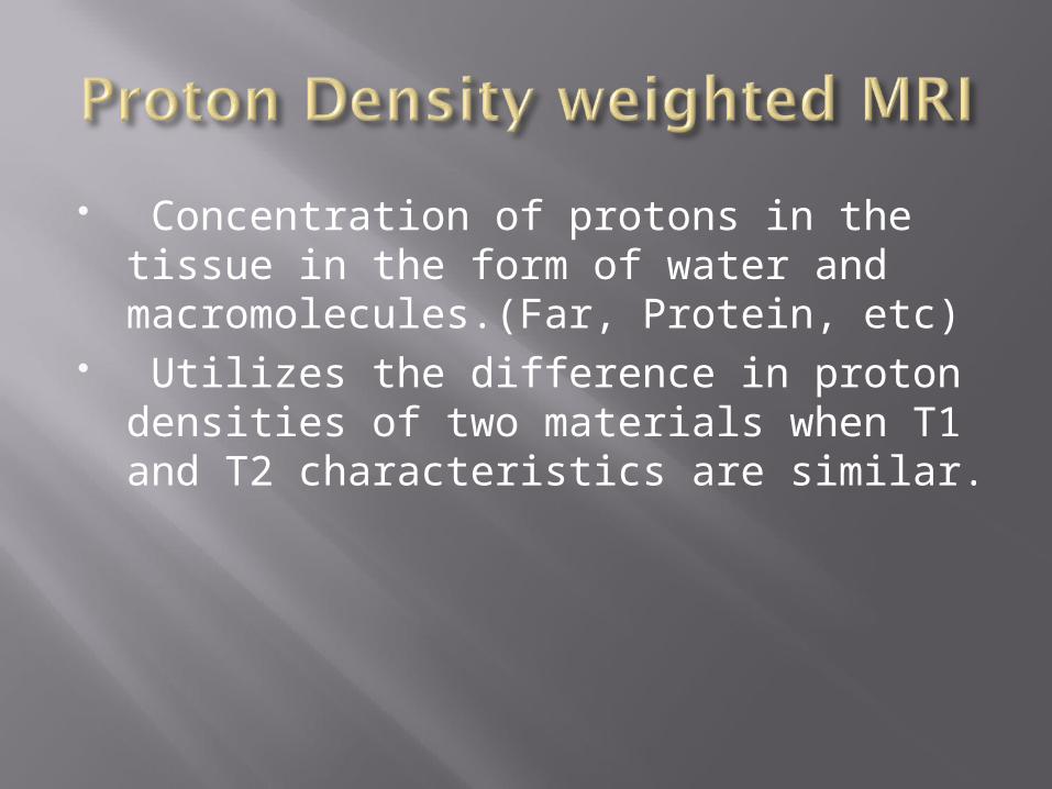
Concentration of protons in the tissue in the form of water and macromolecules.(Far, Protein, etc)
Utilizes the difference in proton densities of two materials when T1 and T2 characteristics are similar.
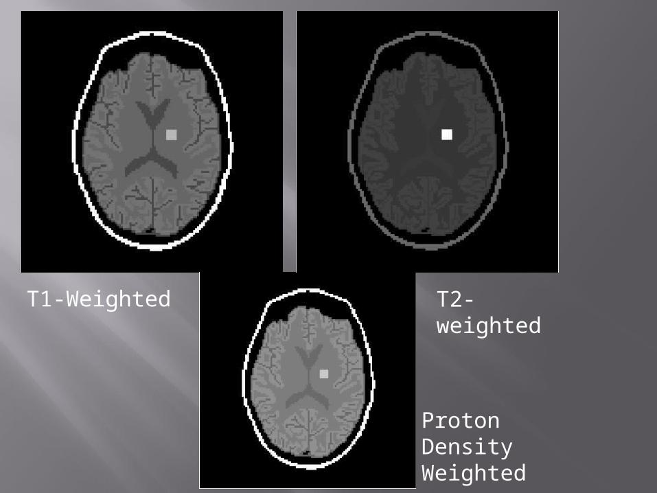
T1-Weighted T2-weighted
Proton Density Weighted
Because of the small space given, some patients experience claustrophobia have difficulty in cooperating during the study.
Some obese patients cannot be examined
Patients with pacemakers and certain ferromagnetic appliances cannot be studied
MRI equipment is expensive to purchase, maintain, and operate
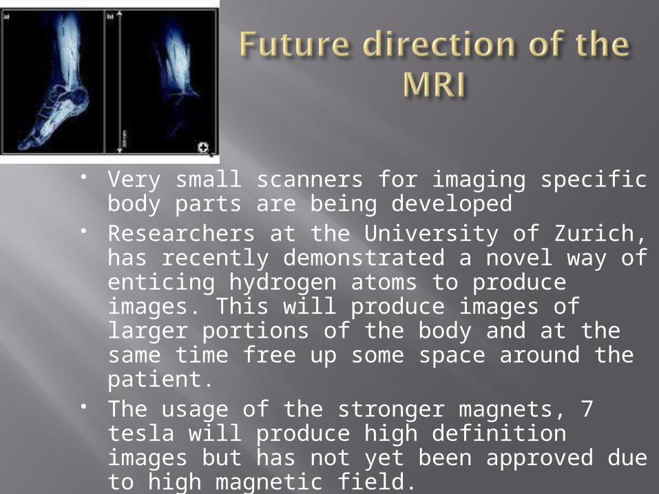
Very small scanners for imaging specific body parts are being developed
Researchers at the University of Zurich, has recently demonstrated a novel way of enticing hydrogen atoms to produce images. This will produce images of larger portions of the body and at the same time free up some space around the patient.
The usage of the stronger magnets, 7 tesla will produce high definition images but has not yet been approved due to high magnetic field.
Department of Health and Human Services
University of Zurich article Discovery Fit & Health Pubmed.gov http://www.medhelp.org http://srinivasarao.webs.com