Embed Size (px)
Citation preview

IOSR Journal of Dental and Medical Sciences (IOSR-JDMS)
e-ISSN: 2279-0853, p-ISSN: 2279-0861.Volume 15, Issue7 Ver. V (July 2016), PP 100-114
www.iosrjournals.org
DOI: 10.9790/0853-15075100114 www.iosrjournals.org 100 | Page
MRI Brain in Perinatal Hypoxia – A Case Series.
Dr. Sanjay Mhalasakant Khaladkar1, Dr. Aditi M.Gujarathi
2, Dr. Vigyat Kamal
3,
Dr. Anubhav Kamal4, Dr. Rajesh Kuber
5
1Professor, Department of Radiology,
2Senior Resident, Department of Radiology,
3PG Student, Department of Radiology,
4Senior Resident, Department of Radiology,
5Professor, Department of Radiology, & Dr. DY Patil Medical College & Research Center, Pimpri, Pune
Abstract:
Background: HIE is one of the most common cause of neonatal deaths and morbidity in children. Due to
limited facilities in India, many a times HIE goes undetected only to present in later stages of life as
handicapped children.
Objective: To study the Magnetic Resonance Imaging (MRI) brain findings in children having suffered
perinatal hypoxia, to analyze MRI findings in brain based on time of perinatal hypoxia.
Material and Methods: A prospective Study was conducted on 100 patients suffered with perinatal hypoxia
on Siemens 1.5 Tesla MAGNATOM AVANTO machine. Patterns of hypoxic ischemic brain injury in preterm
and term infants of were evaluated.
Results: Out of 100 cases, 72% were abnormal and 28% were normal studies. 36 %patients were preterms and
64% had term delivery. In patients with term delivery, 28.1 % were normal and 71.8 % were abnormal. In
patients with preterm delivery, 72.3% cases were found to be abnormal while 27.7 % cases did not show any
significant abnormality.
Conclusion: MRI was a definitive diagnostic modality in perinatal hypoxia. Patterns of brain injury were
determined by nature, timing and severity of insult. Imaging appearances were influenced by sequences used
and time from injury.
I. Introduction
Hypoxic Ischemic Encephalopathy (HIE) refers to a combination of hypoxia, hypercarbia and
metabolic acidosis as a consequence of occlusion of umbilical vessels or interference with placental perfusion in
fetal life and/or due to lack of effective breathing after birth. Hypoxia literally means decreased oxygen supply
and ischemia is a resultant damage to the tissues.Thus any cause which will lead to lack of oxygen supply to the
brain tissues which is extremely necessary for the normal functioning of the brain and thus in turn the other
body system; it will give rise to a series of events that will lead to injury to the brain tissues and certain clinical
manifestations. Almost 40% of deaths under age 5 years in children occur in the neonatal period. Reported
global totals of neonatal deaths due to non-specific conditions of HIE vary from 0.7 million to 1.6 million per
year. Mortality rate in children with severe hypoxia is 25–50%. Incidence of prenatal asphyxia is approximately
3.3% in India. It is usually related to gestational age and birth weight. [1]
Thus, outcomes of HIE are dangerous
and permanent, making it a major burden for the patient, family and the society. Thus, it is very important to
identify, evaluate and analyze the effects of HIE on the brain so as to develop better therapeutic strategies to
decrease the brain injury in newborns and provide them a better future.
II. Materials and Methods A prospective study was conducted on 100 patients in the department of Radio-diagnosis, Padmashree
Dr. D.Y Patil Medical College from July 2012 over a period of 2years to September 2014. The study included
all the patients of paediatric age group (birth to12 years of age) referred for MRI having suffered from perinatal
hypoxia. It included neonates with clinical signs of bradycardia, hypotonia and who gave poor response
to stimulation. In children; developmental delay, cerebral palsy, seizures, hemiplegia/paraplegia/quadriplegia
were the criteria used for performing the MRI. Patients who were on MRI incompatible life support were
excluded. Patients were evaluated by Siemens 1.5 Tesla MAGNATOM AVANTO machine. The bore size was
60 cm and the overall length was 160cm. The MRI system used zero Helium boil off technology. A 32 channel
head coil with iPAT compatibility was used. The proforma was designed on the objective of the study and it was
used after modification.A structured proforma was used to note the detailed clinical history, birth history,
developmental history, immunization details, anthropometry, physical examination findings, systemic
examination findings and investigation findings.Patient was positioned in the supine position and a head coil
was placed around the head to obtain a uniform signal to noise ratio. MRI examination of the brain was
performed in axial, sagittal and coronal planes. A Sagittal acquisition through the mid Sagittal plane was used as

Mri Brain in Perinatal Hypoxia – A Case Series.
DOI: 10.9790/0853-15075100114 www.iosrjournals.org 101 | Page
an initial localizer for subsequent axial and coronal planes. T1 (TR/ TE, 210–710/6–14 ms, flip angle 90°,
matrix- 320 X 320) and T2 (TR/ TE, 1550–5800/90–200ms, 150°, matrix- 320 X 320220 X 320) weighted
images were obtained. Fluid Attenuation Inversion recovery (FLAIR) (TR/ TE, 8050/120 –130/1860–2000ms,
flip angle -150°, matrix- 256 X 320), Diffusion Weighted, Gradient Echo and IR sequences were also
performed. ADC maps of brain were obtained in axial planes. DWI was done by using single-shot spin-echo
echo-planar sequences (TR/TE, 5130–5000/74–68 ms with a b-value of 800–1000 s/mm2, matrix- 190 X 190).
The conventional MR brain protocol included T1-weighted sequences, T2-weighted sequences and Gradient
Echo (GRE) (TR/ TE, 700/26, flip angle-20°, matrix- 132 X 192) sequences. Field of view of 230 mm and slice
thickness of 5mm for routine and 3 mm for IR sequences was used. T1-weighted and inversion recovery
sequences provided excellent anatomic information as well as high contrast between gray and white matter. T2-
weighted sequences provided good contrast resolution between gray matter, unmyelinated white matter and
myelinated white matter. Gradient Echo (GRE) sequences provided increased sensitivity for the detection of T2-
weighted magnetic susceptibility. Three-dimensional T1 IR sequence (3DT1 IR) (TR/TE, 4000-4120/70-74ms,
flip angle-150°,matrix- 154 X 192) and allows thin 1 mm contiguous slices which can be reconstructed in any
anatomic plane. T1-weighted IR sequence is helpful as it gives excellent images of brain anatomy and
maturation. It helps in differentiating myelinated and unmyelinated WM.
III. Observations and Results In our study of 100 cases, the lesions which were accurately diagnosed by MRI included pathologies
such as germinal matrix hemorrhage, periventricular leukomalacia, cystic encephalomalacia, cerebral and
cerebellar atrophy, corpus callosal thinning and agenesis and delayed myelination. Thus, in diagnosis of these
conditions MRI was a definitive diagnostic modality. MRI was able to diagnose these conditions in 72% of the
patients. However, in rest of the 28 % patients, MRI was normal, even though the patient had a history of
perinatal hypoxia or was symptomatic.
Figure-1 Distribution of total number of cases studied
Sex Wise Distribution
In this study MRI of the brain was done in 100 patients out of whom 56 were males and 46 were
females as seen in the chart below. Thus, males were more commonly affected than females.
Figure-2:Sex distribution of children suffered with perinatal hypoxia.

Mri Brain in Perinatal Hypoxia – A Case Series.
DOI: 10.9790/0853-15075100114 www.iosrjournals.org 102 | Page
Age Wise Distribution
The age of patients ranged from 0 years to 12 years. It is clear from the table below that maximum
pathologies were seen in patients who were in the age group of 1 to 5 years.
Figure-3 Age wise distribution of children suffered with perinatal hypoxia.
Gestational Age Wise Distribution
In our case study, 36 patients had a history of preterm delivery and 64 patients were term neonates.
Figure - 4 Distribution of cases according to gestational age.
Distribution of Risk Factors for HIE
There are various risk factors for HIE in a preterm or term neonate. Amongst all maternal and fetal
factors no specific cause was found in around 25% of the cases. It was followed by pre eclampsia and anemia.
The other causes have been charted below.
Risk Factors Number Percentage (n =100)
Indiopathic 25 25 %
Pre eclampsia 20 20 %
Anemia 15 15 %
Placental Factors 10 10 %
Perinatal Infection 8 8 %
Assisted Delivery 7 7 %
Caesarean Section 15 15 %
Table -1 Distribution of risk factors for HIE.
Distribution Of Cases According To The Gravid Status Of The Mothers
Information of gravid status, antenatal examination, hospital or home delivery was obtained of all the mothers of
100 patients.
Gravid status Distribution (n=100)
Primigravida 55 (55%)
No antenatal examination 44 (80%)
Undergone antenatal examination 11 (20%)
Home deliveries 15 (27.2%)
Hospital deliveries 40 (72.8%)
Multigravida 45 (45%)
No antenatal examination 33 (75%)
Undergone antenatal examination 12 (25%)
Home deliveries 14 (31.1%)
Hospital deliveries 31 (68.9%)
Table-2 Distribution of Cases According To the Gravid Status ofthe Mothers.
Mri Brain in Perinatal Hypoxia – A Case Series.
DOI: 10.9790/0853-15075100114 www.iosrjournals.org 103 | Page
Imaging Findings in Term Neonates
64 cases of term neonates who suffered perinatal hypoxia underwent MRI, out of which 46 cases were
abnormal (71.8%). Of them, 36 showed white matter (cortical and/or periventricular) T2 hyperintensities, which
was the most common abnormality found. Other abnormalities were cerebral atrophy, cystic encephalomalacia,
delayed myelination, corpus callosum thinning, cerebellar atrophy, acute infarcts. 18 cases were normal (28.1
%).
Findings Number Percentage (n =64)
White matter hyperintensities 36 60 %
Cerebral atrophy 32 50 %
Delayed myelination 20 31.2 %
Corpus callosum thinning 14 21.8 %
Cystic encephalomalacia 24 37.5 %
Acute infarcts 10 15.6 %
Cerebellar atrophy 10 15.6 %
Normal 18 28.1 %
Table-3MRI findings in HIE in term neonates.
Imaging Findings In Preterm Neonates
36 cases of preterm neonates who suffered perinatal hypoxia were evaluated in our study by MRI, out
of which 10 were normal (27.7 %) and 26 were abnormal (72.3 %). Of the abnormal studies, the most common
were periventricular leucomalacia (12) and cerebral atrophy (12).
Other pathologies which were observed were delayed myelination, corpus callosum thinning, cystic
encephalomalacia, acute infarcts and cerebellar atrophy.
Findings Number Percentage (n =36)
Periventricular leukomalacia 12 33.3 %
Cerebral atrophy 12 33.3 %
Delayed myelination 8 22.2 %
Corpus callosum thinning 8 22.2 %
Cystic encephalomalacia 8 22.2 %
Acute infarcts 4 11.1 %
Cerebellar atrophy 2 5.5 %
Germinal Matrix Hemorrhage 2 5.5 %
Normal 10 27.7 %
Table-4MRI findings in preterm neonates.
Distribution Of Cases According To Symptom Complex
In this study (n=100), maximum cases were of developmental delay (32). The other symptom complexes with
which patients presented were seizures, cerebral palsy, hypotonia and drowsiness.
Table-5Distribution of cases according to symptom complex
Figure-5Distribution of cases according to symptom complex
Sr. No. Pathology No. of cases Percentage
1) Developmental delay 32 32%
2) Seizures 28 28%
3) Cerebral palsy 20 20%
4) Hypotonia 14 14%
5) Drowsiness 6 6%
Total=100

Mri Brain in Perinatal Hypoxia – A Case Series.
DOI: 10.9790/0853-15075100114 www.iosrjournals.org 104 | Page
Distribution of Findings on MRI
Out of 72 cases of HIE findings in perinatal hypoxia, maximum number of cases showed generalized
cerebral atrophy (61 %), followed by T2WI hyperintensities. The other findings in preterm as well as term
neonates were cerebellar atrophy, cystic encephalomalacia, delayed myelination, corpus callosum thinning,
acute infarcts.
Sr. No. Pathology No. of cases(n=72) Percentage
1) T2 hyperintensity 36 50%
2) Cerebral atrophy 44 61%
3) Cystic encephalomalacia 32 44%
4) Delayed myelination 28 39%
5) Corpus callosum thinning 24 33%
6) Acute Infarcts 14 19%
7) Cerebellar atrophy 12 17%
8) Periventricular leukomalacia 12 17%
9) Germinal matrix hemorrhage 2 0.3%
Table 6- Distribution of MRI findings.
Distribution of Findings on MRI According to Areas of Brain Involved:
Sr. No. Site Preterm/Term MRI Findings No. of patients
1. Cortex Term
Preterm
-High signal intensity on T2 and FLAIR
- Infarct - Atrophy
- Atrophy
12
6 32
12
2. Subcortical white matter Term -High signal intensity on T2 and FLAIR 20
3. Periventricular White matter Preterm -High signal intensity on T2 and FLAIR 12
4. Basal ganglia and thalami Preterm Term
-High signal intensity on T2 and FLAIR -Infarct
4 3
5. Hippocampus Term -High signal intensity on T2 and IR 4
6. Cerebellum Preterm -High signal intensity on T2 and FLAIR
-Atrophy
3
2
7. Insula Term -Infarct 1
Table 7- Distribution of findings on MRI according to areas of brain involved
Distribution of Findings on MRI in Patients with Cerebral Palsy
Out of 100 cases of perinatal hypoxia, 20 cases presented with cerebral palsy. Detailed analysis of MRI
findings in these 20 cases is done as follows. 6 (30 %) cases were preterm infants and 14 (70 %) cases were term
infants. Periventricular leukomalacia was the commonest finding in preterm infants, whereas, hyperintense
lesions on T2WI images was the most common findings in term infants. The rest of the findings are given in the
table below.
Table 8- Distribution of findings on MRI in patients with cerebral palsy.
Patterns of Hypoxic Ischemic Injury in Term Infants with Acute Presentation
Out of 64 patients with a history of term delivery, infants presented with acute presentation were 10 in
number. 5 main patterns of distribution of acute ischemic injury have been recognized. Of them, most showed a
watershed predominant pattern.
Mri Brain in Perinatal Hypoxia – A Case Series.
DOI: 10.9790/0853-15075100114 www.iosrjournals.org 105 | Page
Sr.No. Pattern No. of patients (n=10)
1 Basal ganglia thalamus pattern (BGT) 2
2 Watershed predominant pattern of injury (WS) 4
3 White cerebrum pattern 1
4 Periventricular white matter pattern 2
5 Perinatal arterial ischaemic stroke (PAIS) 1
Table9- Distribution of patterns of hypoxic ischemic injury in term infants.
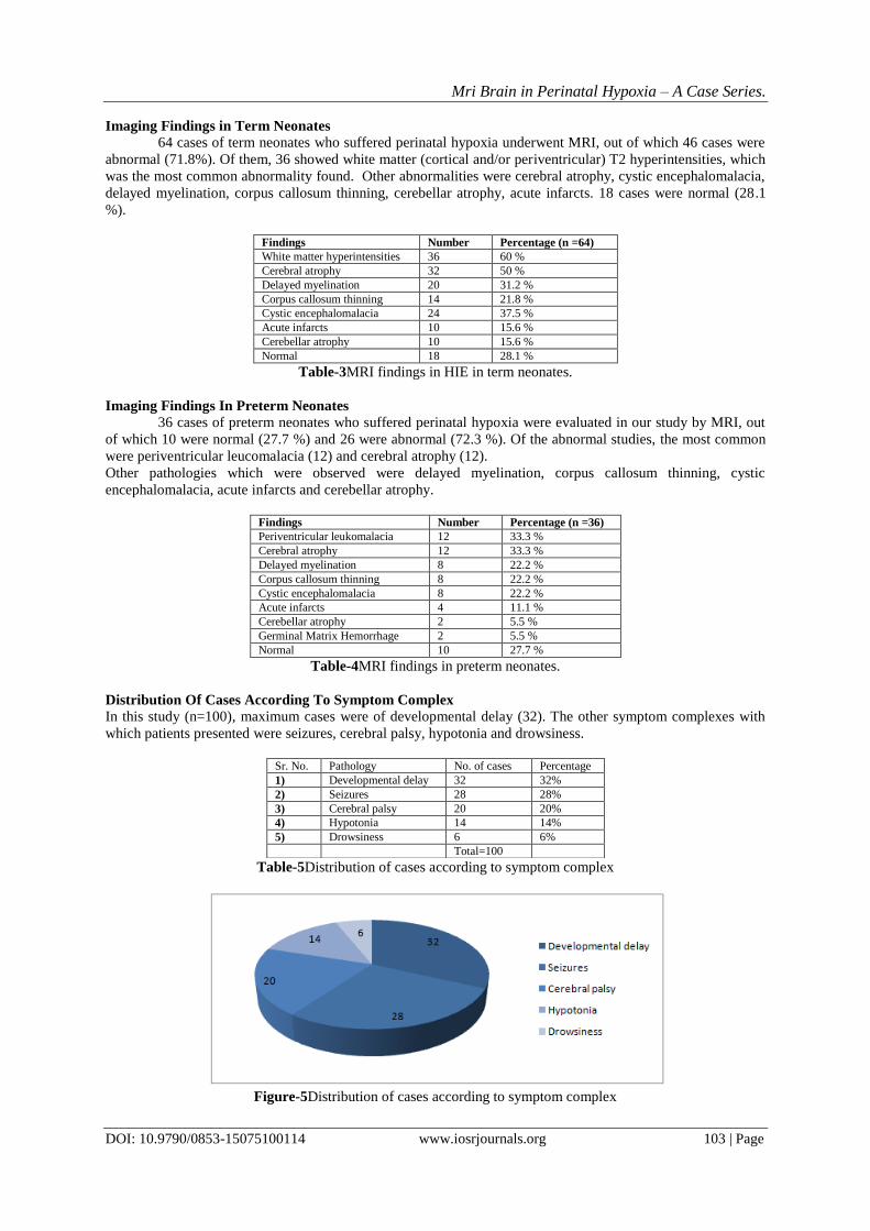
Patterns of Hypoxic Ischemic Injury in Patients with Acute Presentation
Of the total 100 patients, infants that presented with acute presentation with a history of a term delivery
were 10 in number and those with a history of preterm delivery were 4 in number. Risk factors, HIE grades and
MRI findings in 14 patients of acute presentation of HIE are analyzed as follows.
Sr. No. GA HIE Grade
(Sarnat and
Sarnat)
Risk factors MRI Findings (n=14)
1 Preterm I Idiopathic Periventricular white matter pattern
2 Preterm II PROM Periventricular white matter pattern
3 Preterm III Pre-eclampsia Basal ganglia- Thalami and Periventricular
white matter involvement with
4 Preterm III Assessed Deliveries Basal ganglia- Thalami involvement
5 Term I Idiopathic WM lesions
6 Term I Idiopathic WS predominat injury
7 Term I Pre-eclampsia WM lesions
8 Term I Anemia PLIC , WS predominat injury
9 Term II Idiopathic PLIC , WS predominat injury
10 Term II Pre-eclampsia WM lesions
11 Term II Anemia PAIS
12 Term II Antepartum Hemorrhage WS predominat injury
13 Term III Pre-eclampsia Basal ganglia- Thalami involvement
14 Term III Antepartum Hemorrhage Basal ganglia- Thalami involvement
Table10- Patterns of hypoxic ischemic injury in patients with acute presentation.
IV. Discussion Hypoxic-ischemic encephalopathy (HIE) in neonates is an important cause of mortality and morbidity
and neurodevelopmental delay worldwide. It can lead to permanent brain damage and can also cause damage to
other tissues of the body.
Almost 40% of deaths in children under the age 5 years occur in the neonatal period. Decrease in the
mortality rate due to HIE observed in the developed countries could be due to the improved neonatal care,
however the mortality and morbidity due to HIE in the developing countries still remains high and is a
challenge. The total neonatal deaths occurring in the world due to non-specific conditions of HIE vary from 0.7
million7 to 1.6 million per year. In children affected by severe hypoxia, the mortality rate is reportedly 25–50%.
The maximum number of deaths occur in the first week of life as a result of multi- system failure or inadequate
care. The children having severe neurologic deficit die in their infancy from aspiration pneumonia or various
infections. Incidence of prenatal asphyxia is about 3.3% in India and is usually related to gestational age and
birth weight. It occurs in 9% of infants less than 36 weeks gestational age and in 0.5% of infants more than 36
weeks of gestational age accounting for 20% of perinatal deaths (or as high as 50% deaths if still births are
included). [1]
Treatment for children suffered with hypoxic-ischemic encephalopathy was limited to supportive care
for a long time, but now recent advances for effective therapies have been developed. It is a treatable problem
and early identification and intervention is necessary to prevent the long term brain damage.Advances in MRI
technique have made excellent progress over the last few years.MRI and MRS with the help of DTI is useful in
detection of exact patterns of early and late damage in infants with HIE.
Both terminal zones of myelination and perivntricularleukomalacia appear hyperintense on T2WI and
FLAIR and their differentiation is extremely difficult. Terminal zones are the persistent areas of high signal
intensity in the white matter lateral to the bodies of the lateral ventricles and in the dorsal and superior to the
ventricular trigones on T2-weighted images. The difference is best elicited on coronal T2WI and FLAIR images.
The periventricular leukomalacia lesions are more sharply defined. They are present more inferiorly; lateral to
the trigones and near the optic radiations. They are typically brighter on T2WI and FLAIR sequences than
terminal zones of myelination. Periventricular leukomalacia is associated with loss of brain tissue, which results
in the irregularity of the ventricular wall, abnormally deep cortical sulci, sometimes extending down to the
ventricular surface, and thinning of the body of the corpus callosum. A layer of myelinated white matter is
present between the trigone of the ventricle and the terminal zones in normal patients. The very high signal

Mri Brain in Perinatal Hypoxia – A Case Series.
DOI: 10.9790/0853-15075100114 www.iosrjournals.org 106 | Page
intensity of the peritrigonal areas compared to surrounding white matter on FLAIR images with the presence of
local atrophy favor PVL. [2]
Differentiation of hypoxic-ischemic cerebral injury from normal myelination is important for
prediction of neurologic development. T1-weighted images help distinction of infants with hypoxic- ischemic
brain damage from those with normal myelination. The posterior limb of the internal capsule (PLIC) is
myelinated at birth and appears hyperintense on T1-weighted images. The loss of this normal high signal
intensity in the PLIC in infants with HIE, indicates a delay in myelination or insult to previously myelinated
tracts. This loss of high SI in the PLIC, though sometimes subtle, is associated with unfavourable outcome. [3]
Patterns of HIE in preterm and term neonates:
An ischemic event lasting for more than 10 minutes is needed to induce parenchymal changes. The
extent of injury increases with prolonged duration of the insult. Thus the patterns of injury in infants can be
divided into term and preterms depending upon gestational age and being mild to moderate and severe
hypoperfusion injuries, depending upon the severity of insult. [4]
Figure- 6 Patterns of brain injury in mild to moderate hypoperfusion. Schematic of the premature
neonatal brain (left) and that of the term infant (right) illustrates how the vascular supply changes with
maturation and affects the pattern of brain injury in HIE. The premature neonatal brain (left) has a
ventriculopetal vascular pattern, and hypoperfusion results in a periventricular border zone (red shaded area) of
white matter injury. In the term infant (right), a ventriculofugal vascular pattern develops as the brain matures,
and the border zone during hypoperfusion is more peripheral (red shaded area) with subcortical white matter and
parasagittal cortical injury.
(Source- Christeine P. Chao et al. Neonatal Hypoxia Ischemic Encephalopathy: Multimodality Imaging.
Radiographics 2006; 26:159-172.[5]
)
Age of Child Mild to Moderate
Hypotension
Profound Hypotension
Premature neonate (up to 34 postconceptional weeks)
Periventricular white matter injury
Thalamic, basal ganglia, and brainstem injury
Term neonate (~36 to ~56
postconceptional weeks)
Parasagittal
watershed injury
Dorsal brainstem, ventral cerebellar vermis, thalamus,
basal ganglia and perirolandic cortex injury
Older child (more than 4 to 6 postnatal months)
Parasagittal watershed injury
Basal ganglia and diffuse cortical injury
Table-11 Patterns Of Injury In Diffuse Hypoxic-Ischemic Injury
Linda S. de Vries&FlorisGroenendaal in 2014 identified four patterns of acute infarcts in term neonate. [6]
1) Basal ganglia pattern:
It affects bilateral central grey nuclei (ventro lateral thalami and posterior putamina) and perirolandic
cortex. It is generally seen following an acute event, such as a ruptured uterus, placental abruption or umbilical
cord prolapse, and is also referred to as a pattern following ‘acute near total asphyxia. The absence of a normal

Mri Brain in Perinatal Hypoxia – A Case Series.
DOI: 10.9790/0853-15075100114 www.iosrjournals.org 107 | Page
high-signal intensity of the posterior limb of the internal capsule (PLIC) on T1WI is highly predictive of severe
adverse sequelae. On spin echo MRI sequences, inversion of the signal within the PLIC can be seen from 48 to
72 h onwards. DWI will show early changes in the basal ganglia/thalami. Children with the BGT pattern of
injury tend to be severely disabled due to dyskinetic cerebral palsy (CP).
2) Watershed predominant pattern of injury (WS)
t is the type of pattern that can be seen after ‘prolonged partial asphyxia’. The vascular watershed zones
(anterior–middle cerebral artery and posterior–middle cerebral artery) are involved, affecting white matter and
in case of more severe insult also the overlying cortex. The lesions can be uni- or bilateral, posterior and/or
anterior. On spin echo MR images loss of the cortical ribbon and thus the grey–white matter differentiation can
be seen. DWI is however helpful in making an early diagnosis. A follow up MRI may show cystic evolution
with or without atrophy and gliotic changes. This type of insult is usually mild and thus the onset of
neurological signs can be delayed. Severe motor impairment is uncommon in this. Symptomatic parieto-
occipital epilepsy may occur later in childhood, often associated with reduced intelligence quotients and
visuospatial cognitive function.
3) White cerebrum pattern
Marked involvement of the subcortical white matter and cortex is noted with relative sparing of the
immediate periventricular white matter and central grey matter. This is referred to as the ‘white cerebrum’, as
DWI shows completely white cerebrum, contrasted to a normal looking cerebellum. This condition tends to be
fatal, but in case of survival, muticysticencephalomalacia eventually develops.
4) Periventricular white matter pattern.
It is similar to the punctate white matter lesions in the preterm infant. It is associated with a mild degree
of encephalopathy and fewer clinical seizures. Thispattern of brain injury is observed in newborn infants with
congenital heart defects.
5) Perinatal arterial ischemic stroke (PAIS), perinatal haemorrhagic stroke (PHS) and sinovenous
thrombosis.
It is seen in newborns presenting with encephalopathy and/or seizures. Restricted diffusion at the level
of the internal capsule and the middle part of the cerebral peduncle, referred to as ‘pre-Wallerian degeneration’
can be appreciated. It is then followed by Wallerian degeneration at 6–12 weeks and beyond. Presence of
Wallerian degeneration at birth suggests an antenatal onset of the insult.
We carried out a study on 100 patients of perinatal hypoxia presenting with various symptom
complexes. Of them, 72% (72) were abnormal and 28% (28) were normal studies. Our study correlates with
study carried out by M.A. Rutherford. [7]
Our findings correlate well with these studies.
In our study of 100 patients; 56% (56) were males and 46 % (46) were females with a mean age group
of 6 years (age range= 0 to 12 years). Maximum patients were of age group 1 to 5 years. In a study carried out
by AzharMunir Qureshi et al; 79.6% were males and 20.4% were females. [1]
In another study done by D.J.A.
Connolly, the age group range taken into consideration was 1 to 24 years. Of them maximum patients were
between the age group of 1 to 5 years. [8]
Thus, our findings corroborate well this study.
In our study 36 (36 %) patients were preterms and 64 (64%) had term delivery. These findings are in
accordance with study performed by R Yin. He carried out the study on 42 patients in whom 12 were premature
(28.5%) and 30 were full-term (71. 4%).[9]
In another study done by AzharMunir Qureshi et al on 181 infants,
out of 181 neonates 77.9% were full term, 19.1% were premature. Thus, overall, our findings correlate with
these studies. [1]
Various risk factors for HIE have been proposed over the years. In our study, PIH was commonest risk
factor 20 % (20) followed by anaemia 15 % (15). The other risk factors were placental factors 10% (10),
perinatal infections 8% (8), and assessed delivery 7% (7). No risk factors were identified in 25 % (25) cases.
Caesarean section was done in 15 % (15) of the mothers. In the study carried out by AzharMunir Qureshi et al
on 181 patients; the most common risk factor was PIH, observed in 27.7 % of the mothers, followed by anaemia
seen in 16%. Placental causes were present in 18.3%. Assessed delivery was done for 7.2 % of the patients and
in 15.5% no maternal cause was found. 34.3% were delivered by Caesarean section. [1]
Thus, our findings
corroborate well with this study.
Inclusion of antenatal examination, home delivery, hospital delivery was taken into consideration in
our study. We found that around 55 % (55)cases were primigravida. Of them, around 80% (44) patients had not
undergone any antenatal examination. Rest 20 % (11) had undergone antenatal examination. Home delivery
was done in 27.2% (15) of primigravida mothers, whereas hospital deliveries were done in 72.8% (40). In the
remaining 45 % of multigravida, 75% (33) patients did not undergo any antenatal checkup and the rest of the 25

Mri Brain in Perinatal Hypoxia – A Case Series.
DOI: 10.9790/0853-15075100114 www.iosrjournals.org 108 | Page
% (12) underwent antenatal examination. 31.1% (14) of multigravida had undergone home deliveries.Rest of the
68.9 % (31) underwent hospital deliveries. In the study done by AzharMunir Qureshi et al52.5% of the mothers
were primigravidaand of them 5% were managed at home. 47.5 % cases were multigravida and of them, 8 %
were home deliveries. Thus my study is in accordance with this study. [1]
We evaluated 64 (72 %) patients with a history of term delivery. Out of which 28.1 % (18) were
normal studies and 71.8 % (46) cases were abnormal. Out of the abnormal cases cortical and subcortical T2
hyperintensities were noted in 60% (36) cases. Cerebral atrophy was also common and found in 50 % (32)
cases. In patients presenting with acute symptoms, 15.6 % (10) cases showed acute infarcts. Delayed
myelination was seen in 31.2 % (20) of the patients. Mary Rutherford with his colleagues performed a study on
patients with HIE and tried to find the correlation of MR findings with clinical outcome. He found cortical and
subcortical T2 hyperintensities in 50 % of the patients and cerebral atrophy in 50 % of the cases. Basal ganglia
infarction was observed in 18 % cases and 6 % cases showed insular infarcts. Delayed myelination was present
in 50 % of the infants. [10]
The difference in the results of our study and other studies can be because of difference
in the sample size.
We studied 36 (28%) patients who were born with the history of preterm delivery. Of these, 72.3%
cases were found to be abnormal. 33.3% (12) cases showed periventricular leukomalacia and 33.3 % (12)
patients showed cerebral atrophy as the commonest findings. Delayed myelination and corpus callosum thinning
was observed in 22.2 % (8) cases. 27.7 % cases did not show any significant abnormality on MRI. Acute
infarcts were seen in 11.1 % (4) cases. Cerebellar atrophy was present in 5.5 % (2) cases. Germinal matrix
hemorrhage was diagnosed in 5.5 % (2) cases. In a study carried out by Gul Serdaroglu et al, 89 children with
PVL were evaluated. The aim of this study was to find out neurodevelopmental delay in children with
periventricular leukomalacia (PVL). PVL was divided into 3 grades: grade I, unilateral or bilateral areas of
periventricular hyperintensity; grade II, hyperintensity more than 3; grade III, hyperintense lesions more than 3
and ventricular wall irregularity; grade IV, diffuse PVL and ventricular dilatation. Thinning of the corpus
callosum and cortical atrophy was identified respectively in 73% and 47.2% of the patients. Delayed
myelination was noted in 14.3% cases. MRI was normal in 18 % of the infants. [11]
In a similar study done by
PavithraLogitharajah, the major sites of injury were basal ganglia (BG, 75%), white matter (89%), Cortex was
involved (58%) followed by brain stem in 44% cases No abnormality was found in 32%. Significant central
gray matter and brainstem injury was found in many preterm infants with HIE. Neonatal MRI findings allowed
accurate prediction of neurodevelopmental outcome on follow up studies. [12]
The difference in the results of our
study and other studies can be because of different age selection criteria of our study compared to other studies.
We came across various symptom complexes with which patients presented. Of them, maximum
patients presented with developmental delay 32% (32). This was followed by epilepsy seen in 28 % (28)
patients. Cerebral palsy was seen in 20 % (20) cases. Patients presenting with hypotonia were 14 % (14)
whereas 6 % (6) cases had an acute presentation in the form of drowsiness or altered sensorium. Maximum
cases of developmental delay were noted in the study performed by Gul Serdaroglu et al. Epilepsy was found in
33.7% and cerebral palsy in 30.8% of the patients. Approximately 28 % patients had diplegia. [11]
Thus, our
findings are in concordance with this study.
In our study, 20 patients presented with cerebral palsy. Out of these 20 cases 30 % (6) cases were
preterm infants. The most common finding that we encountered was periventricular leukomalacia seen in 50%
(3) of the cases. It was followed by cerebral atrophy and delayed myelination which was seen in 33.3 % (2)
cases. Corpus callosum thinning and infarcts were seen in 16.6 % (1) patients. Whereas 16.6 % (1) cases did not
show any abnormality. 70% (14) cases of term infants presenting with cerebral palsy were studied. The
commonest finding in term infants was hyperintense lesions on T2WI images seen in 64% (8) cases. The next
common finding was cerebral atrophy, which was present in 50 % of the cases (7). Delayed myelination and
acute infarcts were noted in 28.5 % (4) cases. The less common finding was corpus callosum thinning seen in
21.4 % (3) cases. No abnormality was seen in 21.4 % (3) of the cases. Our findings were similar to those studied
by R.Yin. He performed a study of MRI findings in 42 patients, of which 8 were premature (38 %), 13 were
full-terms (62%). Periventricular leukomalacia was seen in 66.6%. Cerebral atrophy was seen in 33.3% cases
and 33.3% children did not demonstrate PVL. The other group he studied included 30 children who were born
between 37 and 42 weeks gestation that is term infants. The MRI findings demonstrated T2 hyperintensities as
the most common finding seen in approx. 30 % patients. It was followed by cerebral atrophy seen in 20 % cases
and other lesions in 20 % cases. Acute infacrts were noted in 13 % of the patients. Delayed myelination was
seen in 7 % cases and corpus callosum thinning in 6 %. [9]
Charles L. Truwit did a study on 40 patients with
cerebral palsy. Of these, 11 were premature and 29 were term infants. Of the 11 patients born prematurely, MR
revealed deep white matter loss, especially in the peritrigonal regions. 81 % of the scans demonstrated thinning
of the corpus callosum. No abnormalities were noted in the basal ganglia or thalami. 27% had diminished
caliber of the brainstem. MR was done in 29 patients born at term, out of which 66% had diminished deep white
matter. 6 % had delayed myelination. In 55 %, the corpus callosum was thinned either focally (involving the

Mri Brain in Perinatal Hypoxia – A Case Series.
DOI: 10.9790/0853-15075100114 www.iosrjournals.org 109 | Page
posterior body and/or splenium) or diffusely. Ventricular abnormalities were seen in 41 % with an irregular
ventricular contour and ventricular enlargement and cerebral atrophy. Cortical thinning was seen in 10% term
patients, one of whom had findings diagnostic of multicysticencephalomalacia.20 % cases had basal ganglia
and/or thalami involvement. [13]
In another case series, RubaBenini studied 126 patients with cerebral palsy. Of
these, 71% had abnormal findings and 36 had normal brain scans. Compared with other CP types, normal-
appearing MRI was more prevalent in dyskinetic CP 72% and less prevalent in spastic hemiplegic CP
10%.Thus, genetic or functional, rather than gross structural lesions, may underlie the pathophysiology of CP.
[14]Overall our findings corroborate with both the studies.
We evaluated 5 patterns of distribution of acute ischemic brain injury in patients presenting with a
history of term delivery. Of the 64 patients with a history of term delivery, 15.6 % presented with acute
symptoms. The watershed predominant injury pattern was the most common finding observed in 40 % (4) of the
patients. Basal ganglia- Thalami pattern and periventricular white matter pattern was noted in 20 % (2) of the
cases respectively. 10% cases showed a white cerebrum pattern and Perinatal Arterial Ischemic Stroke (PAIS)
was seen in 10% cases.84Abnormal signal intensity appearing hypointese on T1WI images was noted in the
posterior limb of internal capsule (PLIC) was noted in 20 % (2) patients. This sign helped in differentiating the
normal myelination from hypoxic injury. [3]
We also studied patterns of acute injury in 11.1% (4 out of 36) patients with a history of preterm
delivery. Of them, 50 % (2) had an abnormal signal intensity in the bilateral periventricular white matter. 50%
patients showed Basal ganglia –Thalami involvement.
An overall interpretation of patient characteristics and findings on the imaging when compared with the
data available according to the review of literature corroborated very well. There are no significant and
inexplicable differences in the range/spectrum of MRI findings in patients presented with HIE.
V. Cases
Figure -7 Axial MRI T2WI (a), FLAIR (b) and (c) show hyperintense areas in the bilateral periventricular white
matter with no restricted diffusion on DWI (d) suggestive of HIE.
Figure-8 Altered signal is seen in bilateral high fronto-parietal region predominantly along cortex and
subcortical white matter and also extending to periventricular white matter appear hypointense on T1WI (a) and
c d

Mri Brain in Perinatal Hypoxia – A Case Series.
DOI: 10.9790/0853-15075100114 www.iosrjournals.org 110 | Page
hyperintense in T2WI (b) and FLAIR (c) with no evidence of restricted diffusion (d) suggestive of gliotic
changes. Ex vacuo dilatation of both lateral ventricles is also seen.
.
Figure-9 Midsagittal MR T1WI (a) and coronal 3D T1WI IR (b) images showing thinning of corpus callosum.
Figure 10 – Axial T2WI MR image showing delayed unmyelinated white matter appearing hyperintense in
bifrontal region.
Figure –11 Axial T1WI (a) and T2WI (b) MR images showing prominence of ventricular system and cortical
sulci suggestive of generalized cerebral atrophy.
Figure – 12 Mid sagittal (a) and axial (b)T1WI MR at the level of basal ganglia showing changes of cystic
encephalomalacia involving both the cerebral hemispheres showing CSF intensity. Note that the deep nuclei are
spared.

Mri Brain in Perinatal Hypoxia – A Case Series.
DOI: 10.9790/0853-15075100114 www.iosrjournals.org 111 | Page
Figure – 13: Multiple areas of cystic encephalomalacia are noted in bilateral cerebral hemispheres appearing
hyperintense on T2WI (a) and suppressed on FLAIR images (b). The basal ganglia and thalami are spared in this
case, thus confirming that it is a term infant.
Figure – 14 Left para sagittal T1WI (a), axial T2WI (b), axial FLAIR (c) and axial DWI (d) MR images at the
level of lateral ventricles showing area of cystic encephalomalacia involving the left MCA territory appearing
hypointense on T1WI and FLAIR, hyperintense on T2WI images with no restriction on DWI.
Figure -15 Axial T1WI (a) and T2WI (b) images showing areas of altered signal intensity in left watershed
areas appearing hypointense on T1WI and hyperintense on T2WI images. These areas are showing restriction on
DWI (c) with corresponding low ADC values (d). This was suggestive of acute infarct in left anterior and
posterior watershed areas suggestive of watershed pattern.

Mri Brain in Perinatal Hypoxia – A Case Series.
DOI: 10.9790/0853-15075100114 www.iosrjournals.org 112 | Page
Figure – 16 Areas of altered signal intensity are seen involving deep nuclei in a preterm infant appearing
hyperintense on T2WI (a) and FLAIR (b) images, showing restriction on DWI (c) with corresponding low ADC
values (d) suggestive of acute infacrts with basal ganglia pattern.
Figure -17 Axial MR T2WI (a) and DWI (b) images at the level of lateral ventricles show areas of altered signal
intensity involving the cerebral white matter with relative sparing of the periventricular white matter and deep
nuclei appearing diffusely hyperintense on T2WI images with corresponding restriction on DWI. This is
suggestive of acute infarct with the white brain pattern of injury.
Figure – 18 Axial DW MR Image at the level of lateral ventricles showing focus of restricted diffusion in the
left frontal periventricular white matter. This is suggestive of periventricular type of pattern of acute infarct.
Mri Brain in Perinatal Hypoxia – A Case Series.
DOI: 10.9790/0853-15075100114 www.iosrjournals.org 113 | Page
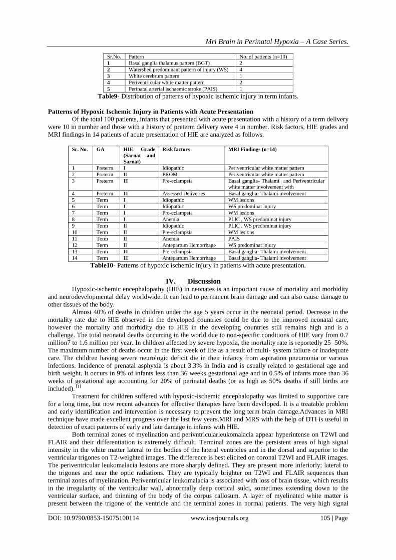
Figure –19 Axial T2WI (a) and DW (b) MR images at the level of lateral ventricles showing acute infarct
involving left MCA territory appearing hyperintense on T2WI and showing restriction on DWI. This is the
perinatal arterial ischemic stroke pattern.
Figure -20 Axial MR images at the level of lateral ventricles showing areas of altered signal intensity in
periventricular areas posteriorly appearing hypointense on T1WI (a). These areas do not show restriction on
DWI (b) suggestive of HIE.
Figure –21 Axial FLAIR MR images at the level of body of lateral ventricles (a) and at the level of corona
radiata (b) showing bilateral periventricular hyperintensities with reduced white matter in both occipital lobes
(forceps major) suggestive of periventricular leukomalacia.
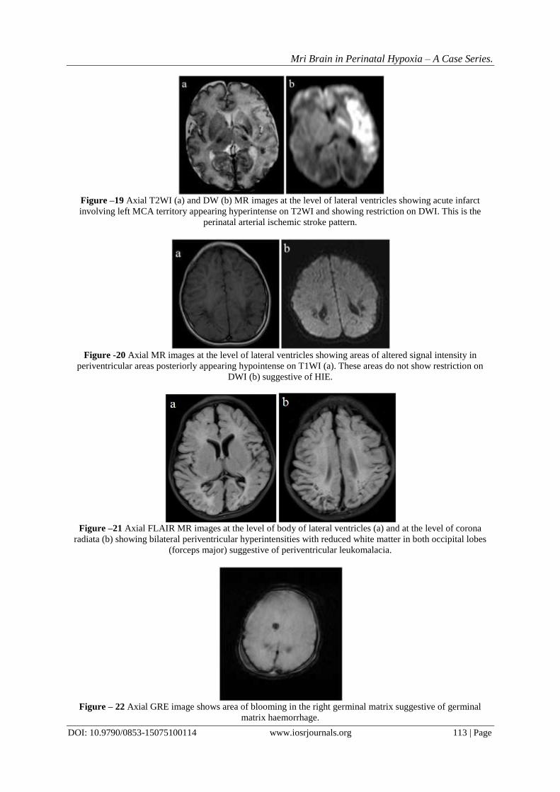
Figure – 22 Axial GRE image shows area of blooming in the right germinal matrix suggestive of germinal
matrix haemorrhage.
Mri Brain in Perinatal Hypoxia – A Case Series.
DOI: 10.9790/0853-15075100114 www.iosrjournals.org 114 | Page
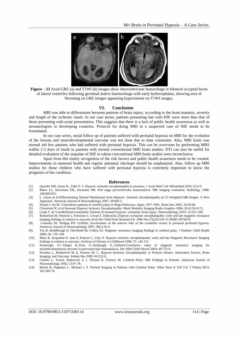
Figure – 23 Axial GRE (a) and T1WI (b) images show intraventricular hemorrhage in bilateral occipital horns
of lateral ventricles following germinal matrix haemorrhage with early hydrocephalus, showing area of
blooming on GRE images appearing hyperintense on T1WI images.
VI. Conclusion MRI was able to differentiate between patterns of brain injury, according to the brain maturity, severity
and length of the ischemic insult. In our case series, patients presenting late with HIE were more than that of
those presenting with acute presentation. This suggests that there is a lack of public health awareness as well as
neonatologists in developing countries. Protocol for doing MRI in a suspected case of HIE needs to be
formulated.
In our case series, serial follow up of patients suffered with perinatal hypoxia on MRI for the evolution
of the lesions and neurodevelopmental outcome was not done due to time constrains. Also, MRI brain was
normal in0 few patients who had suffered with perinatal hypoxia. This can be overcome by performing MRS
within 2-3 days of insult in patients with normal conventional MRI brain studies. DTI can also be useful for
detailed evaluation of the sequelae of HIE in whom conventional MRI brain studies were inconclusive.
Apart from this timely recognition of the risk factors and public health awareness needs to be created.
Improvements in maternal health and regular antenatal checkups should be emphasized. Also, follow up MRI
studies for those children who have suffered with perinatal hypoxia is extremely important to know the
prognosis of the condition.
References [1]. Qureshi AM, Anees R., Tahir S. S. Hypoxic ischemic encephalopathy in neonates. J Ayub Med Coll Abbottabad 2010; 22:4-9.
[2]. Baker LL, Stevenson DK, Enzmann DR. End stage periventricular leukomalacia: MR imaging evaluation. Radiology 1988;
168:809-815. [3]. L. Liauw et al.Differentiating Normal Myelination from Hypoxic- Ischemic Encephalopathy on T1-Weighted MR Images: A New
Approach. American Journal of Neuroradiology 2007; 28:660-5.
[4]. Suzuki J, Ito M. Coincidence patterns of cerebral palsy in Shiga Prefecture, Japan, 1977-1991. Brain Dev 2002; 24:39-48. [5]. Christeine PC.et al Neonatal Hypoxic Ischemic Encephalopathy: Multi Modality Imaging Radio Graphics 2006; 26:S159-S172.
[6]. Linda S. de Vries&FlorisGroenendaal. Patterns of neonatal hypoxic–ischaemic brain injury. Neuroradiology 2010; 52:555–566 [7]. Rutherford M, Pennock J, Schwieso J, Cowan F, DubowitzL.Hypoxic-ischaemic encephalopathy: early and late magnetic resonance
imaging findings in relation to outcome.Arch Dis Child Fetal Neonatal Ed. 1996 Nov;75(3):F145-51.PMID: 8976678.
[8]. Connolly DJ, Widjaja PD. Griffiths. Involvement of the anterior lobe of the cerebellar vermis in perinatal profound hypoxia. American Journal of Neuroradiology 2007; 28(1):16-9.
[9]. Yin R, Reddihough D, Ditchfield M, Collins KJ. Magnetic resonance imaging findings in cerebral palsy. J Paediatr Child Health
2000; 36: 139–144. [10]. Mary R, Jacqueline P, Jane S, Frances C, Lilly D. Hypoxic ischemic encephalopathy: early and late Magnetic Resonance Imaging
findings in relation to outcome. Archives of Disease in Childhood 1996; 75: 145-151
[11]. Serdaroglu G1, Tekgul H, Kitis O, Serdaroglu E, GokbenS.Correlative value of magnetic resonance imaging for neurodevelopmental outcome in periventricular leukomalacia. Dev Med Child Neurol 2004; 46:733-9.
[12]. Pavithra L, Rutherford M A, Frances M. C. Hypoxic-Ischemic Encephalopathy in Preterm Infants: Antecedent Factors, Brain
Imaging, and Outcome. Pediatr Res 2009; 66:222-9. [13]. Charles L. Truwit, Barkovich A J, Thomas K, Ferriero M. Cerebral Palsy: MR Findings in Patients. American Journal of
Neuroradiology 1992; 13:67-78.
[14]. Benini R, Dagenais L, Michael I. S. Normal Imaging in Patients with Cerebral Palsy: What Does It Tell Us? J Pediatr 2013; 162:369-74.