Embed Size (px)
Citation preview
MR Imaging and MR Angiography in the Evaluation of Pulsatile Tinnitus
R. Rex Dietz, Wayne L. Davis, H. Ric Harnsberger, John M. Jacobs, and Duane D. Blatter
PURPOSE: 1) To evaluate the scope of imaging findings seen with spin-echo MR and MR
angiography (MRA) in patients with pulsatile tinnitus; 2) to determine whether MRA adds additional imaging information (to that provided by spin-echo MR) necessary for determining the cause of pulsatile tinnitus; and 3) to suggest MR and MRA imaging techniques for evaluation of patients
with pulsatile tinnitus. METHODS: Forty-nine patients with pulsatile tinnitus were evaluated with
MR and MRA. Seventeen of these patients had conventional angiography. RESULTS: Vascular lesions or paraganglioma were demonstrated in 28 patients. Of these 28 lesions, the majority were
seen best (46%) or only (36%) on MRA. The spectrum of lesions detected included dural arteriovenous fistula (nine), extracranial arteriovenous fistula (three), paraganglioma (five), jugular
bulb variants (three), aberrant internal carotid artery (one), internal carotid artery stenosis (one), tortuous internal carotid artery (one), carotid dissection with pseudoaneurysm (one) , stenosis of
the transverse sinus (two), and arteriovenous malformation (two). CONCLUSIONS: MRA, in conjunction with spin-echo imaging, markedly enhances the ability of MR to diagnose the lesions responsible for pulsatile tinnitus.
Index terms: Magnetic resonance angiography (MRA); Magnetic resonance, comparative studies;
Hearing
AJNR Am J Neuroradiol 15:879-889, May 1994
The spectrum of lesions associated with pulsatile tinnitus makes imaging a challenge, because the differential diagnosis includes congenital and acquired vascular lesions and skull base tumors. In recent years high-resolution computed tomography (CT) with adjunctive angiography has served as the principal radiologic tool for evaluating patients presenting with pulsatile tinnitus (1-3). CT has been shown to be excellent for delineating the bony abnormalities that are associated with some of the vascular diseases and paragangliomas, but angiography is necessary to diagnose or exclude dural arteriovenous fistula (A VF) and intrinsic vascular abnormalities.
Received May 10, 1993; accepted pending revision August 14; revision received October 19.
From the Section of Neuroradiology, Department of Radiology, Uni
versity of Utah Medical Center (R.R.D., W.L.D., H.R.H., J .M.J.), and the Department of Radiology, LDS Hospital (D.D.S.), Salt Lake City, Utah.
Address reprint requests to Wayne L. Davis, MD, Section of Neuro
radiology, Department of Radiology, University of Utah Medical Center,
50 N Medical Dr, Salt Lake City, UT 84132.
AJNR 15:879-889, May 1994 0195-6108/ 94/ 1505-0879 © American Society of Neuroradiology
879
Conventional spin-echo magnetic resonance (MR) has been suggested to be of limited utility in this radiologic evaluation, because it shows poor resolution of both vascular and bony abnormalities (1, 2, 4).
Statistically the most common treatable causes of pulsatile tinnitus are paraganglioma (glomus tympanicum, jugulotympanicum, and jugulare) and dural AVF (1 ). Contrast-enhanced conventional spin-echo MR can diagnose paraganglioma readily (5). With the addition of MR angiography (MRA), MR can also suggest the diagnosis of dural A VF (6). It is possible then that MR with MRA will permit the screening of patients with pulsatile tinnitus with a single radiologic examination.
In this report we present our experience in MR and MRA imaging of 49 patients with pulsatile tinnitus. The goals of our study are to evaluate the scope of imaging findings seen with MR and MRA in patients with pulsatile tinnitus and to determine whether MRA adds additional imaging information (to that provided by conventional MR) necessary for determining the cause of pulsatile tinnitus. In addition, we will propose an
880 DIETZ
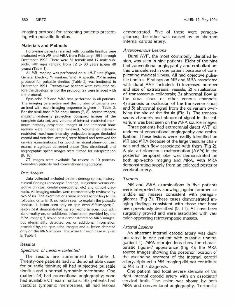
imaging protocol for screening patients presenting with pulsatile tinnitus.
Materials and Methods Forty-nine patients referred with pulsatile tinnitus were
evaluated with MR and MRA from February 1991 through December 1992. There were 31 female and 17 male subjects, with ages ranging from 12 to 80 years (mean 48 years) (Table 1 ).
All MR imaging was performed on a 1.5-T unit (Signa, General Electric, Milwaukee, Wis). A specific MR imaging protocol for pulsatile tinnitus (Table 2) was instituted in December 1991. Twenty-two patients were evaluated before the development of the protocol; 27 were imaged with the protocol.
Spin-echo MR and MRA was performed in all patients. The imaging parameters and the number of patients examined with each imaging sequence is given in Table 2. For the skull-base MRA acquisitions (7 , 8), source images, maximum-intensity projection collapsed images of the complete data set, and volume of interest-restricted maximum-intensity projection images of the temporal bone regions were filmed and reviewed. Volume of interestrestricted maximum-intensity projection images (included carotid and vertebral arteries) were filmed and reviewed for cervical examinations. For two-dimensional phase-contrast exams, magnitude-corrected phase (flow directional) and angiographic speed images were filmed for interpretation (9, 10).
CT images were available for review in 10 patients. Seventeen patients had conventional angiography.
Data Analysis
Data collected included patient demographics, history, clinical findings (otoscopic findings, subjective versus objective tinnitus, cranial neuropathy, etc) and clinical diagnosis. All imaging studies were retrospectively reviewed by two of us. The examinations were scored according to the following criteria: 0, no lesion seen to explain the pulsatile tinnitus; 1, lesion seen only on spin echo MR images; 2, lesion best demonstrated on spin-echo images, but with abnormality on, or additional information provided by, the MRA images; 3, lesion best demonstrated on MRA images, but abnormality detected on, or additional information provided by, the spin-echo images; and 4, lesion detected only on the MRA images. The score for each case is given in Table 1.
Results Spectrum of Lesions Detected
The results are summarized in Table 3. Twenty-one patients had no demonstrable cause for pulsatile tinnitus; all had subjective pulsatile tinnitus and a normal tympanic membrane. One (patient 44) had conventional angiography; none had available CT examinations. Six patients had vascular tympanic membranes; all had lesions
AJNR: 15, May 1994
demonstrated. Five of these were paragangliomas; the other was caused by an aberrant internal carotid artery.
Arteriovenous Lesions Dural AVF, the most commonly identified le
sion, was seen in nine patients. Eight of the nine had conventional angiography and embolization; this was deferred in one patient because of complicating medical illness. All had objective pulsatile tinnitus. Findings on MR and MRA associated with dural AVF included: 1) increased number and size of extracranial vessels; 2) visualization of transosseous collaterals; 3) abnormal flow in the dural sinus or other venous channels; 4) stenosis or occlusion of the transverse sinus; and 5) abnormal signal from the calvarium overlying the site of the fistula (Fig 1 ). The transosseous channels and abnormal signal in the calvarium was best seen on the MRA source images.
Three patients had extracranial direct AVF; all underwent conventional angiography and embolization. These lesions were readily identified on MR and MRA because of the large vascular channels and high flow associated with them (Fig 2) . A pial arteriovenous malformation (A V M) in the posterior temporal lobe was demonstrated on both spin-echo imaging and MRA, with MRA demonstrating supply from an enlarged posterior cerebral artery.
Tumors MR and MRA examinations in five patients
were interpreted as showing jugular foramen or middle ear masses consistent with paragangliomas (Fig 3). These cases demonstrated imaging findings consistent with those that have been previously described (5, 11). All have been surgically proved and were associated with vascular-appearing retrotympanic masses.
Arterial Lesions An aberrant internal carotid artery was dem
onstrated in one patient with pulsatile tinnitw. (patient 1 ). MRA reprojections show the charac .. teristic figure-7 appearance (Fig 4); the MRP. source images showing the posterior location o': the ascending segment of the internal carotid artery. Spin-echo MR imaging did not contribute to MR in this diagnosis.
One patient had focal severe stenosis of the right internal carotid artery with an associated cervical bruit. The lesion was shown by both MRA and conventional angiography. T ortuosit~'
AJNR: 15, May 1994 PULSATILE TINNITUS 881
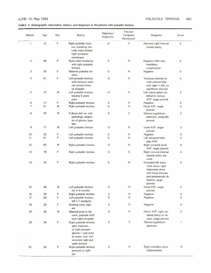
TABLE 1: Demographic information, history, and diagnoses in 49 patients with pulsatile tinnitus
Objective/ Vascular
Patient Age Sex History Tympanic Diagnosis Score Subjective
Membrane?
12 F Right pulsatile tinni- 0 y Aberrant right internal 4 tus , pulsating vas- carotid artery cular mass behind
right tympanic
membrane
2 68 F Right-sided headache s N Negative VBD with 0 with right pulsatile medullary
tinnitus compression
3 30 F Bilateral pu lsatile tin- s N Negative 0 nitus
4 61 F Left pu lsatile tinnitus 0 N Tortuous internal ca- 4 with tortuous inter- rotid arteries bilat-
nal carotid artery eral, right > left, no
on Doppler significant stenosis
5 12 M Left pulsatile t innitus 0 N Left suboccipital ver- 3 trauma 6 years tebral to venous
prior AVF, angio proved
6 17 F Right pulsatile tinnitus s N Negative 0 7 61 M Right pulsatile tinnitus 0 N Dural AVF, angio 4
proved
8 26 M Fullness left ear with s y Glomus jugulotym- 2 pathologic diagno- panicum, surgically
sis of glomus jugu- proved
lare
9 77 M Left pu lsatile tinnitus 0 N Dural AVF, angio 3 proved
10 22 F Left pulsatile t innitus s N Negative 0 II 61 F Left pulsatile tinn itus s N Left temporal lobe 3
pial AVM
12 65 M Right pulsatile tinnitus 0 N Right occipital dural 4 AVF, angio proved
13 78 F Right pu lsatile tinnitus 0 s Right cervica l internal 4 carotid artery ste-
nosis
14 43 F Right pulsatile tinn itus s N Occluded left trans- 4 verse sinus, right
transverse sinus
with focal stenosis
and poststenotic di-
latation, angio
proved
15 58 M Left pulsatile tinnitus 0 N Dural AVF, angio 3 for 3-4 months proved
16 35 F Right pulsatile t innitus s N Negative 0 17 69 F Left pu lsatile tinnitus, s N Negative 0
left C-7 paralysis
18 25 F Rushing noise right s N Negative 0 ear
19 36 M Bilateral bruit in the 0 N Direct AVF, right ver- 3 neck, pulsatile thri ll tebral artery to ve-
over right occipital nous, angio proved
20 39 F Right pulsati le tinnitus s y Glomus jugulotym-
after resection panicum
of r ight paragan-
glioma I year prior
to exam, now with
recurrent right pul-
satile tinnitus
21 50 F Right pulsati le tinnitus s N Right maxillary sinus 0 pressure in right inflammation
ear
882 DIETZ AJNR: 15, May 1994
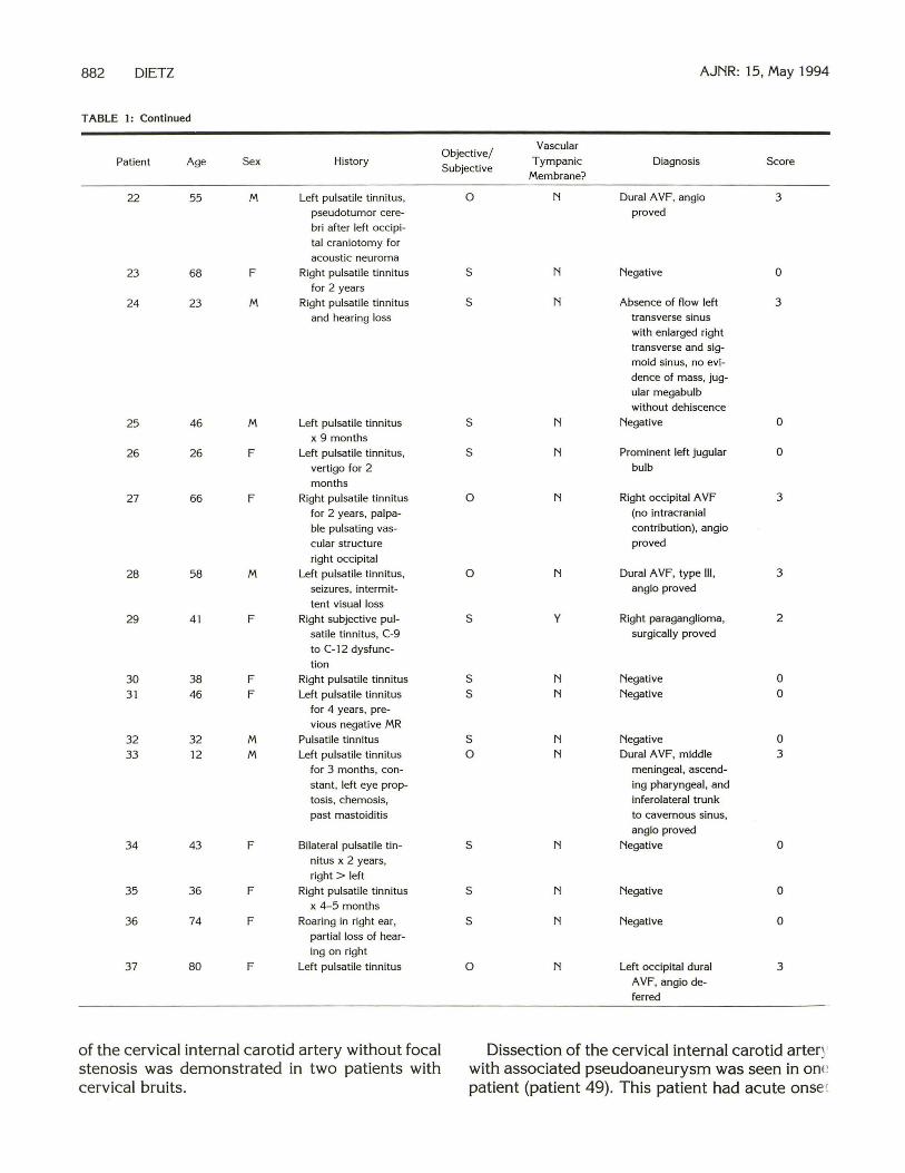
TABLE 1: Continued
Objective/ Vascular
Patient Age Sex History Subjective
Tympanic Diagnosis Score Membrane?
22 55 M Left pulsatile tinnitus, 0 N Dural AVF, angie 3 pseudotumor cere- proved
bri after left occipi-
tal craniotomy for
acoustic neuroma
23 68 F Right pulsatile tinnitus s N Negative 0 for 2 years
24 23 M Right pulsatile tinnitus s N Absence of flow left 3 and hearing Joss transverse sinus
with enlarged right
transverse and sig-
moid sinus, no evi-
dence of mass, jug-
ular megabulb
without dehiscence
25 46 M Left pulsatile tinnitus s N Negative 0 x 9 months
26 26 F Left pulsatile tinnitus, s N Prominent left jugular 0 vertigo for 2 bulb
months
27 66 F Right pulsatile tinnitus 0 N Right occipital A VF 3 for 2 years, palpa- (no intracranial
ble pulsating vas- contribution), angio
cular structure proved
right occipital
28 58 M Left pulsatile tinnitus, 0 N Dural AVF, type Jll, 3 seizures, intermit- angio proved
tent visual Joss
29 41 F Right subjective pul- s y Right paraganglioma, 2 satile tinnitus, C-9 surgically proved
to C-12 dysfunc-
tion
30 38 F Right pulsatile tinnitus s N Negative 0 31 46 F Left pulsatile tinnitus s N Negative 0
for 4 years, pre-
vious negative MR
32 32 M Pulsatile tinnitus s N Negative 0 33 12 M Left pulsatile tinnitus 0 N Dural AVF, middle 3
for 3 months, con- meningeal, ascend-
stan!, left eye prop- ing pharyngeal , and
tosis , chemosis, inferolateral trunk
past mastoiditis to cavernous sinus,
angio proved
34 43 F Bilateral pulsatile tin- s N Negative 0 nitus x 2 years,
right> left
35 36 F Right pulsatile tinnitus s N Negative 0 x 4-5 months
36 74 F Roaring in right ear, s N Negative 0 partial loss of hear-
ing on right
37 80 F Left pulsatile tinnitus 0 N Left occipital dural 3 AVF, angio de-
fer red
of the cervical internal carotid artery without focal Dissection of the cervical internal carotid arter) stenosis was demonstrated in two patients with with associated pseudoaneurysm was seen in one cervical bruits. patient (patient 49). This patient had acute onsec
AJNR: 15, May 1994 PULSATILE TINNITUS 883
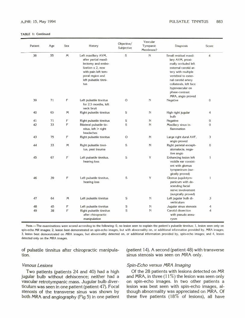
TABLE 1: Continued
Objective/ Vascular
Patient Age Sex History Tympanic Diagnosis Score Subjective
Membrane?
38 55 M Left maxi llary A V M , s N Small residual maxil- 4 after partial maxil- lary AVM, proxi-lectomy and embo- mally occluded left
lization x 2, now external carotid ar-with pain left tern- tery with multiple poral region and vertebral to exter-
left pulsatile tinni- nal carotid artery tus collaterals, left face
hypovascular on
phase-contrast
MRA, angio proved
39 71 F Left pulsatile tinn itus 0 N Negative 0 for 2.5 months, left
neck bruit
40 65 M Right pulsatile tinnitus s N High right jugular 4
bulb
41 71 F Right pulsatile tinnitus s N Negative 0
42 33 F Bilateral pulsatile tin- s N Maxillary sinus in- 0
nitus, left > right flammation
headaches
43 75 F Right pulsatile tinnitus 0 N Large right dural AVF, 3 angio proved
44 33 M Right pulsatile tinni- s N Right parieta l enceph- 0 tus, past trauma alomalacia, nega-
tive angio
45 67 F Left pulsatile tinnitus, s y Enhancing lesion left
hearing loss middle ear consist-
ent with glomus
tympanicum (sur-
gically proved)
46 39 F Left pulsatile tinnitus, s y Glomus jugulotym-
hearing loss panicum with de-
scending facial
nerve involvement
(surgically proved)
47 64 M Left pulsatile tinnitus s N Left jugular bulb di- 3 verticulum
48 45 F Left pulsatile tinnitus s N Jugular sinus stenosis 4
49 38 F Right pulsatile tinnitus s N Carotid dissection 4
after chiropractic with pseudo aneu-
manipulation rysm
Note.- The examinations were scored according to the following: 0 , no lesion seen to explain the patient's pulsatile tinnitus; I , lesion seen only on
spin-echo MR images; 2, lesion best demonstrated on spin-echo images, but with abnormality on , or additional information provided by, MRA images;
3, lesion best demonstrated on MRA images, but abnormality detected on , or additional information provided by , spin-echo images; and 4, lesion
detected only on the MRA images.
of pulsatile tinnitus after chiropractic manipulation.
Venous Lesions
Two patients (patients 24 and 40) had a high jugular bulb without dehiscence; neither had a vascular retrotympanic mass. Jugular bulb diverticulum was seen in one patient (patient 47). Focal stenosis of the transverse sinus was shown by both MRA and angiography (Fig 5) in one patient
(patient 14). A second (patient 48) with transverse sinus stenosis was seen on MRA only.
Spin-Echo versus MRA Imaging
Of the 28 patients with lesions detected on MR and MRA, in three (11 %) the lesion was seen only on spin-echo images. In two other patients a lesion was best seen with spin-echo images, although abnormality was appreciated on MRA. Of these five patients (18% of lesions) , all have
884 DIETZ AJNR: 15, May 1994
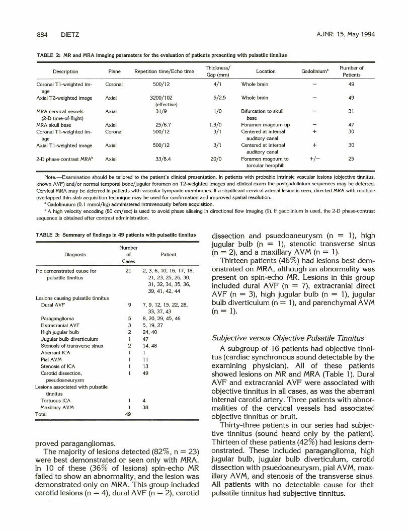
TABLE 2: MR and MRA imaging parameters for the evaluation of patients presenting with pulsatile tinnitus
Description Plane Repetition time/ Echo time
Coronal Tl-weighted im- Coronal 500/ 12 age
A xial T2-weighted image Axial 3200/ 102 (effective)
MRA cervical vessels Axial 31 / 9 (2-D time-of-flight)
MRA skull base Axial 25/ 6.7 Coronal Tl-weighted im- Coronal 500/ 12
age Axial Tl -weighted image Axial 500/ 12
2-D phase-contrast MRAb Axial 33/ 8.4
Thickness/ Gap (mm)
4/ 1
5/ 2.5
1/ 0
1.3/ 0 3/ 1
3/ 1
20/ 0
Location
Whole brain
Whole brain
Bifurcation to skull base
Foramen magnum up Centered at internal
auditory canal Centered at internal
auditory canal
Foramen magnum to torcular herophili
Gadolinium•
+
+
+/ -
Number of
Patients
49
49
31
47
30
30
25
Note.-Examination should be tailored to the patient's clinical presentation. In patients with probable intrinsic vascular lesions (objective tinnitus, known AVF) and/ or normal temporal bone/ jugular foramen on T2-weighted images and clinical exam the postgadolinium sequences may be deferred.
Cervical MRA may be deferred in patients with vascular tympanic membranes. if a significant cervical arterial lesion is seen, directed MRA with multiple
overlapped thin-slab acquisition technique may be used for confirmation and improved spatial resolution. • Gadolinuium (0.1 mmoi/kg) administered intravenously before acquisition. b A high velocity encoding (80 em/ sec) is used to avoid phase aliasing in directional flow imaging (9). If gadolinium is used, the 2-D phase-contrast
sequence is obtained after contrast administration.
TABLE 3: Summary of findings in 49 patients with pulsatile tinnitus
Diagnosis
No demonstrated cause for
pulsatile tinnitus
Lesions causing pulsatile tinnitus
Dural AVF
Paraganglioma
Extracranial A VF High jugular bulb
Jugular bulb diverticulum Stenosis of transverse sinus
Aberrant ICA
Pial AVM Stenosis of ICA
Carotid dissection,
pseudoaneurysm Lesions associated with pulsatile
tinnitus
Tortuous ICA
Maxillary AVM Total
proved paragangliomas.
Number of
Cases
21
9
5 3 2 I 2
49
Patient
2, 3, 6, 10, 16, 17, 18, 21, 23, 25, 26, 30,
31 , 32, 34, 35, 36, 39, 41 , 42, 44
7, 9, 12, 15, 22, 28, 33, 37, 43
8, 20, 29, 45, 46 5, 19, 27
24, 40
47 14, 48
I II 13
49
4 38
The majority of lesions detected (82%, n = 23) were best demonstrated or seen only with MRA. In 10 of these (36% of lesions) spin-echo MR failed to show an abnormality, and the lesion was demonstrated only on MRA. This group included carotid lesions (n = 4), dural AVF (n = 2), carotid
dissection and psuedoaneurysm (n = 1 ), high jugular bulb (n = 1), stenotic transverse sinus (n = 2), and a maxillary A V M (n = 1 ).
Thirteen patients (46%) had lesions best demonstrated on MRA, although an abnormality was present on spin-echo MR. Lesions in this group included dural A VF (n = 7), extracranial direct AVF (n = 3), high jugular bulb (n = 1), jugular bulb diverticulum (n = 1), and parenchymal AVM (n = 1).
Subjective versus Objective Pulsatile Tinnitus A subgroup of 16 patients had objective tinni
tus (cardiac synchronous sound detectable by the exammmg physician). All of these patients showed lesions on MR and MRA (Table 1). Dural A VF and extracranial A VF were associated with objective tinnitus in all cases, as was the aberrant internal carotid artery. Three patients with abnormalities of the cervical vessels had associated objective tinnitus or bruit.
Thirty-three patients in our series had subjective tinnitus (sound heard only by the patient). Thirteen of these patients (42%) had lesions demonstrated. These included paraganglioma, high jugular bulb, jugular bulb diverticulum, carotid dissection with psuedoaneurysm, pial A V M, maxillary A V M, and stenosis of the transverse sinus. All patients with no detectable cause for their pulsatile tinnitus had subjective tinnitus.
AJNR: 15, May 1994 PULSATILE TINNITUS 885
A B
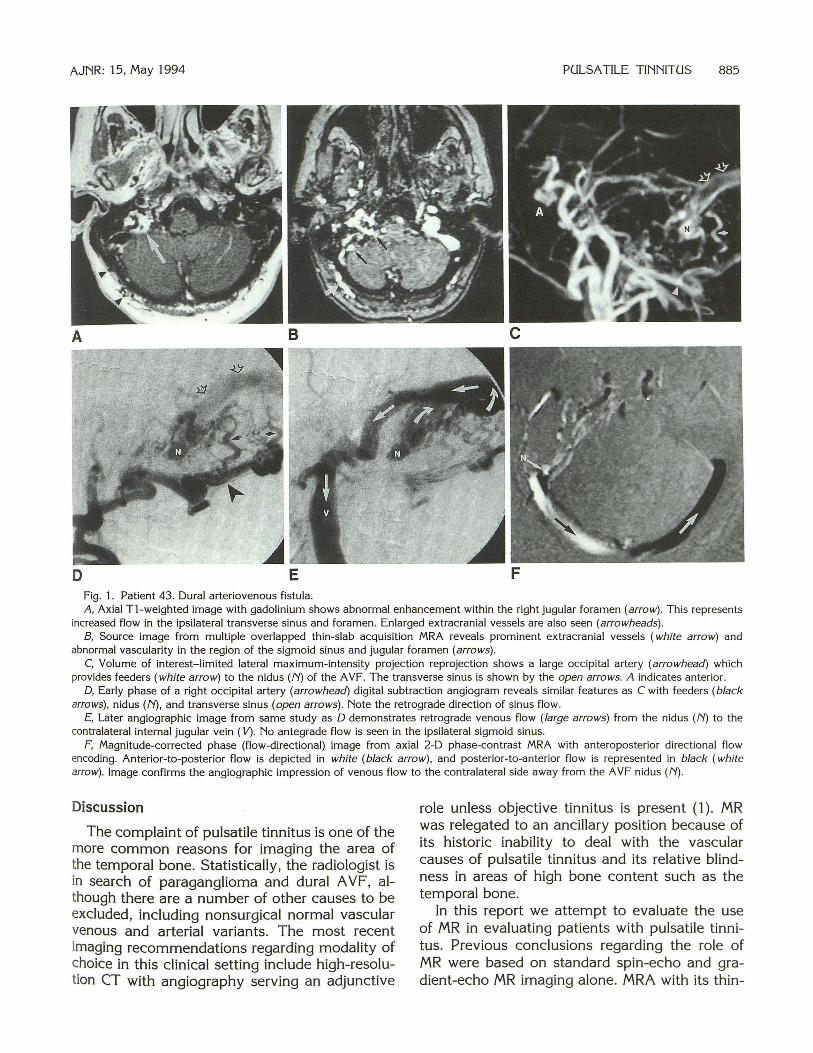
D E Fig. 1. Patient 43. Dural arteriovenous fistula. A, Axial Tl-weighted image with gadolinium shows abnormal enhancement within the right jugular foramen (arrow) . This represents
increased flow in the ipsilateral transverse sinus and foramen . Enlarged extracranial vessels are also seen (arrowheads) . B, Source image from multiple overlapped thin-slab acquisition MRA reveals prominent extracranial vessels (white arrow) and
abnormal vascularity in the region of the sigmoid sinus and jugular foramen (arrows) . C, Volume of interest-limited lateral maximum-intensity projection reprojection shows a large occipital artery (arro whead) which
provides feeders (white arrow) to the nidus (N) of the AVF. The transverse sinus is shown by the open arrows. A indicates anterior . D, Early phase of a right occipital artery (arrowhead) digital subtraction angiogram reveals similar features as C with feeders (black
arrows), nidus (N), and transverse sinus (open arrows). Note the retrograde direction of sinus flow . E, Later angiographic image from same study as D demonstrates retrograde venous flow (large arrows) from the nidus (N) to the
contralateral internal jugular vein ( V). No antegrade flow is seen in the ipsilateral sigmoid sinus. F, Magnitude-corrected phase (flow-directional) image from axial 2-D phase-contrast MRA with anteroposterior directional flow
encoding. Anterior-to-posterior flow is depicted in white (black arrow), and posterior-to-anterior flow is represented in black (white arrow). Image confirms the angiographic impression of venous flow to the contralateral side away from the AVF nidus (N) .
Discussion
The complaint of pulsatile tinnitus is one of the more common reasons for imaging the area of the temporal bone. Statistically, the radiologist is in search of paraganglioma and dural AVF, although there are a number of other causes to be excluded, including nonsurgical normal vascular venous and arterial variants. The most recent imaging recommendations regarding modality of choice in this clinical setting include high-resolution CT with angiography serving an adjunctive
role unless objective tinnitus is present (1). MR was relegated to an ancillary position because of its historic inability to deal with the vascular causes of pulsatile tinnitus and its relative blindness in areas of high bone content such as the temporal bone.
In this report we attempt to evaluate the use of MR in evaluating patients with pulsatile tinnitus. Previous conclusions regarding the role of MR were based on standard spin-echo and gradient-echo MR imaging alone. MRA with its thin-
886 DIETZ AJNR: 15, May 1994
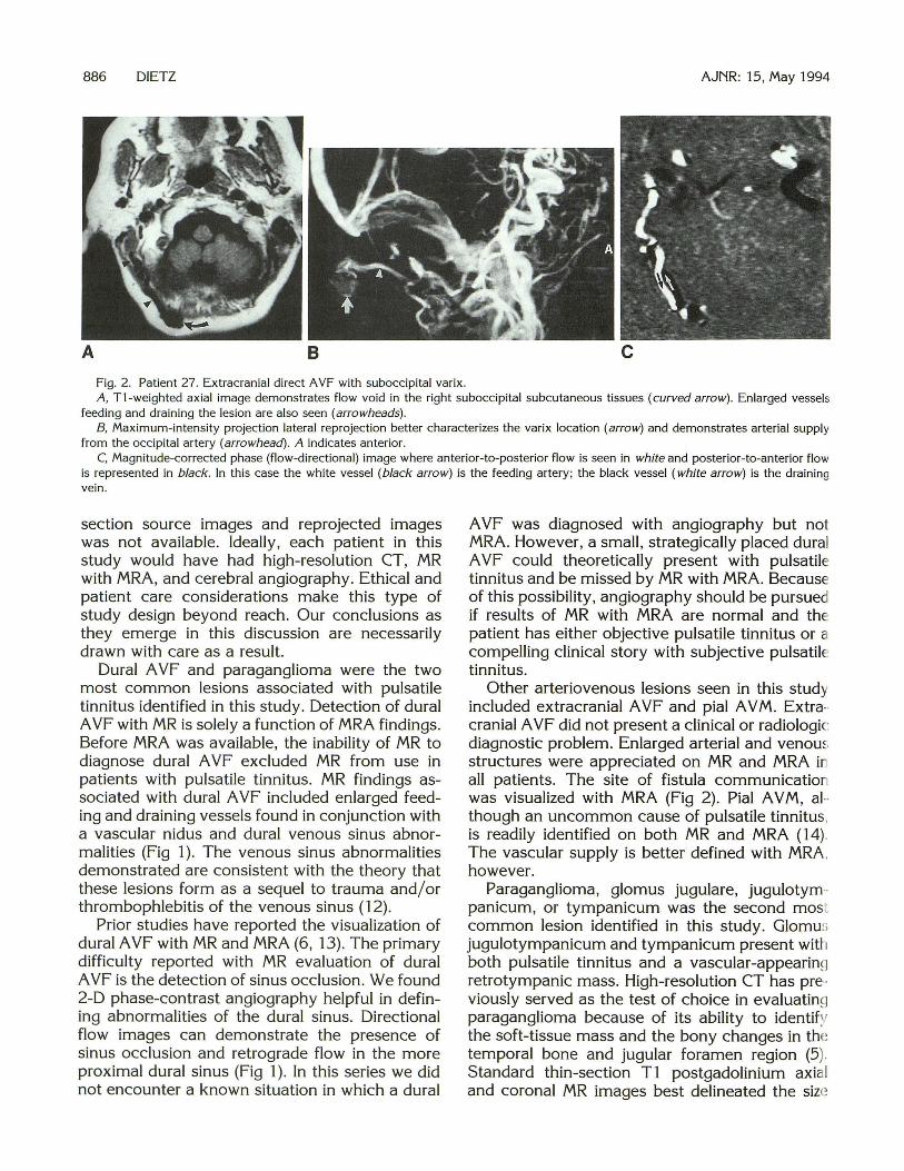
A B c Fig. 2. Patient 27. Extracranial direct A VF with suboccipital varix. A , T 1-weighted axial image demonstrates flow void in the right suboccipital subcutaneous tissues (curved arrow). Enlarged vessels
feeding and draining the lesion are also seen (arrowheads). B, Maximum-intensity projection lateral reprojection better characterizes the varix location (arrow) and demonstrates arterial supply
from the occipital artery (arrowhead). A indicates anterior. C, Magnitude-corrected phase (flow-directional) image where anterior-to-posterior flow is seen in white and posterior-to-anterior flow
is represented in black. In this case the white vessel (black arrow) is the feeding artery; the black vessel (white arrow) is the draining vein.
section source images and reprojected images was not available. Ideally, each patient in this study would have had high-resolution CT, MR with MRA, and cerebral angiography. Ethical and patient care considerations make this type of study design beyond reach. Our conclusions as they emerge in this discussion are necessarily drawn with care as a result.
Dural A VF and paraganglioma were the two most common lesions associated with pulsatile tinnitus identified in this study. Detection of dural A VF with MR is solely a function of MRA findings . Before MRA was available, the inability of MR to diagnose dural A VF excluded MR from use in patients with pulsatile tinnitus. MR findings associated with dural A VF included enlarged feeding and draining vessels found in conjunction with a vascular nidus and dural venous sinus abnormalities (Fig 1 ). The venous sinus abnormalities demonstrated are consistent with the theory that these lesions form as a sequel to trauma and/ or thrombophlebitis of the venous sinus ( 12).
Prior studies have reported the visualization of dural A VF with MR and MRA (6, 13). The primary difficulty reported with MR evaluation of dural AVF is the detection of sinus occlusion. We found 2-D phase-contrast angiography helpful in defining abnormalities of the dural sinus. Directional flow images can demonstrate the presence of sinus occlusion and retrograde flow in the more proximal dural sinus (Fig 1 ). In this series we did not encounter a known situation in which a dural
A VF was diagnosed with angiography but not MRA. However, a small, strategically placed dural A VF could theoretically present with pulsatile tinnitus and be missed by MR with MRA. Because of this possibility, angiography should be pursued if results of MR with MRA are normal and the patient has either objective pulsatile tinnitus or a compelling clinical story with subjective pulsatile tinnitus.
Other arteriovenous lesions seen in this study included extracranial A VF and pial A V M. Extra .. cranial A VF did not present a clinical or radiologic diagnostic problem. Enlarged arterial and venom. structures were appreciated on MR and MRA ir all patients. The site of fistula communicatio was visualized with MRA (Fig 2). Pial A V M, al-· though an uncommon cause of pulsatile tinnitus, is readily identified on both MR and MRA (14). The vascular supply is better defined with MRA, however.
Paraganglioma, glomus jugulare, jugulotympanicum, or tympanicum was the second most common lesion identified in this study. Glomus jugulotympanicum and tympanicum present with both pulsatile tinnitus and a vascular-appearing retrotympanic mass. High-resolution CT has previously served as the test of choice in evaluating paraganglioma because of its ability to identif_, the soft-tissue mass and the bony changes in the temporal bone and jugular foramen region (5). Standard thin-section T1 postgadolinium axial and coronal MR images best delineated the size
AJNR: 15, May 1994
A B
c D
and extent of the paraganglioma. If the lesion extends into a region of adjacent marrow or soft fat, fat-saturation T1 techniques can be useful. MRA typically did not aid in the diagnosis, although a characteristic pattern of abnormal signal was appreciated in several cases (Fig. 3).
Arterial lesions associated with pulsatile tinnitus include internal carotid artery stenosis, ectasia, aberrant internal carotid artery, aneurysm of the petrous internal carotid artery, and fibromuscular dysplasia (3, 4). Pulsatile tinnitus related to this group of arterial lesions probably occurs as a result of marked turbulence and change in velocity of blood flow in the petrous internal carotid artery secondary to vessel caliber change near the skull base. In some patients, however, the vascular noise may be the result of increased blood flow in the ascending pharyngeal artery. Standard MR is unable to detect these lesions. Only MRA can indicate these diagnoses.
PULSATILE TINNITUS 887
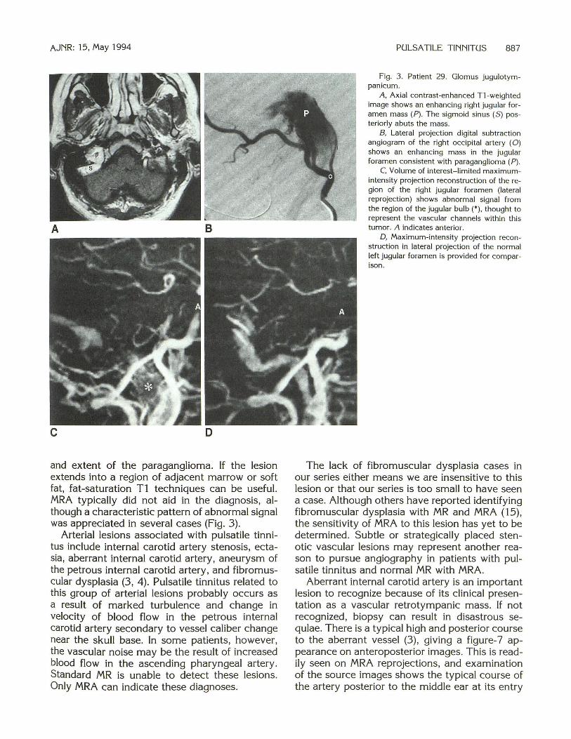
Fig. 3. Patient 29. Glomus jugulotympanicum.
A, Axial contrast-enhanced Tl-weighted image shows an enhancing right jugular foramen mass (P). The sigmoid sinus (S) posteriorly abuts the mass.
8 , Lateral projection digital subtraction angiogram of the right occipital artery ( 0) shows an enhancing mass in the jugular foramen consistent with paraganglioma (P).
C, Volume of interest-limited maximumintensity projection reconstruction of the region of the right jugular foramen (lateral reprojection) shows abnormal signal from the region of the jugular bulb (*), thought to represent the vascular channels within this tumor. A indicates anterior.
D, Maximum-intensity projection reconstruction in lateral projection of the normal left jugular foramen is provided for comparison.
The lack of fibromuscular dysplasia cases in our series either means we are insensitive to this lesion or that our series is too small to have seen a case. Although others have reported identifying fibromuscular dysplasia with MR and MRA (15), the sensitivity of MRA to this lesion has yet to be determined. Subtle or strategically placed stenotic vascular lesions may represent another reason to pursue angiography in patients with pulsatile tinnitus and normal MR with MRA.
Aberrant internal carotid artery is an important lesion to recognize because of its clinical presentation as a vascular retrotympanic mass. If not recognized , biopsy can result in disastrous sequlae. There is a typical high and posterior course to the aberrant vessel (3), giving a figure-7 appearance on anteroposterior images. This is readily seen on MRA reprojections, and examination of the source images shows the typical course of the artery posterior to the middle ear at its entry
888 DIETZ
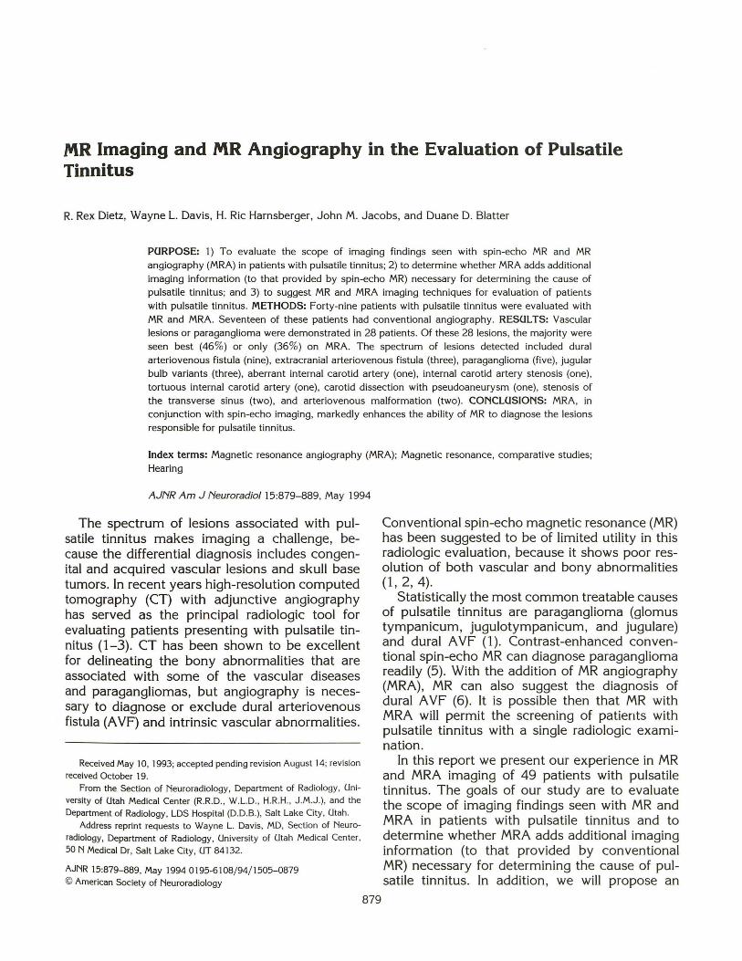
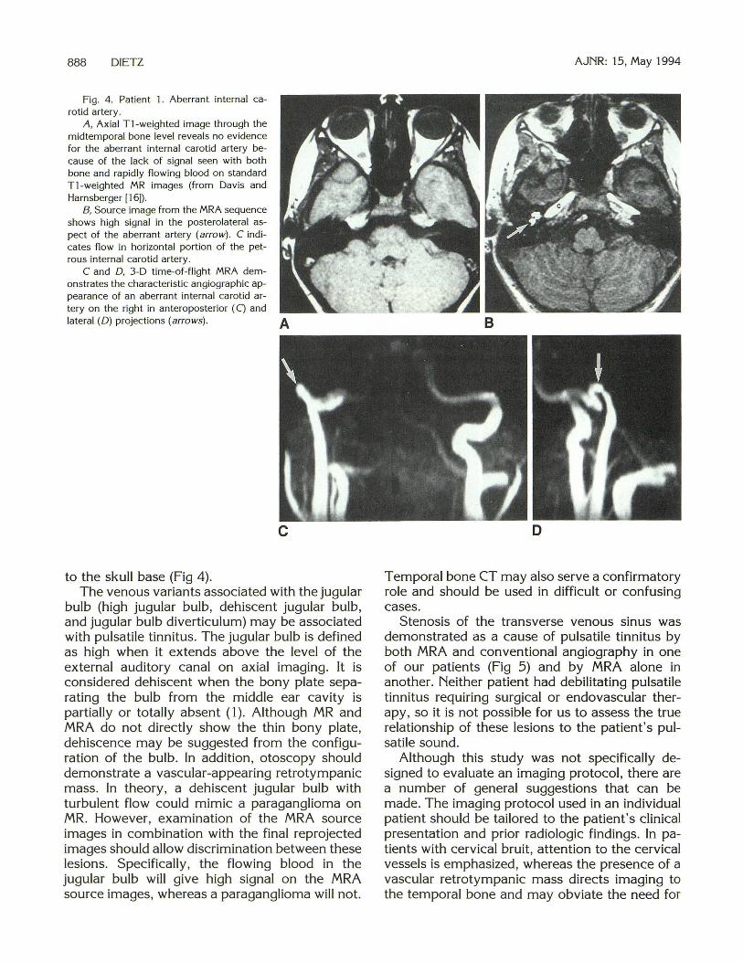
Fig. 4. Patient 1. Aberrant internal carotid artery.
A, Axial T1-weighted image through the midtemporal bone level reveals no evidence for the aberrant internal carotid artery because of the lack of signal seen with both bone and rapidly flowing blood on standard T1-weighted MR images (from Davis and Harnsberger [ 16]).
B, Source image from the MRA sequence shows high signal in the posterolateral aspect of the aberrant artery (arrow). C indicates flow in horizontal portion of the petreus internal carotid artery.
C and D, 3-D time-of-flight MRA demonstrates the characteristic angiographic appearance of an aberrant internal carotid artery on the right in anteroposterior (C) and lateral (D) projections (arrows). A
c
to the skull base (Fig 4). The venous variants associated with the jugular
bulb (high jugular bulb, dehiscent jugular bulb, and jugular bulb diverticulum) may be associated with pulsatile tinnitus. The jugular bulb is defined as high when it extends above the level of the external auditory canal on axial imaging. It is considered dehiscent when the bony plate separating the bulb from the middle ear cavity is partially or totally absent (1) . Although MR and MRA do not directly show the thin bony plate, dehiscence may be suggested from the configuration of the bulb. In addition, otoscopy should demonstrate a vascular-appearing retrotympanic mass. In theory, a dehiscent jugular bulb with turbulent flow could mimic a paraganglioma on MR. However, examination of the MRA source images in combination with the final reprojected images should allow discrimination between these lesions. Specifically, the flowing blood in the jugular bulb will give high signal on the MRA source images, whereas a paraganglioma will not.
AJNR: 15, May 1994
B
Temporal bone CT may also serve a confirmatory role and should be used in difficult or confusing cases.
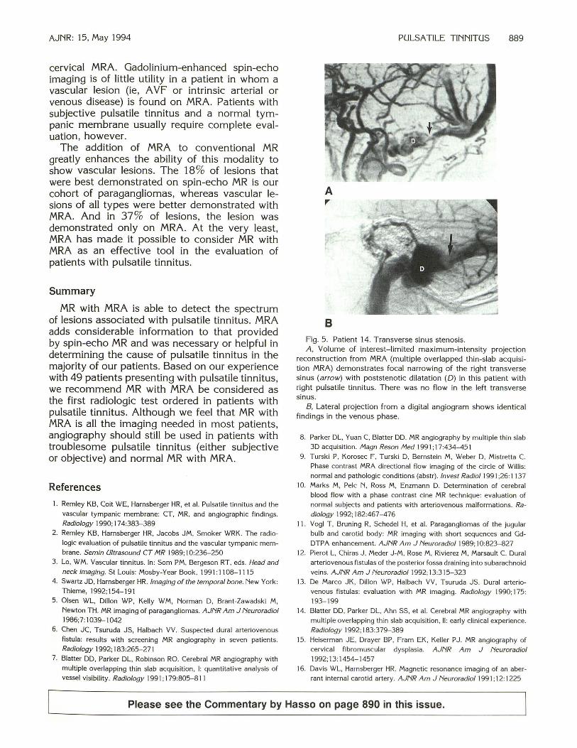
Stenosis of the transverse venous sinus was demonstrated as a cause of pulsatile tinnitus by both MRA and conventional angiography in one of our patients (Fig 5) and by MRA alone in another. Neither patient had debilitating pulsatile tinnitus requiring surgical or endovascular therapy, so it is not possible for us to assess the true relationship of these lesions to the patient's pulsatile sound.
Although this study was not specifically designed to evaluate an imaging protocol, there are a number of general suggestions that can be made. The imaging protocol used in an individual patient should be tailored to the patient's clinical presentation and prior radiologic findings. In patients with cervical bruit, attention to the cervical vessels is emphasized, whereas the presence of a vascular retrotympanic mass directs imaging to the temporal bone and may obviate the need for
AJNR: 15, May 1994
cervical MRA. Gadolinium-enhanced spin-echo imaging is of little utility in a patient in whom a vascular lesion (ie, AVF or intrinsic arterial or venous disease) is found on MRA. Patients with subjective pulsatile tinnitus and a normal tympanic membrane usually require complete evaluation, however.
The addition of MRA to conventional MR greatly enhances the ability of this modality to show vascular lesions. The 18% of lesions that were best demonstrated on spin-echo MR is our cohort of paragangliomas, whereas vascular lesions of all types were better demonstrated with MRA. And in 37% of lesions, the lesion was demonstrated only on MRA. At the very least, MRA has made it possible to consider MR with MRA as an effective tool in the evaluation of patients with pulsatile tinnitus.
Summary
MR with MRA is able to detect the spectrum of lesions associated with pulsatile tinnitus. MRA adds considerable information to that provided by spin-echo MR and was necessary or helpful in determining the cause of pulsatile tinnitus in the majority of our patients. Based on our experience with 49 patients presenting with pulsatile tinnitus, we recommend MR with MRA be considered as the first radiologic test ordered in patients with pulsatile tinnitus. Although we feel that MR with MRA is all the imaging needed in most patients, angiography should still be used in patients with troublesome pulsatile tinnitus (either subjective or objective) and normal MR with MRA.
References
I. Remley KB, Coit WE, Harnsberger HR, et al. Pulsatile tinnitus and the
vascular tympanic membrane: CT, MR, and angiographic findings. Radiology 1990; I 7 4:383-389
2. Remley KB, Harnsberger HR, Jacobs JM, Smoker WRK. The radio
logic evaluation of pulsatile tinnitus and the vascular tympanic membrane. Semin Ultrasound CT MR 1989; 10:236-250
3. Lo, WM. Vascular tinnitus. In : Som PM, Bergeson RT, eds. Head and
neck imaging. St Louis: Mosby-Year Book, 1991:1108-1115 4. Swartz JD, Harnsberger HR. Imaging of the temporal bone. New York :
Thieme, 1992; 154-191
5. Olsen WL, Dillon WP, Kelly WM, Norman D, Brant-Zawadski M,
Newton TH. MR imaging of paragangliomas. AJNR Am J Neuroradiol 1986;7:1039-1042
6. Chen JC, Tsuruda JS, Halbach VV. Suspected dural arteriovenous
fistula : results with screening MR angiography in seven patients. Radiology 1992; 183:265-271
7. Blatter DD, Parker DL, Robinson RO. Cerebral MR angiography with
multiple overlapping thin slab acquisition , 1: quantitative analysis of vessel visibility. Radiology 1991; 179:805-811
A ,.
8
PULSATILE TINNITUS
Fig . 5. Patient 14. Transverse sinus stenosis.
889
A, Volume of i,nterest-limited maximum-intensity projection
reconstruction from MRA (multiple overlapped thin-slab acquisition MRA) demonstrates focal narrowing of the right transverse sinus (arrow) with poststenotic dilatation (D) in this patient with right pulsatile tinnitus. There was no flow in the left transverse sinus.
B, Lateral projection from a digital angiogram shows identical findings in the venous phase.
8. Parker DL, Yuan C, Blatter DD. MR angiography by multiple thin slab 3D acquisition. Magn Reson fried 1991 ;17:434-451
9. Turski P, Korosec F, Turski D, Bernstein M, Weber D, Mistretta C.
Phase contrast MRA directional flow imaging of the circle of Willis: normal and pathologic conditions (abstr) . Invest Radioll991 ;26:1137
10. Marks M, Pelc N, Ross M, Enzmann D. Determination of cerebral
blood flow with a phase contrast cine MR technique: evaluation of normal subjects and patients with arteriovenous malformations. Radiology 1992; 182:467-4 76
11. Yogi T, Bruning R, Schedel H, et al. Paragangliomas of the jugular
bulb and carotid body: MR imaging with short sequences and Gd
DTPA enhancement. AJNR Am J Neuroradiol 1989; I 0:823-827 12. Pierot L, Chiras J , Meder J-M, Rose M, Rivierez M, Marsault C. Dural
arteriovenous fistulas of the posterior fossa draining into subarachnoid
veins. AJNR Am J Neuroradiol 1992; 13:315-323
13. De Marco JK, Dillon WP, Halbach VV, Tsuruda JS. Dural arteriovenous fistulas: evaluation with MR imaging. Radiology 1990; I 75: 193-199
14. Blatter DD, Parker DL, Ahn SS, et al. Cerebral MR angiography with
multiple overlapping thin slab acquisition, II : early clinical experience. Radiology 1992; I 83:379-389
15. Heiserman JE, Drayer BP, Fram EK, Keller PJ. MR angiography of cervical fibromuscular dysplasia. AJNR Am J Neuroradiol 1992; 13:1454-1457
16. Davis WL, Harnsberger HR. Magnetic resonance imaging of an aberrant internal carotid artery. AJNR Am J Neuroradiol 1991 ; 12:1225
Please see the Commentary by Hasso on page 890 in this issue.