Embed Size (px)
Citation preview
Global Siemens HeadquartersSiemens AGWittelsbacherplatz 280333 MuenchenGermany
Global Siemens Healthcare HeadquartersSiemens AGHealthcare SectorHenkestrasse 12791052 ErlangenGermanyPhone: +49 9131 84-0www.siemens.com/healthcare
On account of certain regional limitations of sales rights and service availability, we cannot guarantee that all products included in this brochure are available through the Siemens sales organization worldwide. Availability and packaging may vary by country and are subject to change without prior notice. Some/All of the features and products described herein may not be available in the United States.
All devices listet herein may not be licensed according to Canadian Medical Devices Regulations. The information in this document contains general technical descriptions of specifications and options as well as standard and optional features which do not always have to be present in individual cases.
Siemens reserves the right to modify the design, packaging, specifications, and options described herein without prior notice. Please contact your local Siemens sales representative for the most current information.
Note: Any technical data contained in this document may vary within defined tolerances. Original images always lose a certain amount of detail when reproduced.
Please find fitting accessories: www.siemens.com/medical-accessories
Local Contact Information
In the USASiemens Medical Solutions USA, Inc.51 Valley Stream ParkwayMalvern, PA 19355Phone: +1 888-826-9702Phone: +1 610-448-4500Fax: +1 610-448-2254
In ChinaSiemens Medical Park, Shanghai278, Zhouzhu RoadSIMZ, Nanhui DistrictShanghai, 201318P.R. ChinaPhone: +86-21-38895000Fax: +86-10-28895001
In JapanSiemens-AsahiMedical Technologies Ltd.Takanawa Park Tower 14F20-14, Higashi-Gotanda 3-chomeShinagawa-kuTokyo 141-8644Phone: +81 3 5423 8411
In AsiaSiemens Pte LtdHealthcare SectorRegional HeadquartersThe Siemens Center60 MacPherson RoadSingapore 348615Phone: +65 6490-6000Telefax: +65 6490-6001
Order No. A91MR-9011-6C-7600 | Printed in Germany | CC MR 09111.5 | © 09.2011, Siemens AG
Global Business Unit
Siemens AGHealthcare SectorMagnetic ResonanceHenkestr. 127DE-91052 ErlangenGermanyPhone: +49 9131 84-0
Legal Manufacturer
Siemens AGWittelsbacherplatz 2DE-80333 MuenchenGermany
Siemens Mindit MagneticResonance Ltd. (SMMR)Siemens MRI Center, Gaoxin C. Ave.,2nd, Hi-Tech Industrial ParkShenzen 518057P.R. ChinaPhone: +86 755-26525421Fax: +86 755-26549253
Answers for life.
MAGNETOM AeraImage Gallery
www.siemens.com/aera
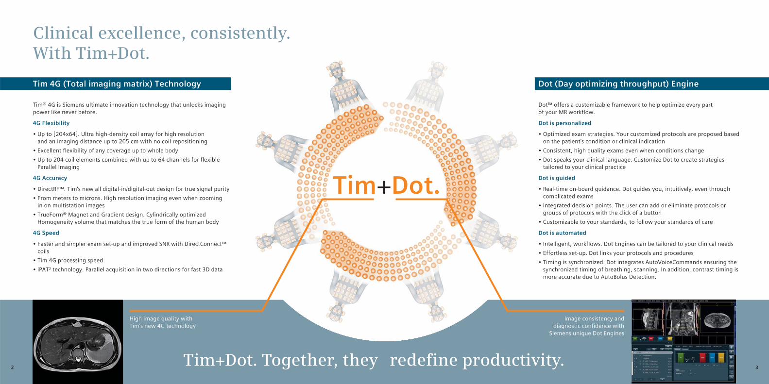
Clinical excellence, consistently.With Tim+Dot.
Tim+Dot.
High image quality with Tim’s new 4G technology
Image consistency and diagnostic confidence with
Siemens unique Dot Engines
Tim+Dot. Together, they redefine productivity.
Tim 4G (Total imaging matrix) Technology
Tim® 4G is Siemens ultimate innovation technology that unlocks imaging power like never before.4G Flexibility• Up to [204x64]. Ultra high-density coil array for high resolution
and an imaging distance up to 205 cm with no coil repositioning • Excellent flexibility of any coverage up to whole body• Up to 204 coil elements combined with up to 64 channels for flexible
Parallel Imaging4G Accuracy• DirectRF™. Tim’s new all digital-in/digital-out design for true signal purity• From meters to microns. High resolution imaging even when zooming
in on multistation images• TrueForm® Magnet and Gradient design. Cylindrically optimized
Homogeneity volume that matches the true form of the human body4G Speed• Faster and simpler exam set-up and improved SNR with DirectConnect™
coils• Tim 4G processing speed• iPAT² technology. Parallel acquisition in two directions for fast 3D data
Dot (Day optimizing throughput) Engine
Dot™ offers a customizable framework to help optimize every part of your MR workflow.Dot is personalized• Optimized exam strategies. Your customized protocols are proposed based
on the patient’s condition or clinical indication• Consistent, high quality exams even when conditions change• Dot speaks your clinical language. Customize Dot to create strategies
tailored to your clinical practiceDot is guided• Real-time on-board guidance. Dot guides you, intuitively, even through
complicated exams • Integrated decision points. The user can add or eliminate protocols or
groups of protocols with the click of a button• Customizable to your standards, to follow your standards of careDot is automated• Intelligent, workflows. Dot Engines can be tailored to your clinical needs• Effortless set-up. Dot links your protocols and procedures• Timing is synchronized. Dot integrates AutoVoiceCommands ensuring the
synchronized timing of breathing, scanning. In addition, contrast timing is more accurate due to AutoBolus Detection.
2 3
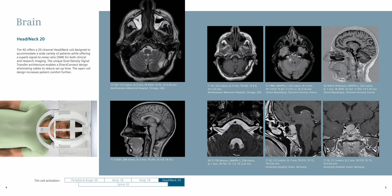
Brain
Head/Neck 20
Tim 4G offers a 20-channel Head/Neck coil designed to accommodate a wide variety of patients while offering a superb signal-to-noise ratio (SNR) for both clinical and research imaging. The unique Dual-Density Signal Transfer architecture enables a DirectConnect design eliminating cables to reduce set-up time. The open coil design increases patient comfort further.
T1 SE, 512 matrix, SL 2 mm, TR 570, TE 15, TA 4:02 minUniversity Hospital, Essen, Germany
3D T2 TSE Restore, GRAPPA 2, 256 matrix,SL 1 mm, TR 750, TE 113, TA 3:22 min
T1 SE, 512 matrix, SL 2 mm, TR 570, TE 15, TA 4:02 minUniversity Hospital, Essen, Germany
3D SPACE IR Restore, GRAPPA 2, 256 matrix,SL 1 mm, TR 3000, TE 257, TI 350, TA 5:30 minCentre Republique, Clermont Ferrand, France
T2 TSE, 512 matrix, SL 5 mm, TR 5000, TE 92, TA 3:35 minNorthwestern Memorial Hospital, Chicago, USA
T1 SE, 320 matrix, SL 5 mm, TR 550, TE 8.9, TA 2:20 minNorthwestern Memorial Hospital, Chicago, USA
T2 TIRM, GRAPPA 2, 320 matrix, SL 4 mm,TR 11570, TE 82, TI 2731.2, TA 3:16 minCentre Republique, Clermont Ferrand, France
T1 FLASH, 288 matrix, SL 5 mm, TR 200, TE 4.8, TA 43 s
Spine 32Body 18 Body 18Peripheral Angio 36 Head/Neck 20Tim coil activation:
4 5
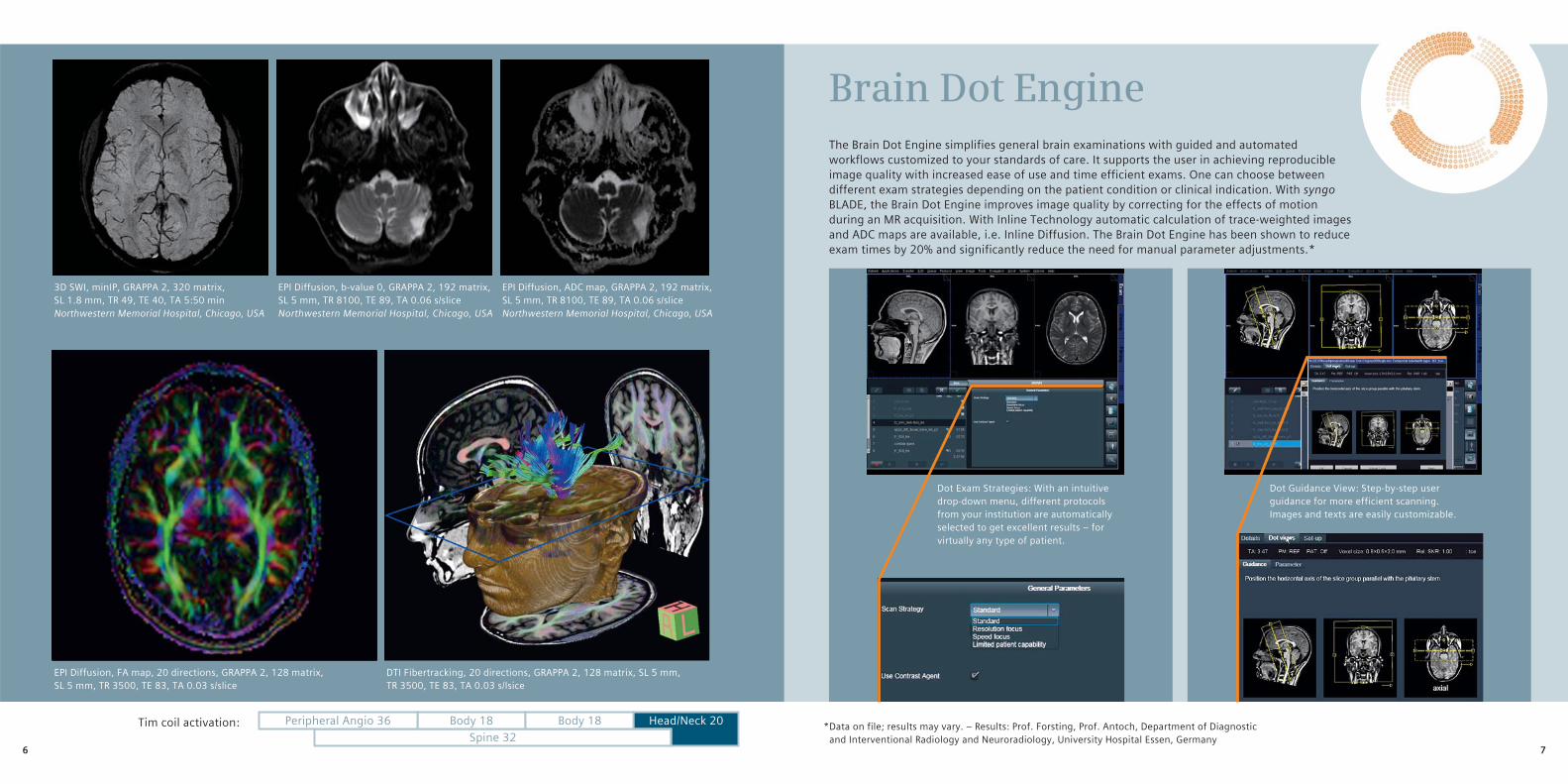
EPI Diffusion, b-value 0, GRAPPA 2, 192 matrix,SL 5 mm, TR 8100, TE 89, TA 0.06 s/sliceNorthwestern Memorial Hospital, Chicago, USA
EPI Diffusion, ADC map, GRAPPA 2, 192 matrix,SL 5 mm, TR 8100, TE 89, TA 0.06 s/sliceNorthwestern Memorial Hospital, Chicago, USA
3D SWI, minIP, GRAPPA 2, 320 matrix,SL 1.8 mm, TR 49, TE 40, TA 5:50 minNorthwestern Memorial Hospital, Chicago, USA
EPI Diffusion, FA map, 20 directions, GRAPPA 2, 128 matrix, SL 5 mm, TR 3500, TE 83, TA 0.03 s/slice
DTI Fibertracking, 20 directions, GRAPPA 2, 128 matrix, SL 5 mm, TR 3500, TE 83, TA 0.03 s/lsice
Spine 32Body 18 Body 18Peripheral Angio 36 Head/Neck 20Tim coil activation:
The Brain Dot Engine simplifies general brain examinations with guided and automated workflows customized to your standards of care. It supports the user in achieving reproducible image quality with increased ease of use and time efficient exams. One can choose between different exam strategies depending on the patient condition or clinical indication. With syngo BLADE, the Brain Dot Engine improves image quality by correcting for the effects of motion during an MR acquisition. With Inline Technology automatic calculation of trace-weighted images and ADC maps are available, i.e. Inline Diffusion. The Brain Dot Engine has been shown to reduce exam times by 20% and significantly reduce the need for manual parameter adjustments.*
Brain Dot Engine
Dot Exam Strategies: With an intuitive drop-down menu, different protocols from your institution are automatically selected to get excellent results – for virtually any type of patient.
* Data on file; results may vary. – Results: Prof. Forsting, Prof. Antoch, Department of Diagnostic and Interventional Radiology and Neuroradiology, University Hospital Essen, Germany
Dot Guidance View: Step-by-step user guidance for more efficient scanning. Images and texts are easily customizable.
76
Neurovascular
Head/Neck 20, Body 18 and Spine 32
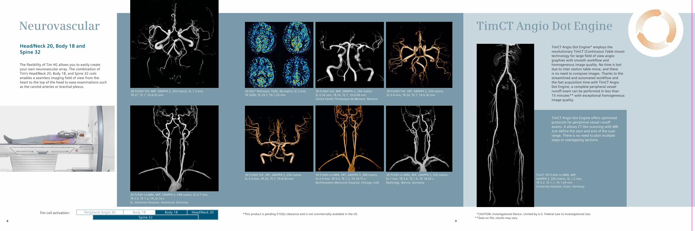
The flexibility of Tim 4G allows you to easily create your own neurovascular array. The combination of Tim’s Head/Neck 20, Body 18, and Spine 32 coils enables a seamless imaging field of view from the heart to the top of the head to ease examinations such as the carotid arteries or brachial plexus.
Spine 32Body 18 Body 18Peripheral Angio 36 Head/Neck 20Tim coil activation:
3D ASL* Perfusion, TGSE, 96 matrix, SL 2 mm, TR 3500, TE 24.7, TA 1:20 min
3D FLASH ToF, MIP, GRAPPA 2, 256 matrix,SL 0.54 mm, TR 24, TE 7, TA 6:08 minCentre Cardio Thoracique de Monaco, Monaco
3D FLASH ce-MRA, MIP, GRAPPA 2, 448 matrix, SL 0.7 mm, TR 3.4, TE 1.2, TA 22.16 sSt. Johannes-Hospital, Dortmund, Germany
3D FLASH ToF, MIP, GRAPPA 2, 320 matrix, SL 1.3 mm, TR 27, TE 7, TA 6:52 min
3D FLASH ToF, VRT, GRAPPA 2, 256 matrix,SL 0.4 mm, TR 24, TE 7, TA 6:30 min
3D FLASH ce-MRA, VRT, GRAPPA 3, 384 matrix,SL 0.9 mm, TR 3.4, TE 1.2, TA 18.71 sNorthwestern Memorial Hospital, Chicago, USA
3D FLASH ce-MRA, MIP, GRAPPA 2, 320 matrix,SL 1 mm, TR 3.4, TE 1.4, TA 18.05 sRadiology, Worms, Germany
TimCT Angio Dot Engine* employs the revolutionary TimCT (Continuous Table move) technology for large field of view angio-graphies with smooth workflow and homogeneous image quality. No time is lost due to inter station table move, and there is no need to compose images. Thanks to the streamlined and automated workflow and the fast acquisition time with TimCT Angio Dot Engine, a complete peripheral vessel runoff exam can be performed in less than 15 minutes** with exceptional homogeneous image quality.
TimCT Angio Dot Engine
TimCT Angio Dot Engine offers optimized protocols for peripheral vessel runoff exams. It allows CT-like scanning with MR: Just define the start and end of the scan range. There is no need to plan multiple steps or overlapping sections.
* *CAUTION: Investigational Device. Limited by U.S. Federal Law to Investigational Use.**Data on file; results may vary.
* This product is pending 510(k) clearance and is not commercially available in the US.
TimCT 3D FLASH ce-MRA, MIP, GRAPPA 3, 320 matrix, SL 1.2 mm, TR 3.2, TE 1.1, TA 1:04 minUniversity Hospital, Essen, Germany
3D FLASH ToF, VRT, GRAPPA 2, 256 matrix,SL 0.4 mm, TR 24, TE 7, TA 6:30 min
FÜR USA SEITE ENTFERNEN!
98
Spine cervical /Neck imaging
Head/Neck 20 and Spine 32
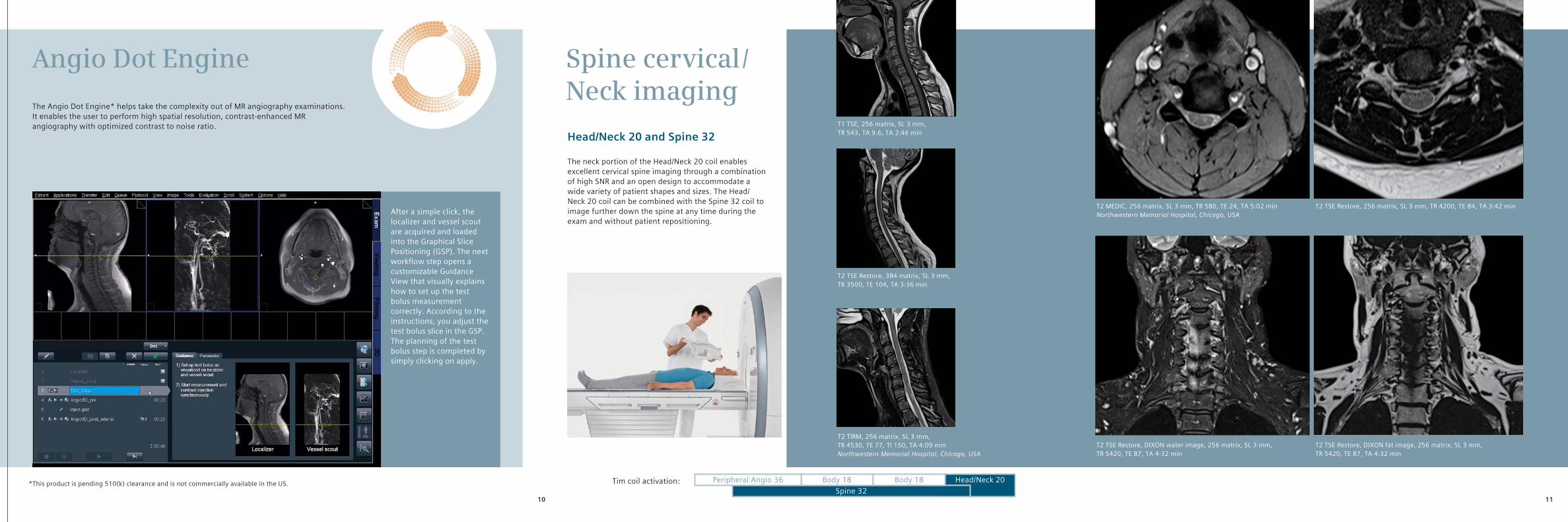
The neck portion of the Head/Neck 20 coil enables excellent cervical spine imaging through a combination of high SNR and an open design to accommodate a wide variety of patient shapes and sizes. The Head/Neck 20 coil can be combined with the Spine 32 coil to image further down the spine at any time during the exam and without patient repositioning.
Spine 32Body 18 Body 18Peripheral Angio 36 Head/Neck 20Tim coil activation:
The Angio Dot Engine* helps take the complexity out of MR angiography examinations. It enables the user to perform high spatial resolution, contrast-enhanced MR angiography with optimized contrast to noise ratio.
Angio Dot Engine
After a simple click, the localizer and vessel scout are acquired and loaded into the Graphical Slice Positioning (GSP). The next workflow step opens a customizable Guidance View that visually explains how to set up the test bolus measurement correctly. According to the instructions, you adjust the test bolus slice in the GSP. The planning of the test bolus step is completed by simply clicking on apply.
* This product is pending 510(k) clearance and is not commercially available in the US.
T1 TSE, 256 matrix, SL 3 mm,TR 543, TA 9.6, TA 2:46 min
T2 TSE Restore, 384 matrix, SL 3 mm,TR 3500, TE 104, TA 3:36 min
T2 TIRM, 256 matrix, SL 3 mm,TR 4530, TE 77, TI 150, TA 4:09 minNorthwestern Memorial Hospital, Chicago, USA
T2 MEDIC, 256 matrix, SL 3 mm, TR 580, TE 24, TA 5:02 minNorthwestern Memorial Hospital, Chicago, USA
T2 TSE Restore, 256 matrix, SL 3 mm, TR 4200, TE 84, TA 3:42 min
T2 TSE Restore, DIXON water image, 256 matrix, SL 3 mm, TR 5420, TE 87, TA 4:32 min
T2 TSE Restore, DIXON fat image, 256 matrix, SL 3 mm, TR 5420, TE 87, TA 4:32 min
FÜR USA SEITE ENTFERNEN!
1110
Spine thoracic lumbarSpine 32
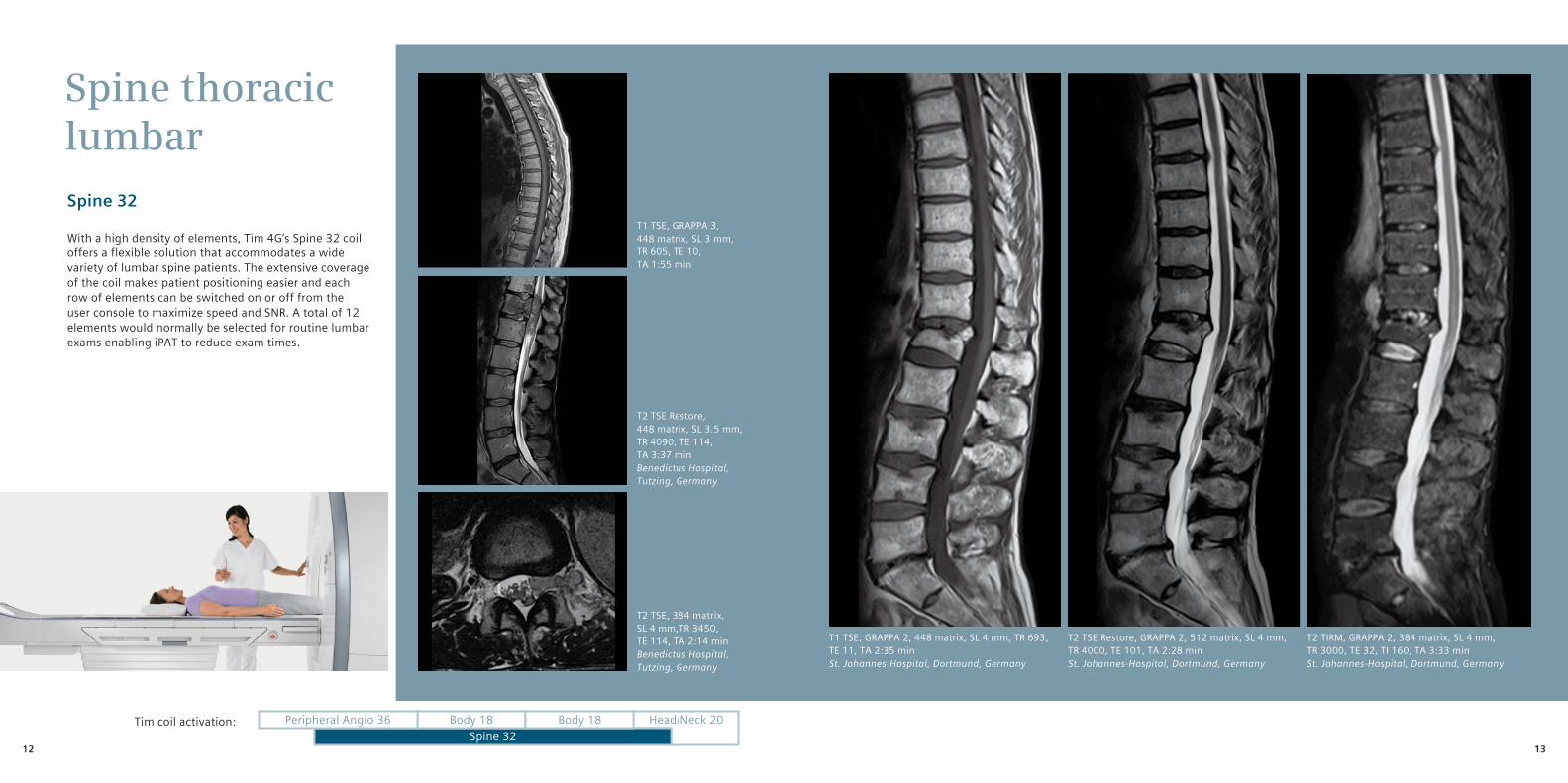
With a high density of elements, Tim 4G’s Spine 32 coil offers a flexible solution that accommodates a wide variety of lumbar spine patients. The extensive coverage of the coil makes patient positioning easier and each row of elements can be switched on or off from the user console to maximize speed and SNR. A total of 12 elements would normally be selected for routine lumbar exams enabling iPAT to reduce exam times.
Spine 32Body 18 Body 18Peripheral Angio 36 Head/Neck 20Tim coil activation:
T1 TSE, GRAPPA 3, 448 matrix, SL 3 mm,TR 605, TE 10, TA 1:55 min
T2 TSE Restore, 448 matrix, SL 3.5 mm,TR 4090, TE 114, TA 3:37 minBenedictus Hospital, Tutzing, Germany
T2 TSE, 384 matrix, SL 4 mm,TR 3450, TE 114, TA 2:14 minBenedictus Hospital, Tutzing, Germany
T1 TSE, GRAPPA 2, 448 matrix, SL 4 mm, TR 693, TE 11, TA 2:35 minSt. Johannes-Hospital, Dortmund, Germany
T2 TSE Restore, GRAPPA 2, 512 matrix, SL 4 mm, TR 4000, TE 101, TA 2:28 minSt. Johannes-Hospital, Dortmund, Germany
T2 TIRM, GRAPPA 2, 384 matrix, SL 4 mm, TR 3000, TE 32, TI 160, TA 3:33 minSt. Johannes-Hospital, Dortmund, Germany
1312
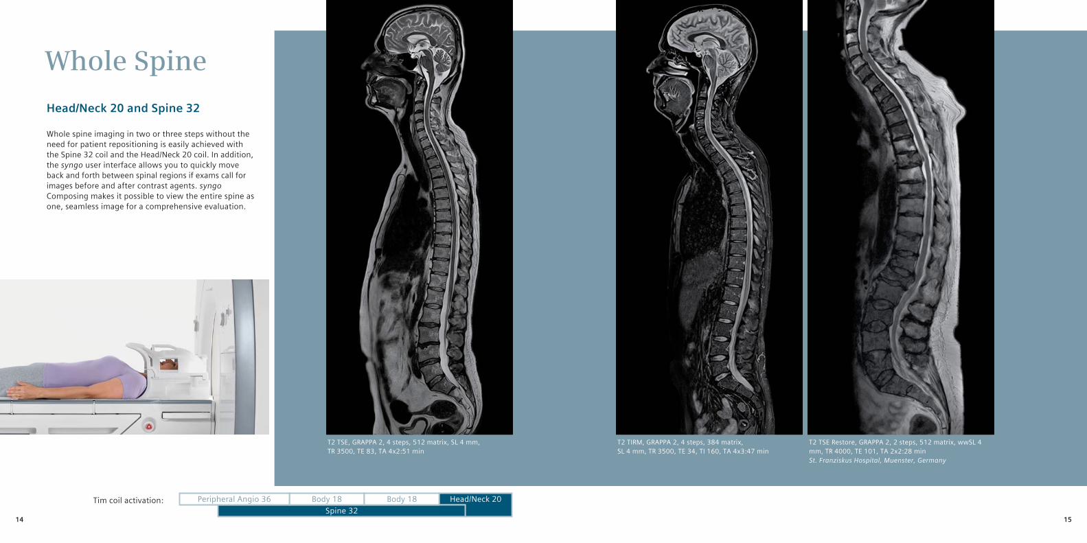
Whole Spine
Head/Neck 20 and Spine 32
Whole spine imaging in two or three steps without the need for patient repositioning is easily achieved with the Spine 32 coil and the Head/Neck 20 coil. In addition, the syngo user interface allows you to quickly move back and forth between spinal regions if exams call for images before and after contrast agents. syngo Composing makes it possible to view the entire spine as one, seamless image for a comprehensive evaluation.
Spine 32Body 18 Body 18Peripheral Angio 36 Head/Neck 20Tim coil activation:
T2 TIRM, GRAPPA 2, 4 steps, 384 matrix,SL 4 mm, TR 3500, TE 34, TI 160, TA 4x3:47 min
T2 TSE Restore, GRAPPA 2, 2 steps, 512 matrix, wwSL 4 mm, TR 4000, TE 101, TA 2x2:28 minSt. Franziskus Hospital, Muenster, Germany
T2 TSE, GRAPPA 2, 4 steps, 512 matrix, SL 4 mm, TR 3500, TE 83, TA 4x2:51 min
1514
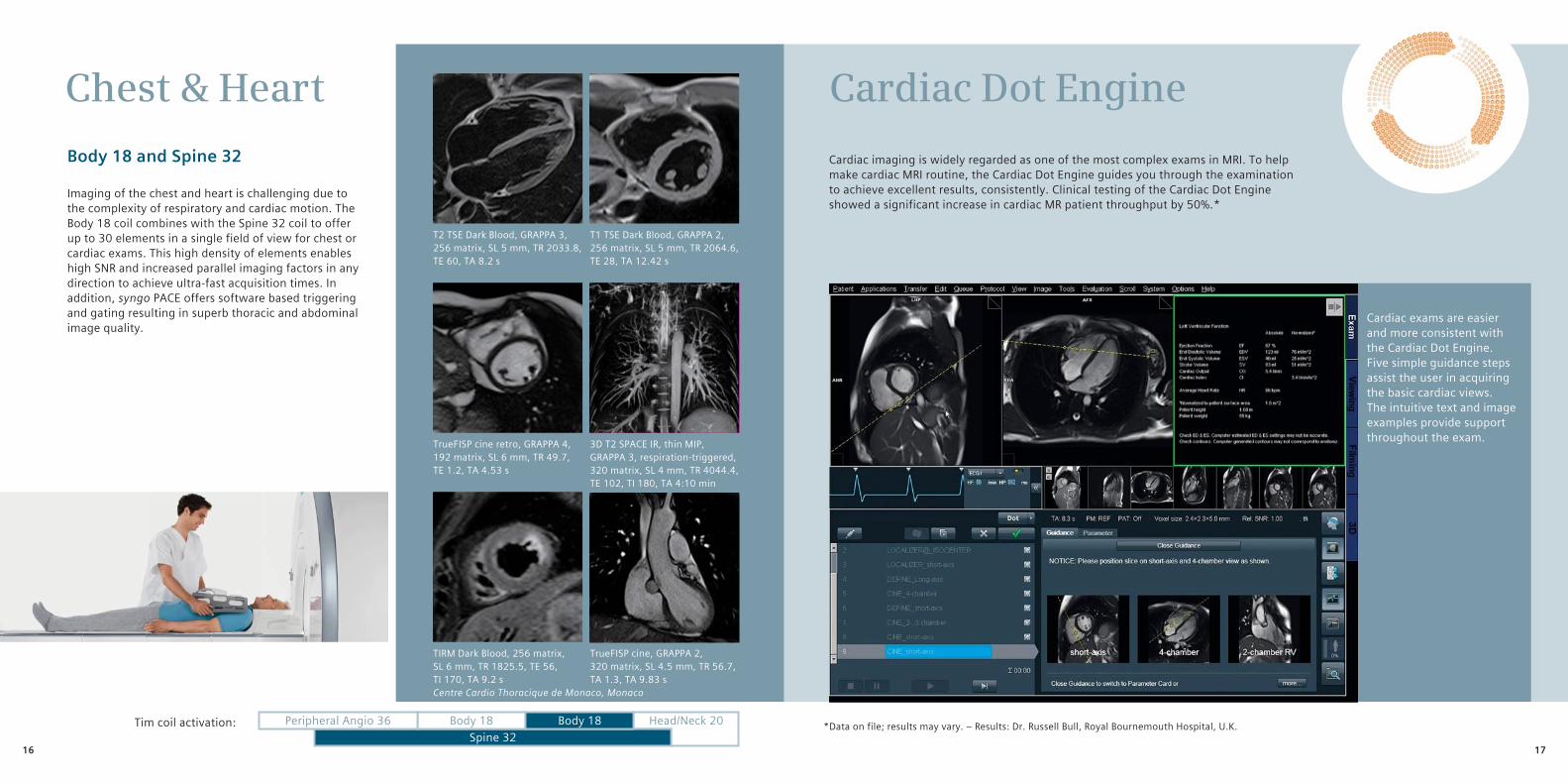
Chest & Heart
Body 18 and Spine 32
Imaging of the chest and heart is challenging due to the complexity of respiratory and cardiac motion. The Body 18 coil combines with the Spine 32 coil to offer up to 30 elements in a single field of view for chest or cardiac exams. This high density of elements enables high SNR and increased parallel imaging factors in any direction to achieve ultra-fast acquisition times. In addition, syngo PACE offers software based triggering and gating resulting in superb thoracic and abdominal image quality.
Spine 32Body 18 Body 18Peripheral Angio 36 Head/Neck 20Tim coil activation:
T1 TSE Dark Blood, GRAPPA 2, 256 matrix, SL 5 mm, TR 2064.6, TE 28, TA 12.42 s
T2 TSE Dark Blood, GRAPPA 3, 256 matrix, SL 5 mm, TR 2033.8, TE 60, TA 8.2 s
TrueFISP cine retro, GRAPPA 4, 192 matrix, SL 6 mm, TR 49.7, TE 1.2, TA 4.53 s
3D T2 SPACE IR, thin MIP, GRAPPA 3, respiration-triggered, 320 matrix, SL 4 mm, TR 4044.4, TE 102, TI 180, TA 4:10 min
TIRM Dark Blood, 256 matrix, SL 6 mm, TR 1825.5, TE 56, TI 170, TA 9.2 sCentre Cardio Thoracique de Monaco, Monaco
Cardiac imaging is widely regarded as one of the most complex exams in MRI. To help make cardiac MRI routine, the Cardiac Dot Engine guides you through the examination to achieve excellent results, consistently. Clinical testing of the Cardiac Dot Engine showed a significant increase in cardiac MR patient throughput by 50%.*
Cardiac Dot Engine
Cardiac exams are easier and more consistent with the Cardiac Dot Engine. Five simple guidance steps assist the user in acquiring the basic cardiac views. The intuitive text and image examples provide support throughout the exam.
* Data on file; results may vary. – Results: Dr. Russell Bull, Royal Bournemouth Hospital, U.K.
TrueFISP cine, GRAPPA 2, 320 matrix, SL 4.5 mm, TR 56.7, TA 1.3, TA 9.83 s
1716
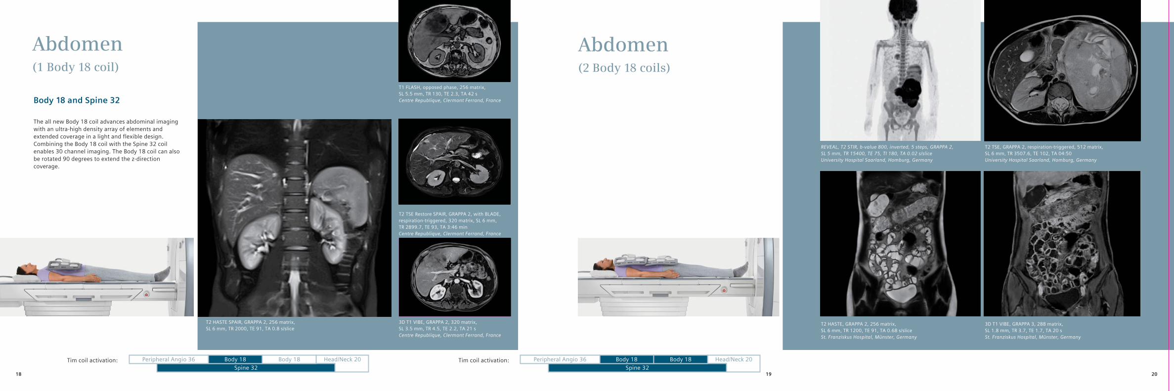
Abdomen(1 Body 18 coil)
Abdomen(2 Body 18 coils)
Body 18 and Spine 32
The all new Body 18 coil advances abdominal imaging with an ultra-high density array of elements and extended coverage in a light and flexible design. Combining the Body 18 coil with the Spine 32 coil enables 30 channel imaging. The Body 18 coil can also be rotated 90 degrees to extend the z-direction coverage.
Spine 32 Spine 32Body 18 Body 18Body 18 Body 18Peripheral Angio 36 Peripheral Angio 36Head/Neck 20 Head/Neck 20Tim coil activation: Tim coil activation:
3D T1 VIBE, GRAPPA 2, 320 matrix,SL 3.5 mm, TR 4.5, TE 2.2, TA 21 sCentre Republique, Clermont Ferrand, France
T2 TSE Restore SPAIR, GRAPPA 2, with BLADE, respiration-triggered, 320 matrix, SL 6 mm, TR 2899.7, TE 93, TA 3:46 minCentre Republique, Clermont Ferrand, France
T1 FLASH, opposed phase, 256 matrix,SL 5.5 mm, TR 130, TE 2.3, TA 42 sCentre Republique, Clermont Ferrand, France
T2 HASTE SPAIR, GRAPPA 2, 256 matrix,SL 6 mm, TR 2000, TE 91, TA 0.8 s/slice
REVEAL, T2 STIR, b-value 800, inverted, 5 steps, GRAPPA 2,SL 5 mm, TR 15400, TE 75, TI 180, TA 0.02 s/sliceUniversity Hospital Saarland, Homburg, Germany
T2 TSE, GRAPPA 2, respiration-triggered, 512 matrix,SL 6 mm, TR 3507.6, TE 102, TA 04:50University Hospital Saarland, Homburg, Germany
T2 HASTE, GRAPPA 2, 256 matrix,SL 6 mm, TR 1200, TE 91, TA 0.68 s/sliceSt. Franziskus Hospital, Münster, Germany
3D T1 VIBE, GRAPPA 3, 288 matrix,SL 1.8 mm, TR 3.7, TE 1.7, TA 20 sSt. Franziskus Hospital, Münster, Germany
201918
Spine 32Body 18 Body 18Peripheral Angio 36 Head/Neck 20Tim coil activation:
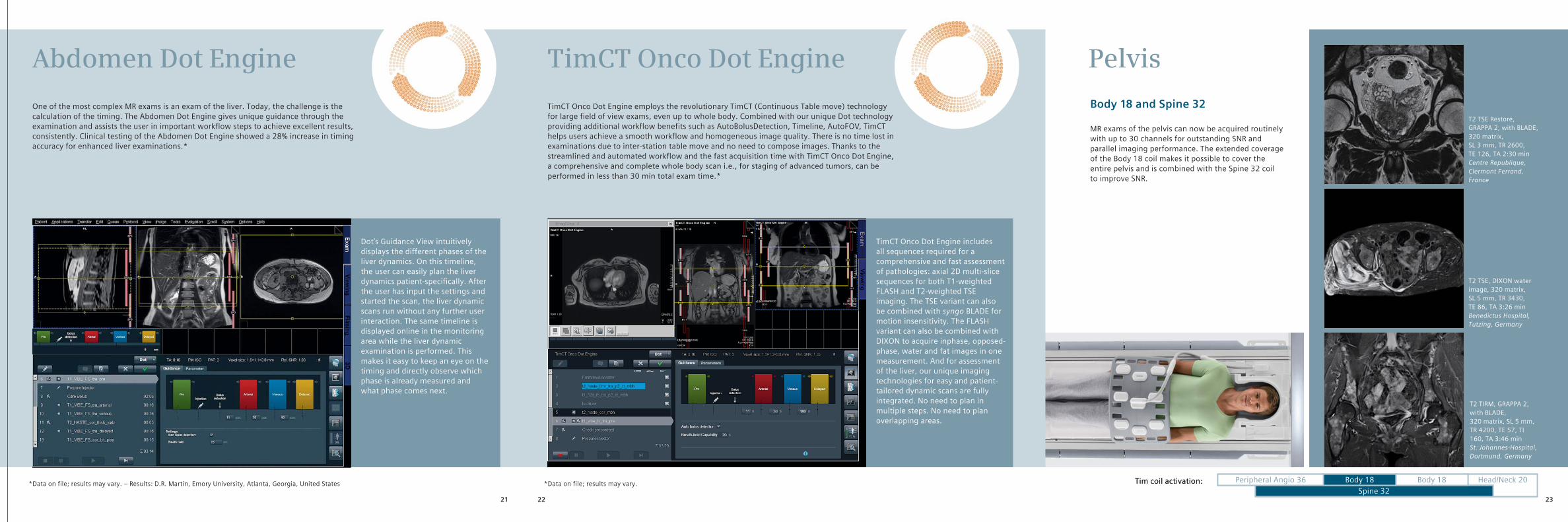
TimCT Onco Dot Engine employs the revolutionary TimCT (Continuous Table move) technology for large field of view exams, even up to whole body. Combined with our unique Dot technology providing additional workflow benefits such as AutoBolusDetection, Timeline, AutoFOV, TimCT helps users achieve a smooth workflow and homogeneous image quality. There is no time lost in examinations due to inter-station table move and no need to compose images. Thanks to the streamlined and automated workflow and the fast acquisition time with TimCT Onco Dot Engine, a compre hensive and complete whole body scan i.e., for staging of advanced tumors, can be performed in less than 30 min total exam time.*
One of the most complex MR exams is an exam of the liver. Today, the challenge is the calculation of the timing. The Abdomen Dot Engine gives unique guidance through the examination and assists the user in important workflow steps to achieve excellent results, consistently. Clinical testing of the Abdomen Dot Engine showed a 28% increase in timing accuracy for enhanced liver examinations.*
Abdomen Dot Engine TimCT Onco Dot Engine
TimCT Onco Dot Engine includes all sequences required for a comprehensive and fast assessment of pathologies: axial 2D multi-slice sequences for both T1-weighted FLASH and T2-weighted TSE imaging. The TSE variant can also be combined with syngo BLADE for motion insensitivity. The FLASH variant can also be combined with DIXON to acquire inphase, opposed-phase, water and fat images in one measurement. And for assessment of the liver, our unique imaging technologies for easy and patient-tailored dynamic scans are fully integrated. No need to plan in multiple steps. No need to plan overlapping areas.
Dot‘s Guidance View intuitively displays the different phases of the liver dynamics. On this timeline, the user can easily plan the liver dynamics patient-specifically. After the user has input the settings and started the scan, the liver dynamic scans run without any further user interaction. The same timeline is displayed online in the monitoring area while the liver dynamic examination is performed. This makes it easy to keep an eye on the timing and directly observe which phase is already measured and what phase comes next.
*Data on file; results may vary.*Data on file; results may vary. – Results: D.R. Martin, Emory University, Atlanta, Georgia, United States
Pelvis
Body 18 and Spine 32
MR exams of the pelvis can now be acquired routinely with up to 30 channels for outstanding SNR and parallel imaging performance. The extended coverage of the Body 18 coil makes it possible to cover the entire pelvis and is combined with the Spine 32 coil to improve SNR.
Spine 32Body 18 Body 18Peripheral Angio 36 Head/Neck 20Tim coil activation:
T2 TSE Restore, GRAPPA 2, with BLADE, 320 matrix,SL 3 mm, TR 2600, TE 126, TA 2:30 minCentre Republique, Clermont Ferrand, France
T2 TSE, DIXON water image, 320 matrix,SL 5 mm, TR 3430, TE 86, TA 3:26 minBenedictus Hospital, Tutzing, Germany
T2 TIRM, GRAPPA 2, with BLADE, 320 matrix, SL 5 mm, TR 4200, TE 57, TI 160, TA 3:46 minSt. Johannes-Hospital, Dortmund, Germany
22 2321
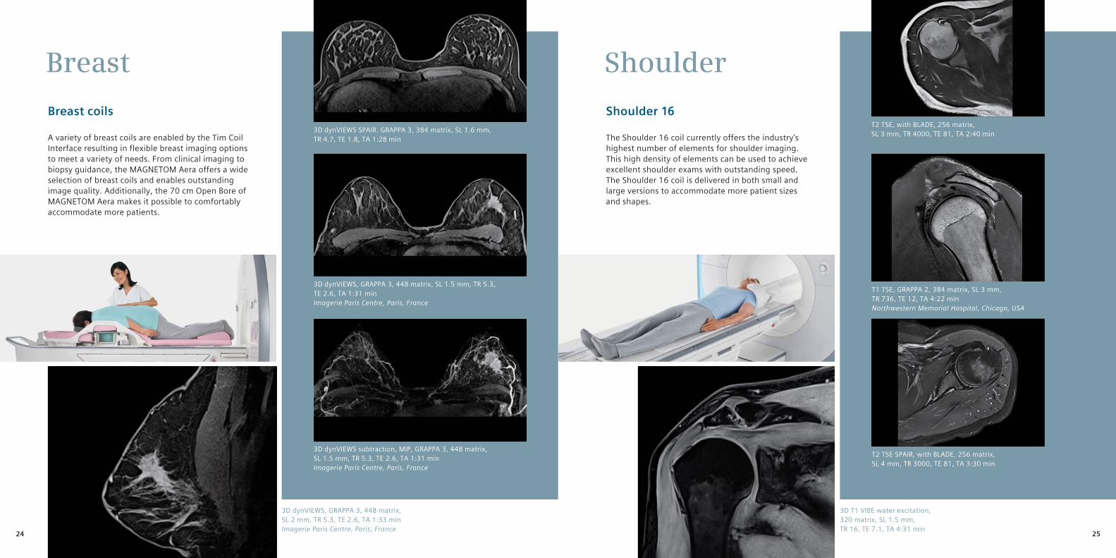
Breast Shoulder
Breast coils
A variety of breast coils are enabled by the Tim Coil Interface resulting in flexible breast imaging options to meet a variety of needs. From clinical imaging to biopsy guidance, the MAGNETOM Aera offers a wide selection of breast coils and enables outstanding image quality. Additionally, the 70 cm Open Bore of MAGNETOM Aera makes it possible to comfortably accommodate more patients.
Shoulder 16
The Shoulder 16 coil currently offers the industry’s highest number of elements for shoulder imaging. This high density of elements can be used to achieve excellent shoulder exams with outstanding speed. The Shoulder 16 coil is delivered in both small and large versions to accommodate more patient sizes and shapes.
3D dynVIEWS, GRAPPA 3, 448 matrix, SL 1.5 mm, TR 5.3, TE 2.6, TA 1:31 minImagerie Paris Centre, Paris, France
3D dynVIEWS SPAIR. GRAPPA 3, 384 matrix, SL 1.6 mm, TR 4.7, TE 1.8, TA 1:28 min
3D dynVIEWS subtraction, MIP, GRAPPA 3, 448 matrix, SL 1.5 mm, TR 5.3, TE 2.6, TA 1:31 minImagerie Paris Centre, Paris, France
T2 TSE, with BLADE, 256 matrix,SL 3 mm, TR 4000, TE 81, TA 2:40 min
T1 TSE, GRAPPA 2, 384 matrix, SL 3 mm,TR 736, TE 12, TA 4:22 minNorthwestern Memorial Hospital, Chicago, USA
T2 TSE SPAIR, with BLADE, 256 matrix,SL 4 mm, TR 3000, TE 81, TA 3:30 min
3D T1 VIBE water excitation, 320 matrix, SL 1.5 mm,TR 16, TE 7.1, TA 4:31 min
3D dynVIEWS, GRAPPA 3, 448 matrix,SL 2 mm, TR 5.3, TE 2.6, TA 1:33 minImagerie Paris Centre, Paris, France 2524
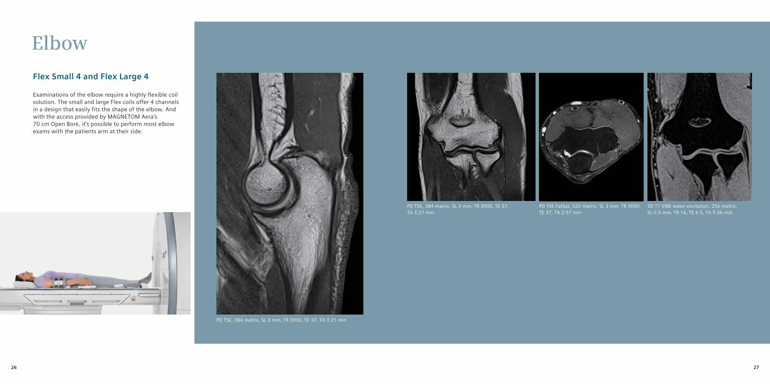
Elbow
Flex Small 4 and Flex Large 4
Examinations of the elbow require a highly flexible coil solution. The small and large Flex coils offer 4 channels in a design that easily fits the shape of the elbow. And with the access provided by MAGNETOM Aera’s 70 cm Open Bore, it’s possible to perform most elbow exams with the patients arm at their side.
PD TSE, 384 matrix, SL 3 mm, TR 3000, TE 37, TA 3:21 min
PD TSE FatSat, 320 matrix, SL 3 mm, TR 3000, TE 37, TA 2:57 min
3D T1 VIBE water excitation, 256 matrix,SL 0.5 mm, TR 16, TE 6.5, TA 5:36 min
PD TSE, 384 matrix, SL 3 mm, TR 3000, TE 37, TA 3:21 min
2726
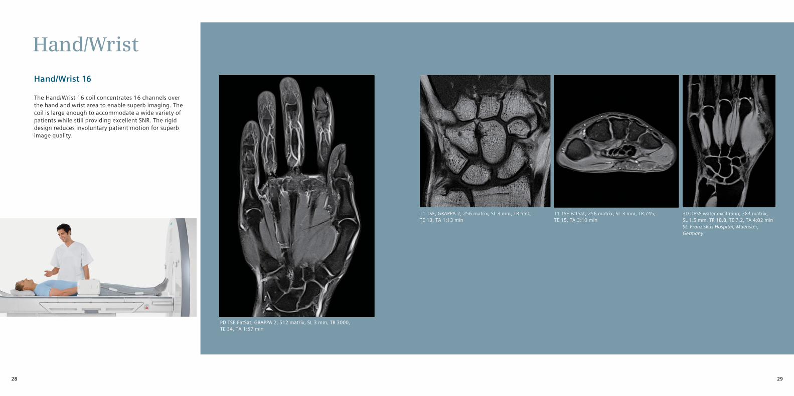
Hand/Wrist
Hand/Wrist 16
The Hand/Wrist 16 coil concentrates 16 channels over the hand and wrist area to enable superb imaging. The coil is large enough to accommodate a wide variety of patients while still providing excellent SNR. The rigid design reduces involuntary patient motion for superb image quality.
T1 TSE, GRAPPA 2, 256 matrix, SL 3 mm, TR 550, TE 13, TA 1:13 min
PD TSE FatSat, GRAPPA 2, 512 matrix, SL 3 mm, TR 3000, TE 34, TA 1:57 min
3D DESS water excitation, 384 matrix, SL 1.5 mm, TR 18.8, TE 7.2, TA 4:02 minSt. Franziskus Hospital, Muenster, Germany
T1 TSE FatSat, 256 matrix, SL 3 mm, TR 745, TE 15, TA 3:10 min
2928

KneeHip
Tx/Rx 15-Channel Knee Coil
The Tx/Rx 15-Channel Knee Coil enables parallel imaging in any direction to power 2D and 3D knee exams with excellent resolution and a fast exam time. The coil’s large architecture accommodates a wide variety of patients. If necessary, the Body 18 coil can also be used to image knees.
Body 18 and Spine 32
The Body 18 coil combines a high-density array with large or small field of view coverage for hip imaging. Hip exams with high SNR and high resolution are easily achieved. The coil is generally large enough to cover both hips for bilateral imaging or can be wrapped around one side to increase the element density when examining unilaterally.
PD TSE Restore FatSat, GRAPPA 2, 384 matrix,SL 2.5 mm, TR 3220, TE 29, TA 3:29 minBenedictus Hospital, Tutzing, Germany
T1 TSE, 512 matrix, SL 3 mm, TR 551, TE 20, TA 3:31 min
T2 TSE Restore FatSat, GRAPPA 2, 320 matrix,SL 3 mm, TR 5940, TR 63, TA 4:04 min
T1 FLASH FatSat, GRAPPA 2, 512 matrix,SL 3 mm, TR 710, TE 7.1, TA 4:25 min
PD TSE FatSat, 320 matrix, SL 3 mm, TR 5300, TE 49, TA 2:34 min
PD TSE Restore, 320 matrix, SL 3 mm,TR 2100, TE 23, TA 4:10 min
Spine 32Body 18 Body 18Peripheral Angio 36 Head/Neck 20Tim coil activation:
PD TSE Restore, 320 matrix, SL 3 mm,TR 2720, TE 20, TA 5:25 min
T2 TSE, 448 matrix, SL 3 mm,TR 4000, TE 77, TA 3:24 min
3130
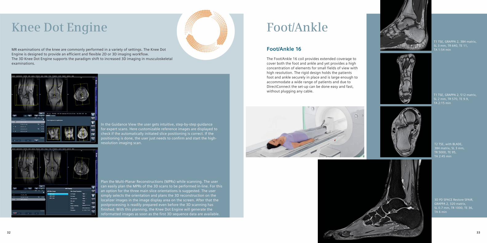
MR examinations of the knee are commonly performed in a variety of settings. The Knee Dot Engine is designed to provide an efficient and flexible 2D or 3D imaging workflow. The 3D Knee Dot Engine supports the paradigm shift to increased 3D imaging in musculoskeletal examinations.
Knee Dot Engine
In the Guidance View the user gets intuitive, step-by-step guidance for expert scans. Here customizable reference images are displayed to check if the automatically initiated slice positioning is correct. If the positioning is done, the user just needs to confirm and start the high-resolution imaging scan.
Plan the Multi-Planar Reconstructions (MPRs) while scanning. The user can easily plan the MPRs of the 3D scans to be performed in-line. For this an option for the three main slice orientations is suggested. The user simply selects the orientation and plans the 3D reconstruction on the localizer images in the image display area on the screen. After that the postprocessing is readily prepared even before the 3D scanning has finished. With this planning, the Knee Dot Engine will generate the reformatted images as soon as the first 3D sequence data are available.
Foot/Ankle
Foot/Ankle 16The Foot/Ankle 16 coil provides extended coverage to cover both the foot and ankle and yet provides a high concentration of elements for small fields of view with high resolution. The rigid design holds the patients foot and ankle securely in place and is large enough to accommodate a wide range of patients and due to DirectConnect the set-up can be done easy and fast, without plugging any cable.
T1 TSE, GRAPPA 2, 384 matrix,SL 3 mm, TR 640, TE 11, TA 1:54 min
T1 TSE, GRAPPA 2, 512 matrix,SL 2 mm, TR 570, TE 9.9, TA 2:15 min
T2 TSE, with BLADE, 384 matrix, SL 3 mm, TR 5000, TE 95, TA 2:45 min
3D PD SPACE Restore SPAIR, GRAPPA 2, 320 matrix,SL 0.7 mm, TR 1000, TE 36, TA 6 min
3332
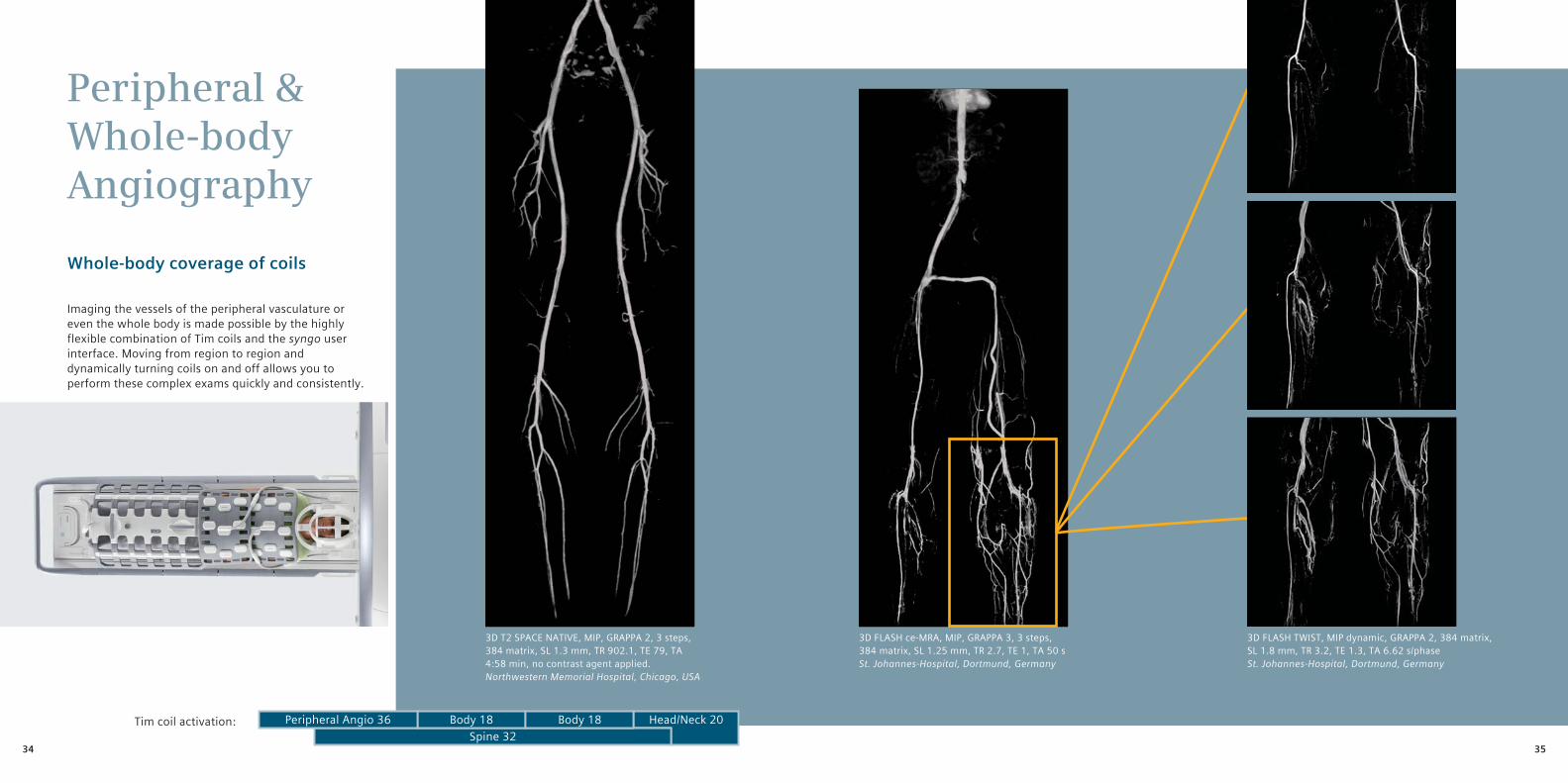
Peripheral & Whole-body Angiography
Whole-body coverage of coils
Imaging the vessels of the peripheral vasculature or even the whole body is made possible by the highly flexible combination of Tim coils and the syngo user interface. Moving from region to region and dynamically turning coils on and off allows you to perform these complex exams quickly and consistently.
Spine 32Body 18 Body 18Peripheral Angio 36 Head/Neck 20Tim coil activation:
3D FLASH ce-MRA, MIP, GRAPPA 3, 3 steps, 384 matrix, SL 1.25 mm, TR 2.7, TE 1, TA 50 sSt. Johannes-Hospital, Dortmund, Germany
3D FLASH TWIST, MIP dynamic, GRAPPA 2, 384 matrix,SL 1.8 mm, TR 3.2, TE 1.3, TA 6.62 s/phaseSt. Johannes-Hospital, Dortmund, Germany
3D T2 SPACE NATIVE, MIP, GRAPPA 2, 3 steps, 384 matrix, SL 1.3 mm, TR 902.1, TE 79, TA 4:58 min, no contrast agent applied.Northwestern Memorial Hospital, Chicago, USA
3534
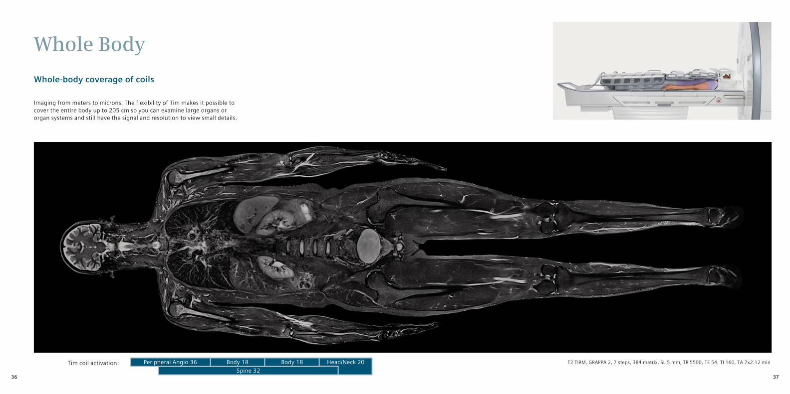
Whole Body
Whole-body coverage of coils
Imaging from meters to microns. The flexibility of Tim makes it possible to cover the entire body up to 205 cm so you can examine large organs or organ systems and still have the signal and resolution to view small details.
Spine 32Body 18 Body 18Peripheral Angio 36 Head/Neck 20Tim coil activation: T2 TIRM, GRAPPA 2, 7 steps, 384 matrix, SL 5 mm, TR 5500, TE 54, TI 160, TA 7x2:12 min
3736
Global Siemens HeadquartersSiemens AGWittelsbacherplatz 280333 MuenchenGermany
Global Siemens Healthcare HeadquartersSiemens AGHealthcare SectorHenkestrasse 12791052 ErlangenGermanyPhone: +49 9131 84-0www.siemens.com/healthcare
On account of certain regional limitations of sales rights and service availability, we cannot guarantee that all products included in this brochure are available through the Siemens sales organization worldwide. Availability and packaging may vary by country and are subject to change without prior notice. Some/All of the features and products described herein may not be available in the United States.
All devices listet herein may not be licensed according to Canadian Medical Devices Regulations. The information in this document contains general technical descriptions of specifications and options as well as standard and optional features which do not always have to be present in individual cases.
Siemens reserves the right to modify the design, packaging, specifications, and options described herein without prior notice. Please contact your local Siemens sales representative for the most current information.
Note: Any technical data contained in this document may vary within defined tolerances. Original images always lose a certain amount of detail when reproduced.
Please find fitting accessories: www.siemens.com/medical-accessories
Local Contact Information
In the USASiemens Medical Solutions USA, Inc.51 Valley Stream ParkwayMalvern, PA 19355Phone: +1 888-826-9702Phone: +1 610-448-4500Fax: +1 610-448-2254
In ChinaSiemens Medical Park, Shanghai278, Zhouzhu RoadSIMZ, Nanhui DistrictShanghai, 201318P.R. ChinaPhone: +86-21-38895000Fax: +86-10-28895001
In JapanSiemens-AsahiMedical Technologies Ltd.Takanawa Park Tower 14F20-14, Higashi-Gotanda 3-chomeShinagawa-kuTokyo 141-8644Phone: +81 3 5423 8411
In AsiaSiemens Pte LtdHealthcare SectorRegional HeadquartersThe Siemens Center60 MacPherson RoadSingapore 348615Phone: +65 6490-6000Telefax: +65 6490-6001
Order No. A91MR-9011-6C-7600 | Printed in Germany | CC MR 09111.5 | © 09.2011, Siemens AG
Global Business Unit
Siemens AGHealthcare SectorMagnetic ResonanceHenkestr. 127DE-91052 ErlangenGermanyPhone: +49 9131 84-0
Legal Manufacturer
Siemens AGWittelsbacherplatz 2DE-80333 MuenchenGermany
Siemens Mindit MagneticResonance Ltd. (SMMR)Siemens MRI Center, Gaoxin C. Ave.,2nd, Hi-Tech Industrial ParkShenzen 518057P.R. ChinaPhone: +86 755-26525421Fax: +86 755-26549253
Answers for life.
MAGNETOM AeraImage Gallery
www.siemens.com/aera