Embed Size (px)
Citation preview

MPFL SystemTM
Surgical Technique Manual
For Medial Patellofemoral Ligament Reconstruction
0086
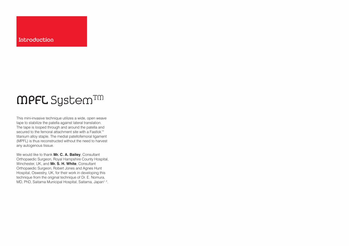
MPFL SystemTM
Introduction
This mini-invasive technique utilizes a wide, open weave tape to stabilize the patella against lateral translation. The tape is looped through and around the patella and secured to the femoral attachment site with a Fastlok™ titanium alloy staple. The medial patellofemoral ligament (MPFL) is thus reconstructed without the need to harvest any autogenous tissue.
We would like to thank Mr. C. A. Bailey, Consultant Orthopaedic Surgeon, Royal Hampshire County Hospital, Winchester, UK, and Mr. S. H. White, Consultant Orthopaedic Surgeon, Robert Jones and Agnes Hunt Hospital, Oswestry, UK, for their work in developing this technique from the original technique of Dr. E. Nomura, MD, PhD, Saitama Municipal Hospital, Saitama, Japan1, 2.

INDICATIONThe MPFL System is indicated for the following:• Reconstruction of the medial patellofemoral
ligament in cases of recurrent patellar subluxation or dislocation
Contraindications to surgery include:• Known hypersensitivity to implant materials. If the
patient is suspected of having any foreign body sensitivity, appropriate tests should be made prior to implantation.
• Infections or any structural or pathological condition of the bone or soft tissue that would be expected to impair healing or secure fixation.
• Patients unable or unwilling to restrict activities to prescribed levels or follow a rehabilitation programme during the healing period.
• Skeletally immature patients. The MPFL System will not elongate with growth and must not bridge or disrupt the growth plate.
• Poly-Tapes integrate well with the patient’s tissue and consequently may not be suitable where eventual removal of the tape is anticipated.
Caution should be advised when patients present with the following conditions:• A Q angle greater than 25º• Severe patella alta (patella height assessed by the
Insall-Salvati index greater than 1.3)

Product Overview
IMPLANTSThe MPFL System uses a 10 mm x 500 mm Poly-Tape in conjunction with a 6 mm Fastlok fixation device.
The Poly-Tape is a flexible textile device which has the following advantages:• Manufactured from polyester, a biocompatible
material that has been in use for the reconstruction of ligaments and tendons for over 25 years
• Obviates the need for harvesting autograft; avoids donor site morbidity and additional incisions
The Poly-Tape has several structural features intended to facilitate the surgery and improve its outcome:• Continuous longitudinal yarns of the device provide it
with sufficient strength to allow early mobilization• Flat open weave section acts as a scaffold that
encourages tissue ingrowth3
The Fastlok is made from a titanium alloy and consists of a staple and buckle. It provides a unique triple clamping action to minimize slippage under repeated loading.
6 mm Fastlok
10 mm x 500 mm Poly-Tape

INSTRUMENTATION The following instruments are packaged with the implant set:• 20 cm malleable probe with eye for passing the Poly-
Tape through the bone tunnel• 3.2 mm diameter drill bit (plain shank) Fastlok Instruments:• Impactor/Extractor• Sliding Hammer

PREPARATION AND INSPECTIONThe procedure is performed with the patient in the supine position under general or spinal anaesthesia with a tourniquet inflated. Preoperative antibiotics are administered.
The knee is positioned using a side support and a sandbag so the limb can be flexed and held at 90º.
RECOMMENDED APPROACHA medial longitudinal incision is made two thirds of the distance between the medial epicondyle and the medial border of the patella. The incision is 4-6 cm long and should give access to the adductor tubercle and the lateral border of the patella.
Superficial dissection is performed to the medial patellofemoral retinaculum.
NOTE: Take care not to damage the infrapatellar branch of the saphenous nerve.
The entry point for the patellar bone tunnel is marked on the medial border of the patella within the centre of the MPFL footprint. This is approximately at the junction of the proximal and middle thirds of the patella.
NOTE: To avoid tunnel “blow out” do not place the tunnel too far anterior in the sagittal plane.
Surgical Technique
1 2
MPFL
Entry point for the patellar bone tunnel approximately 1/3 patella height
Proximal 1/3
Middle 1/3
Distal 1/3
MPFL
Vastus medialis
Incision 4-6 cm long

A flap of the medial patellofemoral retinaculum 10 mm wide and 6-8 cm long is extended from the point marked on the medial border of the patella to the point of origin of the ligament. The flap is then raised using sharp dissection and left attached to the patella. This usually passes just distal to the vastus medialis muscle. If the synovial lining of the knee joint is breached it may be repaired using a 2/0 vicryl suture.
Femoral fixation is located within the footprint of the origin of the medial patellofemoral ligament. The origin is just distal to the adductor tubercle and lies approximately equidistant to the medial epicondyle and the adductor tubercle.
NOTE: Take care to diathermy blood vessels at that site (branches of the descending geniculate and superior medial geniculate arteries).
Using sharp dissection and then a Bohler bone rongeur, soft tissue and a slither of cortical bone are removed over this point 12 mm in length by 3 mm in width. This should be approximately 45º to the long axis of the femur. This is to allow insertion and burial of the Fastlok fixation device, the undue prominence of which leads to pain and tenderness.
With the knee in extension, the medial border of the patella can be identified and a 3.2 mm drill bit (provided) is passed from the medial side, emerging from the patella at its anterolateral border.
If the exit point and orientation of the drill hole are not correct, the tunnel can be repositioned by over-drilling using a 4.5 mm drill bit.
If a larger tunnel is preferred for threading the Poly-Tape, the 3.2 mm drill hole can be expanded using a 4.5 mm drill bit.
NOTE: Where possible, round the tunnel edges to prevent abrasion of the Poly-Tape.
3 54
Vastus medialis
Retinaculum flap
Adductor tubercle
Medial epicondyle
Origin of the MPFL
Remove soft tissue to allow for insertion of Fastlok
Direction of MPFL

7 8
With the knee in extension, the probe (provided) is used to pass the 10 mm x 500 mm Poly-Tape through the patellar tunnel from medial to lateral. The Crile artery forceps are passed across the anterior surface of the patella, deep to the anterior retinaculum and the Poly-Tape is passed back across the patella from lateral to medial.
The knee is then flexed and extended while the Poly-Tape is held to the attachment site to ensure the patella tracks well throughout the range of flexion and the Poly-Tape is not too tight. In some cases an isometric position can be achieved. When isometric, the knee should be positioned at 60o flexion when performing the fixation. This allows a normal position for the patella to be judged. However, the greatest tension is usually found in full flexion and less often in full extension. Fixation of the Poly-Tape should then be performed with the knee positioned at whichever angle creates the greatest tension, to avoid over-tightening the Poly-Tape.
NOTE: Placement of the femoral attachment site too proximally may cause tightening of the reconstruction in flexion. Placement too distal may cause tightening of the reconstruction in extension.
The Poly-Tape is then threaded to the Fastlok as follows:
Figure 7a: The staple is gripped firmly within the Impactor. The two free ends of the Poly-Tape are threaded through the Fastlok buckle, which is then held at its middle with forceps, so that both strands of the tape are suspended from the buckle. The Impactor is held above the buckle so that the legs of the staple are parallel with the buckle.
Figure 7b: The staple is brought down past the side of the buckle over which the Poly-Tape is folded, so that the legs of the staple straddle the four strands. The staple (via the Impactor) is turned through 90º in the horizontal plane.
Figure 7c: The buckle is then flipped down through 180º over the prongs of the staple. The staple legs are checked to ensure they still straddle both strands of the tape and that the legs do not pierce the tapes.
For a detailed description of the Fastlok technique please see the Instructions For Use leaflet packaged with the Fastlok (LAB 108).
The Fastlok is then introduced to the femoral attachment site as follows:
The staple is rotated so the sharp prongs are orientated approximately 45º to the long axis of the femur and slightly forwards towards the patella, avoiding entry into the knee joint.
Figure 8a: The two ends of the Poly-Tape are simultaneously pulled while pushing the staple (via the Impactor) along the tape, forwards toward the chosen site for implanting the Fastlok, until the prongs reach the bone surface.
NOTE: During this step do not straighten the tape. Its two ends must be at the angle shown, otherwise it may be difficult to push the staple forward.
Figure 8b: Keeping the tape between the staple legs, the Impactor is adjusted so that its axis is perpendicular to the surface of the bone at the implantation site.
6
Ensure the Poly-Tape lies flat on the anterior surface of the patella. Avoid twisting or rolling of the Poly-Tape. Figure 7a
Figure 7b
Figure 7c
Free ends of tape
90º
180º
Figure 8a
Figure 8b
Push Impactor
Pull ends of tape
Tension
Pull
Pull
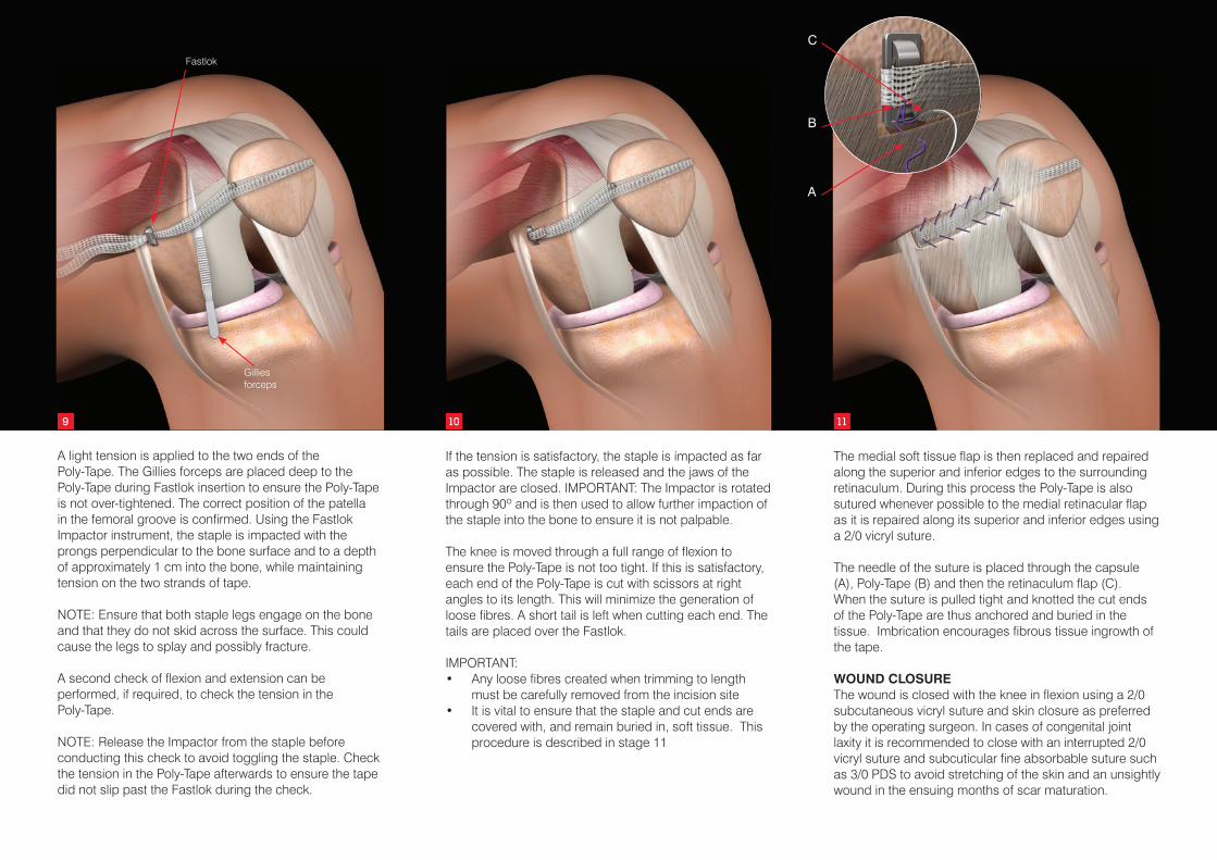
A light tension is applied to the two ends of the Poly-Tape. The Gillies forceps are placed deep to the Poly-Tape during Fastlok insertion to ensure the Poly-Tape is not over-tightened. The correct position of the patella in the femoral groove is confirmed. Using the Fastlok Impactor instrument, the staple is impacted with the prongs perpendicular to the bone surface and to a depth of approximately 1 cm into the bone, while maintaining tension on the two strands of tape.
NOTE: Ensure that both staple legs engage on the bone and that they do not skid across the surface. This could cause the legs to splay and possibly fracture.
A second check of flexion and extension can be performed, if required, to check the tension in the Poly-Tape.
NOTE: Release the Impactor from the staple before conducting this check to avoid toggling the staple. Check the tension in the Poly-Tape afterwards to ensure the tape did not slip past the Fastlok during the check.
If the tension is satisfactory, the staple is impacted as far as possible. The staple is released and the jaws of the Impactor are closed. IMPORTANT: The Impactor is rotated through 90º and is then used to allow further impaction of the staple into the bone to ensure it is not palpable.
The knee is moved through a full range of flexion to ensure the Poly-Tape is not too tight. If this is satisfactory, each end of the Poly-Tape is cut with scissors at right angles to its length. This will minimize the generation of loose fibres. A short tail is left when cutting each end. The tails are placed over the Fastlok.
IMPORTANT:• Any loose fibres created when trimming to length
must be carefully removed from the incision site• It is vital to ensure that the staple and cut ends are
covered with, and remain buried in, soft tissue. This procedure is described in stage 11
The medial soft tissue flap is then replaced and repaired along the superior and inferior edges to the surrounding retinaculum. During this process the Poly-Tape is also sutured whenever possible to the medial retinacular flap as it is repaired along its superior and inferior edges using a 2/0 vicryl suture.
The needle of the suture is placed through the capsule (A), Poly-Tape (B) and then the retinaculum flap (C). When the suture is pulled tight and knotted the cut ends of the Poly-Tape are thus anchored and buried in the tissue. Imbrication encourages fibrous tissue ingrowth of the tape.
WOUND CLOSUREThe wound is closed with the knee in flexion using a 2/0 subcutaneous vicryl suture and skin closure as preferred by the operating surgeon. In cases of congenital joint laxity it is recommended to close with an interrupted 2/0 vicryl suture and subcuticular fine absorbable suture such as 3/0 PDS to avoid stretching of the skin and an unsightly wound in the ensuing months of scar maturation.
9 10 11
B
C
APull ends of tape
Gilliesforceps
Fastlok

POSTOPERATIVE MANAGEMENT
The rehabilitation programme (below) provides only an outline of the prescribed regime. For a full description refer to the document entitled “MPFL System Rehabilitation Programme for Medial Patellofemoral Ligament Reconstruction” (LAB 135).
The rehabilitation programme should be supervised by a specialist physiotherapist. All mobilization and exercises should be performed within the pain free range of movement.
As in any implant surgery, satisfactory wound healing is of paramount importance.
The patient should be warned not to exceed the prescribed activity levels or to overload the repair before complete healing has occurred.
This rehabilitation programme was developed in conjunction with Ian Horsley MSc, MCSP, Clinical Lead Physiotherapist, English Institute of Sport (EIS) North West, of BackinAction Physiotherapy and Sports Injury Clinic, Wakefield, UK.
Immediate Postoperative Actions• Cast immobilization is unnecessary. • A cricket pad splint is applied in theatre and the
patient usually stays in hospital overnight, followed by consultation with a physiotherapist prior to discharge the following morning.
Weeks 0-2• The patient may fully weight bear with crutches
initially and is instructed to perform isometric exercises.
Week 2• The patient is seen in clinic at two weeks to discard
the splint, inspect the wound and remove the stitches.
REFERENCES1. Nomura E, Horiuchi Y, Kihara M. A mid-term
follow-up of medial patellofemoral ligament reconstruction using an artificial ligament for recurrent patellar dislocation. The Knee. 2000;7(4):211-215.
2. Nomura E, Inoue M, Kobayashi S. Long-term follow-up and knee osteoarthritis change after medial patellofemoral ligament reconstruction for recurrent patellar dislocation. Am J Sports Med. 2007;35(11):1851-1858.
3. Nomura E, Inoue M, Sugiura H. Histological evaluation of medial patellofemoral ligament reconstructed using the Leeds-Keio ligament prosthesis. Biomaterials. 2005;26(15):2663-2670.
Weeks 2-6• Physiotherapy continues with knee flexion exercises
and glute medius exercises. Heel walking and toe walking are commenced.
Weeks 6-12• Light exercise may be undertaken such as walking,
running on a treadmill and cycling.
Week 12 Onwards• On agreement with the physiotherapist the patient
can commence functional training and return to competitive sport.

102-1060 MPFL System Implant Set, includes:
Poly-Tape, 10 mm x 500 mm (supplied sterile) Fastlok, 6 mm x 23 mm (supplied sterile)
Packaged with the following disposables:
Malleable Probe with eye, stainless steel, 20 cm (supplied sterile)Drill Bit, plain shank to fit Jacobs Chuck, 3.2 mm diameter (supplied sterile)
Fastlok Instruments:
202-1137 Impactor/Extractor (non-sterile) 202-1118 Sliding Hammer (non-sterile)
Individual re-order codes
102-1010 Poly-Tape, 10 mm x 500 mm (supplied sterile)102-1380 Fastlok, 6 mm x 23 mm (supplied sterile)202-3026 Malleable Probe with eye, stainless steel, 20cm (supplied sterile)
Please refer to the Instructions for Use leaflets packaged with the Poly-Tape and Fastlok for essential information including Use, Sterility, Indications, Contraindications, Warnings and Precautions, Potential Adverse Effects and Storage. Additional copies may be obtained from the Neoligaments™ Sales Department, or downloaded from www.neoligaments.com
Ordering Information

Neoligaments™ A division of Xiros™ Springfield House Whitehouse Lane Leeds LS19 7UE
Tel. +44 (0) 113 238 7202 Fax. +44 (0) 113 238 7201 [email protected] www.neoligaments.com
Xiros Limited, registered in England No. 1664824.
All rights reserved. © Neoligaments™ 2017. Worldwide patents and patents pending. Neoligaments, MPFL System, Fastlok and Xiros are trademarks of Xiros.
Developed and manufactured by
LAB 137 4.00