Embed Size (px)
Citation preview
Vol. 191, No. 4S, Supplement, Sunday, May 18, 2014 THE JOURNAL OF UROLOGY� e379
Kidney Cancer: Evalution/Staging III
Moderated Poster
Sunday, May 18, 2014 1:00 PM-3:00 PM
MP36-01CAN MULTIPHASE CT SCAN DISTINGUISH BETWEEN TYPE 1AND TYPE 2 PAPILLARY RENAL CELL CARCINOMA? ARETROSPECTIVE ANALYSIS
Michelle McDonald*, Hak Lee, Vipulkumar Dadhania, Song Wang,Ryan Kopp, Alp Tuna Beksac, Lejla Aganovic, Fiona Hughes,Jessica Wang-Rodriguez, Ahmed Shabaik, Donna Hansel,Ithaar Derweesh, San Diego, CA
INTRODUCTION AND OBJECTIVES: Advances in imagingtechnology and understanding differences in renal tumor histology haveled to improved management of small renal masses. Several studieshave suggested that papillary renal cell carcinoma Type 1 (T1-pRCC)and Type 2 (T2-pRCC) have distinct behaviors and that prediction ofmorphology may be possible by imaging. We investigated imagingcharacteristics of T1- and T2-pRCC and their pathological correlates todetermine utility of multiphase computerized tomography (CT), and theCT enhancement washout formula, in differentiating between T1 andT2-pRCC subtypes.
METHODS: Retrospective analysis was performed on 39 pa-tients (18 T1-pRCC/21 T2-pRCC) who underwent surgical extirpationand had multiphase CT at our institution from 12/2007-7/2012. Pa-thology was confirmed and data was analyzed between subgroupsbased on histology. Multiphase CT was analyzed and tumor size,morphology, and attenuation in Hounsfield Units (HU) were recorded.Change in HU (dHU) was calculated between noncontrast (NC), cor-ticomedullary (CM), nephrographic (N) and delayed (D) phases.Enhancement washout was calculated by formula (N HU-D HU)/(NHU-NC HU).
RESULTS: There was no difference in median tumor size(T1-pRCC 2.8 vs. T2-pRCC 2.0 cm, p¼0.520). Significantly greaterproportion of high grade tumors (III/IV) were noted in pRCC-T2(42.9%) vs. pRCC-T1 (5.6%) (p¼0.011). There were no imagingdifferences between pRCC subtypes with respect to: frequency ofirregular borders (5.6% vs. 14.3%, p¼0.609), presence of calcifi-cations (11.1% vs. 14.3%, p¼1.000), presence of necrosis (11.1%vs. 28.6%, p¼0.247), or heterogeneous enhancement (16.7% vs.28.8%, p¼0.464). There was no difference in dHU between CM-NC(p¼0.126), and D-NC (p¼0.065). However, T2-pRCC had higherdHU between N-NC (42.7) vs. T1-pRCC (27.8, p¼0.036). Similarproportions of T1- (61.1%) and T2-pRCC (52.4%) tumors had anenhancement washout <0 (p¼0.584).
CONCLUSIONS: In our well-matched pRCC groups withrespect to size, there was substantial overlap of key radiographicfindings, despite T2-pRCC having greater proportion of high grade tu-mors. Caution should be exercised in utilization of CT to determinebetween pRCC subtypes. If pRCC is suspected on multiphase CT andrisk stratification is necessary prior to offering active treatment, percu-taneous biopsy should be strongly considered prior to placing a patienton an active surveillance protocol. Further prospective investigation isrequisite to confirm these findings.
Source of Funding: None
MP36-02VALIDATION AND GENOMIC INTERROGATION OF THE METVARIANT RS11762213 AS A PREDICTOR OF ADVERSEOUTCOMES IN CLEAR CELL RENAL CELL CARCINOMA
A Ari Hakimi*, Irina Ostrovnaya, Anders Jacobsen, Jonathan Coleman,Paul Russo, Roy Mano, Alex Sankin, Robert J. Motzer, New York, NY;Mark Purdue, Bethesda, MD; Mark Pomerantz, Matthew Freedman,Toni Choueiri, Boston, MA; James J. Hsieh, Robert J. Klein, New York, NY
INTRODUCTION AND OBJECTIVES: The exonic single nucle-otide variant rs11762213 located in the MET oncogene has recently beenidentified as a prognostic marker in clear cell renal cell carcinoma (ccRCC).We sought to validate this finding using the Cancer Genome Atlas cohortand explore the biologic implications utilizing the available genomic data.
METHODS: The variant call file (VCF) was available for 287 pa-tients ofwhom272hadexpression dataavailable.We thenextracted thedatafor rs11762213 as follows for the "normal" sample: allelic fraction for variantallele > 80% is homozygous variant while allelic fraction for variant allelebetween 30% and 70% is heterozygous. Paired tumor-normal materials,genomic data (whole exome, RNAseq, and DNA methylation) and clinicalinformation were acquired from publically available ccRCC TCGA datasets.
Associations between binary variables, and between binary andcontinuous variables, were assessed using Fisher’s Exact and MannWhitney two tailed tests, respectively. Kaplan-Meier method was usedto estimate the survival probabilities. Cancer specific survival (CSS)was analyzed using the competing risk method and Cox proportionalhazard regression was used for analysis of time to recurrence. Multi-variate competing risk models were also fitted in the TCGA cohort inorder to adjust for the validated Mayo Clinic SSIGN score.
RESULTS: Overall the variant allele of rs11762213 was detec-ted in 10.3% of the cohort and was associated with higher nuclear grade(p¼0.03) and trended toward higher clinical stage (p¼0.07). Afteradjusting for the prognostic SSIGN score, the risk allele remained asignificant predictor for adverse cancer specific survival (CSS; p<0.0001;Odds Ratio [OR] 3.88, 95% confidence interval [CI] 1.99-7.56) and fortime to recurrence (TTR; p¼0.003; OR 2.97; 95% CI 1.44-6.2). RNAsequencing data for MET did not reveal differences in tumor mRNAexpression when stratified by risk allele, but did show differences in thenormal kidney expression (p¼0.02) in a smaller cohort (n¼61).
CONCLUSIONS: The exonic MET variant rs11762213 is anindependent predictor of adverse CSS and TTR in ccRCC and shouldbe integrated into clinical practice for prognostic stratification. Geneexpression analysis suggests a biologic effect on the normal kidneyMET mRNA expression. Further external validation and biologicalinterrogation is necessary.
Source of Funding: This work has been supported by the PaulaMoss Trust for the research into the cure and treatment of kidneycancer (Hsieh), the Sidney Kimmel Center for Prostate andUrologic Cancers, by funds provided by David H. Koch throughthe Prostate Cancer Foundation, the National Cancer InstituteT32 CA082088-12 training grant (Hakimi), and the Stephen PHanson Family Fund Fellowship in Kidney Cancer (Hakimi).
MP36-03MAJOR HISTOCOMPATIBILITY COMPLEX CLASS I EXPRESSIONIN CLEAR CELL RENAL CELL CARCINOMA: CORRELATION WITHCLINICAL OUTCOME
Sarah Holzman*, Adeboye Osunkoya, Brian Pollack, Kenneth Ogan,Viraj Master, Atlanta, GA
INTRODUCTION AND OBJECTIVES: The majority of clear cellrenal cell carcinoma (CCRCC) patients initially present with localizeddisease with the potential for curative nephrectomy. Unfortunately,about one-third subsequently develop metastatic disease. Becausecurrent models of CCRCC recurrence have poor predictive power, thereis a need to develop better predictors of metastasis. Prior studies haveshown that CCRCC progression is susceptible to immune systemmodulation. Notably, tumor cell expression of major histocompatibilitycomplex class I (MHCI) has been shown to be critical for recognition
e380 THE JOURNAL OF UROLOGY� Vol. 191, No. 4S, Supplement, Sunday, May 18, 2014
and suppression by the host immune system. In this study we analyzedMHCI expression in CCRCC with emphasis on correlation with clin-ical outcome.
METHODS: We identified CCRCC patients that had radical ne-phrectomy as monotherapy, and had at least four years of follow-up data.Patients with T4 disease or metastasis at presentation were excluded. Allslides were reviewed by a Urologic Pathologist; a block with tumor andadjacent renal parenchyma was selected from each case. Immunohis-tochemical staining for MHCI was performed. We utilized whole slidescanning and automated image analysis; representative areas of tumorand normal kidney were selected and averaged using Aperio imageanalysis software (positive pixel count version 9). Unpaired t-test andone-way ANOVA were performed in GraphPad Prism.
RESULTS: In this pilot study, 34 patients that met our criteriawere selected for analysis. Fuhrman nuclear grades (FNG) were asfollows: FNG 2 10/34 patients (29%), FNG 3 20/34 patients (59%) andFNG 4 4/34 patients (11%). Although there was no statistically signifi-cant correlation with Fuhrman nuclear grade (ANOVA, p¼0.800), pa-tients who were alive at follow up had increased MHCI expression(80.1% average positivity score) than those who died of disease (53%average positivity score; t-test, p<0.0001). Patients who were alive withrecurrence had increased MHCI expression (81.3% positivity score)compared to those who succumbed to disease recurrence (53.2%positivity score; t-test, p<0.0001).
CONCLUSIONS: This study demonstrates that MHCI expressionmay be an important prognostic factor in CCRCC for recurrence freesurvival and for the prognosis of patients with recurrence. Our study is thefirst to demonstrate that increased MHCI expression is a favorable prog-nostic indicator in patients with metastatic CCRCC. Additionally, our resultssuggest that MHCI expression plays an important role in tumor-host im-mune system interaction in CCRCC and deserves further investigation.
Source of Funding: none
MP36-04QUANTIFICATION OF RENAL CELL OPTICAL BIOMARKERSUSING SECOND HARMONIC GENERATION IMAGING
Sara Best*, Terra Thimm, Yuming Liu, Matthew Houlihan,Jeremy Bredfelt, Kevin Eliceiri, Madison, WI
INTRODUCTION AND OBJECTIVES: Initial investigationsexamining Second Harmonic Generation (SHG) based optical bio-markers have revealed subjective differences in intrinsic collagen sig-nals between malignant and benign renal tissue. Improvements indigital analytic technology and software are permitting quantification ofthis SHG based optical biomarker. Since SHG signatures have beenlinked to prognosis and outcomes in ovarian and breast cancer, wesought to characterize renal cell carcinoma (RCC) using SHG andimage quantification
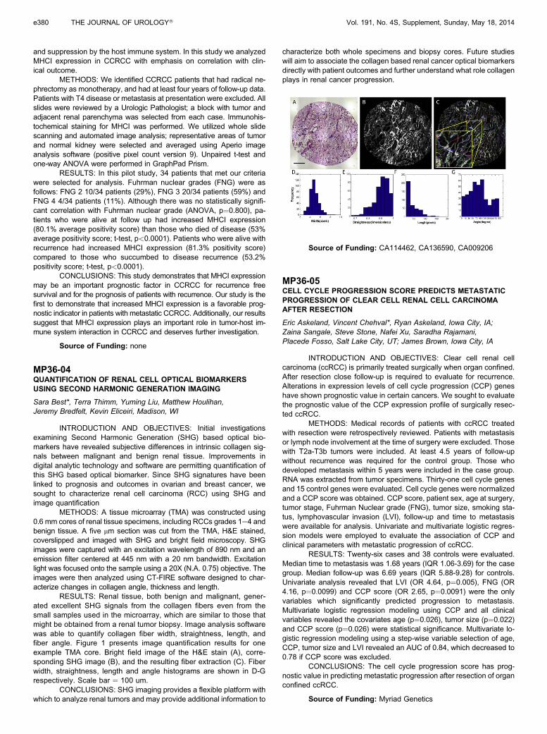
METHODS: A tissue microarray (TMA) was constructed using0.6 mm cores of renal tissue specimens, including RCCs grades 1e4 andbenign tissue. A five mm section was cut from the TMA, H&E stained,coverslipped and imaged with SHG and bright field microscopy. SHGimages were captured with an excitation wavelength of 890 nm and anemission filter centered at 445 nm with a 20 nm bandwidth. Excitationlight was focused onto the sample using a 20X (N.A. 0.75) objective. Theimages were then analyzed using CT-FIRE software designed to char-acterize changes in collagen angle, thickness and length.
RESULTS: Renal tissue, both benign and malignant, gener-ated excellent SHG signals from the collagen fibers even from thesmall samples used in the microarray, which are similar to those thatmight be obtained from a renal tumor biopsy. Image analysis softwarewas able to quantify collagen fiber width, straightness, length, andfiber angle. Figure 1 presents image quantification results for oneexample TMA core. Bright field image of the H&E stain (A), corre-sponding SHG image (B), and the resulting fiber extraction (C). Fiberwidth, straightness, length and angle histograms are shown in D-Grespectively. Scale bar ¼ 100 um.
CONCLUSIONS: SHG imaging provides a flexible platform withwhich to analyze renal tumors and may provide additional information to
characterize both whole specimens and biopsy cores. Future studieswill aim to associate the collagen based renal cancer optical biomarkersdirectly with patient outcomes and further understand what role collagenplays in renal cancer progression.
Source of Funding: CA114462, CA136590, CA009206
MP36-05CELL CYCLE PROGRESSION SCORE PREDICTS METASTATICPROGRESSION OF CLEAR CELL RENAL CELL CARCINOMAAFTER RESECTION
Eric Askeland, Vincent Chehval*, Ryan Askeland, Iowa City, IA;Zaina Sangale, Steve Stone, Nafei Xu, Saradha Rajamani,Placede Fosso, Salt Lake City, UT; James Brown, Iowa City, IA
INTRODUCTION AND OBJECTIVES: Clear cell renal cellcarcinoma (ccRCC) is primarily treated surgically when organ confined.After resection close follow-up is required to evaluate for recurrence.Alterations in expression levels of cell cycle progression (CCP) geneshave shown prognostic value in certain cancers. We sought to evaluatethe prognostic value of the CCP expression profile of surgically resec-ted ccRCC.
METHODS: Medical records of patients with ccRCC treatedwith resection were retrospectively reviewed. Patients with metastasisor lymph node involvement at the time of surgery were excluded. Thosewith T2a-T3b tumors were included. At least 4.5 years of follow-upwithout recurrence was required for the control group. Those whodeveloped metastasis within 5 years were included in the case group.RNA was extracted from tumor specimens. Thirty-one cell cycle genesand 15 control genes were evaluated. Cell cycle genes were normalizedand a CCP score was obtained. CCP score, patient sex, age at surgery,tumor stage, Fuhrman Nuclear grade (FNG), tumor size, smoking sta-tus, lymphovascular invasion (LVI), follow-up and time to metastasiswere available for analysis. Univariate and multivariate logistic regres-sion models were employed to evaluate the association of CCP andclinical parameters with metastatic progression of ccRCC.
RESULTS: Twenty-six cases and 38 controls were evaluated.Median time to metastasis was 1.68 years (IQR 1.06-3.69) for the casegroup. Median follow-up was 6.69 years (IQR 5.88-9.28) for controls.Univariate analysis revealed that LVI (OR 4.64, p¼0.005), FNG (OR4.16, p¼0.0099) and CCP score (OR 2.65, p¼0.0091) were the onlyvariables which significantly predicted progression to metastasis.Multivariate logistic regression modeling using CCP and all clinicalvariables revealed the covariates age (p¼0.026), tumor size (p¼0.022)and CCP score (p¼0.026) were statistical significance. Multivariate lo-gistic regression modeling using a step-wise variable selection of age,CCP, tumor size and LVI revealed an AUC of 0.84, which decreased to0.78 if CCP score was excluded.
CONCLUSIONS: The cell cycle progression score has prog-nostic value in predicting metastatic progression after resection of organconfined ccRCC.
Source of Funding: Myriad Genetics