Embed Size (px)
Citation preview

Moyamoya Disease Presenting with Intracranial Haemorrhàge
Pages with reference to book, From 349 To 351 Ali Akbar,Aftab Qureshi,Rashid Jooma ( Department of Neurosurgery, Jinnah Postgraduate Medical Centre, Karachi. )
Moyamoya disease is a spontaneous occlusion of the circle of Willis with abnonnal compensatory
anastamotic vascular networks at the base of the brain. Early reports characterised the condition as a
cause of cerebral ischaemia with progressive neurological deficits in infants and children or intracranial
haemorrhage in adolescents and adults of Japanese Community1,2. However, it is now well known to
occur in the other parts of the world as well - though being contingent on angiographic findings, the
diagnosis depend on the level of the nation’s health care provision. Two cases of moyamoya diseases
admitted to neurosurgical unit with intracranial haemorrhage are being presented here.
Case Reports
Case 1
This previously healthy 38 year old male suffered an acute episode of headache with vomiting and
within few minutes became unresponsive and was deeply unconscious. He was admitted to the district
hospital where on conservative management he regained consciousness after 7 days without focal
signs. However, he again suffered a sudden attack of headache with vomiting resulting in
unconsciousness. Thirty-six hours after improvement from the initial episode, the patient was then
shifted to our hospital.
On admission his vital signs were nonnal and general physical examinationwas unremarkable. He was
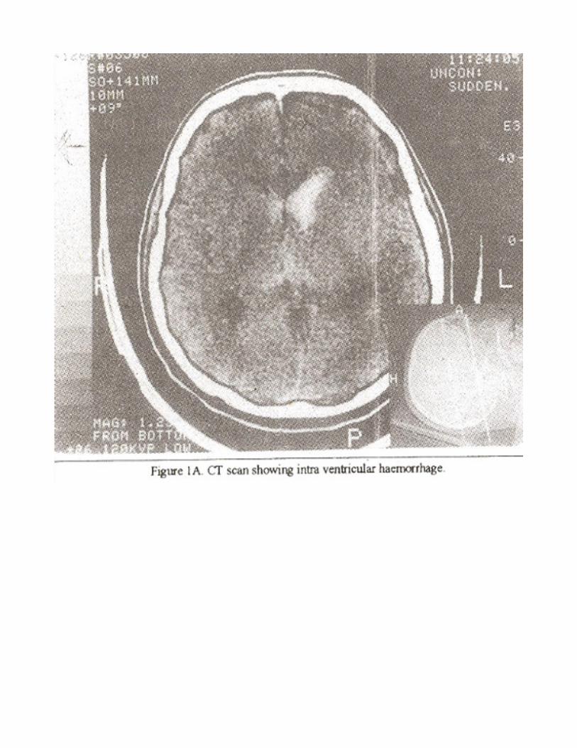
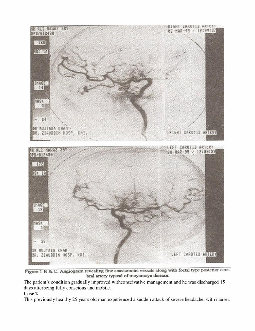
unconscious but responding to painful stimuli (Glasgow Coma Score 10/15). Computerized
Tomography (CT) Scan showed an intraventricular haemorrhage. Carotid angiography revealed the
occlusion of the supra clinoid portion of the internal carotid arteries onboth sides. There were foetal
type posterior cerebral arteries and fme anastamotic vessels in the form rete, irngating the territory of
anterior Circle of Wills characteristic
of moyamoya disease (Figure 1A, B, C).
The patient’s condition gradually improved withconseivative management and he was discharged 15
days afterbeing fully conscious and mobile.
Case 2This previously healthy 25 years old man experienced a sudden attack of severe headache, with nausea

and vomiting. Twelve hours later he developed neck stuffiness and became drowsy. He was taken to a
near by hospital where spinal tap yielded bloody cerebrospinal fluid. He was mimediately referred to
our department. On admission his vital signs were nonnal and his general physical status was good. He
was drowsy (Glasgow Coma Score 13/1 5) had stiff neck and right sided haemiparesis.
CT Scan showed a small left intracerebral haematoma along with subarachnoid haemorrhage. Carotid
angiogram demonstrated occlusion of the supra clinoid carotid artery with profuse anastomosis
between the ophthalmic artery and intracranial vessels, in the manner of rete typical of moyamoya
disease (Figure 2A, B, C).

Non-surgical management resulted in return of consciousness over 20 days and when seen at six month
follow-up be was without deficit though the family reported aggressive behaviour.
Discussion

The term moyomoya means “puff of smoke” in Japanese and the term was first used by Suzuki et al3,
for a disease involving occlusion of moderate sized arteries at or near the Circle of Willis. Such
occlusive basal vasculopathies are chronic and are accompanied by a pathologic attempt at
collatemlization, involving proliferation of arterioles and small penetrating artenes nearthe perforating
substance. Such proliferation of deep collaterals provides the typical angiographic appearance of a
“puff of smoke”. There may be other attempts atcollateralization also via transdural collateral channels,
The aetiology of the disease is still not well established. It has been hypothesized that the carotid
stenosis to be the primaiy condition and moyamoya vessels to be the result of it1,4,5, while others
believe that the disease results from progressive occlusion of vessels with stenosis initially involving
internal carotid artery then reaching the antenor and middle cerebral arteries and at last involving the
communicating and posterior cerebral arteries along with an abnormal vescular net work of moyamoya
vessels1,3.
This disease manifests in two forms, Juvenile form characterised clinically by seizures or progressive
neurological deficits due to ischemia and infarctions and the adults form, usually made manifest by
intracranial haemorrhage, which may be subarachnoid, intracerebral or intraventricular. Subarachnoid
haemorrhage may result from rupture of basal pathologic perforating vessels which are highly friable or
ruptured aneurysms or pseudoaneurysms6-8. Moyamoya is a cause of 4-5% ischaemic strokes
inpaediatnc age groups9. Many children who have recurrent ischaemic strokes develop progressive
mental deterioration10.
The purpose of reporting these two cases is that this disease does exist in our community but due to
lack of diagnostic facilities, the paediatric cases are not being diagnosed, only adult patients who
present with strokes are being referred to tertiary care hospitals for investigations and management.
Hence children who present with neurologic deficits and strokes should be accurately evaluated and
referred to neurosurgical centres for revascularization procedures.
References
1. Handa J, Hands H. Progressive cerebral arterial occlusive disease, an analysis of 27 cases.
Neuroradiology, 1972;3:119-33.
2. Carlson CB, Harvey FH. Loop J. Progressive alternating hemiplegia in early childhood with basal
arterial anastornosis and telangiectasia (moyarnoya syndrome). Neurology, 1973:23:734-44.
3. Suzuki J, Takaku A. Cerebrovascular ‘moyamoya’ disease. Disease showing abnormal net like
vessels in the base of the brain. Arch. Neurol., 1969;20:288-99.
4. Kudo T. Spontaneous occlusion of the circle of Willis. Neurol. Surg., 1975;3:711-24.
5. Vulia 0, Alexianu M, Gabor S. Hypoplasia and obstruction of the circle of Willis in a case ofatypical
cerebral haemorrhage and its relationship to the Nishimoto’s disease. Neurology, 1970;20:36 1-67.
6. Oka K, Yamashita M, Sadoshima S, et al. Cerebral haemorrhage in moyamoya disease at autopsy.
Virchos Arch., 1981 ;392:247-51 .
7. Jun-Ichiro H, Nobuo H, Tetsuya T. Moyamoya disease with repeated intraventricular haemorrhage
due to aneuiysm rupture. Report of two cases: J. Neurosurg., 1994;80:328-31.
8. Hiroshi Y, Sumitak T. Cerebrovascular moyamoya disease associated with an intracranial
pseudoaneurysm. J. Neurosurg., 1982;56:131- 34.
9. SchoenbergB, Mellinger J. Schoenberg DG. Cerebrovascular disease in infants and children. A study
of incidence. Clinical features and survival. Neurology, 1978;28:763-68.
10. Ishii R, Takeuchi S. Ibayashi K, et al. Intelligence in children with moyamoya disease. Evaluation
after surgical treatment with special reference to changes in cerebral blood flow. Stroke, 1984; 15:873-
77.