Embed Size (px)
Citation preview
MOUTHPART AND DIGESTIVE TRACT MORPHOLOGY OF THE
SYNCHRONIZED FIREFLY, Pteroptyx tener (COLEOPTERA: LAMPYRIDAE)
Nurul Wahida, O.*, Nur Hudawiyah, A., Roslim, R., Nur Khairunnisa, S. & Norela, S.
Centre for Insect Systematics, Faculty of Science and Technology,
Universiti Kebangsaan Malaysia, 43600 Bangi, Selangor, Malaysia *Corresponding author: [email protected]
ABSTRACT
The firefly, Pteroptyx tener (Coleoptera: Lampyridae) lives in the mangrove area and is
economically valuable for ecotourism because of its unique synchronous flashing behavior. It
has been observed that the adult species does not have the appearance of sucking type
mouthpart morphology though it is believed that its diet depends on the plant sap. This study
focused on the morphology of the mouthpart and, anatomy and histology of the digestive
tract of P. tener. From gross morphology observation, P. tener has hypognathus mouthpart
with a crescent-like narrow apicle mandible. Scanning Transmission Electron Microscope
(STEM) image showed that P. tener has a special food canal inside its mandible that runs
from the tip to the end part of the mandible. The tip of the mandible could be used as piercing
equipment for P. tener to suck plant sap for feeding purposes. Morphological investigations
of the digestive tract showed that this species have a small crop, large ventriculus and long
intestinal tract that is suitable for a liquid feeder insect. Histological sections show that the
digestive tract consist of thick muscle layer especially on the hindgut and thin epithelial layer
on all parts of the gut.
Keywords: Fireflies, mouthpart, digestive tract, morphology, histology, Pteroptyx tener.
ABSTRAK
Kelip-kelip, Pteropyx tener (Coleoptera: Lampyridae) hidup di paya bakau dan mempunyai
nilai ekonomi dalam ekopelancongan kerana keunikan kelakuannya berkelip secara
bersinkroni. Spesies dewasanya telah diperhatikan tidak menunjukkan morfologi alat mulut
jenis menghisap walaupun dipercayai bahawa spesies ini bergantung kepada sap tumbuhan.
Kajian ini memfokuskan terhadap morfologi alat mulut, anatomi dan histologi saluran
penghadamannya. Berdasarkan pemerhatian morfologinya, alat mulut P. tener merupakan
jenis hipognatus dengan mandibel berhujung tunggal seperti bulan sabit. Imej dari Mikroskop
Transmisi Pengimbas Elektron (STEM) menunjukkan bahawa P. tener mempunyai saluran
pemakanan khas di dalam mandibelnya bermula dari hujung tajam mandibel hingga ke
hujung lebar. Hujung tajam mandibel ini dipercayai digunakan untuk mencucuk ke dalam
tumbuhan bagi menghisap sap untuk mendapatkan makanan. Kajian morfologi terhadap
saluran penghadamannya menyokong hipotesis bahawa P. tener merupakan pemakan cairan
walaupun mempunyai alat mulut jenis mengunyah dengan memiliki tembolok yang kecil,
ventrikulus yang besar dan saluran usus yang panjang. Histologi sel menunjukkan saluran
Serangga 23(2):170-182 Nurul Wahida et al.
ISSN 1394-5130 171
penghadamannya terdiri daripada lapisan otot tebal terutama di bahagian usus belakang dan
lapitan epitelium nipis di keseluruhan sistem penghadaman.
Kata kunci: Kelip-kelip, alat mulut, saluran penghadaman, morfologi, histologi, Pteroptyx
tener
INTRODUCTION
Pteroptyx tener (Coleoptera: Lampyridae) is one of the firefly species that can be found in
Kg. Kuantan, Kuala Selangor, Malaysia. They prefer damp soils (usually mangrove) as its
main habitat and can be found along Sungai Selangor’s riverbank which is vegetated by
Berembang tree (Sonneratia caseolaris), sagu (Metroxylon sagu) and others wild mangrove
plants (Kazama et al. 2007; Khoo et al. 2009; Norzeana & Norela 2011; Wan Faridah et al.
2010; Wan Juliana et al. 2012).
Previously, some researchers believed that Berembang tree is the food sources for P.
tener (Mokhtar et al. 2009, 2010; Wan Juliana et al. 2012) as it contains 20% sucrose. It also
has been speculated that this beetle sucks the nectar of Berembang tree (Buck 1988;
Nallakumar 2003). Recently in 2017, Cheng et al. has confirmed that there is no S. caseolaris
cDNA detected in the gut of adult P. tener though it spends most of its adult life on this host
plant. They obtained single plant DNA that is identical to the rbcL sequence of Heritiera
littoralis (Malvaceae) that found along riverian area, which they speculate that this species
travels further for its feeding session.
Some short lived firefly such as Photinus ignitus Fall do not feed in the adult stage but
some long-life adult firefly such as Ellychnia corrusca Linneus which last for 10 months fed
during its adulthood (Lewis & Cratsley 2008). The adult of P. tener is reported to live for 3 to
4 weeks (Nada et. al 2012). The larva and adult firefly has been suggested to adapt for fluid
feeding. The larvae of firefly are being reported to suck the liquefy food through its canal on
the mandible with the toxins from the digestive enzyme (Fu & Ballantyne 2008; Labandeira
1997). However, no one ever reported on adult P. tener mouthpart to confirm whether these
canals retains on the adults’ firefly and supported fluid feeding. Therefore, mouthpart and
digestive tract morphology study Fare crucial to confirm the feeding type of adult P. tener.
The information about P. tener digestive morphology is vital for its conservation
efforts to ensure the survival of P. tener in Kg. Kuantan, Kuala Selangor that is now threaten
by human activity (Norzeana & Norela 2011; Mokhtar et al. 2009). According to Enger and
Smith (2004), survival rate of a species depends on several factors; some of them are
competition among species for food and habitat. Hence, information on type of food taken by
P. tener based on its digestive morphology could confirm its food preferences and this
additional information could be used in its conservation in future.
MATERIALS AND METHOD
Sampling
Adult fireflies, P. tener (Coleoptera, Lampyridae) were collected at night by using sweep net
at Kg. Kuantan, Kuala Selangor (3°21'38.78"N 101°18'4.67"E) in October 2012 and were
reared in plastic containers (175 x120 x 70 mm). Few pieces of berembang’s leaves were
placed in the plastic containers provided with wet tissues to retain moisture and few drops of
pure honey were dropped on the wet tissue.
Serangga 23(2):170-182 Nurul Wahida et al.
ISSN 1394-5130 172
Mouthpart Morphology
Pteroptyx tener head was separated from its body by using a small scissor and forceps under
Leica stereomicroscope. External Mouthparts of P. tener were observed under Tabletop
Scanning Electron Microscope (TM-1000 Hitachi). For observation under Scanning
Transmission Electron Microscope (STEM Jeol JSM-6400), the mouthpart was embedded in
resin before the block being sectioned (1 µm) using ultra-microtome to observe the food
canal in the mandible.
Digestive Tract Morphology
Pteroptyx tener was placed in Phosphate Buffer Solution (PBS: 136.9 mM NaCl, 8.1 mM
Na2HPO4, 2.7 mM KH2PO4), with pH 7.1. Dissection was done by using micro dissecting kit
under Leica stereomicroscope. The internal organ was first stained with Methylene blue
solution. The cuticle and fat body was separated to observe the digestive tract. Gross
morphology image of digestive tract was taken by using Carl Zeiss Image Analyzer
Microscope with Axiocam MRc software.
Digestive Tract Histology
Digestive tract was fixed in formalin fixative overnight. Then, the samples were put through a
series of ethanol (70%, 80%, 90% and 100 %.) to dehydrate then followed by xylene
immersion. Tissues were embedded in paraffin wax. Blocks of waxed tissue were then
sectioned (4 µm) by Leica RM2245 microtome. Four to five sections were placed on a slide
and dried in oven overcnight. Sectioned tissues were then stained according to hematoxylin &
eosin (H&E) method (Behmer et al. 1976; Junqueira & Junqueira 1983). Microphotography
was taken using Zeiss Axio Scope with iSolutionLite software on 20x magnification after the
tissues were mounted.
RESULTS AND DISCUSSION
Pteroptyx tener Mouthpart Morphology
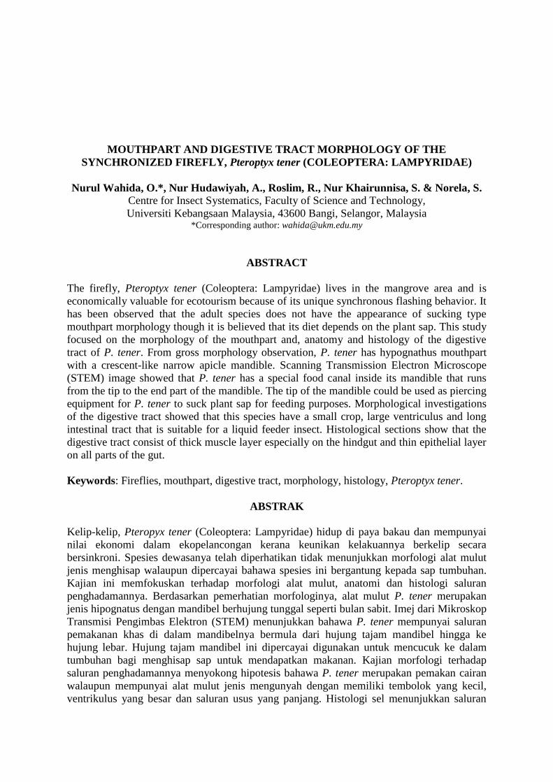
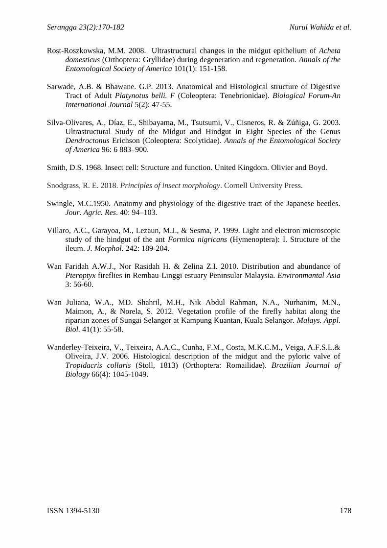
The mouthpart of P. tener consists of maxillary palps, labial palps, mandible, labrum and
labium. From the frontal view, the whole mouthparts were furnished with hairy structure
called seta (Figure 1). These hairs would act as a sense organ that used to detect the suitable
food type and to manipulate the food. These data collection supports Harald et al. (2005) who
state that hair of maxillae assist an insect to absorb and push the food into its mouth. Figure
1B shows the mandible morphology that is crescent in shape with narrow apical. Mandibles
usually move laterally in order to bite and chew food (Mohamad Salleh 1983). However, in
this case, P. tener mandible has a pointed tip that is suitable for piercing.
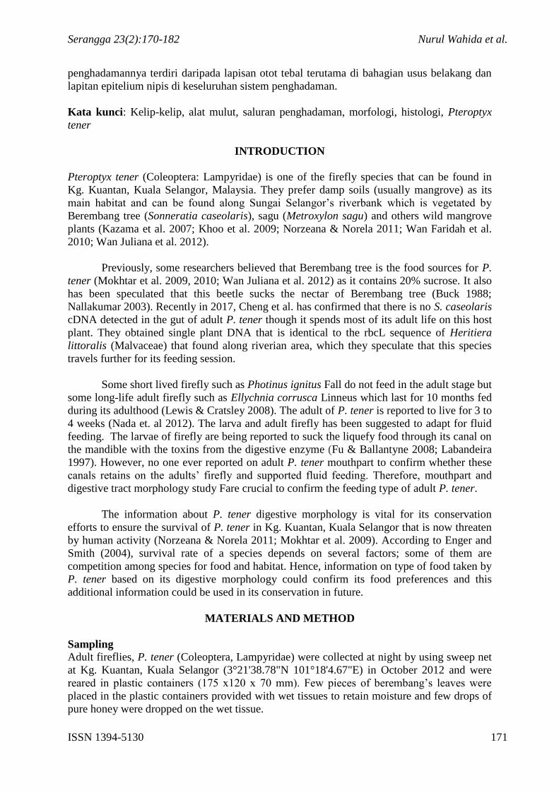
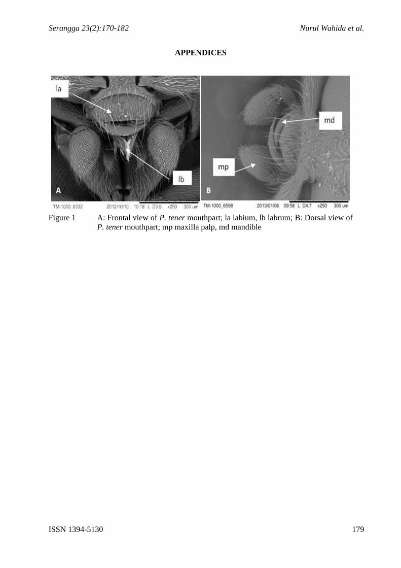
From STEM image, it is clearly observed that P. tener mandible has a special canal
running from the tips of the mandible towards the end of the mandible (Figure 2B).
According to Ballantyne and Menayah (2000; 2002), P. tener has chewing type mouthparts.
The function of haustellate type mouthpart has been replaced by the canal in the mandible.
Cicero (1994) reported for some beetles’ species, especially lampyridae, the mandibles
contain food canal which function to absorb nectar or liquid (Figure 3A). This statement was
proven in this study by using STEM technique that portrays a similar food canal in the
mandible of P. tener.
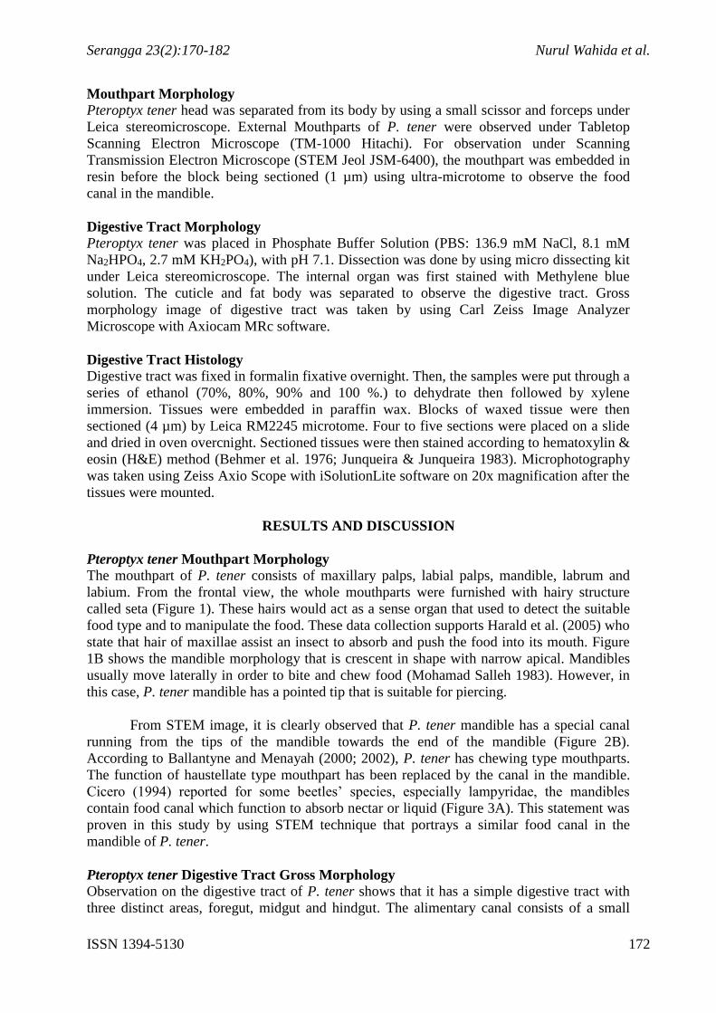
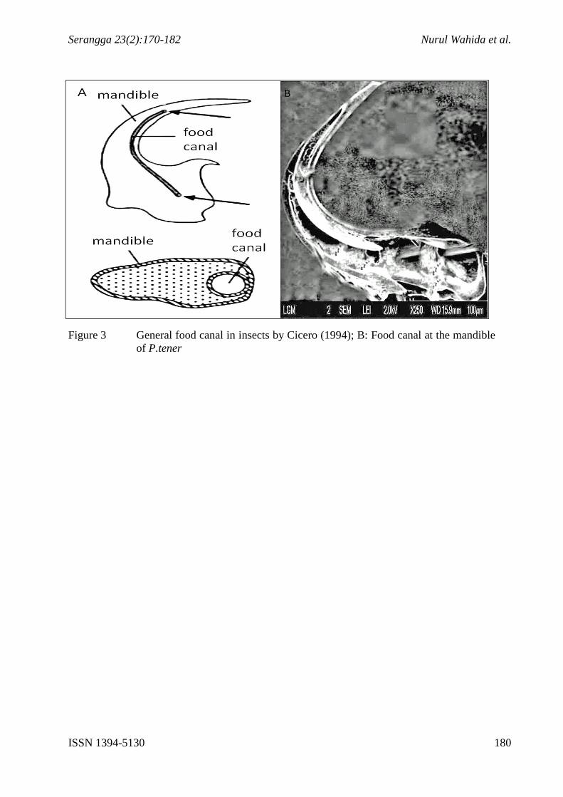
Pteroptyx tener Digestive Tract Gross Morphology
Observation on the digestive tract of P. tener shows that it has a simple digestive tract with
three distinct areas, foregut, midgut and hindgut. The alimentary canal consists of a small
Serangga 23(2):170-182 Nurul Wahida et al.
ISSN 1394-5130 173
crop that is the foregut area, a large midgut and a long hindgut that opens outside through
anus (Figure 4). A few strands of Malphigian tubules are found to be the initial part of a
hindgut that clearly separates these two regions. P. tener has a small crops compared to the
ventriculus at the midgut area. Crops functions mainly as a food-storage organ, before being
digested in the midgut. The foregut does not contain any proventriculus. This finding
supports the hypothesis that P. tener do not fed on solid food. Nevertheless, the midgut is
having a large ventriculus while the hindgut is quite long and narrow consists of ileum and
rectum. Length of the digestive gut depends on the feeding habit of the insect (Sarwade &
Bhawarne 2013). For a liquid feeder, it is expected that hindgut structure longer than other
features to support the filtration process of large amount of liquid that allows maximal
contact with liquid food (Chapman 1998). Hence, this proves that adult P. tener fed on easily
digested materials such as liquid or nectars. According to Mohamed Salleh (1983), feeding
type affect the digestive tract morphology. For example, solid feeders usually have
proventriculus that function to grind the solid food. For P. tener, proventiculus is absent at
the foregut area. This probably due to the feeding habit of this species that does not need this
structure to grind the food that supports our hypothesis that this species is a liquid feeder by
having a longer hindgut compared to the midgut and foregut which is needed in the fast
filtration process of liquid type of food. The small size of P. tener foregut indicates that the
food does not need to be stored long before it being digest in the large area of midgut. This
also have proved that the adult does not stored solid food in the foregut area like other insect
that fed on solid food such as crickets. Previously researchers reported that adult firefly does
not fed (Buck 1988; Nallakumar 2003). It is also being speculated before that firefly probably
fed on plant sap or nectar. The speculation has ended when a rearing experiment by Faust
(2008) proved that individual adult firefly, Photunis carolinus green fed on a fruit that has
increased its lifespan. Cheng et al. (2017) also had investigated the gut contain of P.tener
confirmed few DNA food plants, that has strengthen our hypothesis that P. tener is a liquid
feeder and fed during its adult stage.
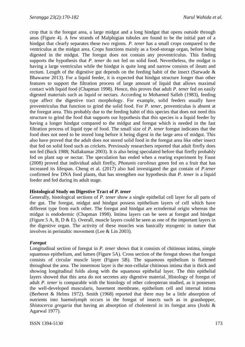
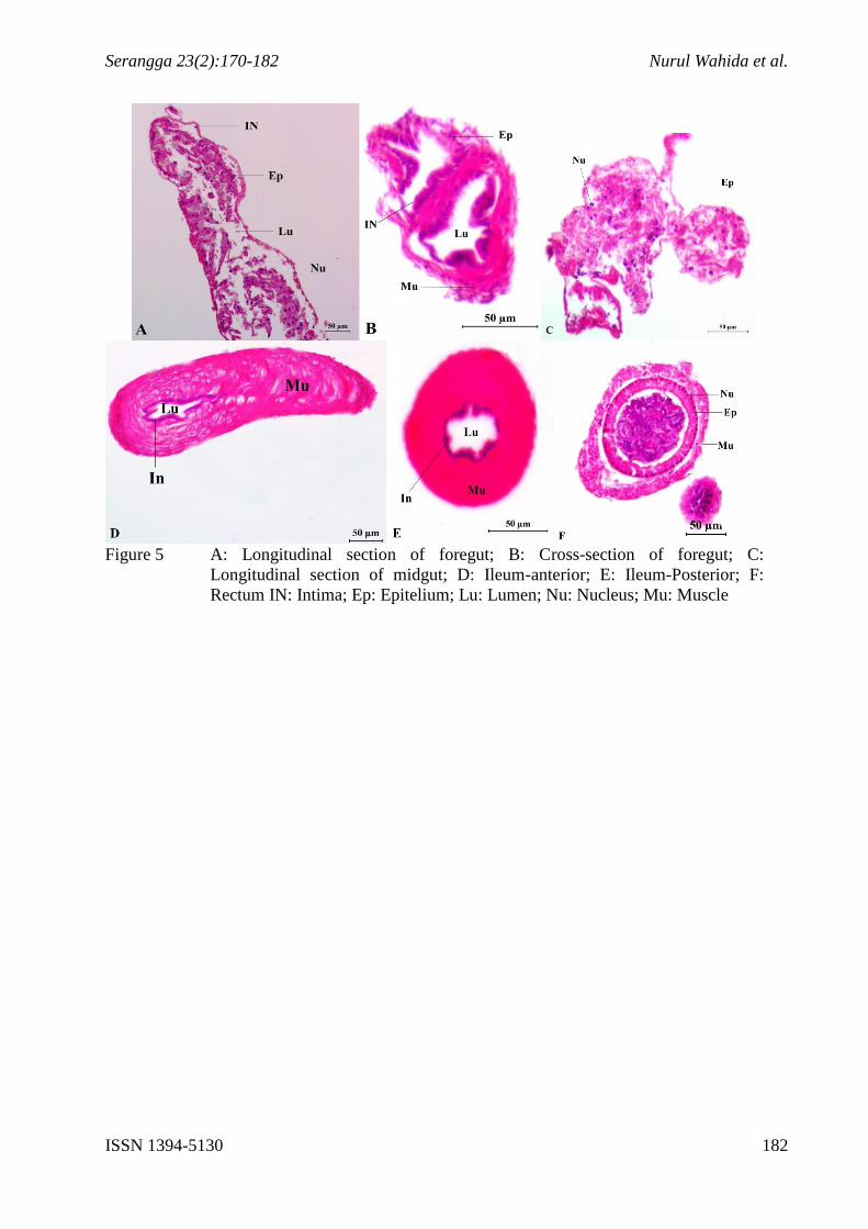
Histological Study on Digestive Tract of P. tener
Generally, histological sections of P. tener show a single epithelial cell layer for all parts of
the gut. The foregut, midgut and hindgut possess epithelium layers of cell which have
different type from each other. The foregut and hindgut are ectodermal origin whereas the
midgut is endodermic (Chapman 1998). Intima layers can be seen at foregut and hindgut
(Figure 5 A, B, D & E). Overall, muscle layers could be seen as one of the important layers in
the digestive organ. The activity of these muscles was basically myogenic in nature that
involves in peristaltic movement (Lee & Lin 2003).
Foregut
Longitudinal section of foregut in P. tener shows that it consists of chitinous intima, simple
squamous epithelium, and lumen (Figure 5A). Cross section of the foregut shows that foregut
consists of circular muscle layer (Figure 5B). The squamous epithelium is flattened
throughout the area. The innermost layer is the non-cellular chitinous intima that is thick and
showing longitudinal folds along with the squamous epithelial layer. The thin epithelial
layers showed that this area do not secretes any digestive material. Histology of foregut of
adult P. tener is comparable with the histology of other coleopteran studied, as it possesses
the well-developed muscularis, basement membrane, epithelium cell and internal intima
(Berberet & Helms 1972). Smith (1968) reported that there may be a little absorption of
nutrients into haemolymph occurs in the foregut of insects such as in grasshopper,
Shistocerca gregaria that having an absorption of cholesterol in its foregut area (Joshi &
Agarwal 1977).
Serangga 23(2):170-182 Nurul Wahida et al.
ISSN 1394-5130 174
Midgut
Figure 5C shows the longitudinal section of midgut P. tener. As midgut is endodermal origin,
it shows the absence of chitinous lining. Columnar cells, light striated muscle and connective
tissue could be detected in this area. The ventriculus of P. tener is larger than the foregut and
this suggests that the liquid food is being digested and absorb rapidly in this area before being
transferred to the hindgut. Insect midgut is the region for both digestion of food particle as
well as absorption of nutrients, and is a very metabolically active tissue. It may be the longest
part of alimentary canal and its diameter decreases as it tapers towards hindgut showing
anterior and posterior midgut morphologically (Sarwade & Bhawane 2013) but not in the
case of P. tener as it has shorter midgut compared to the hindgut. Some authors (Bution et al.
2006; Rost-Roszkowska 2008; Wanderley-Texiera et al. 2006) reported the midgut
epithelium arranged as a single layer and composed of columnar and regenerative cell, thus
confirmed the results obtained in this research, which is found that the epithelium as a
columnar epithelium type. The columnar epithelium cell in insect usually covered with
mikrovilli at the lumen area, as they are known to be responsible for both secretions of
digestive enzymes and absorption transfer of nutrients into the haemolymph.
Hindgut
The histology of adult P. tener hindgut only consists of ileum (anterior hindgut) and rectum
(posterior hindgut) in this study. This part is the longest and narrowed compared to foregut
and midgut.
I. Ileum
Histologically the muscularis include the single thick layer of outer circular muscle through
the longitudinal and cross section image (Figure 5D and 5E). The epithelium consists of
cuboidal cells supported by basement membrane. The nuclei of epithelium cell are round and
small. The epithelium shows longitudinal folds of cuboidal cells with prominent nuclei. The
epithelium is lined internally by thick muscularis similar with reported in other studies
(Gressitt 1953; Jones 1940; Snodgrass 2018; Swingle 1930). The presence of single layer of
circular muscle in the ileum of adult P. tener investigated here different in some beetle
species of Dendroctonus such as, in which the external muscle has two layers in which the
cryptonephredial systems is located (Silva-Olivares et al. 2003). The presence of Malphigian
tubules in the initial part of the hindgut shows that the function of primary urine production in
this species has been taken care of. Nevertheless, the presence of single layer of circular
muscle has also been reported in ants (Villaro et al. 1999).
II. Rectum
The epithelium of rectums bears double broad folds of muscularis in which the inner layer
consists of large flattened cuboidal epithellium cells with large round nuclei (Figure 5F). The
epithelium is supported by distinct basal membrane. The epithelium structure is much less
similar like epithelium structure in the ileum but the nuclei in the epithelium of rectum are
larger.
CONCLUSION
From this study, we would like to suggest that P. tener is a liquid feeder that probably fed on
plant saps by piercing through its mandible that has a special canal which then being directly
forward to its digestive gut. Digestive tract morphological study supports the hypothesis on
liquid-sucking type feeding in P. tener by having large ventriculus in the midgut and a long
hindgut with the absence of important organ for grinding solid food that is proventiculus.
Serangga 23(2):170-182 Nurul Wahida et al.
ISSN 1394-5130 175
ACKNOWLEDGEMENTS
We also would like to thank Mrs. Habibah for helping us with the histological procedures and
techniques and Mr. Hisham Bin Mahat from Kg Kuantan for helping us in sample collection.
We would like to express our gratitude to Centre for Insect Systematics, School of
Environmental and Natural Resource Sciences in Universiti Kebangsaan Malaysia (UKM)
and Microscopy unit, School of Bioscience and Biotechnology, UKM for allowing us to do
this research using their facilities. The work was funded through the allocation from
Universiti Kebangsaan Malaysia using research grants (ST-2017-013, LAUREATE-2013-
002, KOMUNITI-2013-005, KOMUNITI-2012-010, KOMUNITI-2011-030 and ST-2013-
005).
Serangga 23(2):170-182 Nurul Wahida et al.
ISSN 1394-5130 176
REFERENCES
Ballantyne, L.A. & Menayah R. 2000. Redescription of the synchronously firefly, Pteroptyx
tener Oliver (Coleoptera: Lampyridae) of Kampung Kuantan, Selangor. Malayan
Nature Journal 54(4): 323-328.
Ballantyne, L.A. & Menayah R. 2002. A description of larvae and redescription of adults of
the firefly Pteroptyx valida oliver in Selangor, Malaysia (Coleoptera: Lampyridae:
Luciolinae), with notes on Luciolinae larvae. The Raffles Bulletin of Zoology 50(1):
101-109.
Behmer, A. O., Talosa, C. M. E., Neto, F. G. A. 1976. Manual De Técnicas Para Histologia
Normal e Patológica. EDART. Editora da Universidade de São Paulo. SP.
Berberet, C. & Helms, T.J. 1972. Comparative anatomy and histology of selective systems in
larva and adult Phyllophaga anxia (Coleoptera: Scarabaeidae). Ann: Entol. Soc. Am.
65(5).
Bution, M.L., Caetano, F.H., Britto, F.B., Tomaino Gomes, G.A. & Zara, F.J. 2006.
Histology and histochemistry of the ventriculus of Dolichoderus (Monacis bispinosus
(Oliver, 1972) (Hymenoptera: Formicidae). Micron 37: 249-254.
Buck, J. 1988. Synchronous rhythmic flashing of fireflies. II. Quarterly review of biology
265–289.
Chapman R.F. 1998. The Insects, Structure and Function. 4th Edition. U.K: Cambridge
University Press.
Cheng, S., Chan, K. M., Ishak, S. F., Khoo, V. & Chew, M. Y. 2017. Elucidating food plants
of the aggregative, synchronously flashing Southeast Asian firefly, Pteroptyx tener
Olivier (Coleoptera, Lampyridae). BioRisk 12-25.
Cicero, J. M. 1994. Composite, haustellate mouthparts in netwinged beetle and firefly larvae
(Coleoptera, Cantharoidea: Lycidae, Lampyridae). Journal of morphology 219(2): 183-
192.
Enger, E.D. & Smith, B.F. 2004. Environmental Science: A Study of Interrelationships. U.K.:
McGraw Hill-Higher Education.
Faust, L. 2008. The Synchronous Firefly (Photinus carolinus) of the Great Smoky Mountains
National Park (Coleoptera: Lampyridae). Thailand International Firefly Symposium
Queen Sirikit Botanical Gardens.
Fu, X. H., & Ballantyne, L. .2008. Taxonomy and behaviour of lucioline fireflies
(Coleoptera: Lampyridae: Luciolinae) with redefinition and new species of Pygoluciola
Wittmer from mainland China and review of Luciola LaPorte. Zootaxa,1733(1), 1-44.
Gressit, J.L. 1953. The content rhinocerous beetle (Oryctes rhinocerous) with particular
reference to the Palan Islands. Bishop Mus Bull. 212: 157.
Serangga 23(2):170-182 Nurul Wahida et al.
ISSN 1394-5130 177
Harald, W.K., John, D.P. & Nikolaus, U.S. 2005. Mouthparts of flower-visiting insects.
Anthropod Structure & Development 34: 1-40.
Jones, C.R. 1940. The alimentary canal of Diplotax liberta Germ. (Scarabidae: Coleoptera).
Ohio. Jour. Sci. 40: 94-103
Joshi, M. & Agarwal, H. C. 1977. Site of cholesterol absorption in some insects. Journal of
Insect Physiology 23(3): 403-404.
Junqueira, L. C. U., & Junqueira, L. M. M. S. (1983). Técnicas básicas de citologia e
histologia. São Paulo: Santos.
Kazama, S., Matsumoto, S., Priyantha S.R., Hamamoto, H. & Sawamoto, M. 2007.
Characterization of firefly habitat using a geographical information system with
hydrological simulation. Ecological Modeling 209: 392-400.
Khoo, V., Kirton, L.G. & Nada, B. 2009. The fate of Pterotyx tener. A Bulletin Supporting
Plant and Animal Conservation in Malaysia 10: 4-6.
Labandeira, C. C. (1997). Insect mouthparts: ascertaining the paleobiology of insect feeding
strategies. Annual Review of Ecology and Systematics, 28(1), 153-193.
Lee, W. Y. & Lin, T. L. 2003. Morphology and ultrastructure of the hindgut of the oriental
fruit fly Bactrocera dorsalis (Diptera: Tephritidae). Formosan Entomol. 23: 113-128
Lewis S.M. & Cratsley C.K. 2008. Flash signal evolution, mate choice, and predation in
fireflies. Annu Rev Entomol. 53: 293–321.
Mohamad Salleh, M.S. 1983. Pengenalan Serangga, Kota Kinabalu, Malaysia: Japan
Overseas Cooperation Volunteers.
Mokhtar, J., Asmah, A. & Zaini, S. 2010. Kemandirian industry eko-pelancongan: Kes
tarikan pelancong keli-kelip Kg. Kuantan. Malaysia Journal of Society and Space 6(3):
89-97.
Mokhtar, J., Asmah, A., Zaini, S., Maimon A., Norela, S. & Normukhnun M. 2009. Penilaian
IKA Sg. Selangor pasca pembinaaan empangan Sg. Selangor. Malaysia Journal of
Society and Space 5(2): 126-130.
Nada, B., Kirton, L. G., Norma-Rashid, Y., Cheng, S., Shahlinney, L., & Phon, C. K. (2012).
Monitoring the fireflies of the Selangor River. Mangrove and coastal environment of
Selangor, Malaysia, University of Malaya Press, Malaysia, 153-162.
Nallakumar, K. 2003 The synchronously flashing aggregative fireflies of Peninsular
Malaysia. Biodiversity 4(2): 11–16.
Norzeana, R. & Norela, S. 2011. Kajian pembiakan kelip-kelip Pteroptyx tener di makmal.
Prosiding Seminar Hasil Penyelidikan Sains Sekitaran Sesi 2010/2011, Universiti
Kebangsaan Malaysia, pp. 214-218.
Serangga 23(2):170-182 Nurul Wahida et al.
ISSN 1394-5130 178
Rost-Roszkowska, M.M. 2008. Ultrastructural changes in the midgut epithelium of Acheta
domesticus (Orthoptera: Gryllidae) during degeneration and regeneration. Annals of the
Entomological Society of America 101(1): 151-158.
Sarwade, A.B. & Bhawane. G.P. 2013. Anatomical and Histological structure of Digestive
Tract of Adult Platynotus belli. F (Coleoptera: Tenebrionidae). Biological Forum-An
International Journal 5(2): 47-55.
Silva-Olivares, A., Díaz, E., Shibayama, M., Tsutsumi, V., Cisneros, R. & Zúñiga, G. 2003.
Ultrastructural Study of the Midgut and Hindgut in Eight Species of the Genus
Dendroctonus Erichson (Coleoptera: Scolytidae). Annals of the Entomological Society
of America 96: 6 883–900.
Smith, D.S. 1968. Insect cell: Structure and function. United Kingdom. Olivier and Boyd.
Snodgrass, R. E. 2018. Principles of insect morphology. Cornell University Press.
Swingle, M.C.1950. Anatomy and physiology of the digestive tract of the Japanese beetles.
Jour. Agric. Res. 40: 94–103.
Villaro, A.C., Garayoa, M., Lezaun, M.J., & Sesma, P. 1999. Light and electron microscopic
study of the hindgut of the ant Formica nigricans (Hymenoptera): I. Structure of the
ileum. J. Morphol. 242: 189-204.
Wan Faridah A.W.J., Nor Rasidah H. & Zelina Z.I. 2010. Distribution and abundance of
Pteroptyx fireflies in Rembau-Linggi estuary Peninsular Malaysia. Environmantal Asia
3: 56-60.
Wan Juliana, W.A., MD. Shahril, M.H., Nik Abdul Rahman, N.A., Nurhanim, M.N.,
Maimon, A., & Norela, S. 2012. Vegetation profile of the firefly habitat along the
riparian zones of Sungai Selangor at Kampung Kuantan, Kuala Selangor. Malays. Appl.
Biol. 41(1): 55-58.
Wanderley-Teixeira, V., Teixeira, A.A.C., Cunha, F.M., Costa, M.K.C.M., Veiga, A.F.S.L.&
Oliveira, J.V. 2006. Histological description of the midgut and the pyloric valve of
Tropidacris collaris (Stoll, 1813) (Orthoptera: Romailidae). Brazilian Journal of
Biology 66(4): 1045-1049.
Serangga 23(2):170-182 Nurul Wahida et al.
ISSN 1394-5130 179
APPENDICES
Figure 1 A: Frontal view of P. tener mouthpart; la labium, lb labrum; B: Dorsal view of
P. tener mouthpart; mp maxilla palp, md mandible
Serangga 23(2):170-182 Nurul Wahida et al.
ISSN 1394-5130 180
Figure 3 General food canal in insects by Cicero (1994); B: Food canal at the mandible
of P.tener
Serangga 23(2):170-182 Nurul Wahida et al.
ISSN 1394-5130 181
Figure 4 Gross morphology of P. tener digestive tract. Fg: Foregut; V: Ventriculus, MT:
Malphigian tubules; I: Ileum; R: Rectum
Serangga 23(2):170-182 Nurul Wahida et al.
ISSN 1394-5130 182
Figure 5 A: Longitudinal section of foregut; B: Cross-section of foregut; C:
Longitudinal section of midgut; D: Ileum-anterior; E: Ileum-Posterior; F:
Rectum IN: Intima; Ep: Epitelium; Lu: Lumen; Nu: Nucleus; Mu: Muscle