Embed Size (px)
Citation preview
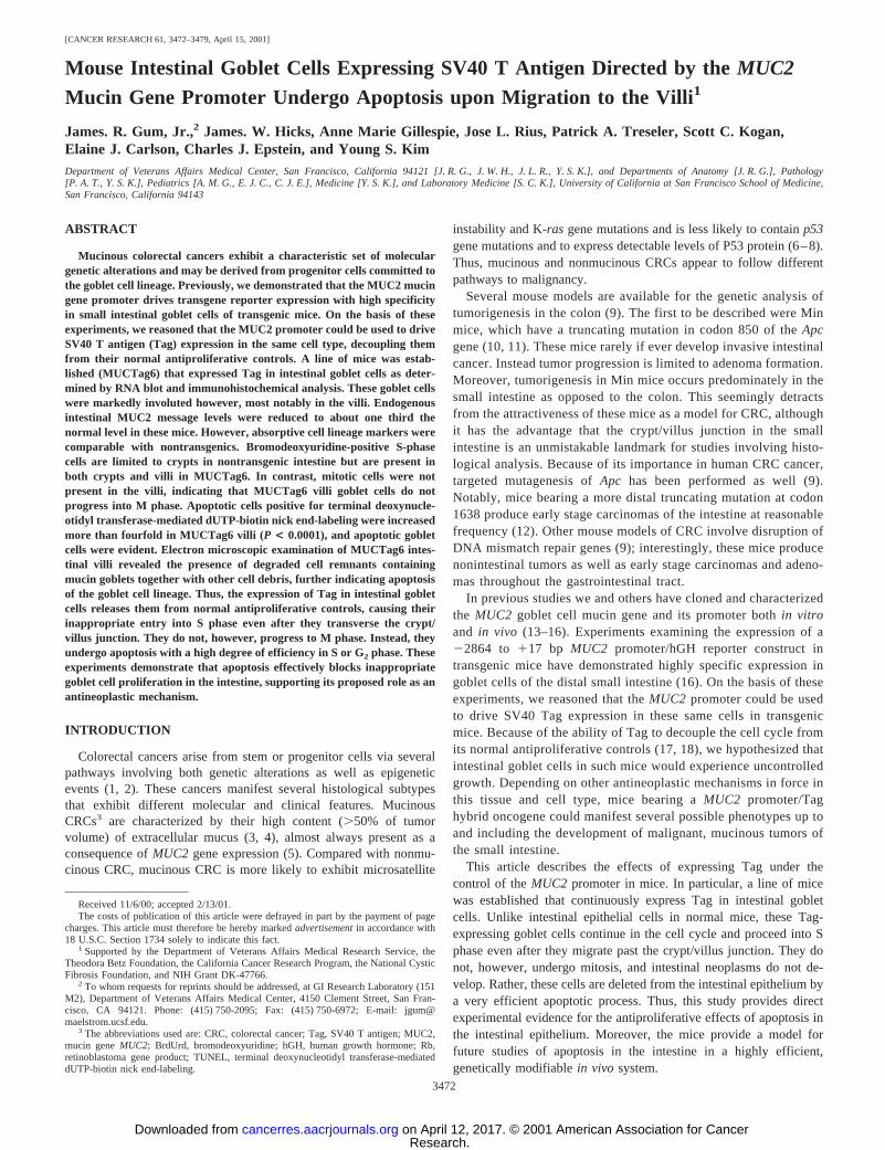
[CANCER RESEARCH 61, 3472–3479, April 15, 2001]
Mouse Intestinal Goblet Cells Expressing SV40 T Antigen Directed by theMUC2
Mucin Gene Promoter Undergo Apoptosis upon Migration to the Villi1
James. R. Gum, Jr.,2 James. W. Hicks, Anne Marie Gillespie, Jose L. Rius, Patrick A. Treseler, Scott C. Kogan,Elaine J. Carlson, Charles J. Epstein, and Young S. KimDepartment of Veterans Affairs Medical Center, San Francisco, California 94121 [J. R. G., J. W. H., J. L. R., Y. S. K.], and Departments of Anatomy [J. R. G.], Pathology[P. A. T., Y. S. K.], Pediatrics [A. M. G., E. J. C., C. J. E.], Medicine [Y. S. K.], and Laboratory Medicine [S. C. K.], University of California at San Francisco School of Medicine,San Francisco, California 94143
ABSTRACT
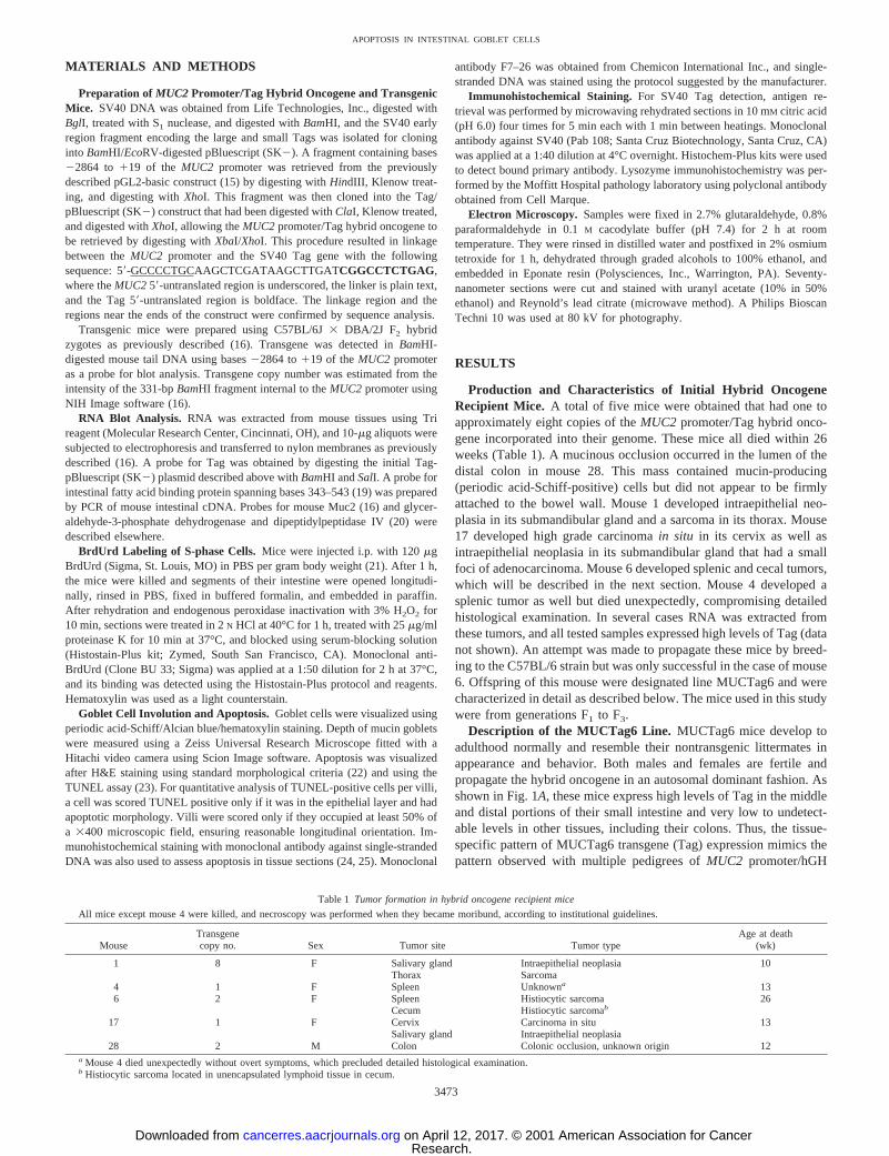
Mucinous colorectal cancers exhibit a characteristic set of moleculargenetic alterations and may be derived from progenitor cells committed tothe goblet cell lineage. Previously, we demonstrated that the MUC2 mucingene promoter drives transgene reporter expression with high specificityin small intestinal goblet cells of transgenic mice. On the basis of theseexperiments, we reasoned that the MUC2 promoter could be used to driveSV40 T antigen (Tag) expression in the same cell type, decoupling themfrom their normal antiproliferative controls. A line of mice was estab-lished (MUCTag6) that expressed Tag in intestinal goblet cells as deter-mined by RNA blot and immunohistochemical analysis. These goblet cellswere markedly involuted however, most notably in the villi. Endogenousintestinal MUC2 message levels were reduced to about one third thenormal level in these mice. However, absorptive cell lineage markers werecomparable with nontransgenics. Bromodeoxyuridine-positive S-phasecells are limited to crypts in nontransgenic intestine but are present inboth crypts and villi in MUCTag6. In contrast, mitotic cells were notpresent in the villi, indicating that MUCTag6 villi goblet cells do notprogress into M phase. Apoptotic cells positive for terminal deoxynucle-otidyl transferase-mediated dUTP-biotin nick end-labeling were increasedmore than fourfold in MUCTag6 villi (P < 0.0001), and apoptotic gobletcells were evident. Electron microscopic examination of MUCTag6 intes-tinal villi revealed the presence of degraded cell remnants containingmucin goblets together with other cell debris, further indicating apoptosisof the goblet cell lineage. Thus, the expression of Tag in intestinal gobletcells releases them from normal antiproliferative controls, causing theirinappropriate entry into S phase even after they transverse the crypt/villus junction. They do not, however, progress to M phase. Instead, theyundergo apoptosis with a high degree of efficiency in S or G2 phase. Theseexperiments demonstrate that apoptosis effectively blocks inappropriategoblet cell proliferation in the intestine, supporting its proposed role as anantineoplastic mechanism.
INTRODUCTION
Colorectal cancers arise from stem or progenitor cells via severalpathways involving both genetic alterations as well as epigeneticevents (1, 2). These cancers manifest several histological subtypesthat exhibit different molecular and clinical features. MucinousCRCs3 are characterized by their high content (.50% of tumorvolume) of extracellular mucus (3, 4), almost always present as aconsequence ofMUC2 gene expression (5). Compared with nonmu-cinous CRC, mucinous CRC is more likely to exhibit microsatellite
instability and K-rasgene mutations and is less likely to containp53gene mutations and to express detectable levels of P53 protein (6–8).Thus, mucinous and nonmucinous CRCs appear to follow differentpathways to malignancy.
Several mouse models are available for the genetic analysis oftumorigenesis in the colon (9). The first to be described were Minmice, which have a truncating mutation in codon 850 of theApcgene (10, 11). These mice rarely if ever develop invasive intestinalcancer. Instead tumor progression is limited to adenoma formation.Moreover, tumorigenesis in Min mice occurs predominately in thesmall intestine as opposed to the colon. This seemingly detractsfrom the attractiveness of these mice as a model for CRC, althoughit has the advantage that the crypt/villus junction in the smallintestine is an unmistakable landmark for studies involving histo-logical analysis. Because of its importance in human CRC cancer,targeted mutagenesis ofApc has been performed as well (9).Notably, mice bearing a more distal truncating mutation at codon1638 produce early stage carcinomas of the intestine at reasonablefrequency (12). Other mouse models of CRC involve disruption ofDNA mismatch repair genes (9); interestingly, these mice producenonintestinal tumors as well as early stage carcinomas and adeno-mas throughout the gastrointestinal tract.
In previous studies we and others have cloned and characterizedthe MUC2 goblet cell mucin gene and its promoter bothin vitroand in vivo (13–16). Experiments examining the expression of a22864 to 117 bp MUC2 promoter/hGH reporter construct intransgenic mice have demonstrated highly specific expression ingoblet cells of the distal small intestine (16). On the basis of theseexperiments, we reasoned that theMUC2 promoter could be usedto drive SV40 Tag expression in these same cells in transgenicmice. Because of the ability of Tag to decouple the cell cycle fromits normal antiproliferative controls (17, 18), we hypothesized thatintestinal goblet cells in such mice would experience uncontrolledgrowth. Depending on other antineoplastic mechanisms in force inthis tissue and cell type, mice bearing aMUC2 promoter/Taghybrid oncogene could manifest several possible phenotypes up toand including the development of malignant, mucinous tumors ofthe small intestine.
This article describes the effects of expressing Tag under thecontrol of theMUC2 promoter in mice. In particular, a line of micewas established that continuously express Tag in intestinal gobletcells. Unlike intestinal epithelial cells in normal mice, these Tag-expressing goblet cells continue in the cell cycle and proceed into Sphase even after they migrate past the crypt/villus junction. They donot, however, undergo mitosis, and intestinal neoplasms do not de-velop. Rather, these cells are deleted from the intestinal epithelium bya very efficient apoptotic process. Thus, this study provides directexperimental evidence for the antiproliferative effects of apoptosis inthe intestinal epithelium. Moreover, the mice provide a model forfuture studies of apoptosis in the intestine in a highly efficient,genetically modifiablein vivo system.
Received 11/6/00; accepted 2/13/01.The costs of publication of this article were defrayed in part by the payment of page
charges. This article must therefore be hereby markedadvertisementin accordance with18 U.S.C. Section 1734 solely to indicate this fact.
1 Supported by the Department of Veterans Affairs Medical Research Service, theTheodora Betz Foundation, the California Cancer Research Program, the National CysticFibrosis Foundation, and NIH Grant DK-47766.
2 To whom requests for reprints should be addressed, at GI Research Laboratory (151M2), Department of Veterans Affairs Medical Center, 4150 Clement Street, San Fran-cisco, CA 94121. Phone: (415) 750-2095; Fax: (415) 750-6972; E-mail: [email protected].
3 The abbreviations used are: CRC, colorectal cancer; Tag, SV40 T antigen; MUC2,mucin geneMUC2; BrdUrd, bromodeoxyuridine; hGH, human growth hormone; Rb,retinoblastoma gene product; TUNEL, terminal deoxynucleotidyl transferase-mediateddUTP-biotin nick end-labeling.
3472
Research. on April 12, 2017. © 2001 American Association for Cancercancerres.aacrjournals.org Downloaded from
MATERIALS AND METHODS
Preparation of MUC2 Promoter/Tag Hybrid Oncogene and TransgenicMice. SV40 DNA was obtained from Life Technologies, Inc., digested withBglI, treated with S1 nuclease, and digested withBamHI, and the SV40 earlyregion fragment encoding the large and small Tags was isolated for cloninginto BamHI/EcoRV-digested pBluescript (SK2). A fragment containing bases22864 to 119 of the MUC2 promoter was retrieved from the previouslydescribed pGL2-basic construct (15) by digesting withHindIII, Klenow treat-ing, and digesting withXhoI. This fragment was then cloned into the Tag/pBluescript (SK2) construct that had been digested withClaI, Klenow treated,and digested withXhoI, allowing theMUC2 promoter/Tag hybrid oncogene tobe retrieved by digesting withXbaI/XhoI. This procedure resulted in linkagebetween theMUC2 promoter and the SV40 Tag gene with the followingsequence: 59-GCCCCTGCAAGCTCGATAAGCTTGATCGGCCTCTGAG,where theMUC259-untranslated region is underscored, the linker is plain text,and the Tag 59-untranslated region is boldface. The linkage region and theregions near the ends of the construct were confirmed by sequence analysis.
Transgenic mice were prepared using C57BL/6J3 DBA/2J F2 hybridzygotes as previously described (16). Transgene was detected inBamHI-digested mouse tail DNA using bases22864 to119 of theMUC2 promoteras a probe for blot analysis. Transgene copy number was estimated from theintensity of the 331-bpBamHI fragment internal to theMUC2 promoter usingNIH Image software (16).
RNA Blot Analysis. RNA was extracted from mouse tissues using Trireagent (Molecular Research Center, Cincinnati, OH), and 10-mg aliquots weresubjected to electrophoresis and transferred to nylon membranes as previouslydescribed (16). A probe for Tag was obtained by digesting the initial Tag-pBluescript (SK2) plasmid described above withBamHI andSalI. A probe forintestinal fatty acid binding protein spanning bases 343–543 (19) was preparedby PCR of mouse intestinal cDNA. Probes for mouse Muc2 (16) and glycer-aldehyde-3-phosphate dehydrogenase and dipeptidylpeptidase IV (20) weredescribed elsewhere.
BrdUrd Labeling of S-phase Cells. Mice were injected i.p. with 120mgBrdUrd (Sigma, St. Louis, MO) in PBS per gram body weight (21). After 1 h,the mice were killed and segments of their intestine were opened longitudi-nally, rinsed in PBS, fixed in buffered formalin, and embedded in paraffin.After rehydration and endogenous peroxidase inactivation with 3% H2O2 for10 min, sections were treated in 2N HCl at 40°C for 1 h, treated with 25mg/mlproteinase K for 10 min at 37°C, and blocked using serum-blocking solution(Histostain-Plus kit; Zymed, South San Francisco, CA). Monoclonal anti-BrdUrd (Clone BU 33; Sigma) was applied at a 1:50 dilution for 2 h at 37°C,and its binding was detected using the Histostain-Plus protocol and reagents.Hematoxylin was used as a light counterstain.
Goblet Cell Involution and Apoptosis. Goblet cells were visualized usingperiodic acid-Schiff/Alcian blue/hematoxylin staining. Depth of mucin gobletswere measured using a Zeiss Universal Research Microscope fitted with aHitachi video camera using Scion Image software. Apoptosis was visualizedafter H&E staining using standard morphological criteria (22) and using theTUNEL assay (23). For quantitative analysis of TUNEL-positive cells per villi,a cell was scored TUNEL positive only if it was in the epithelial layer and hadapoptotic morphology. Villi were scored only if they occupied at least 50% ofa 3400 microscopic field, ensuring reasonable longitudinal orientation. Im-munohistochemical staining with monoclonal antibody against single-strandedDNA was also used to assess apoptosis in tissue sections (24, 25). Monoclonal
antibody F7–26 was obtained from Chemicon International Inc., and single-stranded DNA was stained using the protocol suggested by the manufacturer.
Immunohistochemical Staining. For SV40 Tag detection, antigen re-trieval was performed by microwaving rehydrated sections in 10 mM citric acid(pH 6.0) four times for 5 min each with 1 min between heatings. Monoclonalantibody against SV40 (Pab 108; Santa Cruz Biotechnology, Santa Cruz, CA)was applied at a 1:40 dilution at 4°C overnight. Histochem-Plus kits were usedto detect bound primary antibody. Lysozyme immunohistochemistry was per-formed by the Moffitt Hospital pathology laboratory using polyclonal antibodyobtained from Cell Marque.
Electron Microscopy. Samples were fixed in 2.7% glutaraldehyde, 0.8%paraformaldehyde in 0.1M cacodylate buffer (pH 7.4) for 2 h at roomtemperature. They were rinsed in distilled water and postfixed in 2% osmiumtetroxide for 1 h, dehydrated through graded alcohols to 100% ethanol, andembedded in Eponate resin (Polysciences, Inc., Warrington, PA). Seventy-nanometer sections were cut and stained with uranyl acetate (10% in 50%ethanol) and Reynold’s lead citrate (microwave method). A Philips BioscanTechni 10 was used at 80 kV for photography.
RESULTS
Production and Characteristics of Initial Hybrid OncogeneRecipient Mice. A total of five mice were obtained that had one toapproximately eight copies of theMUC2 promoter/Tag hybrid onco-gene incorporated into their genome. These mice all died within 26weeks (Table 1). A mucinous occlusion occurred in the lumen of thedistal colon in mouse 28. This mass contained mucin-producing(periodic acid-Schiff-positive) cells but did not appear to be firmlyattached to the bowel wall. Mouse 1 developed intraepithelial neo-plasia in its submandibular gland and a sarcoma in its thorax. Mouse17 developed high grade carcinomain situ in its cervix as well asintraepithelial neoplasia in its submandibular gland that had a smallfoci of adenocarcinoma. Mouse 6 developed splenic and cecal tumors,which will be described in the next section. Mouse 4 developed asplenic tumor as well but died unexpectedly, compromising detailedhistological examination. In several cases RNA was extracted fromthese tumors, and all tested samples expressed high levels of Tag (datanot shown). An attempt was made to propagate these mice by breed-ing to the C57BL/6 strain but was only successful in the case of mouse6. Offspring of this mouse were designated line MUCTag6 and werecharacterized in detail as described below. The mice used in this studywere from generations F1 to F3.
Description of the MUCTag6 Line. MUCTag6 mice develop toadulthood normally and resemble their nontransgenic littermates inappearance and behavior. Both males and females are fertile andpropagate the hybrid oncogene in an autosomal dominant fashion. Asshown in Fig. 1A, these mice express high levels of Tag in the middleand distal portions of their small intestine and very low to undetect-able levels in other tissues, including their colons. Thus, the tissue-specific pattern of MUCTag6 transgene (Tag) expression mimics thepattern observed with multiple pedigrees ofMUC2 promoter/hGH
Table 1 Tumor formation in hybrid oncogene recipient mice
All mice except mouse 4 were killed, and necroscopy was performed when they became moribund, according to institutional guidelines.
MouseTransgenecopy no. Sex Tumor site Tumor type
Age at death(wk)
1 8 F Salivary gland Intraepithelial neoplasia 10Thorax Sarcoma
4 1 F Spleen Unknowna 136 2 F Spleen Histiocytic sarcoma 26
Cecum Histiocytic sarcomab
17 1 F Cervix Carcinoma in situ 13Salivary gland Intraepithelial neoplasia
28 2 M Colon Colonic occlusion, unknown origin 12a Mouse 4 died unexpectedly without overt symptoms, which precluded detailed histological examination.b Histiocytic sarcoma located in unencapsulated lymphoid tissue in cecum.
3473
APOPTOSIS IN INTESTINAL GOBLET CELLS
Research. on April 12, 2017. © 2001 American Association for Cancercancerres.aacrjournals.org Downloaded from
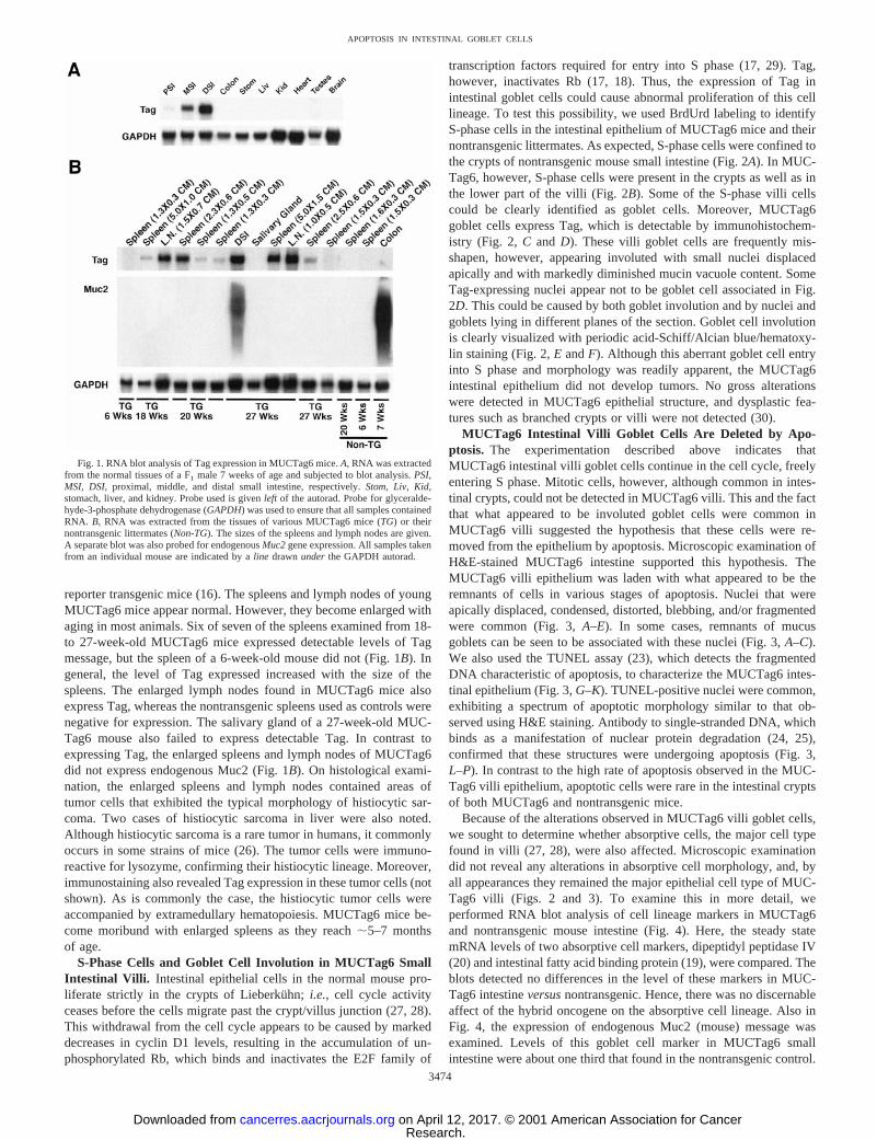
reporter transgenic mice (16). The spleens and lymph nodes of youngMUCTag6 mice appear normal. However, they become enlarged withaging in most animals. Six of seven of the spleens examined from 18-to 27-week-old MUCTag6 mice expressed detectable levels of Tagmessage, but the spleen of a 6-week-old mouse did not (Fig. 1B). Ingeneral, the level of Tag expressed increased with the size of thespleens. The enlarged lymph nodes found in MUCTag6 mice alsoexpress Tag, whereas the nontransgenic spleens used as controls werenegative for expression. The salivary gland of a 27-week-old MUC-Tag6 mouse also failed to express detectable Tag. In contrast toexpressing Tag, the enlarged spleens and lymph nodes of MUCTag6did not express endogenous Muc2 (Fig. 1B). On histological exami-nation, the enlarged spleens and lymph nodes contained areas oftumor cells that exhibited the typical morphology of histiocytic sar-coma. Two cases of histiocytic sarcoma in liver were also noted.Although histiocytic sarcoma is a rare tumor in humans, it commonlyoccurs in some strains of mice (26). The tumor cells were immuno-reactive for lysozyme, confirming their histiocytic lineage. Moreover,immunostaining also revealed Tag expression in these tumor cells (notshown). As is commonly the case, the histiocytic tumor cells wereaccompanied by extramedullary hematopoiesis. MUCTag6 mice be-come moribund with enlarged spleens as they reach;5–7 monthsof age.
S-Phase Cells and Goblet Cell Involution in MUCTag6 SmallIntestinal Villi. Intestinal epithelial cells in the normal mouse pro-liferate strictly in the crypts of Lieberkuhn;i.e., cell cycle activityceases before the cells migrate past the crypt/villus junction (27, 28).This withdrawal from the cell cycle appears to be caused by markeddecreases in cyclin D1 levels, resulting in the accumulation of un-phosphorylated Rb, which binds and inactivates the E2F family of
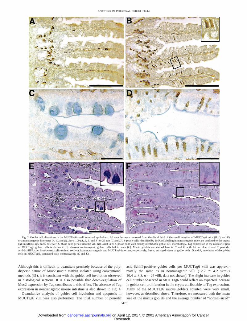
transcription factors required for entry into S phase (17, 29). Tag,however, inactivates Rb (17, 18). Thus, the expression of Tag inintestinal goblet cells could cause abnormal proliferation of this celllineage. To test this possibility, we used BrdUrd labeling to identifyS-phase cells in the intestinal epithelium of MUCTag6 mice and theirnontransgenic littermates. As expected, S-phase cells were confined tothe crypts of nontransgenic mouse small intestine (Fig. 2A). In MUC-Tag6, however, S-phase cells were present in the crypts as well as inthe lower part of the villi (Fig. 2B). Some of the S-phase villi cellscould be clearly identified as goblet cells. Moreover, MUCTag6goblet cells express Tag, which is detectable by immunohistochem-istry (Fig. 2, C and D). These villi goblet cells are frequently mis-shapen, however, appearing involuted with small nuclei displacedapically and with markedly diminished mucin vacuole content. SomeTag-expressing nuclei appear not to be goblet cell associated in Fig.2D. This could be caused by both goblet involution and by nuclei andgoblets lying in different planes of the section. Goblet cell involutionis clearly visualized with periodic acid-Schiff/Alcian blue/hematoxy-lin staining (Fig. 2,E andF). Although this aberrant goblet cell entryinto S phase and morphology was readily apparent, the MUCTag6intestinal epithelium did not develop tumors. No gross alterationswere detected in MUCTag6 epithelial structure, and dysplastic fea-tures such as branched crypts or villi were not detected (30).
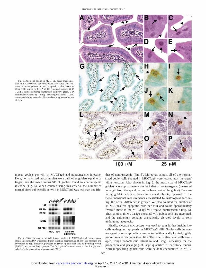
MUCTag6 Intestinal Villi Goblet Cells Are Deleted by Apo-ptosis. The experimentation described above indicates thatMUCTag6 intestinal villi goblet cells continue in the cell cycle, freelyentering S phase. Mitotic cells, however, although common in intes-tinal crypts, could not be detected in MUCTag6 villi. This and the factthat what appeared to be involuted goblet cells were common inMUCTag6 villi suggested the hypothesis that these cells were re-moved from the epithelium by apoptosis. Microscopic examination ofH&E-stained MUCTag6 intestine supported this hypothesis. TheMUCTag6 villi epithelium was laden with what appeared to be theremnants of cells in various stages of apoptosis. Nuclei that wereapically displaced, condensed, distorted, blebbing, and/or fragmentedwere common (Fig. 3,A–E). In some cases, remnants of mucusgoblets can be seen to be associated with these nuclei (Fig. 3,A–C).We also used the TUNEL assay (23), which detects the fragmentedDNA characteristic of apoptosis, to characterize the MUCTag6 intes-tinal epithelium (Fig. 3,G–K). TUNEL-positive nuclei were common,exhibiting a spectrum of apoptotic morphology similar to that ob-served using H&E staining. Antibody to single-stranded DNA, whichbinds as a manifestation of nuclear protein degradation (24, 25),confirmed that these structures were undergoing apoptosis (Fig. 3,L–P). In contrast to the high rate of apoptosis observed in the MUC-Tag6 villi epithelium, apoptotic cells were rare in the intestinal cryptsof both MUCTag6 and nontransgenic mice.
Because of the alterations observed in MUCTag6 villi goblet cells,we sought to determine whether absorptive cells, the major cell typefound in villi (27, 28), were also affected. Microscopic examinationdid not reveal any alterations in absorptive cell morphology, and, byall appearances they remained the major epithelial cell type of MUC-Tag6 villi (Figs. 2 and 3). To examine this in more detail, weperformed RNA blot analysis of cell lineage markers in MUCTag6and nontransgenic mouse intestine (Fig. 4). Here, the steady statemRNA levels of two absorptive cell markers, dipeptidyl peptidase IV(20) and intestinal fatty acid binding protein (19), were compared. Theblots detected no differences in the level of these markers in MUC-Tag6 intestineversusnontransgenic. Hence, there was no discernableaffect of the hybrid oncogene on the absorptive cell lineage. Also inFig. 4, the expression of endogenous Muc2 (mouse) message wasexamined. Levels of this goblet cell marker in MUCTag6 smallintestine were about one third that found in the nontransgenic control.
Fig. 1. RNA blot analysis of Tag expression in MUCTag6 mice.A, RNA was extractedfrom the normal tissues of a F1 male 7 weeks of age and subjected to blot analysis.PSI,MSI, DSI, proximal, middle, and distal small intestine, respectively.Stom, Liv, Kid,stomach, liver, and kidney. Probe used is givenleft of the autorad. Probe for glyceralde-hyde-3-phosphate dehydrogenase (GAPDH) was used to ensure that all samples containedRNA. B, RNA was extracted from the tissues of various MUCTag6 mice (TG) or theirnontransgenic littermates (Non-TG). The sizes of the spleens and lymph nodes are given.A separate blot was also probed for endogenousMuc2gene expression. All samples takenfrom an individual mouse are indicated by aline drawnunder the GAPDH autorad.
3474
APOPTOSIS IN INTESTINAL GOBLET CELLS
Research. on April 12, 2017. © 2001 American Association for Cancercancerres.aacrjournals.org Downloaded from
Although this is difficult to quantitate precisely because of the poly-disperse nature of Muc2 mucin mRNA isolated using conventionalmethods (31), it is consistent with the goblet cell involution observedin histological sections. It is also possible that down-regulation ofMuc2 expression by Tag contributes to this effect. The absence of Tagexpression in nontransgenic mouse intestine is also shown in Fig. 4.
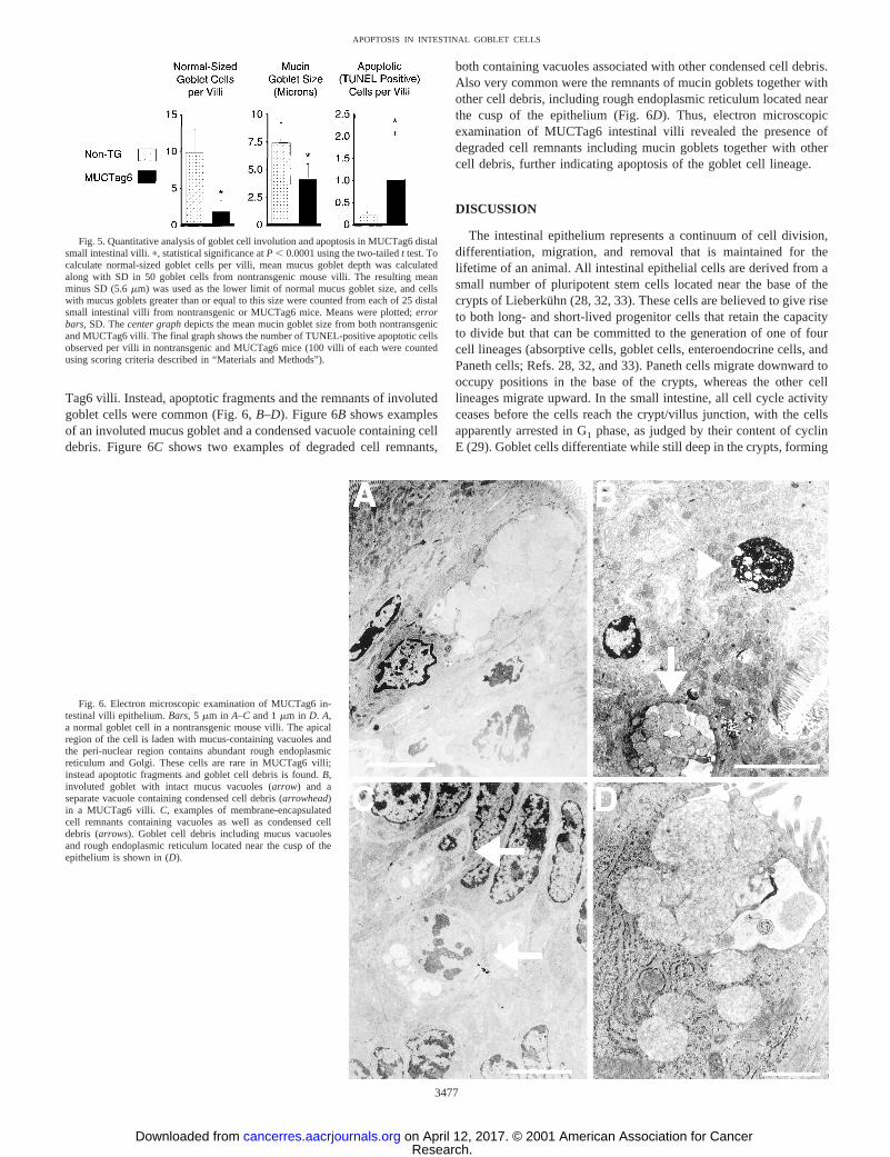
Quantitative analysis of goblet cell involution and apoptosis inMUCTag6 villi was also performed. The total number of periodic
acid-Schiff-positive goblet cells per MUCTag6 villi was approxi-mately the same as in nontransgenic villi (12.26 4.2 versus10.46 3.3,n 5 25 villi; data not shown). The slight increase in gobletcell number observed in MUCTag6 could reflect an expected increasein goblet cell proliferation in the crypts attributable to Tag expression.Many of the MUCTag6 mucus goblets counted were very small,however, as described above. Therefore, we measured both the meansize of the mucus goblets and the average number of “normal-sized”
Fig. 2. Goblet cell alterations in the MUCTag6 small intestinal epithelium. All samples were removed from the distal third of the small intestine of MUCTag6 mice (B, D,andF)or a nontransgenic littermate (A, C,andE). Bars, 100 (A, B, E,andF) or 25mm (C andD). S-phase cells identified by BrdUrd labeling in nontransgenic mice are confined to the crypts(A); in MUCTag6 mice, however, S-phase cells persist into the villi (B).Insetin B, S-phase cells with clearly identifiable goblet cell morphology. Tag expression in the nuclear regionof MUCTag6 goblet cells is shown inD, whereas nontransgenic goblet cells fail to stain (C). Mucin goblets are stained blue inC and D with Alcian blue. E and F, periodicacid-Schiff/Alcian blue/hematoxylin-stained sections from nontransgenic and MUCTag6 intestine, respectively;insets, enlarged views of goblet cells.D andF, involution of the gobletcells in MUCTag6, compared with nontransgenic (CandE).
3475
APOPTOSIS IN INTESTINAL GOBLET CELLS
Research. on April 12, 2017. © 2001 American Association for Cancercancerres.aacrjournals.org Downloaded from
mucus goblets per villi in MUCTag6 and nontransgenic intestine.Here, normal-sized mucus goblets were defined as goblets equal to orlarger than the mean minus SD of goblets found in nontransgenicintestine (Fig. 5). When counted using this criteria, the number ofnormal-sized goblet cells per villi in MUCTag6 was less than one fifth
that of nontransgenic (Fig. 5). Moreover, almost all of the normal-sized goblet cells counted in MUCTag6 were located near the crypt/villus junction. Also shown in Fig. 5, the mean size of MUCTag6goblets was approximately one half that of nontransgenic (measuredin length from the apical part to the basal part of the goblet). Becauseliving goblet cells are three-dimensional objects, opposed to thetwo-dimensional measurements necessitated by histological section-ing, the actual difference is greater. We also counted the number ofTUNEL-positive apoptotic cells per villi and found approximatelyfivefold more in the MUCTag6 villiversusnontransgenic (Fig. 5).Thus, almost all MUCTag6 intestinal villi goblet cells are involuted,and the epithelium contains dramatically elevated levels of cellsundergoing apoptosis.
Finally, electron microscopy was used to gain further insight intocells undergoing apoptosis in MUCTag6 villi. Goblet cells in non-transgenic mouse epithelium are packed with apically located, tightlypacked mucus vacuoles (Fig. 6A). These cells also have well-devel-oped, rough endoplasmic reticulum and Golgi, necessary for theproduction and packaging of large quantities of secretory mucus.Normal-appearing goblet cells were seldom encountered in MUC-
Fig. 4. RNA blot analysis of cell lineage markers in MUCTag6 and nontransgenicmouse intestine. RNA was isolated from intestinal segments, and blots were prepared andhybridized to Tag, dipeptidyl peptidase IV (DPPIV), intestinal fatty acid binding protein(FABPI), and mouse Muc2 probes. The final pair of autorads were probed for glyceral-dehyde-3-phosphate dehydrogenase (GAPDH).
Fig. 3. Apoptotic bodies in MUCTag6 distal small intes-tinal villi. Arrowheads,apoptotic bodies associated with rem-nants of mucus goblets;arrows, apoptotic bodies devoid ofidentifiable mucus goblets.A–E,H&E-stained sections.G–K,TUNEL-stained sections; counterstain is methyl green.L–P,immunohistochemistry using anti-single-stranded DNA;counterstain is hematoxylin. Size markers are given at bottomof figure.
3476
APOPTOSIS IN INTESTINAL GOBLET CELLS
Research. on April 12, 2017. © 2001 American Association for Cancercancerres.aacrjournals.org Downloaded from
Tag6 villi. Instead, apoptotic fragments and the remnants of involutedgoblet cells were common (Fig. 6,B–D). Figure 6Bshows examplesof an involuted mucus goblet and a condensed vacuole containing celldebris. Figure 6Cshows two examples of degraded cell remnants,
both containing vacuoles associated with other condensed cell debris.Also very common were the remnants of mucin goblets together withother cell debris, including rough endoplasmic reticulum located nearthe cusp of the epithelium (Fig. 6D). Thus, electron microscopicexamination of MUCTag6 intestinal villi revealed the presence ofdegraded cell remnants including mucin goblets together with othercell debris, further indicating apoptosis of the goblet cell lineage.
DISCUSSION
The intestinal epithelium represents a continuum of cell division,differentiation, migration, and removal that is maintained for thelifetime of an animal. All intestinal epithelial cells are derived from asmall number of pluripotent stem cells located near the base of thecrypts of Lieberkuhn (28, 32, 33). These cells are believed to give riseto both long- and short-lived progenitor cells that retain the capacityto divide but that can be committed to the generation of one of fourcell lineages (absorptive cells, goblet cells, enteroendocrine cells, andPaneth cells; Refs. 28, 32, and 33). Paneth cells migrate downward tooccupy positions in the base of the crypts, whereas the other celllineages migrate upward. In the small intestine, all cell cycle activityceases before the cells reach the crypt/villus junction, with the cellsapparently arrested in G1 phase, as judged by their content of cyclinE (29). Goblet cells differentiate while still deep in the crypts, forming
Fig. 5. Quantitative analysis of goblet cell involution and apoptosis in MUCTag6 distalsmall intestinal villi.p, statistical significance atP , 0.0001 using the two-tailedt test. Tocalculate normal-sized goblet cells per villi, mean mucus goblet depth was calculatedalong with SD in 50 goblet cells from nontransgenic mouse villi. The resulting meanminus SD (5.6mm) was used as the lower limit of normal mucus goblet size, and cellswith mucus goblets greater than or equal to this size were counted from each of 25 distalsmall intestinal villi from nontransgenic or MUCTag6 mice. Means were plotted;errorbars, SD. Thecenter graphdepicts the mean mucin goblet size from both nontransgenicand MUCTag6 villi. The final graph shows the number of TUNEL-positive apoptotic cellsobserved per villi in nontransgenic and MUCTag6 mice (100 villi of each were countedusing scoring criteria described in “Materials and Methods”).
Fig. 6. Electron microscopic examination of MUCTag6 in-testinal villi epithelium.Bars, 5mm in A–Cand 1mm in D. A,a normal goblet cell in a nontransgenic mouse villi. The apicalregion of the cell is laden with mucus-containing vacuoles andthe peri-nuclear region contains abundant rough endoplasmicreticulum and Golgi. These cells are rare in MUCTag6 villi;instead apoptotic fragments and goblet cell debris is found.B,involuted goblet with intact mucus vacuoles (arrow) and aseparate vacuole containing condensed cell debris (arrowhead)in a MUCTag6 villi. C, examples of membrane-encapsulatedcell remnants containing vacuoles as well as condensed celldebris (arrows). Goblet cell debris including mucus vacuolesand rough endoplasmic reticulum located near the cusp of theepithelium is shown in (D).
3477
APOPTOSIS IN INTESTINAL GOBLET CELLS
Research. on April 12, 2017. © 2001 American Association for Cancercancerres.aacrjournals.org Downloaded from
characteristic apically located mucus-containing goblets (34, 35).They then migrate for 2–3 days until they reach the tip of the villiwhere they are deleted by a sloughing process that may also involveapoptotic mechanisms and the partial reclamation of cellular material(22). The colonic epithelium experiences a pattern of cell renewalsimilar to what occurs in the small intestine, although the process isless well studied (36). In colon cancer, genetic and epigenetic eventsoccur that cause the dysregulation of genes involved in controllingvarious aspects of this cell renewal process (1, 2, 37).
In this study we sought to perturb the regulation of intestinal gobletcell proliferation via the expression of Tag under the control of theMUC2 promoter in transgenic mice. We have previously shown thatthisMUC2promoter directs reporter gene expression to goblet cells ofthe distal portion of the mouse small intestine (16). Reporter expres-sion initiates deep in the crypts, where goblet cells are still activelydividing (34, 35). The principle effect of Tag on the cell cycle is tobind and inactivate the Rb protein, thus releasing E2F transcriptionfactor and driving the passage of cells from G1 to S phase (17, 29).Our hypothesis was that this would cause the uncontrolled prolifera-tion of goblet cells, leading to the expansion of the goblet cell lineageas well as priming for further mutations that would ultimately lead tothe development of mucinous CRC. A further hypothesis was that theexpression of Tag in still-dividing, deep crypt cells, as opposed toquiescent villi cells, may be important in the initiation of tumorigen-esis. The formation of these hypotheses was guided by previousstudies in which intestinal cell lineage-restricted promoters were usedto drive Tag expression in mice (21, 38, 39). Use of theFabpipromoter resulted in Tag expression in the villi of the small intestine(21). This caused reentry of quiescent villi absorptive cells into thecell cycle but had no measurable effect on absorptive cell numbers orvilli cell lineage distribution, nor did neoplasms arise. Interestingly,however, an increase in apoptosis was noted in the villi of these mice(29, 30). Mice with SV40 Tag driven by the cryptdin-2 promoterexperienced Paneth cell ablation in the small intestine instead ofproliferation and surprisingly developed neuroendocrine tumors of theprostate, derived from a second cell type in which the cryptdin-2promoter is also active (38). A similar attempt using a glucagonpromoter resulted in endocrine tumors in the colon and the pancreas,where Tag was expressed in endocrine cells (39).
Forced expression of Tag did release intestinal goblet cells from atleast one antiproliferative control. As shown in Fig. 2B, MUCTag6goblet cells fail to undergo G1 arrest and continue into S phaseinappropriately as they migrate up the crypts and into the villi. Thisalone, however, does not lead to intestinal neoplasia. It appears rather,that these S-phase cells are removed by a very efficient apoptoticprocess in lieu of entering mitosis. The evidence for this is as follows:(a) the absence of mitotic cells in the MUCTag6 villi despite thepresence of S phase goblet cells; (b) the presence of large numbers ofTUNEL and anti-single-stranded, DNA-positive, and histologicallyevident apoptotic cell bodies in MUCTag6 villi, some of which retainmorphological traces of their goblet cell origins (Figs. 3 and 6); and(c) the involution of MUCTag6 mucin goblets (visualized by periodicacid-Schiff/Alcian blue staining), resulting in their near-completeremoval as they migrate away from the crypt/villus junction (Figs. 2,E andF, and 6). At the final stages of this process, some material fromthe degraded cells, especially the mucus, appears to be extruded intothe intestinal lumen, whereas some apoptotic cellular material appearsto be engulfed by neighboring cells (Figs. 2,E andF, and 6). Thus, thecellular debris from intestinal apoptosis, at least in this setting, is bothendocytosed and sloughed. This suggests the possibility that bothprocesses may be used to delete intestinal epithelial cells from theextrusion zone near the villi tips at the end of their natural life span(22).
The antineoplastic effects of apoptosis in the intestine has beensuggested by other studies, including analysis of cells in culture andthe observation that the proapoptotic geneBAX is frequently mutatedin microsatellite unstable CRCs (40, 41). It is also been observed thatboth CRCs and adenomas have increased apoptotic indices (42, 43).Furthermore, the colonic epithelium of azoxymethane-treated ratsexhibits abundant apoptotic indices (44). This study provides directexperimental evidence for the antiproliferative effects of apoptosis inthe intestine in a well-defined,in vivosystem. Because goblet cells aremorphologically distinct from other intestinal cell lineages, it is clearthat complete or near-complete ablation of these cells has occurred bythe time of their migration to the upper part of the villi. Thus,intestinal apoptosis can be extremely efficient, and the bulk of theinvivo evidence suggests that it occurs as an adaptive response to theuncontrolled proliferation of intestinal epithelial cells.
The development of histiocytic sarcomas in MUCTag hybrid on-cogene-bearing mice was unexpected.MUC2 gene expression is verytightly regulated, and to the best of our knowledgeMUC2 has notbeen reported to be expressed in macrophages, the presumed precur-sors of histiocytic sarcomas. It is likely that the hybrid oncogeneexpresses Tag in a population of macrophages and that this leads toexpansion of this cell lineage. The correlation of Tag expression withspleen size in MUCTag6 supports this hypothesis (Fig. 1B). Furtherepigenetic and genetic changes may then be required for histiocyticsarcoma formation. The observation that endogenous Muc2 messageis not expressed in the histiocytic sarcomas (Fig. 1B) indicates that thehybrid oncogene can be active in a population of cells, albeit aselected population, that does not express endogenousMUC2. Thedevelopment of histiocytic sarcomas and ensuing limitation of lifespan in MUCTag6 may preclude the possibility of intestinal neoplasmformation, which could require antiapoptotic genetic changes. Thenotion that additional genetic changes may be required for neoplasiain Tag-expressing intestinal epithelial cells is supported by the obser-vation that cell lines developed from Tag-expressing intestinal epi-thelial cells do not form colonies in soft agar or produce tumors whentransplanted into nude mice (45). Furthermore, decreased apoptosisfailed to increase intestinal tumor formation in the genetically initiatedApc1638 mouse model (46). It is also possible that in the MUCTag6model, Tag is not expressed early enough in the intestinal goblet celldifferentiation pathway to effect neoplasia formation, although deepcrypt goblet cells retain the ability to divide (34).
In addition to Rb inactivation, Tag is known to inactivate P53 (17,18). Because P53 has proapoptotic activity (47), a cell expressing Tagmay be expected to be apoptosis deficient. On the other hand, P53-independent pathways for apoptosis are known to be operative inmany cell types (48). Most relevant to this study, intestinal absorptivecells forced to reenter the cell cycle by Tag expression in the villiundergo apoptosis with equal efficiency in P53 wild-type and P53 nullmice (29, 30). Thus, it is most likely that the goblet cell apoptosisdemonstrated in this study proceeds via a P53- independent pathway.
In summary, this study demonstrates the effectiveness of apoptosisin counterbalancing uncontrolled proliferation in the intestine using awell-defined, in vivo system. MUCTag6 intestinal goblet cells con-tinue into S phase during their migration from the crypts to the villi.Rather than progressing to mitosis, however, these cells undergoapoptosis, resulting in the near complete ablation of the goblet celllineage in the upper reaches of the villi. Thus, these experimentsdemonstrate that apoptosis counteracts uncontrolled proliferation inthe intestine and support the hypothesis that apoptosis functions as animportant antineoplastic mechanism in this tissue. In addition, theMUCTag6 line provides a model that will be useful for the examina-tion of several parameters important in regulating apoptosis in thelower gastrointestinal tract, including the roles of the BAX, APC, and
3478
APOPTOSIS IN INTESTINAL GOBLET CELLS
Research. on April 12, 2017. © 2001 American Association for Cancercancerres.aacrjournals.org Downloaded from
E2F1 gene products and the determination of the signaling pathwaysthat initiate the apoptotic process. These studies will have directrelevance to understanding and perhaps enhancing through chemo-preventive intervention the anticancer effects of apoptosis in thehuman colon.
ACKNOWLEDGMENTS
We thank Sandra Huling and Ivy Hsieh for their aid with electron micros-copy.
REFERENCES
1. Vogelstein, B., Fearon, E. R., Hamilton, S. R., Kern, S. E., Preisinger, A. C., Leppert,M., Nakamura, Y., White, R., Smits, A. M., and Bos, J. L. Genetic alterations duringcolorectal-tumor development. N. Engl. J. Med.,319: 525–532, 1988.
2. Potter, J. D. Colorectal cancer: molecules and populations. J. Natl. Cancer Inst.,91:916–932, 1999.
3. Symonds, D. A., and Vickery, A. L. Mucinous carcinoma of the colon and rectum.Cancer (Phila.),37: 1891–1900, 1976.
4. Jass, J. R., and Sobin, L. H. Histological typing of intestinal tumors. Berlin: Springer-Verlag, 1990.
5. Hanski, C., Hofmeier, M., Schmitt-Graff, A., Riede, E., Hanski, M. L., Borchard, F.,Sieber, E., Niedobitek, F., Foss, H-D., Stein, H., and Riecken, E. O. Overexpressionor ectopic expression of MUC2 is the common property of mucinous carcinomas ofthe colon, pancreas, breast, and ovary. J. Pathol.,182: 385–391, 1997.
6. Hanski, C., Tiecke, F., Hummel, M., Hanski, M. L., Ogorek, D., Rolfs, A., Schmitt-Graff, A., Stein, H., and Riecken, E. O. Low frequency ofp53 gene mutation andprotein expression in mucinous colorectal carcinomas. Cancer Lett.,103: 163–170,1996.
7. Messerini, L., Vitelli, F., De Vitis, L. R., Mori, S., Calzolari, A., Palmirotta, R.,Calabro, A., and Papi, L. Microsatellite instability in sporadic mucinous colorectalcarcinomas: relationship to clinico-pathological variables. J. Pathol.,182: 380–384,1997.
8. Zhang, H., Evertsson, S., and Sun, X. Clinicopathological and genetic characteristicsof mucinous carcinomas in the colorectum. Int. J. Oncol.,14: 1057–1061, 1999.
9. Heyer, J., Yang, K., Lipkin, M., Edelmann, W., and Kucherlapati, R. Mouse modelsfor colorectal cancer. Oncogene,18: 5325–5333, 1999.
10. Moser, A. R., Pitot, H. C., and Dove, W. F. A dominant mutation that predisposes tomultiple intestinal neoplasia in the mouse. Science (Wash DC),247: 322–324, 1990.
11. Su, L. K., Kinzler, K. W., Vogelstein, B., Preisinger, A. C., Moser, A. R., Luongo, C.,Gould, K. A., and Dove, W. F. Multiple intestinal neoplasia caused by a mutation inthe murine homolog of theAPC gene. Science (Wash DC),256: 668–670, 1992.
12. Fodde, R., Edelmann, W., Yang, K., van Leeuwen, C., Carlson, C., Renault, B.,Breukel, C., Alt, E., Lipkin, M., Khan, P. M., and Kucherlapati, R. A targetedchain-termination mutation in the mouseApc gene results in multiple intestinaltumors. Proc. Natl. Acad. Sci. USA,91: 8969–8973, 1994.
13. Gum, J. R., Hicks, J. W., Toribara, N. W., Siddiki, B., and Kim, Y. S. Molecularcloning of human intestinal mucin (MUC2) cDNA: identification of the aminoterminus and overall sequence similarity to pre-pro-von Willebrand factor. J. Biol.Chem.,269: 2440–2446, 1994.
14. Velcich, A., Palumbo, L., Selleri, L., Evans, G., and Augenlicht, L. Organization andregulatory aspects of the human intestinal mucin gene (MUC2) locus. J. Biol. Chem.,272: 7968–7976, 1997.
15. Gum, J. R., Hicks, J. W., and Kim, Y. S. Identification and characterization of theMUC2 (human intestinal mucin) gene 59-flanking region. Promoter activity in cul-tured cells. Biochem. J.,324: 259–267, 1997.
16. Gum, J. R., Hicks, J. W., Gillespie, A. M., Carlson, E. J., Komuves, L., Karnik, S.,Hong, J. C., Epstein, C. J., and Kim, Y. S. Goblet cell-specific expression mediatedby the MUC2 mucin gene promoter in the intestine of transgenic mice. Am. J.Physiol.,276: G666–G676, 1999.
17. Lundberg, A. S., and Weinberg, R. A. Control of the cell cycle and apoptosis. Eur. J.Cancer,35: 531–539, 1999.
18. Pardee, A. B. G1 events and regulation of cell proliferation. Science (Wash DC),246:603–608, 1989.
19. Green, R. P., Cohn, S. M., Sacchettini, J. C., Jackson, K. E., and Gordon, J. I. Themouse intestinal fatty acid binding protein gene: nucleotide sequence, pattern ofdevelopmental and regional expression, and proposed structure of its protein product.DNA Cell Biol., 11: 31–41, 1992.
20. Gum, J. R., Erickson, R. H., Hicks, J. W., Rius, J. L., and Kim, Y. S. Analysis ofdipeptidyl peptidase IV gene regulation in transgenic mice: DNA elements sufficientfor promoter activity in the kidney, but not the intestine, reside on the proximalportion of the gene 59-flanking region. FEBS Lett.,482: 49–53, 2000.
21. Hauft, S. M., Kim, S. H., Schmidt, G. H., Pease, S., Rees, S., Harris, S., Roth, K. A.,Hansbrough, J., Cohn, S. M., Ahnen, D. J., Wright, N. A., Goodlad, R. A., andGordon, J. I. Expression of SV-40 T antigen in the small intestinal epithelium oftransgenic mice results in proliferative changes in the crypt and reentry of villus-associated enterocytes into the cell cycle but has no apparent effect on cellulardifferentiation programs and does not cause neoplastic transformation. J. Cell Biol.,117: 825–839, 1992.
22. Hall, P. A., Coates, P. J., Ansari, B., and Hopwood, D. Regulation of cell number inthe mammalian gastrointestinal tract: the importance of apoptosis. J. Cell Sci.,107:3569–3577, 1994.
23. Gavrieli, Y., Sherman, Y., and Ben-Sasson, S. A. Identification of programmed celldeathin situ via specific labeling of nuclear DNA fragmentation. J. Cell Biol.,119:493–501, 1992.
24. Frankfurt, O. S., Robb, J. A., Sugarbaker, E. V., and Villa, L. Monoclonal antibodyto single-stranded DNA is a specific and sensitive cellular marker of apoptosis. Exp.Cell Res.,226: 387–397, 1996.
25. Frankfurt, O. S., Robb, J. A., Sugarbaker, E. V., and Villa, L. Apoptosis in breastcarcinomas detected with monoclonal antibody to single-stranded DNA: Relation tobcl-2 expression, hormone receptors, and lymph node metastases. Clin. Cancer Res.,3: 465–471, 1997.
26. Frith, C. H., Ward, J. M., Frederickson, T., and Harleman, J. H. Neoplastic lesions ofthe hematopoietic system.In: U. Mohr, D. L. Dungworth, C. C. Capen, W. W.Carlton, J. P. Sundberg, and J. M. Ward (eds.), Pathobiology of the Aging Mouse, pp.219–235. Washington, DC: ILSI Press, 1996.
27. Cheng, H., and Leblond, C. P. Origin, differentiation and renewal of the four mainepithelial cell types in the mouse small intestine. V. Unitarian theory of the origin ofthe four epithelial cell types. Am. J. Anat.,141: 537–561, 1974.
28. Wong, M. H., Stappenbeck, T. S., and Gordon, J. I. Living and commuting inintestinal crypts. Gastroenterology,116: 208–210, 1999.
29. Chandrasekaran, C., Coopersmith, C. M., and Gordon, J. I. Use of normal andtransgenic mice to examine the relationship between terminal differentiation ofintestinal epithelial cells and accumulation of their cell cycle regulators. J. Biol.Chem.,271: 28414–28421, 1996.
30. Coopersmith, C. M., Chandrasekaran, C., McNevin, M. S., and Gordon, J. I. Bi-transgenic mice reveal that K-rasVal12 augments a p53-independent apoptosis whensmall intestinal villus enterocytes reenter the cell cycle. J. Cell Biol.,138: 167–179,1997.
31. Debailleul, V., Laine, A., Huet, G., Mathon, P., d’Hooghe, M. C., Aubert, J. P., andPorchet, N. Human mucin genesMUC2, MUC3, MUC4, MUC5AC, MUC5B,andMUC6 express stable and extremely large mRNAs and exhibit a variable lengthpolymorphism. An improved method to analyze large mRNAs. J. Biol. Chem.,273:881–890, 1998.
32. Gordon, J. I., Schmidt, G. H., and Roth, K. A. Studies of intestinal stem cells usingnormal, chimeric, and transgenic mice. FASEB J.,6: 3039–3050, 1992.
33. Gordon, J. I. Intestinal epithelial differentiation: new insights from chimeric andtransgenic mice. J. Cell Biol.,108: 1187–1194, 1989.
34. Paulus, U., Loeffler, M., Zeidler, J., Owen, G., and Potten, C. S. The differentiationand lineage development of goblet cells in the murine small intestinal crypt: exper-imental and modelling studies. J. Cell Sci.,106: 473–483, 1993.
35. Bjerknes, M., and Cheng, H. Clonal analysis of mouse intestinal epithelial progeni-tors. Gastroenterology,116: 7–14, 1999.
36. Schmidt, G. H., Wilkinson, M. M., and Ponder, B. A. Cell migration pathway in theintestinal epithelium: anin situ marker system using mouse aggregation chimeras.Cell, 40: 425–429, 1985.
37. Stappenbeck, T. S., Wong, M. H., Saam, J. R., Mysorekar, I. U., and Gordon, J. I.Notes from some crypt watchers: regulation of renewal in the mouse intestinalepithelium. Curr. Opin. Cell Biol.,10: 702–709, 1998.
38. Garabedian, E. M., Humphrey, P. A., and Gordon, J. I. A transgenic mouse model ofmetastatic prostate cancer originating from neuroendocrine cells. Proc. Natl. Acad.Sci. USA,95: 15382–15387, 1998.
39. Lee, Y. C., Asa, S. L., and Drucker, D. J. Glucagon gene 59-flanking sequences directexpression of simian virus 40 large T antigen to the intestine, producing carcinoma ofthe large bowel in transgenic mice. J. Biol. Chem.,267: 10705–10708, 1992.
40. Morin, P. J., Sparks, A. B., Korinek, V., Barker, N., Clevers, H., Vogelstein, B., andKinzler, K. W. Activation ofb-catenin-Tcf signaling in colon cancer by mutations inb-catenin or APC. Science (Wash DC),275: 1787–1790, 1997.
41. Rampino, N., Yamamoto, H., Ionov, Y., Li, Y., Sawai, H., Reed, J. C., and Perucho,M. Somatic frameshift mutations in theBAX gene in colon cancers of the microsat-ellite mutator phenotype. Science (Wash DC),275: 967–969, 1997.
42. Koike, M. Significance of spontaneous apoptosis during colorectal tumorigenesis.J. Surg. Oncol.,62: 97–108, 1996.
43. Watanabe, I., Toyoda, M., Okuda, J., Tenjo, T., Tanaka, K., Yamamoto, T.,Kawasaki, H., Sugiyama, T., Kawarada, Y., and Tanigawa, N. Detection of apoptoticcells in human colorectal cancer by two different in situ methods: antibody againstsingle-stranded DNA and terminal deoxynucleotidyl transferase-mediated dUTP-biotin nick end-labeling (TUNEL) methods. Jpn. J. Cancer Res.,90: 188–193, 1999.
44. Samaha, H. S., Kelloff, G. J., Steele, V., Rao, C. V., and Reddy, B. S. Modulation ofapoptosis by sulindac, curcumin, phenylethyl-3-methylcaffeate, and 6-phenylhexylisothiocyanate: apoptotic index as a biomarker in colon cancer chemoprevention andpromotion. Cancer Res.,57: 1301–1305, 1997.
45. D’Abaco, G. M., Whitehead, R. H., and Burgess, A. W. Synergy betweenAPCmin andan activatedras mutation is sufficient to induce colon carcinomas. Mol. Cell. Biol.,16: 884–891, 1996.
46. Augenlicht, L. H., Anthony, G. M., Church, T. L., Edelmann, W., Kucherlapati, R.,Yang, K., Lipkin, M., and Heerdt, B. G. Short-chain fatty acid metabolism, apoptosis,and Apc-initiated tumorigenesis in the mouse gastrointestinal mucosa. Cancer Res.,59: 6005–6009, 1999.
47. Ahnen, D. J. Stalking the guardian of the genome: p53 in colorectal carcinogenesis.Am. J. Gastroenterol.,91: 3–6, 1996.
48. Liebermann, D. A., Hoffman, B., and Steinman, R. A. Molecular controls of growtharrest and apoptosis: p53-dependent and -independent pathways. Oncogene,11:199–310, 1995.
3479
APOPTOSIS IN INTESTINAL GOBLET CELLS
Research. on April 12, 2017. © 2001 American Association for Cancercancerres.aacrjournals.org Downloaded from
2001;61:3472-3479. Cancer Res James. R. Gum, Jr., James. W. Hicks, Anne Marie Gillespie, et al. Apoptosis upon Migration to the Villi
Mucin Gene Promoter UndergoMUC2Directed by the Mouse Intestinal Goblet Cells Expressing SV40 T Antigen
Updated version
http://cancerres.aacrjournals.org/content/61/8/3472
Access the most recent version of this article at:
Cited articles
http://cancerres.aacrjournals.org/content/61/8/3472.full.html#ref-list-1
This article cites 45 articles, 25 of which you can access for free at:
Citing articles
/content/61/8/3472.full.html#related-urls
This article has been cited by 3 HighWire-hosted articles. Access the articles at:
E-mail alerts related to this article or journal.Sign up to receive free email-alerts
Subscriptions
Reprints and
To order reprints of this article or to subscribe to the journal, contact the AACR Publications
Permissions
To request permission to re-use all or part of this article, contact the AACR Publications
Research. on April 12, 2017. © 2001 American Association for Cancercancerres.aacrjournals.org Downloaded from