Embed Size (px)
Citation preview
http://events.embo.org/11-mouse-phenotypingents.embo.org/11-mouse-phenotyping/eveve
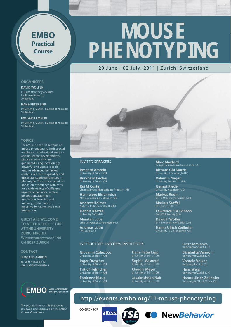
EMBOPracticalCourse
MOUSEPHENOTYPING
20 June - 02 July , 2011 | Zur ich, Switzer land
ORGANISERS
DAVID WOLFER
ETH and University of ZürichInstitute of AnatomySwitzerland
University of Zürich, Institute of AnatomySwitzerland
IRMGARD AMREIN
University of Zürich, Institute of AnatomySwitzerland
CONTACT
IRMGARD AMREIN
Tel 0041 44 635 53 [email protected]
INVITED SPEAKERS
INSTRUCTORS AND DEMONSTRATORS
European Molecular Biology Organization
GUEST ARE WELCOME
TO ATTEND THE LECTURE
AT THE UNIVERSITY
ZURICH-IRCHEL
Winterthurerstrasse 190
CH-8057 ZURICH
The programme for this event was reviewed and approved by the EMBO Course Committee.
Irmgard Amrein
Burkhard Becher
Rui M Costa
Hannelore Ehrenreich
Andrew Holmes
Dennis Kaetzel
Maarten Loos
Andreas Lüthi
Marc Mayford
Richard GM Morris
Valentin Nägerl
Gernot Riedel
Markus Rudin
Lawrence S Wilkinson
David P Wolfer
Hanns Ulrich Zeilhofer
University of Zürich (CH)
University of Zürich (CH)
Champalimaud Neuroscience Program (PT)
MPI Exp Medicine Göttingen (DE)
National Institute of Health (US)
University Oxford (UK)
Vrije Universiteit Amsterdam (NL)
FMI Basel (CH)
Scripps Research Institute La Jolla (US)
University of Edinburgh (UK)
University Bordeaux 2 (FR)
University Aberdeen (UK)
ETH & University of Zürich (CH)
ETH Zurich (CH)
University & ETH of Zürich (CH)
Giovanni Colacicco
Fritjof Helmchen
Fabienne Klaus
Elisabetta Vannoni
Vootele Voikar
Hans Welzl
Hanns Ulrich Zeilhofer
University of Zürich (CH)
University of Zürich (CH)
University of Zürich (CH)
Sophie Masneuf
University of Zürich (CH)
University Helsinki (FI)
University of Zürich (CH)
University & ETH of Zürich (CH)
TOPICSThis course covers the topic of mouse phenotyping with special emphasis on behavioral analysis and on recent developments. Mouse models that are generated using increasingly powerful and versatile tools require advanced behavioral analysis in order to quantify and
phenotype. This course provides hands-on experience with tests
aspects of behavior, such as perception, attention, motivation, learning and memory, motor control, ingestive behavior, and social interaction.
CO-SPONSOR
University of Zürich (CH)
Lutz SlomiankaUniversity of Zürich (CH)
Hans-Peter LippUniversity of Zürich (CH)
Cardiff University (UK)
Inger DrescherUniversity of Zürich (CH)
University of Zürich (CH)
University of Zürich (CH)
Claudia Meyer
Jayakrishnan Nair
HANS-PETER LIPP
Markus Stoffel
ETH & University of Zürich (CH)
1
European Molecular Biology Organization EMBO
Practical Course on Mouse Phenotyping
June 20 – July 2, 2011
Division of Functional Neuroanatomy
Division of Neuroanatomy and Behaviour
Institute of Anatomy
University Zürich‐Irchel
Switzerland
Supported by TSE, Zeiss and NewBehavior
2
Table of Contents General Information .......................................................................................................................................6
Student participants ...................................................................................................................................6
Course Staff ................................................................................................................................................8
Organizers...............................................................................................................................................8
Local staff ...............................................................................................................................................8
Invited lecturers and demonstrators .....................................................................................................9
Domestic arrangements .......................................................................................................................... 12
Course activities .................................................................................................................................. 12
Tram 10 time table ............................................................................................................................. 15
Accommodation .................................................................................................................................. 16
Excursion to central Switzerland and Mount Pilatus on Sunday, June 26, 2011 ................................ 17
Course content ............................................................................................................................................ 19
Projects .................................................................................................................................................... 19
Demonstrations ....................................................................................................................................... 20
Lectures ................................................................................................................................................... 21
Timetable overview ..................................................................................................................................... 23
Abstracts ...................................................................................................................................................... 24
Projects .................................................................................................................................................... 24
Project A – Spatial reference memory in the water‐maze .................................................................. 24
Project B – Spatial working memory on dry mazes ............................................................................. 25
Project C – Learning related to defensive and social behaviors ......................................................... 26
Project D – Olfactory and gustatory learning ...................................................................................... 27
IntelliCage project ............................................................................................................................... 28
Demonstrations ....................................................................................................................................... 29
Demonstration 01 – Testing pain in mice ........................................................................................... 29
Demonstration 02 – Motor function ................................................................................................... 30
Demonstration 03 – Exploratory behavior and anxiety ...................................................................... 31
Demonstration 04 – EEG monitoring in freely moving mice ............................................................... 32
Demonstration 05 – Two‐photon microscopy of the living brain ....................................................... 33
Demonstration 06 – Stereology: Phenotyping structural correlates of functional changes ............... 34
Lectures ................................................................................................................................................... 35
Lecture 01 – Contributions of mouse transgenics to learning and memory ....................................... 35
Lecture 02 – Genetics and the use of mice as models of anxiety and stress‐related disorders ......... 36
Lecture 03 – Phenomics: High‐throughput phenotypic screening ...................................................... 37

3
Lecture 04 – Pain phenotyping of mice ............................................................................................... 38
Lecture 05 – Multiple phenotypes of CNS inflammation! ................................................................... 39
Lecture 06 – Molecular neuroimaging in rodents ............................................................................... 40
Lecture 07 – MicroRNAs and their role in metabolism ....................................................................... 41
Lecture 08 – Assaying dissociable aspects of response inhibition in mouse models .......................... 42
Lecture 09 – Genetic control of active neural circuits ........................................................................ 43
Lecture 10 – Adult neurogenesis and behavior................................................................................... 44
Lecture 11 – Defining the neuronal circuitry of fear ........................................................................... 45
Lecture 12 – Interaction between mutations, genetic background and environment ....................... 46
Lecture 13 – Novel genetic x environmental models of psychiatric diseases: the challenge to diagnose schizophrenia and autism in a mouse .................................................................................. 47
Lecture 14 – Electrophysiological correlates of learning in vivo ......................................................... 48
Lecture 15 – Using mouse models to investigate the neural bases of action learning ....................... 49
Lecture 16 – Optogenetic dissection of neural circuits in mice .......................................................... 50
Lecture 17 – Superresolution imaging of living synapses ................................................................... 51
Poster abstracts ....................................................................................................................................... 52
Poster 01 – In vivo studies on GDNF and its receptor GFR α1 using conditional knock‐out and conditional knock‐in mice ................................................................................................................... 52
Poster 02 – Influence of IntelliCage testing on subsequent behavioral measures; Intra‐test and inter‐test relationships between automated measures of home cage behavior and behavioral measures from conventional tests ...................................................................................................................... 53
Poster 03 – The role of histone deacetylases 1 and 2 in neural development ................................... 54
Poster 04 – Automated motor function phenotyping in mice via voluntary wheel running .............. 55
Poster 05 – BAC transgenic mouse lines for studying BDNF gene regulation ..................................... 56
Poster 06 – Impairment of adult brain neurogenesis alters hippocampus‐dependent behavioural tasks without reducing learning ability ............................................................................................... 57
Poster 07 – Functional role of steroid receptors expressed in vomeronasal receptor epithelium in house mouse ....................................................................................................................................... 58
Poster 08 – Activity‐dependent recycling of synaptotagmin 1 by stonin 2 at the mammalian synapse ............................................................................................................................................................. 59
Poster 09 – The metabotropic mGlu5 receptor positive allosteric modulator, CDPPB, enhances social discrimination in a developmental model of schizophrenia, neonatal phencyclidine‐treated rats ... 60
Poster 10 – Behavioral and autonomic function abnormalities in mice lacking central serotonin .... 61
Poster 11 – Molecular dissection of the critical role of CRTC1‐CREB in regulating neuronal immediate early gene expression .......................................................................................................................... 62
Poster 12 – Different strategies of choices in the Rat Gambling Task reveal individual profiles related to human psychiatric disorders ........................................................................................................... 63
Poster 13 – Regulators of proteotoxicity in mouse models for neurodegenerative diseases ............ 64
4
Poster 14 – Molecular and behavioral patterns related to stress responsivity and associated with elevated risk for PTSD ......................................................................................................................... 65
Poster 15 – Role of different gamma secretase enzymes in prepulse inhibition (PPI) and memory in mouse brain ......................................................................................................................................... 66
Poster 16 – Phosphorylation of SCG10/stathmin‐2 determines multipolar stage exit and neuronal migration rate ...................................................................................................................................... 67
Project Protocols ......................................................................................................................................... 68
Project A .................................................................................................................................................. 68
Place and cue navigation in the water‐maze ...................................................................................... 68
Project B .................................................................................................................................................. 74
Radial‐ maze spatial working memory procedure .............................................................................. 74
T‐maze spontaneous alternation ........................................................................................................ 79
Project C .................................................................................................................................................. 83
Contextual and delay (tone) fear conditioning ................................................................................... 83
Trace fear conditioning ....................................................................................................................... 87
Three‐chamber test of sociability and preference to social novelty ................................................... 90
Project D .................................................................................................................................................. 93
Conditioned taste aversion ................................................................................................................. 93
Social transmission of food preferences ............................................................................................. 95
Burrowing test ..................................................................................................................................... 97
Nest construction test ......................................................................................................................... 99
IntelliCage Project ................................................................................................................................. 101
Free adaptation, nose‐poke adaptation, drinking session adaptation – Experiment 1 .................... 103
Delay discounting task ‐ Experiment 2 .............................................................................................. 105
Demonstration Protocols .......................................................................................................................... 108
Demonstration 01 – Testing pain in mice ............................................................................................. 108
Models of thermal nociception: hot plate and plantar test .............................................................. 108
von Frey Filament Test ...................................................................................................................... 113
Capsaicin model ................................................................................................................................ 115
Demonstration 02 – Motor function ..................................................................................................... 117
Rotarod test ....................................................................................................................................... 117
Beam walking test ............................................................................................................................. 118
Foot print test .................................................................................................................................... 120
Grip test ............................................................................................................................................. 121
Demonstration 03 – Exploratory behavior and anxiety ........................................................................ 122
Open Field test .................................................................................................................................. 122

5
Light‐dark transition test ................................................................................................................... 124
Elevated null‐maze ............................................................................................................................ 126
Emergence and novel object tests .................................................................................................... 129
Demonstration 04 – EEG monitoring of the living brain ....................................................................... 132
Demonstration 05 – Two‐photon microscopy of the living brain ......................................................... 132
Demonstration 06 – Stereology: Phenotyping structural correlates of functional changes ................. 132

6
General Information
Student participants Amberg Carolina University of Helsinki (Finland) Institute of Biotechnology Viikinkaari 9 00014 Helsinki, Finland [email protected]
Codita Alina Karolinska Institutet (Romania) NVS Department, KI‐ADRC Novum, plan 5 14186 Stockholm, Sweden [email protected]
Hagelkruys Astrid Medical University of Vienna/MFPL (Austria) Dr. Bohrgasse 9/2 1030 Wien, Austria [email protected]
Heise Ines, PhD Mediaca Research Council, Mammalian Genetics Unit (Germany) Harwell Science and Innovation Campus OX11 0RD Didcot, United Kingdom [email protected]
Jaason Kaur Tallinn University of Technology (Estonia) Institute of Gene Technology Akadeemia tee 15 12618 Tallinn, Estonia [email protected]
Jedynak Paulina Nencki Institute of Experimental Biology (Poland) 3 Pasteur 02‐093 Warsaw, Poland [email protected]
Kvasha Ilya A. N. Severtzov Institute of Ecology & Evolution (Russia) Laboratory of Comparative Neurobiology Leninskij prosp 33 119071 Moscow, Russia [email protected]
Maritzen Tanja, PhD Freie Universität Berlin (Germany) Takustr. 6 14195 Berlin, Germany [email protected]

7
Morisot Nadège Université Bordeaux 2 (France) CNRS UMR 5287 Batiment 4A, 3ème étage, porte 8 146 rue Léo Saignat 33000, Bordeaux, France [email protected]
Mosienko, Valentina Max‐Delbrueck Center for Molecular Medicine (Belarus) Robert‐Roessle str.10 13125 Berlin, Germany valentina.mosienko@mdc‐berlin.de
Nonaka Mio, PhD University of Tokyo (Japan) S605, Igakubu‐3‐gokan building 7‐3‐1 hongo, Bunkyo‐ku 113‐0033 Tokyo, Japan [email protected]‐tokyo.ac.jp
Rivalan Marion, PhD Charité Universitätsmedizin Berlin (France) Exzellenzcluster NeuroCure Kognitive Neurobiologie Dorotheenstr.94 10117 Berlin, Germany [email protected]
Sin Olga University Medical Center Groningen (Portugal) PO box 30.001 9700 RB Groningen, Netherland [email protected]
Szklarczyk Klaudia Institute of Pharmacology Polish Academy of Sciences (Poland) Smętna 12 31‐321 Cracow, Poland [email protected]
Visser Aline, PhD Catholic University of Leuven (Belgum) O&N1 Herestraat 49 box 602 3000, Leuven, Belgum [email protected]‐kuleuven.be
Zdrojewska Justyna Turku Centre for Biotechnology (Poland) Tykistokatu 6 20521 Turku, Finland [email protected]
8
Course Staff
Organizers Wolfer David P, MD ETH Zürich, Institute of Human Movement Science and Sports University of Zürich, Institute of Anatomy Division of Functional Neuroanatomy Winterthurerstrasse 190 8057 Zürich, Switzerland [email protected] Lipp Hans‐Peter, PhD University of Zürich, Institute of Anatomy Division of Neuroanatomy and Behavior Winterthurerstrasse 190 8057 Zürich, Switzerland [email protected] Amrein Irmgard, PhD University of Zürich, Institute of Anatomy Division of Functional Neuroanatomy/Neuroanatomy and Behavior Winterthurerstrasse 190 8057 Zürich, Switzerland [email protected]
Local staff Colacicco Giovanni, MD [email protected] Drescher Inger [email protected] Nair Jayakrishnan [email protected] Klaus Fabienne [email protected] Masneuf Sophie, PhD [email protected] Meyer Claudia [email protected] Rhyner Astrid [email protected] Sebele Monika [email protected] Slomianka Lutz [email protected] Vannoni Elisabetta, PhD [email protected] Welzl Hans, PhD [email protected]

9
Invited lecturers and demonstrators Becher Burkhard, PhD University of Zürich Pathologie and Exper. Immunologie, Y44 J 92 Winterthurerstrasse 190 8057 Zürich, Switzerland [email protected] Costa Rui M, DVM/PhD Champalimaud Neuroscience Program Champalimaud Foundation Champalimaud Centre for the Unknown Av. Brasília 1400‐038 Lisbon, Portugal [email protected] Ehrenreich Hannelore, MD/DVM Max Planck Institute for Experimental Medicine (MPI Exp Medicine) Göttingen Hermann‐Rein‐Str. 3 37075 Göttingen, Germany [email protected] Helmchen Fritjof, PhD University of Zurich Institute of Brain Research, Y55 H 66 Winterthurerstrasse 190 8057 Zürich, Switzerland [email protected] Holmes Andrew, PhD National Institute of Health NIH National Institute on Alcohol Abuse and Alcoholism (NIAAA) 5635 Fishers Lane, MSC 9304 Bethesda, MD 20892‐9304, USA [email protected] Kaetzel Dennis, PhD Miesenboeck Lab Department of Physiology, Anatomy and Genetics University of Oxford Sherrington Building Parks Road Oxford , OX1 3PT, United Kingdom [email protected] Loos Maarten, PhD Vrije Univ Amsterdam Neuroscience Campus Amsterdam B4 wing – W&N building De Boelelaan 1085, Nederland [email protected]
10
Lüthi Andreas, PhD Friedrich Miescher Institute for Biomedical Research (FMI) Basel Maulbeerstrasse 66 P.O. Box 2543 4002 Basel , Switzerland [email protected] Mayford Marc, PhD Scripps Research Institute La Jolla Department of Cell Biology California Campus 10550 N. Torrey Pines Rd. Mail Drop TPC‐11 La Jolla, CA 92037, USA [email protected] Morris Richard GM, PhD FRS, CBS University of Edinburgh Royal Society/Wolfson Professor of Neuroscience 1 George Square Edinburgh, EH8 9JZ, United Kingdom [email protected] Nägerl U Valentin, PhD University of Bordeaux 2 Institut interdisciplinaire de Neurosciences Synaptic Plasticity and Superresolution Microscopy – AVENIR Group CNRS UMR 5297 Université Bordeaux 2 146, rue Léo‐Saignat 33077 Bordeaux cédex, France valentin.nagerl@u‐bordeaux2.fr Riedel Gernot, PhD University of Aberdeen Institute of Medical Sciences Foresterhill Aberdeen ∙ AB25 2ZD, United Kingdom [email protected] Rudin Markus, PhD University & ETH Zurich Institute für Pharmakologie und Toxikologie, HI1 E22.4 Wolfgang‐Pauli‐Strasse 27 8093 Zürich, Switzerland [email protected] Stoffel Markus, MD ETH Zurich Institut für Molekulare Systembiologie, HPT E73 Wolfgang‐Pauli‐Str. 16 8093 Zürich [email protected]
11
Voikar Vootele, MD/PhD University of Helsinki Neuroscience Center P.O. Box 56, FI‐00014 [email protected] Wilkinson Lawrence S, PhD Cardiff University School of Medicine Professor of Behavioural Genetics Department of Psychological Medicine & Neurology 2nd Floor, Henry Wellcome Building Heath Park Cardiff CF14 4XN, United Kingdom [email protected] Zeilhofer Hanns Ulrich, MD University & ETH Zurich Institut für Pharmakologie u Toxikologie, Y17 J 68 Winterthurerstrasse 190 8057 Zürich, Switzerland [email protected]
12
Domestic arrangements
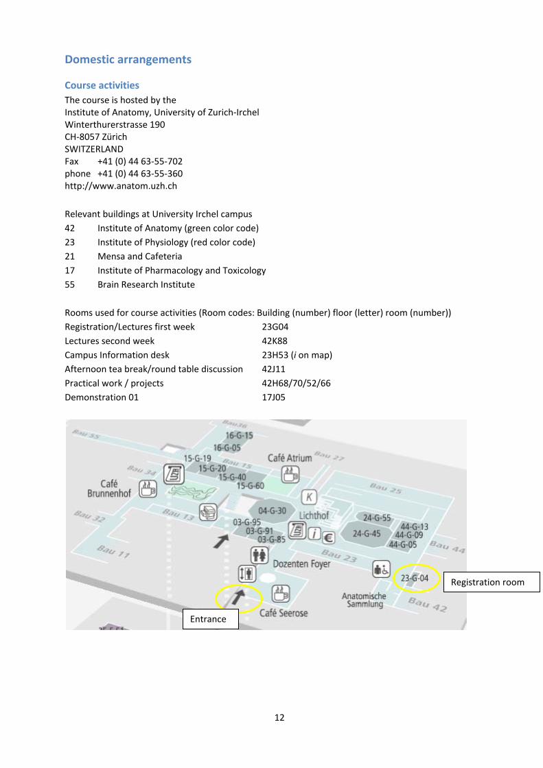
Course activities The course is hosted by the Institute of Anatomy, University of Zurich‐Irchel Winterthurerstrasse 190 CH‐8057 Zürich SWITZERLAND Fax +41 (0) 44 63‐55‐702 phone +41 (0) 44 63‐55‐360 http://www.anatom.uzh.ch Relevant buildings at University Irchel campus 42 Institute of Anatomy (green color code) 23 Institute of Physiology (red color code) 21 Mensa and Cafeteria 17 Institute of Pharmacology and Toxicology 55 Brain Research Institute Rooms used for course activities (Room codes: Building (number) floor (letter) room (number)) Registration/Lectures first week 23G04 Lectures second week 42K88 Campus Information desk 23H53 (i on map) Afternoon tea break/round table discussion 42J11 Practical work / projects 42H68/70/52/66 Demonstration 01 17J05
Entrance
Registration room
13
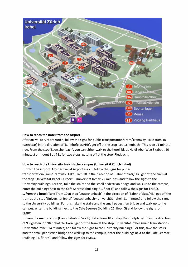
How to reach the hotel from the Airport After arrival at Airport Zurich, follow the signs for public transportation/Tram/Tramway. Take tram 10 (streetcar) in the direction of ‘Bahnhofplatz/HB’, get off at the stop 'Leutschenbach'. This is an 11 minute ride. From the stop 'Leutschenbach', you can either walk to the hotel ibis at Heidi‐Abel‐Weg 5 (about 10 minutes) or mount Bus 781 for two stops, getting off at the stop 'Riedbach'. How to reach the University Zurich Irchel campus (Universität Zürich Irchel) … from the airport: After arrival at Airport Zurich, follow the signs for public transportation/Tram/Tramway. Take Tram 10 in the direction of ‘Bahnhofplatz/HB’, get off the tram at the stop 'Universität Irchel' (Airport – Universität Irchel: 22 minutes) and follow the signs to the University buildings. For this, take the stairs and the small pedestrian bridge and walk up to the campus, enter the buildings next to the Café Seerose (building 21, floor G) and follow the signs for EMBO. … from the hotel: Take Tram 10 at stop ‘Leutschenbach’ in the direction of ‘Bahnhofplatz/HB’, get off the tram at the stop 'Universität Irchel' (Leutschenbach– Universität Irchel: 11 minutes) and follow the signs to the University buildings. For this, take the stairs and the small pedestrian bridge and walk up to the campus, enter the buildings next to the Café Seerose (building 21, floor G) and follow the signs for EMBO. … from the main station (Hauptbahnhof Zürich): Take Tram 10 at stop ‘Bahnhofplatz/HB’ in the direction of ‘Flughafen’ or ‘Bahnhof Oerlikon’, get off the tram at the stop 'Universität Irchel' (main train station ‐ Universität Irchel: 14 minutes) and follow the signs to the University buildings. For this, take the stairs and the small pedestrian bridge and walk up to the campus, enter the buildings next to the Café Seerose (building 21, floor G) and follow the signs for EMBO.

14
(Please note that there is a second large building complex of the University in the city centre, near the main station called ‘Universität Zürich Hauptgebäude’. This is NOT the place where the EMBO course is taking place!) How to reach the hotel from the railway main station (Hauptbahnhof Zurich) If you arrive by train, take the Tram 10 (direction ‘Flughafen’ or ‘Bahnhof Oerlikon’). Get off the tram at stop 'Leutschenbach' (about 25 minutes) and you can either walk to the hotel ibis at Heidi‐Abel‐Weg 5 (about 10 minutes) or mount Bus 781 for two stops, getting off at the stop 'Riedbach'.
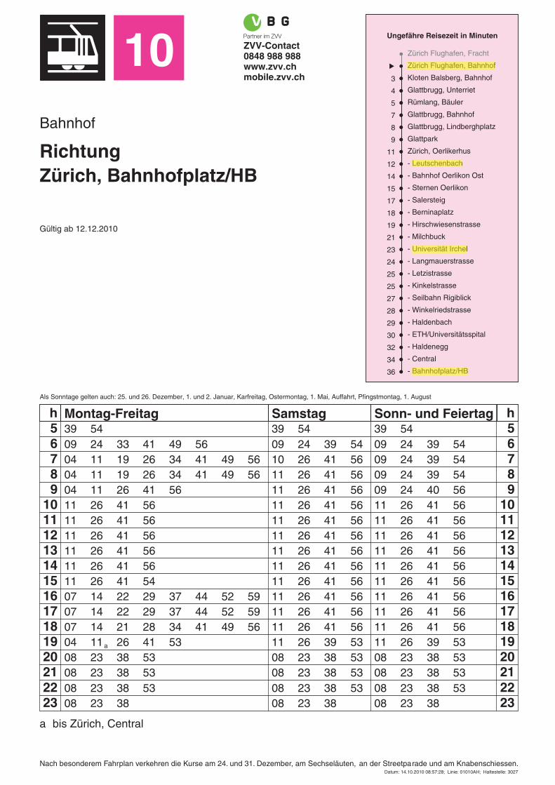
10 ZVV-Contact0848 988 988www.zvv.chmobile.zvv.ch
Bahnhof
RichtungZürich, Bahnhofplatz/HB
Gültig ab 12.12.2010
Ungefähre Reisezeit in Minuten
Zürich Flughafen, Fracht
Zürich Flughafen, Bahnhof
Kloten Balsberg, Bahnhof3
Glattbrugg, Unterriet4
Rümlang, Bäuler5
Glattbrugg, Bahnhof7
Glattbrugg, Lindberghplatz8
Glattpark9
Zürich, Oerlikerhus11
- Leutschenbach12
- Bahnhof Oerlikon Ost14
- Sternen Oerlikon15
- Salersteig17
- Berninaplatz18
- Hirschwiesenstrasse19
- Milchbuck21
- Universität Irchel23
- Langmauerstrasse24
- Letzistrasse25
- Kinkelstrasse25
- Seilbahn Rigiblick27
- Winkelriedstrasse28
- Haldenbach29
- ETH/Universitätsspital30
- Haldenegg32
- Central34
- Bahnhofplatz/HB36
Als Sonntage gelten auch: 25. und 26. Dezember, 1. und 2. Januar, Karfreitag, Ostermontag, 1. Mai, Auffahrt, Pfingstmontag, 1. August
h Montag-Freitag Samstag Sonn- und Feiertag h5 56 67 78 89 9
10 1011 1112 1213 1314 1415 1516 1617 1718 1819 1920 2021 2122 2223 23
39 5409 24 33 41 49 5604 11 19 26 34 41 49 5604 11 19 26 34 41 49 5604 11 26 41 5611 26 41 5611 26 41 5611 26 41 5611 26 41 5611 26 41 5611 26 41 5407 14 22 29 37 44 52 5907 14 22 29 37 44 52 5907 14 21 28 34 41 49 5604 11 26 41 5308 23 38 5308 23 38 5308 23 38 5308 23 38
39 5409 24 39 5410 26 41 5611 26 41 5611 26 41 5611 26 41 5611 26 41 5611 26 41 5611 26 41 5611 26 41 5611 26 41 5611 26 41 5611 26 41 5611 26 41 5611 26 39 5308 23 38 5308 23 38 5308 23 38 5308 23 38
39 5409 24 39 5409 24 39 5409 24 39 5409 24 40 5611 26 41 5611 26 41 5611 26 41 5611 26 41 5611 26 41 5611 26 41 5611 26 41 5611 26 41 5611 26 41 5611 26 39 5308 23 38 5308 23 38 5308 23 38 5308 23 38
a
Nach besonderem Fahrplan verkehren die Kurse am 24. und 31. Dezember, am Sechseläuten, an der Streetparade und am Knabenschiessen.
a bis Zürich, Central
Datum: 14.10.2010 08:57:28; Linie: 01010AH; Haltestelle: 3027
16
Accommodation Participants, lecturers and demonstrators from outside Switzerland are accommodated at the
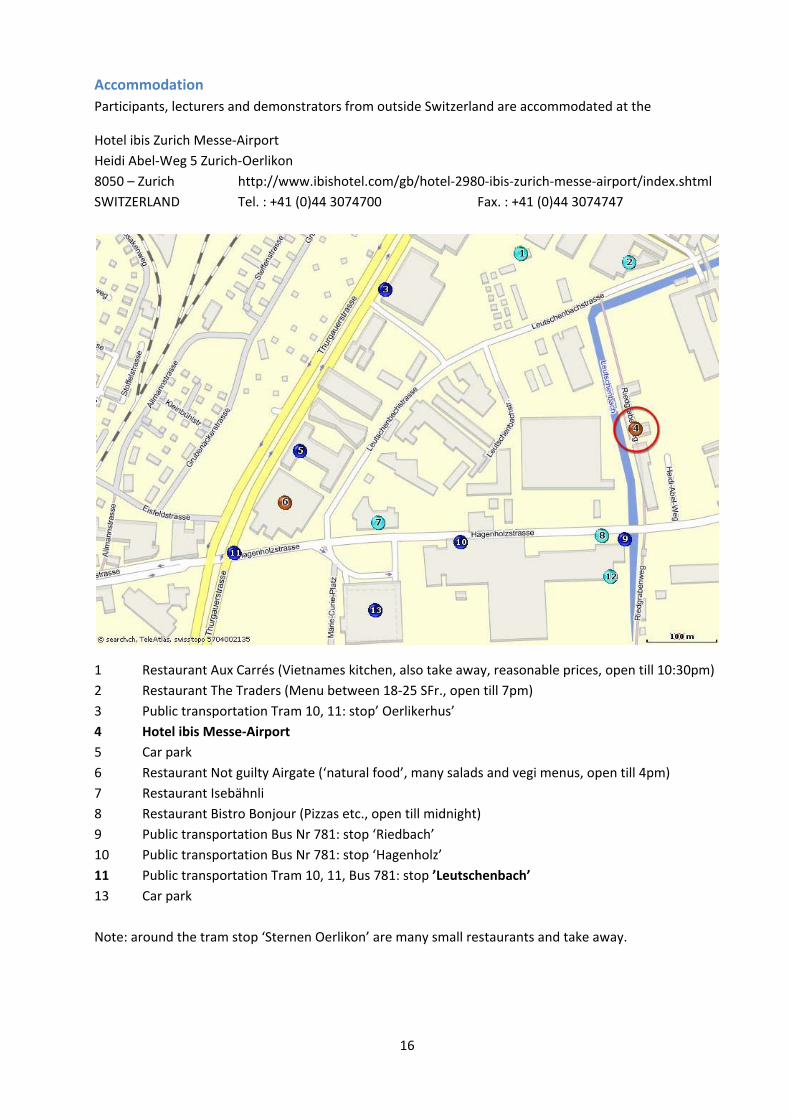
Hotel ibis Zurich Messe‐Airport Heidi Abel‐Weg 5 Zurich‐Oerlikon 8050 – Zurich http://www.ibishotel.com/gb/hotel‐2980‐ibis‐zurich‐messe‐airport/index.shtml SWITZERLAND Tel. : +41 (0)44 3074700 Fax. : +41 (0)44 3074747
1 Restaurant Aux Carrés (Vietnames kitchen, also take away, reasonable prices, open till 10:30pm) 2 Restaurant The Traders (Menu between 18‐25 SFr., open till 7pm) 3 Public transportation Tram 10, 11: stop’ Oerlikerhus’ 4 Hotel ibis Messe‐Airport 5 Car park 6 Restaurant Not guilty Airgate (‘natural food’, many salads and vegi menus, open till 4pm) 7 Restaurant Isebähnli 8 Restaurant Bistro Bonjour (Pizzas etc., open till midnight) 9 Public transportation Bus Nr 781: stop ‘Riedbach’ 10 Public transportation Bus Nr 781: stop ‘Hagenholz’ 11 Public transportation Tram 10, 11, Bus 781: stop ’Leutschenbach’ 13 Car park Note: around the tram stop ‘Sternen Oerlikon’ are many small restaurants and take away.
17
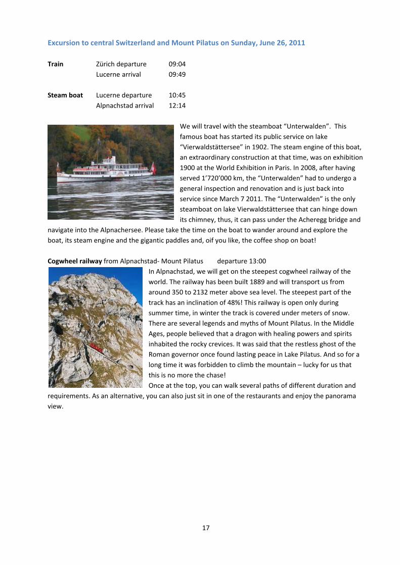
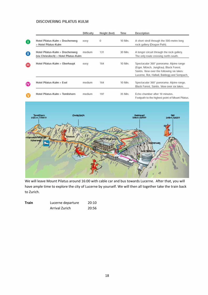
Excursion to central Switzerland and Mount Pilatus on Sunday, June 26, 2011 Train Zürich departure 09:04 Lucerne arrival 09:49 Steam boat Lucerne departure 10:45 Alpnachstad arrival 12:14
We will travel with the steamboat “Unterwalden”. This famous boat has started its public service on lake “Vierwaldstättersee” in 1902. The steam engine of this boat, an extraordinary construction at that time, was on exhibition 1900 at the World Exhibition in Paris. In 2008, after having served 1’720’000 km, the “Unterwalden” had to undergo a general inspection and renovation and is just back into service since March 7 2011. The “Unterwalden” is the only steamboat on lake Vierwaldstättersee that can hinge down its chimney, thus, it can pass under the Acheregg bridge and
navigate into the Alpnachersee. Please take the time on the boat to wander around and explore the boat, its steam engine and the gigantic paddles and, oif you like, the coffee shop on boat! Cogwheel railway from Alpnachstad‐ Mount Pilatus departure 13:00
In Alpnachstad, we will get on the steepest cogwheel railway of the world. The railway has been built 1889 and will transport us from around 350 to 2132 meter above sea level. The steepest part of the track has an inclination of 48%! This railway is open only during summer time, in winter the track is covered under meters of snow. There are several legends and myths of Mount Pilatus. In the Middle Ages, people believed that a dragon with healing powers and spirits inhabited the rocky crevices. It was said that the restless ghost of the Roman governor once found lasting peace in Lake Pilatus. And so for a long time it was forbidden to climb the mountain – lucky for us that this is no more the chase! Once at the top, you can walk several paths of different duration and
requirements. As an alternative, you can also just sit in one of the restaurants and enjoy the panorama view.
18
We will leave Mount Pilatus around 16:00 with cable car and bus towards Lucerne. After that, you will have ample time to explore the city of Lucerne by yourself. We will then all together take the train back to Zurich. Train Lucerne departure 20:10 Arrival Zurich 20:56
19
Course content
Projects
Projects / Group work
Four different mini‐projects with mice, embedded in ongoing projects at the institute, will run through the entire course and be carried out by the participants themselves in groups of four. The projects will be instructed and supervised by experienced teachers. Each group will have a main project but will be given the opportunity to interact with the other projects. Project work will mainly be done in the afternoon. In the evening labs will remain open for those who need more time to complete their work. The projects will familiarize the participants with the following behavioral assays of learning and memory (from planning and design to analysis and statistics of the collected data). Project A – Spatial reference memory in the water‐maze Instructors Colacicco Giovanni and Meyer Claudia Participants Heise Ines, Sin Olga, Codita Alina and Zdrojewska Justyna Location 42H68 Project B – Spatial working memory on dry mazes Instructors Masneuf Sophie and Drescher Inger Participants Maritzen Tanja, Visser Alina, Jaanson Kaur and Hagelkruys Astrid Location 42H70 Project C – Learning related to defensive and social behaviors Instructors Voikar Vootele Participants Mosienko Valentina, Morisot Nadège, Szklarczyk Klaudia and Amberg Carolina Location 42H52a Project D – Olfactory and gustatory learning Instructors Welzl Hans Participants Jedynak Paulina, Kvasha Ilya, Rivalan Marion and Nonaka Mio Location 42H66b In parallel to the experimental work listed above, all students will run behavioral tests with socially housed mice using the home‐cage automated system IntelliCage. IntelliCage Project Instructor Vannoni Elisabetta Participants All Location 42H52b
20
Demonstrations To complement project work and to familiarise the course participants with a broader range of procedures and techniques, a number of practical demonstrations are given during the course which will be attended by all course participants in groups of eight students. Some demonstrations are live, others are based on video material. Demo 01 – Testing pain in mice Instructors Di Lio Alessandra, Ralvenius William and Zeilhofer Hanns Ulrich Location 17J05 Demo 02 – Motor function Instructors Welzl Hans Location 42H66b Demo 03 – Exploratory behavior and anxiety Instructors Voikar Vootele Location 42H70 Demo 04 – EEG monitoring in freely moving mice Instructors Lipp Hans‐Peter and Nair Jayakrishnan Location 42J44 Demo 05 – Two‐photon microscopy of the living brain Instructors Helmchen Fritjof Location Demo 06 – Stereology: Phenotyping structural correlates of functional changes Instructors Klaus Fabienne Klaus and Slomianka Lutz Location 42 J 11 and 42 J 45
21
Lectures The lectures are for all participants. Guests from the Institute of Anatomy and other institutes of the University and ETH are welcome.
Lecture 01 – Contributions of mouse transgenics to learning and memory Speaker Morris Richard GM Location/ time 23G04, June 20, 16:30 Lecture 02 – Genetics and the use of mice as models of anxiety and stress‐related disorders Speaker Holmes Andrew Location/ time 23G04, June 21, 9:00 Lecture 03 – High throughput phenotypic screening Speaker Loos Maarten Location/ time 23G04, June 21, 10:45 Lecture 04 – Pain phenotyping of mice Speaker Zeilhofer Hanns Ulrich Location/ time 23G04, June 22, 9:00 Lecture 05 – Multiple Phenotypes of CNS inflammation! Speaker Becher Burkhard Location/ time 23G04, June 22, 10:45 Lecture 06 – Molecular neuroimaging in rodents Speaker Rudin Markus Location/ time 23G04, June 23, 9:00 Lecture 07 – MicroRNAs and their role in metabolism Speaker Stoffel Markus Location/ time 23G04, June 23, 10:45 Lecture 08 – Assaying dissociable aspects of response inhibition in mouse models Speaker Wilkinson Lawrence S Location/ time 23G04, June 24, 9:00 Lecture 09 – Genetic control of active neural circuits Speaker Mayford Marc Location/ time 23G04, June 24, 10:45

22
Lecture 10 – Adult neurogenesis and behavior Speaker Amrein Irmgard Location/ time 42K88, June 27, 9:00 Lecture 11 – Defining the neuronal circuitry of fear Speaker Lüthi Andreas Location/ time 42K88, June 27, 10:45 Lecture 12 – Interaction between mutations, genetic background and environment Speaker Wolfer David P Location/ time 42K88, June 28, 9:00 Lecture 13 – Novel genetic x environmental models of psychiatric diseases: the challenge to diagnose schizophrenia and autism in a mouse Speaker Ehrenreich Hannelore Location/ time 42K88, June 28, 10:45 Lecture 14 – Electrophysiological correlates of learning in vivo Speaker Riedel Gernot Location/ time 42K88, June 29, 9:00 Lecture 15 – Using mouse models to investigate the neural bases of action learning Speaker Costa Rui M Location/ time 42K88, June 29, 10:45 Lecture 16 – Optogenetic dissection of neural circuits in mice Speaker Kaetzel Dennis Location/ time 42K88, June 30, 9:00 Lecture 17 – Superresolution imaging of living synapses Speaker Nägerl Valentin U Location/time 42K88, June 30, 10:45

EMBO Practical Course on Mouse Phenotyping 2011
Mon June 20 Tue June 21 Wed June 22 Thu June 23 Fri June 24 Sat June 25 Sun June 2609:00 - 10:15 23G04 L02 23G04 L04 23G04 L06 23G04 L08 Groups BCD visit Project A
Holmes, Andrew Zeilhofer, Hanns U Rudin, Markus Wilkinson, Lawrence S (09:00 - 10:30)Genetics and the use of mice as Pain phenotyping of mice Molecular neuroimaging in Assaying dissociable aspects of 42H68models of anxiety and stress- rodents response inhibition in mouse related disorders models
10:15 - 10:45 Coffee break Coffee break Coffee break Coffee break 10:30-11:00 Coffee break10:45 - 12:00 23G04 L03 23G04 L05 23G04 L07 23G04 L09 Groups ACD visit project B Excursion to Central
Loos, Maarten Becher, Burkhard Stoffel, Markus Mayford, Marc (11:00 -12:30) Switzerland
20.06.2011
Loos, Maarten Becher, Burkhard Stoffel, Markus Mayford, Marc (11:00 -12:30) SwitzerlandHigh throughput phenotypic Multiple Phenotypes of CNS Phenotyping energy Genetic control of active neural 42H70 City and lake Lucerne screening inflammation! homeostasis circuits Pilatus
12:00 - 13:00 Lunch Lunch Lunch Lunch Lunch13:00 23G04 Registration opens 13:00 - 15:00 42H52b IntelliCage Project Group work Demo01 (AB), Demo02 (CD) Demo02 (AB), Demo01 (CD) Groups ABD visit Project C14:00 Formation of interest groups Vannoni, Elisabetta A 42H68 17J05 D01 Zeilhofer, Hanns U (13:30-15:00)16:00 23G04 Welcome, general info B 42H70 Testing pain in mice 42H52a16:30 - 18:00 C 42H52a 42H66b D02 Welzl, Hans23G04 L01 D 42H66b Motor functionM i Ri h d GM 15 00 16 00 42J11 T b k d t bl 42J11 T b k d t bl 42J11 T b k d t bl 42J11 T b k d t bl 42J11 T b kMorris, Richard GM 15:00 - 16:00 42J11 Tea break, round table 42J11 Tea break, round table 42J11 Tea break, round table 42J11 Tea break, round table 42J11 Tea breakContributions of mouse transgenics discussion with speakers discussion with speakers discussion with speakers discussion with speakersto learning and memory 16:00 - 18:30 42H52b IntelliCage Project Group work (ABCD) Group work (ABCD) Group work (ABCD) Groups ABC visit Project DAfterwards: get together party 18:30 - 20:00 42J11 Wine, cheese and posters 42J11 Wine, cheese and posters (15:30 - 17:00)42J11 Medihof of groups A and B of groups C and D 42H66b
42G53 reserved for work/study 42G53 reserved for work/study 42G53 reserved for work/study 42G53 reserved for work/study 42G53 reserved for work/studytill 17:00 all day all day all day all day
Mon June 27 Tue June 28 Wed June 29 Thu June 30 Fri July 01 Sat July 0209:00 - 10:15 42K88 L10 42K88 L12 42K88 L14 42K88 L16 Groups ABCD Groups ABCD
Amrein, Irmgard Wolfer, David Riedel, Germot Kaetzel, Dennis Analye data and prepare Presentation of projectsAdult neurogenesis and behavior Interaction between mutations, Electrophysiological correlates Optogenetic dissection of presentations 23G04
genetic background and of learning in vivo neural circuits in mice 42J11; 42G53environment
10:15 - 10:45 Coffee break Coffee break Coffee break Coffee break10:15 - 10:45 Coffee break Coffee break Coffee break Coffee break10:45 - 12:00 42K88 L11 42K88 L13 42K88 L15 42K88 L17
Lüthi, Andreas Ehrenreich, Hannelore Costa, Rui M Nägerl, U ValentinDefining the neuronal circuitry Novel genetic x environmental Using mouse models to investi- Superresolution imaging of living of fear models of psychiatric diseases: gate the neural bases of action synapses
the challenge to diagnose schizo- learningphrenia and autism in a mouse
12:00 - 13:00 Lunch Lunch Lunch Lunch Lunch Concluding remarks, 13:00 - 15:00 Demo03 (AB), Demo04 (CD) Demo04 (AB), Demo03 (CD) Demo05 (AB), Demo06 (CD) D06 (AB), D05 (CD) Groups ABCD course evaluation
42H68/42G 3 D03 V ik V l f42H68/42G53 D03 Voikar, Vootele D05 Helmchen, Fritjof Analye data and prepareExploratory behavior and fear Two-photon microscopy of the living brain presentations42J44 D04 Lipp, Hans-Peter 42J45/11 D06 Klaus, Fabienne 42J11; 42G53EEG monitoring in freely moving mice Stereology: Phenotyping structural correlates of functional changes
15:00 - 16:00 42J11 Tea break, round table 42J11 Tea break, round table 42J11 Tea break, round table 42J11 Tea break, round table discussion with speakers discussion with speakers discussion with speakers discussion with speakers
afterwards Group work (ABCD) Group work (ABCD) Group work (ABCD) Group work (ABCD)42G53 reserved for work/study 42G53 reserved for work/study 42G53 reserved for work/study 42G53 reserved for work/studyall day all day all day all dayall day all day all day all day
20.06.2011
24
Abstracts
Projects
Project A – Spatial reference memory in the water‐maze Instructors Colacicco Giovanni and Meyer Claudia Participants Heise Ines, Sin Olga, Codita Alina and Zdrojewska Justyna Location 42H68 Protocol
• Place and cue navigation in the water‐maze Mice: strain comparison DBA/2 versus C57BL/6 In the water‐maze place navigation task, introduced by Richard G.M. Morris in 1981 as a test for spatial reference memory of rats, an animal is released repeatedly from varying locations into a pool of water that has been made opaque by addition of milk or nontoxic white paint. To escape from the water, the subject has to locate a platform that is hidden underneath the water surface at a constant location. Because the goal is invisible and no local cues are available inside the pool, the subject must learn to navigate using multiple distant cues arranged in the room around the pool. Mice can learn the water‐maze place navigation task as well. During the last two decades it has become one of the most frequently used tests to assess hippocampal function and spatial memory in genetically modified mice. Place navigation is disrupted by lesions of the hippocampus, but the paradigm is also sensitive to genetic changes which reduce behavioural flexibility, disrupt exploratory behavior or affect motivation. Intact function of sensory and motor systems is typically verified using the cue navigation task in which the platform is marked with a local cue and placed at random locations. The place navigation protocol that will be used during the course will also include a phase of reversal learning. That is, after three days of training for a fixed platform location, its position will be changed to test the animals’ capacity to adapt their spatial orientation and to learn a second platform location. This reversal phase may reveal milder deficits in animals that are still able to learn a single platform location. Other protocols that have been developed for the water‐maze, such as serial reversal or delayed matching‐to‐place tasks, may also be presented and discussed during the course.
25
Project B – Spatial working memory on dry mazes Instructors Masneuf Sophie and Drescher Inger Participants Maritzen Tanja, Visser Alina, Jaanson Kaur and Hagelkruys Astrid Location 42H70 Protocols
• Radial‐ maze spatial working memory procedure
• T‐maze spontaneous alternation Mice: strain comparison DBA/2 versus C57BL/6 In the radial maze working memory procedure that will be used in this project, the subject is released on the central platform of the elevated eight‐arm maze on which it can move freely to collect small food pellets from the ends of the arms. The subject has to develop a strategy allowing it to collect all eight pellets without re‐entering an emptied arm. Radial maze tasks have been popularized as tests of hippocampus‐dependent spatial memory in 1976 by Olton and Samuelson who showed that rats rely on extramaze cues for spatial orientation, rather than utilizing intramaze odor cues or repetitive choice patterns. As mice behave in similar ways, radial maze tasks have become a quite popular alternative or complement of the water‐maze place navigation task for testing hippocampal function in genetically modified mice. When all arms are re‐baited before each trial, the memory for already visited arms is only valid within a trial and not from one trial to the next. Therefore, this procedure is said to measure spatial working memory rather than spatial reference memory. But, as will be discussed during the course, with modified protocols the eight‐arm radial maze can also be used to assess spatial reference memory. During the course we will also demonstrate how the mice are placed under a restricted feeding regime in order to increase their motivation to retrieve the food pellets.
Spontaneous alternation on the T‐maze is a simple and rapid protocol that exploits the innate motivation of rodents to explore novel places. In a sample trial, the subject is allowed to freely choose the left or right arm of the T‐maze. Once it has made a choice, it is confined to the chosen arm and allowed to explore it. After a short delay, a choice trial is run in which the animal can again freely choose between the left and right arm. It will typically choose the arm not visited before, indicating that it remembers the choice made during the sample trial. This spontaneous alternation behavior is highly sensitive to disruption of hippocampal function, but also to other manipulations of the brain including those that reduce exploratory drive.

26
Project C – Learning related to defensive and social behaviors Instructors Voikar Vootele Participants Mosienko Valentina, Morisot Nadège, Szklarczyk Klaudia and Amberg Carolina Location 42H52a Protocols
• Contextual and delay (tone) fear conditioning
• Trace fear conditioning
• Three‐chamber test of sociability and preference to social novelty Mice: Strain comparison DBA/2 versus C57BL/6, half of the mice were single housed before testing, the rest in group cages Fear conditioning, a form of classical Pavlovian conditioning (associative learning), is often used to study emotional learning and memory in rodents, with the freezing response typically measured as behavioural endpoint (conditioned response, CR). In cued and contextual fear conditioning the CR appears following pairing of an unconditioned stimulus (US, foot shock) with a conditioned stimulus (CS, cue, e.g. tone) when the animal is re‐exposed to the CS or training context. There are two types of cue conditioning. In delay conditioning procedures the US co‐terminates with the CS. Trace conditioning inserts a trace interval between CS and US, requiring the learning of a temporal relationship between the two stimuli. Fear conditioning is generally disrupted by lesions of the amygdala. The hippocampus is needed for contextual memory (association of context and US) and for trace fear conditioning. New memories are stabilized by a process called consolidation. However, reactivation (retrieval) of a memory trace can induce an additional labile phase that requires re‐stabilization of a memory by a process named re‐consolidation. Inhibition of fear is studied by exposing a conditioned animal to the CS many times in absence of the US. The resulting decline of the conditioned fear response is attributed to a process called fear extinction. However, there are several situations in which extinguished fear responses reappear. Reinstatement refers to reappearance of extinguished fear following unsignaled presentations of the US. Renewal refers to reappearance of the extinguished CR when animals are tested in a context different from the one in which extinction training took place. Spontaneous recovery refers to a reappearance of extinguished CR with the passage of time following extinction training in the absence of any further explicit training. Mice are social animals and the study of exploratory and aggressive social interactions has become an important aspect of behavioral phenotyping, for example in the context of autism research. It has also been recognized that social interaction is an important source of enrichment in laboratory conditions and that social deprivation can alter cognitive as well as emotional behavior of mice. In this project, mice will be examined in a recently introduced three‐chamber sociability and social recognition test. In a first phase of this test, the subject is confronted with a unknown mouse and a novel object in order to measure its preference for a social stimulus. In a second phase, social recognition memory is assessed by confronting the subject with a novel and a previously encountered mouse and measuring its preference to interact with the novel mouse.
27
Project D – Olfactory and gustatory learning Instructors Welzl Hans Participants Jedynak Paulina, Kvasha Ilya, Rivalan Marion and Nonaka Mio Location 42H66b
Protocols
• Conditioned taste aversion
• Social transmission of food preferences
• Burrowing test
• Nest construction test Mice: Strain comparison DBA/2 versus C57BL/6 Conditioned taste aversion (CTA) is a well established learning and memory paradigm in rats and mice that is considered to be a special form of classical conditioning. Rodents – as well as many other species including man – learn to associate a novel taste (conditioned stimulus CS) of food or liquid with nausea (unconditioned stimulus US) they experienced after its consumption. As a consequence, they avoid drinking fluid or consuming food with this specific taste. Advantages of this paradigm are its relative independence of motor behaviour, as well as the wealth of available anatomical and pharmacological data implying several brain structures, pathways, neurotransmitters and their receptors, and cellular processes in CTA. At the level of the forebrain, this form of CTA depends among other structures on the integrity of the amygdala and insular cortex, but is not disrupted by hippocampal lesions. To establish CTA, animals will first be adapted to a specific drinking schedule, then aversively conditioned to saccharin solution by intraperitoneal injection of lithium chloride which causes nausea. After an interval, their avoidance of saccharin solution is tested in a choice session. Social transmission of food preferences takes advantage of the fact that rodents develop a preference for foods they have recently smelled on the breath of another rodent. There is evidence that this process not only relies on the olfactory system, but also requires an intact hippocampus. In preparation for this experiment, involved mice are adapted to eating ground food from special food cups and a demonstrator mouse is selected from each cage. The demonstrator mouse is first fed a specifically flavoured ground food and then allowed to interact with the remaining cage mates, called the observer mice. After an interval, the observer mice are tested in a choice situation between the demonstrated food and another unfamiliar food.
Burrowing (displacement of food pellets from a tube in the home cage) and nest construction are species‐typical behaviours that are very sensible to hippocampal lesions. Quantitative assessment of these behaviours in genetically modified mice is a valuable complement of hippocampus‐dependent learning tasks.
28
IntelliCage project Instructor Vannoni Elisabetta Participants All Location 42H52b Protocols
• Free adaptation, nose‐poke adaptation, drinking session adaptation
• Delay discounting task
Mice: Strain comparison DBA/2 versus C57BL/6. The IntelliCage system enables automatic monitoring of mice behavior over an extended period of time in a homecage‐like environment within social groups. It includes four conditioning corners. The animals are implanted with a passive transponder recognized by reader antennas every time they enter a corner. Number and duration of corner visits is automatically monitored individually. Each chamber contains 2 holes leading to water bottles by means of nose‐pokes. Access to reward is controlled by motorized movable doors and can be regulated for individual mice according to pre‐programmed computer‐operated experimental schedules. Lickometers measure licking behavior whereas food is provided ab libitum. During this project, two IntelliCages, each containing 8 DBA/2 and 8 C57BL/6 mice, will be used. In the first IntelliCage, naive mice will be introduced and will run through three different adaptation phases: free adaptation (FA), nose‐poke adaptation (NPA) and drinking session adaptation (DSA). During FA and NPA, animals will acquire information about the novel environment by means of exploratory activity indexed by the number of corner visits and nose‐pokes. Anxiety‐related behaviors possibly affecting exploration, spontaneous spatial preferences, and circadian activity will also be assessed. During the DSA, mice will learn to access the reward during two 1‐hour sessions per day. Time learning capabilities of the animals will be evaluated by looking at the activity profile before, during and after the session. The second IntelliCage houses mice that are already familiar with the system and that have been previously exposed to sweet solution. After evaluation of the preference for the sweet solution by looking at licking pattern of the mice, a delay discounting task will be started. During this task, mice will have a choice between two alternative rewards: plain water (small reward) available immediately after corner entrance and the sweet solution (large reward), only available after certain delay. As the delay to the large reward increases, mice normally tend to switch their preference towards immediate delivery of small reward despite lower payoff in the long term. An earlier switching point will indicate a lower tolerance to delay and higher impulsivity. 1. Helms CM, Reeves JM, Mitchell SH (2006) Impact of strain and D‐amphetamine on impulsivity (delay discounting) in
inbred mice. Psychopharmacology 188: 144‐151. 2. Koot S, Adriani W, Saso L, van den Bos R, Laviola G (2009). Home cage testing of delay discounting in rats. Behavior
Research Methods 41(4): 1169‐1176. 3. Krackow S, Vannoni E, Codita A, Mohammed AH, Cirulli F, Branchi I, Alleva E, Reichelt A, Willuweit A, Voikar V, Colacicco
G, Wolfer DP, Buschmann JU, Safi K & Lipp HP (2010) Consistent behavioral phenotype differences between inbred mouse strains in the IntelliCage. Genes Brain Behav 9:722‐731.
4. Voikar V, Colacicco G, Gruber O, Vannoni E, Lipp HP & Wolfer DP (2010) Conditioned response suppression in the IntelliCage: assessment of mouse strain differences and effects of hippocampal and striatal lesions on acquisition and retention of memory. Behavioural brain research 213:304‐312.
29
Demonstrations
Demonstration 01 – Testing pain in mice Instructors Di Lio Alessandra, Ralvenius William and Zeilhofer Hanns Ulrich University of Zurich, Institute of Pharmacology and Toxicology Location 17J05 Protocols
• Hot plate test (thermal nociception)
• Plantar test (thermal nociception)
• Von Frey filaments (mechanical nociception)
• Capsaicin‐induced nociception (chemical) The assessment of behavioral responses to potentially tissue‐damaging (noxious) stimuli is an important aspect of the behavioral phenotyping of mutant mice. It is frequently done in the search for new genes involved in the perception or processing of noxious stimuli or pain, or to assess possible analgesic drug actions. Even if the primary aim of the study is not in the pain field, it may be necessary to exclude a major impairment of the well‐being of genetically modified mice by pain unintentionally caused by the genetic manipulation. Genetic manipulations may affect the responses of a mouse to an acute noxious stimulus such as heat or intense mechanical stress. This demonstration will introduce different paradigms used to quantify behavioral responses of mice to noxious sensory stimuli. Sensitivity to noxious heat stimuli can be assessed in the hot plate test or the plantar test. Mice are either placed on a heated (52°C warm) metal plate (hot plate test) or one of their hind paws is exposed to a defined radiant heat source (plantar test). In both cases, the response latency until a nocifensive (withdrawal) reaction occurs is measured. Sensitivity to painful mechanical stimuli is tested with conventional calibrated or electronic von Frey filaments and quantified according to the force required to induce a reliable nocifensive response. Acute chemical nociception can be assessed by subcutaneous injection of the TRPV1 receptor agonist capsaicin, which specifically excites nociceptors. Capsaicin‐induced nociception is typically quantified as the time the mouse spends licking or biting the injected paw. In many mouse mutants, pain phenotypes may however only become apparent after induction of inflammation or neuropathy. Both conditions can dramatically increase the sensitivity to noxious sensory stimulation (i.e. cause hyperalgesia) and can also lead to nocifensive reactions triggered by innocuous stimuli – a phenomenon called allodynia. Two mouse models of inflammatory and neuropathic pain sensitization will be demonstrated. Subcutaneous injection of the yeast extract zymosan A and the chronic constriction injury of the sciatic nerve will be introduced as models of inflammatory and neuropathic pain. 1. Sandkühler (2009) Models and mechanisms of hyperalgesia and allodynia. Physiol Rev 89:707‐758.
30
Demonstration 02 – Motor function Instructors Welzl Hans University of Zurich, Institute of Anatomy Location 42H66b Protocols
• Rotarod test
• Beam walking test
• Food print test
• Grip strength test Massive alterations of sensory or motor function may already be evident during an initial neurological examination of a mouse. Milder deficits, by contrast, will be overlooked unless specifically designed tests are used. If the purpose of a genetically modified mouse is to study motor functions, or to model human diseases that affect these functions, a battery of specialised and sensitive behavioural tests is needed to verify that the expected motor impairments are present. However, virtually all learning tests and paradigms for the examination of exploratory behaviour and fear are based on the assessment of a motor response to some sensory stimulus. So, any description of a learning deficit or change of emotional behaviour will be incomplete without a careful check of relevant motor functions and sensory modalities. Control experiments to test relevant sensory function will be demonstrated as part of each project. The assessment of locomotor activity will be discussed in demo 03 on exploratory activity and fear. In addition, various behavioural tasks will be shown which test motor coordination and muscle force. In the rotarod test, mice must maintain balance on a rotating rod whose speed increases linearly from 4 to 40 revolutions per minute during an observation period of 5 minutes. Motor coordination, particularly of the hindlimbs, is also tested in the beam walking test, where mice have to traverse an elevated narrow beam which is suspended between a start platform and their home cage. The difficulty of this task can be varied by using beams with different shapes and widths. The foot print test permits quantitative assessment of the geometry of gait. The hindpaws are dipped in black ink before the animal traverses a tunnel which is placed on a sheet of white paper. The foot print pattern can be digitised using a graphic table and then analysed quantitatively. Finally, the demo will show a simple device that can be used to measure forepaw grip strength.
31
Demonstration 03 – Exploratory behavior and anxiety Instructors Voikar Vootele Neuroscience Centre, University of Helsinki Location 42H68 / 42G53 Protocols
• Open field test
• Light‐dark transition test
• Elevated null‐maze test
• Emergence and object exploration test Exploration generally refers to behaviours that are triggered off by novelty. Exploration permits the animal to gain information about a novel environment, which may have an adaptive value and/or reinforcing properties. Exploration‐related responses include a large number of behaviours such as scanning, sniffing, walking, rearing, leaning, jumping, digging, dragging objects etc. Exploration depends upon different determinants, such as size of the apparatus, lighting conditions, presence of attractive or aversive stimuli in the environment, complexity of the environment, conflict between unknown and familiar space, degree of satiety etc. The assessment of responses to novelty and to aversive environments is an important aspect of the behavioural investigation of mutant mice. This may be the main focus of the study, or part of the phenotyping battery, because alterations of emotional behaviour can interfere with the other behavioural domains. This demonstration will introduce a variety of paradigms that have proven useful in the assessment of exploratory and anxiety‐like behaviour in mice. They can be classified as unconditioned or ethological models of anxiety, that are based on conflict situation between the innate tendency of mice to explore the novel environments or objects and their natural avoidance of potentially dangerous situations (bright illumination, open space, cliffs). The tests differ, however, with respect to the overall aversiveness of the situation and use different kinds of aversive stimuli. In the open field test, the mouse is brought into a large, shelter‐less open arena and its locomotor behaviour is recorded. In the light‐dark transition test, the subject is introduced into an unfamiliar apparatus where it is offered a choice between a brightly lit compartment and a smaller, dark refuge. The elevated nullmaze is a narrow circular runway which is made aversive by elevating it 50 cm above the ground. The tested subject can freely choose between two opposed 90° sectors that are open and two sectors that are protected by side walls. This test can also be run in an elevated plus‐maze configuration with two protected and two open arms. In the emergence test, the mouse is to explore an unfamiliar, open arena. Overall aversiveness is reduced by offering the possibility to escape into a familiar nest box that was introduced into the subjects home cage at least 24h before the test. The object exploration test, finally, examines the reaction of a mouse to the introduction of a small novel object into a familiar arena.
32
Demonstration 04 – EEG monitoring in freely moving mice Instructors Lipp Hans‐Peter and Nair Jayakrishnan University of Zurich, Institute of Anatomy Location 42J44 Why measuring neural activity of mice in the context of phenotyping? 1. Behavioral phenotyping of mice measures primarily movements or their suppression, the results
being often interpreted according to hypotheses related to memory processing. Since there is no quantitative behavioral measure of memory, disturbances of cognitive functions are often masked by changes in executive functions introduced by genetic manipulations or experimental treatments. Thus, it is necessary to find endophenotypes that are independent of motor activities, such as altered EEG or heart rate conditioning. For example, a mouse exposed to a fear conditioning procedure and exposed to the test chamber 24 h later may still perceive the environment as threatening, but could also choose a motor escape strategy.
2. We have chosen to implement EEG recording in mice moving freely in a variety of environments, alone or together. Besides from recording neural activity during standard task situation, the EEG also permits assessment of neuronal excitability (e.g., epilepsy) and sleep patterns.
We have adapted a technique using miniature data loggers on freely flying homing pigeons to the use with mice. Thus, mice are implanted with electrodes linked to a permanent connector embedded in dental cement. Animals are habituated to wear the devices by using dummies of similar weight (2.7 g inclusive batteries). When habituated, the mice can carry neurologgers that record simultaneously on 4 channels EEG with a frequency of 500 Hz per channel for up to 60 h. In addition, the device records movement of the mice and store synchronizing signals through an infrared receiver. Since no radio signals are involved, mice can be kept together in one cage, or alone in many small cages in a rack. After the recording, the neurologger is removed, and data downloaded do a computer for further analysis by means of commercially available programs such Spike2. We aim to use the technology for screening in the home cage and for analyzing EEG during conditioning and tests of social behavior. The demo will include mounting neurologgers, short recordings in open field situations, download of the data followed by an analysis of the EEG parameters during that session. 1. Etholm L, Arabadzisz D, Lipp HP & Heggelund P (2010) Seizure logging: A new approach to synchronized cable‐free EEG
and video recordings of seizure activity in mice. J Neurosci Methods 192:254‐260. 2. Vyssotski AL, Dell'Omo G, Dell'Ariccia G, Abramchuk AN, Serkov AN, Latanov AV, Loizzo A, Wolfer DP & Lipp HP (2009)
EEG responses to visual landmarks in flying pigeons. Curr Biol 19:1159‐66. 3. Jeon D, Kim S, Chetana M, Jo D, Ruley HE, Lin SY, Rabah D, Kinet JP & Shin HS (2010) Observational fear learning involves
affective pain system and Cav1.2 Ca2+ channels in ACC. Nat Neurosci, 13:482‐488.

33
Demonstration 05 – Two‐photon microscopy of the living brain Instructors Helmchen Fritjof University of Zurich, Institute of Brain Research Location to be announced Animal behavior emerges from neural computations in complex microcircuits of a large number of excitatory and inhibitory nerve cells. To understand the principles of such microcircuit operation, we need to identify and monitor ensembles of local neuronal populations and ultimately reveal their dynamic properties when the animals are performing meaningful tasks. Two‐photon microscopy using calcium‐sensitive dyes is currently the prevailing optical method for probing neuronal ensembles in vivo. In this laboratory demonstration, we will first give a brief theoretical introduction into the basic working principles of two‐photon microscopy, present different types of synthetic and genetically encoded fluorescent markers, and discuss several modes of recording optical signals. Afterwards, we will practically demonstrate applications of two‐photon microscopy of neuronal networks using the calcium‐sensitive dye Oregon Green BAPTA and the astrocyte‐specific dye sulforhodamine 101 in an acute in vivo experiment of rodent neocortex. 1. Stosiek C, Garaschuk O, Holthoff K, Konnerth A (2003) In vivo two‐photon calcium imaging of neuronal networks. PNAS
100(12):7319‐24. 2. Dombeck DA, Khabbaz AN, Collman F, Adelman TL, Tank DW (2007) Imaging large‐scale neural activity with cellular
resolution in awake, mobile mice. Neuron 56(1):43‐57.
3. Göbel W & Helmchen F (2007) In vivo calcium imaging of neural network function. Physiology (Bethesda) 22: 358‐65.
34
Demonstration 06 – Stereology: Phenotyping structural correlates of functional changes Instructors Klaus Fabienne and Slomianka Lutz University of Zürich, Institute of Anatomy Location 42 J 11 and 42 J 45 Phenotyping possible structural correlates of behavioral changes may provide important clues towards the underlying mechanisms. Identifying structural changes may even be a first pointer at which behavioral domain is likely to be affected. Keys to obtaining valid measures of structural features of the brain are statistically representative sampling (independent random or uniform random systematic) and the application of the correct probe. Probes have been developed that return methodologically unbiased estimates of all cardinal geometrical properties of 3‐dimensional structures. Global estimators return the numbers (disector, fractionator, Sterio, 1984, West et al., 1991), surface (virtual cycloids, Gokhale et al., 2004), length (iSector, spaceballs, Løkkegaard et al., 2001, Mouton et al., 2002) or volume (Cavalieri estimator, Løkkegaard et al., 2001) of structures of interest. Using simple mathematical relationship equations, counts of interactions of the probe with the structural feature of interest will return estimates that approach the true value with increased sampling. A common denominator of these methods is that the dimensions of probe and feature MUST sum up to at least three. E.g. if we want to estimate the number (0‐dimensional) of cells or synapses, the probe must be a volume (3‐dimensional). Although independent random sampling is statistically valid, uniform random systematic sampling is typically more efficient. In addition to an increase in efficiency, it also allows to evaluate the data with regard to improvements in study design (CE estimators, Slomianka and West, 2005), i.e. to determine how we with the least possible effort can test for biologically relevant effects. In this demonstration we will first briefly introduce the theory of the methods, the pitfalls of older but still commonly used approaches. The theoretical introduction will be follow by a practical one to the methods that we routinely apply in our laboratory. 1. Gokhale AM, Evans RA, Mackes JL, Mouton PR (2004) Design‐based estimation of surface area in thick tissue sections of
arbitrary orientation using virtual cycloids. J Microscopy 216:25‐31. 2. Løkkegaard A, Nyengaard JR, West MJ (2001) Stereological estimates of number and length of capillaries in subdivisions
of the human hippocampal region. Hippocampus 11:726‐740. 3. Mouton PR, Gokhale AM, Ward NL, West MJ (2002) Stereological length estimation using spherical probes. J Microscopy
206:54‐64. 4. Slomianka L, West MJ (2005) Estimators of the precision of stereological estimates: an example based on the CA1
pyramidal cell layer of rats. Neuroscience 136:757‐767. 5. Sterio DC (1984) The unbiased estimation of number and sizes of arbitrary particles using the disector. J Microscopy
134:127‐136.
6. West MJ, Slomianka L, Gundersen HJG (1991) Unbiased stereological estimation of the total number of neurons in the subdivisions of rat hippocampus using the optical fractionator. Anat Rec 231:482‐497.
35
Lectures
Lecture 01 – Contributions of mouse transgenics to learning and memory Morris Richard G M The University of Edinburgh (UK) Study of the neurobiological mechanisms of learning and memory forms a central part of contemporary neuroscience, with research teams organised to focus on both the diverse brain systems that mediate different types of memory (e.g. declarative vs. non‐declarative) and the distinct stages of memory formation (e.g. encoding, storage, consolidation and retrieval). In animal studies, much of this work has been done in non‐human primates and rats, with murine studies now making a major contribution by virtue of the excellent opportunities they afford for examining molecular‐genetic mechanisms. In this lecture, I shall lay out a framework for thinking about such studies and then draw on published from various laboratories, including my own, to illustrate how gene‐targeting approaches (transgenics, knock‐outs and now optogenetics) are contributing to the field. I shall also contrast the virtues and drawbacks of standardised vs. analytic approaches to experimental design, and touch on the question of how physiological, pharmacological and molecular‐genetic approaches can work hand‐in‐hand to yield deeper insights than can be secured using only behavioural approaches. Work in both rats and mice will be described. By way of example, I will consider how studies targeting the NMDA receptor added to earlier pharmacological work in rats to yield insights about the role of this receptor in memory encoding. I will move on to consider how neuromodulation at the time of encoding can have a profound impact on the persistence of memory. Third, I will touch on the issue of within‐subject designs to yield insights into gradual neurodegenerative changes in memory and the value of these in helping to test novel therapeutics. Finally, I shall touch on work focusing on memory retrieval, how it can be modulated and how retrieval can sometimes set in train events that lead to alterations in stored memory traces (‘reconsolidation’). 1. Morris RGM (2001) Episodic‐like memory in animals: psychological criteria, neural mechanisms and the value of
episodic‐like tasks to investigate animals models of neurodegenerative disease. Phil Trans Roy Soc London B 356:1‐12. 2. Nakazawa K, McHugh TJ, Wilson MA and Tonegawa S (2004) NMDA receptors, place cells and hippocampal spatial
memory. Nat Rev Neurosci 5:361‐372. 3. Chen G et al. (2007) Active A‐beta immunization restores spatial learning in PDAPP mice displaying very low levels of
Beta‐amyloid. J Neurosci 27:2654‐2662. 4. Wang S‐H and Morris RGM (2010) Hippocampal‐neocortical interactions in memory formation, consolidation and
reconsolidation. Ann Rev Psychol 61:49‐79.
36
Lecture 02 – Genetics and the use of mice as models of anxiety and stress‐related disorders Holmes Andrew National Institute of Health (USA) Exposure to psychological trauma and stress are risk factors for mood and anxiety disorders, but individuals differ widely in their susceptibility/resilience to stress. This lecture will involve discussion of various rodent preclinical methods that have been developed to study 1) anxiety‐related behaviors in assays such as the light/dark exploration test and elevated plus‐maze , 2) learned fear and learned fear inhibition as measured by Pavlovian fear conditioning and extinction, 3) the behavioral effects of stressors including restraint and forced swimming. The lecture will also cover some of the major approaches that can be used to study individual differences and genetic influences on anxiety‐, fear‐ and stress‐related phenotypes in mice. These include strain comparisons, recombinant inbred strains, phenotype‐selected lines, and engineered mutant lines. Students will be encouraged to think critically about both the strengths and caveats associated with current methods, and consider strategies for mitigating weaknesses of specific methods by for example employing multiple, complimentary approaches. 1. Caspi A, Hariri AR, Holmes A, Uher R & Moffitt TE (2010) Genetic Sensitivity to the Environment: The Case of the
Serotonin Transporter Gene and Its Implications for Studying Complex Diseases and Traits. Am J Psychiatry 167:509‐527. 2. Holmes A (2008) Genetic variation in cortico‐amygdala serotonin function and risk for stress‐related disease. Neurosci
Biobehav Rev 32:1293‐1314. 3. Holmes A & Wellman CL (2009) Stress‐induced prefrontal reorganization and executive dysfunction in rodents. Neurosci
Biobehav Rev 33:773‐783. 4. Hefner K, Whittle N, Juhasz J, Norcross M, Karlsson RM, Saksida LM, Bussey TJ, Singewald N & Holmes A (2008) Impaired
fear extinction learning and cortico‐amygdala circuit abnormalities in a common genetic mouse strain. J Neurosci 28:8074‐8085.
5. Mozhui K, Karlsson RM, Kash TL, Ihne J, Norcross M, Patel S, Farrell MR, Hill EE, Graybeal C, Martin KP, Camp M, Fitzgerald PJ, Ciobanu DC, Sprengel R, Mishina M, Wellman CL, Winder DG, Williams RW & Holmes A (2010) Strain Differences in Stress Responsivity Are Associated with Divergent Amygdala Gene Expression and Glutamate‐Mediated Neuronal Excitability. J Neurosci 30:5357‐5367.
6. Cryan JF & Holmes A (2005) The ascent of mouse: Advances in modelling human depression and anxiety. Nat Rev Drug Discovery 4:775‐790.
37
Lecture 03 – Phenomics: High‐throughput phenotypic screening Loos Maarten VU University, Department of Molecular and Cellular Neurobiology, Neuroscience Campus Amsterdam (NL) The impact of genetic mutations or pharmacological treatment on the molecular composition of the brain can be studied on a large scale using ‐omics technologies. In contrast, to investigate behavioral consequences of these manipulations, researchers rely on labor intensive and time consuming behavioral assays that target a fraction of the behaviors displayed by mice. I will present examples of large scale academic screening projects employing serial test batteries of these conventional tests, to allow for phenotyping larger numbers of animals in a wide range of behavioral domains. Behavioral phenotyping in an automated home cage environmental has been proposed as an alternative method to reduce the required amount of time and resources involved with behavioral screening. In addition, home cage testing will circumvent the multitude of human‐animal interactions involved in test batteries, thereby holding the promise to decrease idiosyncratic results obtained across laboratories. When examining at sufficient detail, spontaneous home cage behavior of mice appears to be highly discriminative between genotypes (e.g. 3, 5) and generates new hypotheses on the function and interaction of behavioral and physiological systems (4). Conceptually, methods to analyze spontaneous home cage behavior are still in their infancy. I will present novel alleys that have been entered to better detect and understand spontaneous behaviors (e.g. 1). In addition to measuring spontaneous behavior, tasks can be implemented in instrumented home cages to assess specific behavioral domains. The Intellicage was probably the first instrumented home cage and over the last few years several different experimental protocols have been designed and validated by lesions and pharmacological interventions. Instrumented home cages have been developed for singly housed male mice, in which we recently developed several learning paradigms. In conclusion, the throughput and depth of behavioral phenotyping can increase by incorporating automated home cage observations in behavioral laboratories. This will challenge the scientists to development new testing protocols, databases, novel analyses methods to ultimately gain novel insights into mouse behavior. 1. Benjamini Y, Fonio E, Galili T, Havkin GZ & Golani I (2011) Quantification of Behavior Sackler Colloquium: Quantifying the
buildup in extent and complexity of free exploration in mice. PNAS doi:10.1073/pnas.1014837108 2. de Visser L, van den Bos R, Kuurman WW, Kas MJ & Spruijt BM (2006) Novel approach to the behavioural
characterization of inbred mice: automated home cage observations. Genes Brain Behav, 5:458‐466. 3. Goulding EH, Schenk AK, Juneja P, MacKay AW, Wade JM & Tecott LH (2008) A robust automated system elucidates
mouse home cage behavioral structure. PNAS, 105:20575‐20582. 4. Jhuang H, Garrote E, Yu X, Khilnani V, Poggio T, Steele AD & Serre T (2010) Automated home‐cage behavioural
phenotyping of mice. Nat Commun, 1, 68. 5. Lad HV, Liu L, Paya‐Cano JL, Parsons MJ, Kember R, Fernandes C & Schalkwyk LC (2010) Behavioural battery testing:
evaluation and behavioural outcomes in 8 inbred mouse strains. Physiology & behavior 99:301‐316. 6. Loos M, van der Sluis S, Bochdanovits Z, van Zutphen IJ, Pattij T, Stiedl O, Smit AB & Spijker S (2009) Activity and
impulsive action are controlled by different genetic and environmental factors. Genes Brain Behav 8:817‐828. 7. Philip VM, Duvvuru S, Gomero B, Ansah TA, Blaha CD, Cook MN, Hamre KM, Lariviere WR, Matthews DB, Mittleman G,
Goldowitz D & Chesler EJ (2010) High‐throughput behavioral phenotyping in the expanded panel of BXD recombinant inbred strains. Genes Brain Behav 9:129‐159.
38
Lecture 04 – Pain phenotyping of mice Zeilhofer Hanns Ulrich University of Zurich, Institute of Pharmacology and Toxicology (CH) Potentially tissue‐damaging (noxious) stimuli are sensed by specialized nerve cells called nociceptors. Their physiological function is to indicate potential tissue damage and to trigger escape or withdrawal responses. This acute nociceptive pain is of outmost importance for the maintenance of physical integrity and survival of organisms. However, inflammation, neuropathy, and dysfunctions of the peripheral or central nervous system can sensitize the nociceptive system and lead to pathological syndromes, characterized by increased sensitivity to noxious stimuli (hyperalgesia), by pain evoked by input from non‐nociceptive fibers (allodynia) and by spontaneous pain occurring in the absence of any sensory stimulation. A comprehensive analysis of a possible “pain phenotype” of genetically modified mice needs to take into account different types of pain (acute nociceptive, inflammatory, and neuropathic) and different types of sensory stimuli (thermal, mechanical, chemical) (4). Most classical methods of pain assessment in rodents rely on the quantification of withdrawal responses evoked by acute stimuli such as noxious heat or cold, intense mechanical, and chemical stimuli. In rodents, subcutaneous injections of zymosan A, complete Freund’s adjuvant, or carrageenan are frequently used to induce inflammatory pain. Ligation or partial transection of the sciatic nerve or ligation of a spinal nerve are established models of neuropathic pain in mice. Models, which mimic typical diseases accompanied by chronic pain in humans, include collagen‐induced arthritis, and diabetes or chemotherapy induced neuropathy. Unfortunately, most of these rodent pain models cannot be applied in human volunteers, which limits translational studies. UV‐induced sun‐burns (1) and subcutaneous capsaicin injections are exceptions which are also applicable in humans. Possible caveats of pain models using withdrawal responses include a possible poor correlation of changes in withdrawal reactions and cortical pain perception. Furthermore, withdrawal responses do not allow the measurement of pain evoked by suprathreshold stimuli, or spontaneous and on‐going pain. Alternative tests include the recording of spontaneous vocalizations, conditioned place preference (2), telemetric measurement of heart rate and heart rate variability, and the assessment of facial expressions (3). Noxious stimulus evoked changes in the activation of the pain matrix measured in rodent f‐MRI experiments correlate probably best with the conscious experience of pain, but have other inherent limitations such as the requirement of anesthesia and the restriction to the measurement of evoked pain. In addition to these technical aspects, it is important to keep in mind that different mouse strains differ in their pain sensitivity and pain behavior. Care must therefore be taken with respect to differences in the genetic background between genetically modified and wild‐type control mice. 1. Bishop et al., (2007) Characterisation of ultraviolet‐B‐induced inflammation as a model of hyperalgesia in the rat. Pain
131:70‐82. 2. King et al. (2009) Unmasking the tonic‐aversive state in neuropathic pain. Nat Neurosci 12:1364‐1366. 3. Langford et al. (2010) Coding of facial expressions of pain in the laboratory mouse. Nat Methods 7:447‐449. 4. Sandkühler (2009) Models and mechanisms of hyperalgesia and allodynia. Physiol Rev 89:707‐758.
39
Lecture 05 – Multiple phenotypes of CNS inflammation! Schreiner Bettina, Locatelli Giuseppe, Greter Melanie and Becher Burkhard University of Zurich, Institute of Experimental Immunology (CH) Multiple Sclerosis (MS) is generally modeled in susceptible animals by immunization with a myelin antigen. This procedure elicits an autoimmune‐response leading to CNS inflammation, demyelination, axonal damage and ultimately neurologic deficit. The animal model for CNS inflammation is termed experimental autoimmune encephalomyelitis (EAE) and captures most pathological aspects of MS. EAE can be monophasic (chronic) or relapsing‐remitting and usually affects predominantly the spinal cord and to a minor extent the meninges of the cerebellum. Paralysis i.e. motor‐function can be monitored and I will present a simple and reliable scoring system. In this context, we serendipitously discovered a mutant mouse in which the traditional clinical scoring system for EAE with spinal predominance is insufficient and the monitoring of additionally parameters became a necessity. We will discuss the classical and atypical clinical phenotype (also called “rotatory EAE” with cerebellar foci) and how to best assess and quantify it. Lastly, I will go over some new mouse models of demyelination, in which conditional ablation of specific CNS glial cells result in different qualities and degrees of motor impairment. In these experimental paradigms, new clinical scoring systems were developed in order to better describe the observed progressive ataxia, tremor and other clinical symptoms. 1. Codarri L, Gyülveszi G, Magnenat L, Hesske L, Fontana A, Suter T & Becher B (2011) RORγt‐dependent GM‐CSF secretion
by TH cells is essential for the effector phase of EAE pathogenesis. Nature Immunol 12:560–567. 2. Kebir H, Kreymborg K, Ifergan I, Dodelet‐Devillers A, Cayrol R, Giuliani F, Arbour N, Becher B, Prat A (2007) IL‐23‐
dependent IL‐22+ human TH‐17 memory lymphocytes promote CNS inflammation and neuronal killing. Nature Med 13:1173‐5.
3. Greter M, Heppner F, Lemos MP, Odermatt BM, Goebels N, Laufer T, Noelle RJ, Becher B (2005) Dendritic cells permit immune invasion of the CNS during experimental autoimmune encephalomyelitis. Nature Med 11:328‐334.
4. Schreiner B, Heppner FL & Becher B (2009) Modeling multiple sclerosis in animals. Seminars in Immunopathology 31: 479‐95.

40
Lecture 06 – Molecular neuroimaging in rodents Rudin Markus Institute for Biomedical Engineering UZH/ETH & Institute of Pharmacology and Toxicology, UZH (CH) Non‐invasive imaging techniques are central for studying the structure‐function relationships of the central nervous system (CNS) and as such have become important tools for studying basic aspects of brain function, for characterizing CNS pathologies and monitoring the efficacy of therapeutic interventions. More recently, specific imaging strategies have been developed that allow annotating CNS structures with cellular and molecular information. Examples comprise the mapping of receptor distribution, assessment of receptor function, or visualization of the infiltration and migration of labeled cells into CNS structures. Depending on whether structural, functional or molecular issues are addressed, the imaging strategy has to fulfill different requirements regarding spatial resolution, temporal resolution, sensitivity, molecular specificity, etc. These aspects cannot be met by a single imaging modality, but rather by combining information provided by complementary methods. Mice and in particular genetically engineered mice have become essential in biomedical research both for basic research studies and as model of human diseases. The challenge in mouse imaging is the need for high spatial resolution and correspondingly the need for high sensitivity (i.e. high signal to noise ratio SNR), which will demand for special imaging solutions. High SNR can be achieved by increasing the signal by increasing the amount of signal generating moieties, or by reducing the noise e.g. by reducing the thermal noise of the detector. In addition, stable physiological conditions have to be maintained. Today, these problems are largely solved and high resolution images can be obtained from anaesthetized mice. The lecture will illustrate the potential of non‐invasive multimodal imaging to phenotype the murine CNS with regard structural, functional/physiological and molecular properties.
41
Lecture 07 – MicroRNAs and their role in metabolism Stoffel Markus Institute of Molecular System Biology, ETH Zurich (CH) MicroRNAs (miRNAs) are an abundant class of short non‐coding RNAs that have been identified in the genomes of a wide range of multi‐cellular life forms as well as viruses. I will discuss three microRNAs, miR‐122, miR‐375 and miR‐103, which play essential roles in cholesterol synthesis, pancreatic b‐cell growth and insulin sensitivity, respectively. Furthermore, the concept of pharmacologically targeting miRNAs to regulate protein networks that are involved in disease etiologies will be discussed. 1. Poy M N, Eliasson L, Krutzfeld J, Kuwajima S, Ma X, Macdonald P E, Pfeffer S, Tuschl T, Rajewsky N, Rorsman P et al.
(2004) A pancreatic islet‐specific microRNA regulates insulin secretion. Nature 432:226‐30. 2. Jordan SD, Krüger M, Willmes DM, Redemann N, Wunderlich NF, Brönneke HS et al. (2011) Obesity‐induced
overexpression of miRNA‐143 inhibits insulin‐stimulated AKT activation and impairs glucose metabolism. Nature Cell Biology 13:434‐446.
3. Trajkovski M, Hausser J, Soutschek J, Bhat B, Akin A et al. (2011) MicroRNAs 103 and 107 regulate insulin sensitivity. Nature (08 June 2011).
42
Lecture 08 – Assaying dissociable aspects of response inhibition in mouse models Wilkinson Lawrence S Cardiff University School of Medicine (UK) Inhibition is a fundamental property of behaviour required for flexible responding and humans have evolved executive brain systems that can engage inhibitory processes in order to reduce interference from irrelevant distracting stimuli, block unwanted memories and emotions and suppress inappropriate choices and actions. Without the efficient operation of these inhibitory mechanisms behaviour can become maladaptive, as seen in a large range of disorders where subjects exhibit impulsive responding, such as ADHD, mania, chronic substance abuse and schizophrenia. Animal models are making an increasing contribution to our understanding of the psychology and underlying neurobiology of behavioural inhibition and impulsivity. Here, I will discuss recent progress in exploiting the potential of genetically engineered mice using novel behavioural approaches. The data so far emphasise the relatively high translational relevance of animal models in this area of behavioural neuroscience. The findings add weight to the existence of dissociable components of impulsive behaviour, they inform the human literature, and may be of significant use in the development of drug therapies to treat the many disorders where failures in behavioural inhibition are prominent. 1. Winstanley CA, Eagle DM, Robbins TW (2006) Behavioral models of impulsivity in relation to ADHD: translation between
clinical and preclinical studies. Clin Psychol Rev 26(4):379‐95. 2. Mar AC, Walker AL, Theobald DE, Eagle DM, Robbins TW (2011) Dissociable effects of lesions to orbitofrontal cortex
subregions on impulsive choice in the rat. J Neurosci 31(17):6398‐404. 3. Bizot JC, David S, Trovero F (2011) Effects of atomoxetine, desipramine, d‐amphetamine and methylphenidate on
impulsivity in juvenile rats, measured in a T‐maze procedure. Neurosci Lett. 489(1):20‐4. 4. Isles AR, Humby T, Wilkinson LS (2003) Measuring impulsivity in mice using a novel operant delayed reinforcement task:
effects of behavioural manipulations and d‐amphetamine. Psychopharmacology 170(4):376‐82. 5. Isles AR, Humby T, Walters E, Wilkinson LS (2004) Common genetic effects on variation in impulsivity and activity in
mice. J Neurosci 24(30):6733‐40. 6. Lambourne SL, Humby T, Isles AR, Emson PC, Spillantini MG, Wilkinson LS (2007) Impairments in impulse control in mice
transgenic for the human FTDP‐17 tauV337M mutation are exacerbated by age. Hum Mol Genet 16(14):1708‐19.
43
Lecture 09 – Genetic control of active neural circuits Mayford Marc Scripps Research Institute La Jolla, California (USA) When we learn new information we use only a tiny fraction of the neurons in our brain for that particular memory trace. This sparse encoding makes it difficult to study the cellular and molecular changes associated with learning. In this lecture I will discuss recent results from our lab and others that seek to develop genetic tools to target the sparse subset of neurons associated with a particular specific memory trace. In one approach we combine elements of the Tet‐system with a promoter that is stimulated by high level neural activity (the cfos promoter) to generate mice in which a genetic tag can be introduced into neurons based their activity at a given point in time. Using this approach we found that neurons activated during learning were reactivated during recall of the memory and that the behavioral performance was correlated with the strength of the reactivation. In a second set of studies we used this activity based approach to examine learning induced molecular and cellular changes specifically in neural circuits activated with learning. We found learning induced increased trafficking of glutamate receptors to the synapse in a manner and a structural decrease in dendritic spines specifically in activated neurons. These opposing mechanisms could work to store information while maintaining homeostasis on total synaptic drive. Finally, I will discuss studies that seek to determine the underlying circuit structure of a memory representation. Here we use the cfos‐promoter based system to drive expression of a mutant muscarinic receptor hM3Dq (DREADD) into neurons activated by environmental stimuli. Neurons expressing the hM3Dq can be stimulated to fire action potentials by administration of a specific chemical ligand. We found that mice can incorporate anatomically dispersed artificial stimulation of neurons into a discrete memory trace. These results suggest a surprising degree of flexibility in the incorporation of neural activity into memory representations. (Matsuo et al., 2008; Reijmers and Mayford, 2009; Reijmers et al., 2007; Silva et al., 2009; Yasuda and Mayford, 2006) 1. Matsuo N, Reijmers L & Mayford M (2008) Spine‐type‐specific recruitment of newly synthesized AMPA receptors with
learning. Science 319:1104‐1107. 2. Reijmers L & Mayford M (2009) Genetic control of active neural circuits. Front Mol Neurosci 2:27. 3. Reijmers LG, Perkins BL, Matsuo N & Mayford M (2007) Localization of a stable neural correlate of associative memory.
Science 317:1230‐1233. 4. Silva AJ, Zhou Y, Rogerson T, Shobe J & Balaji J (2009) Molecular and cellular approaches to memory allocation in neural
circuits. Science 326: 391‐395. 5. Yasuda M &Mayford MR (2006) CaMKII activation in the entorhinal cortex disrupts previously encoded spatial memory.
Neuron 50:309‐318.
44
Lecture 10 – Adult neurogenesis and behavior Amrein Irmgard University of Zurich, Institute of Anatomy (CH) While adult hippocampal neurogenesis (AHN) had been known for ~40 years, it is only in the late 90ties when the observation of an increase in neurogenesis associated with improvements in cognitive performance caught interest. Moreover, the increase was observed after the rather innocuous provision of an enriched environment to the mice. Subsequently, many studies, including work done in our laboratory, showed that access to a running wheel, which is part of many enriched environments, in itself powerfully increased AHN in laboratory rodents. So far, the studies have shown that voluntary running performance is species and strain specific, and also basal neurogenesis varies between species and strains, indicating that both processes have a strong genetic component. While physical activity has a transient effect on cell proliferation, it appears that differentiation and survival of young neurons greatly benefit from exercising. Thus, the idea that physical challenges would keep our brains young had finally found its place in science. Running has indeed been found to have beneficial effect on the injured or senescent brain of laboratory rodents. Physical activity is, however, not always beneficial. Running after ischemia attenuated AHN in rats and increased plaque burden in a transgenic mouse model of Alzheimer’s disease without positive cognitive effects. Also, we and others have demonstrated that the effect of running is context‐dependent. Conditions creating a necessity for the animals to run or motivating them to run more than they would do voluntarily does not translate into increased neurogenesis. In addition to environment and exercise, several types of natural behavioral patterns have been shown to influence or correlate with neurogenesis: experiencing maternal care, reproductive experience from sex to motherhood, aggressive behaviors or social interactions. These observations indicate that the tempting assumption that, through physical exercise neurogenesis increases and hence cognition improves, might fall short of more complex effects and interactions. Here, I will discuss that motivation may be a key factor in determining whether exercise will positively affect neurogenesis in the hippocampus of laboratory animals. In addition, our results of an exercise‐ and context‐ independent regulation of AHN in genetically heterogeneous wild mouse species indicate that AHN in wild rodents is stabilized to the sum of transient pleasant and aversive stimuli characterizing a natural environment. 1. Klaus F, Hauser T, Slomianka L, Lipp HP & Amrein I (2009) A reward increases running‐wheel performance without
changing cell proliferation, neuronal differentiation or cell death in the dentate gyrus of C57BL/6 mice. Behav Brain Res 204:175‐181.
2. Hauser T, Klaus F, Lipp HP & Amrein I (2009) No effect of running and laboratory housing on adult hippocampal neurogenesis in wild caught long‐tailed wood mouse. BMC Neurosci 10:43.
3. Leuner B & Gould E (2010) Structural Plasticity and Hippocampal Function. Ann Rev Psychology 61:111‐140. 4. Klaus F, Amrein I (2011) Running in laboratory and wild rodents: Differences in context sensitivity and plasticity of
hippocampal neurogenesis. Behav Brain Res. In Press
45
Lecture 11 – Defining the neuronal circuitry of fear Lüthi Andreas Friedrich Miescher Institute for Biomedical Research (FMI) Basel (CH) Classical fear conditioning is one of the most powerful models for studying the neuronal substrates of associative learning and for investigating how plasticity in defined neuronal circuits causes behavioral changes. In animals and humans, the amygdala is a key brain structure within a larger neuronal network mediating the acquisition, expression and extinction of fear memories. This presentation will review emerging concepts in the organization and function of the neuronal circuitry of fear learning and extinction. In particular, I will summarize recent progress in understanding how switches in the activity of distinct types of amygdala neurons mediate rapid changes in fear behavior. I will show that functionally, anatomically and genetically defined types of amygdala neurons are precisely connected within the local circuitry and within larger‐scale neuronal networks and that they contribute to specific aspects of fear learning and extinction. Finally, I will discuss how local inhibitory circuits contribute to the acquisition and expression of fear and extinction memories by multiple mechanisms and at multiple levels both in the amygdala and in other brain areas. The lecture aims at illustrating how the convergence of molecular, electrophysiological and optical approaches has enriched our understanding of the neuronal basis of fear conditioning and of learning and memory in general. 1. Herry C, Singewald N, Ferraguti F, Letzkus J, Ehrlich I, Lüthi A (2010) Neuronal circuits of fear extinction. Eur J Neurosci
31:599‐612. 2. Ehrlich I, Humeau Y, Grenier F, Ciocchi S, Herry C, Lüthi A (2009) Amygdala inhibitory circuits and the control of fear
memory. Neuron 62:757‐771.
46
Lecture 12 – Interaction between mutations, genetic background and environment Wolfer David P Institute of Anatomy and Zurich Center for Integrative Human Physiology, University of Zurich, Switzerland; Institute for Human Movement Sciences, ETH Zurich, Switzerland Highly sophisticated molecular genetic tools are used to generate mutant mouse models for the study of human biology and disease, including higher brain function and mental illness. The phenotyping of these mouse models, in particular at the level of behavior, requires careful consideration of genetic background as well as environmental factors. Because many inbred laboratory mouse strains are the result of selective breeding for extreme behavioral or morphological traits, genetic background alone can produce sufficient variation to span the range of behavioral variables in many tests. Environmental influences during the history of the animal and at the time of behavioral testing have a large impact as well. Both, genetic background and environment, can interfere with a behavioral test by introducing noise and/or bias. While noise can be handled, at least to a certain degree, by statistics, bias cannot and must be prevented by appropriate experimental design in order to avoid false negative or false positive results. Both genetic background and environmental factors must be controlled and documented as precisely as possible. Genetic background can be managed most efficiently if (i) mutations are backcrossed to and maintained in one or (if possible) two well‐characterized, commonly available inbred strains as congenic lines and (ii) if mutant and wild‐type littermates are analyzed on a well‐defined genetic background that can be reproduced at any time from the inbred stocks. This may be inbred mice, F1 hybrids or a F2 generation, depending on the genetic model and the hypothesis being tested. Double and triple mutant models may require custom solutions. These recommendations do not eliminate the so called “flanking allele problem”, genetic bias resulting from genetic linkage between the targeted locus and neighboring genes. If desired, such bias can be removed using simple modifications of the standard breeding schemes. 1. Crusio WE, Goldowitz D, Holmes A, Wolfer DP (2009) Standards for the publication of mouse mutant studies. Genes
Brain Behav 8:1‐4. 2. Taft RA, Davisson M, Wiles MV (2006) Know thy mouse. Trends Genet 22:649‐653. 3. Lathe R (2004) The individuality of mice. Genes Brain Behav 3:317‐327. 4. Wolfer DP, Litvin O, Morf S, Nitsch RM, Lipp HP, Wurbel H (2004) Laboratory animal welfare: cage enrichment and
mouse behaviour. Nature 432: 821‐822. 5. Wahlsten D, Rustay NR, Metten P, Crabbe JC (2003) In search of a better mouse test. Trends Neurosci 26:132‐136. 6. Wolfer DP, Crusio WE, Lipp HP (2002) Knockout mice: simple solutions to the genetic background and the flanking gene
problems. Trends Neurosci 25:336‐340. 7. Crabbe JC, Wahlsten D, Dudek BC. (1999) Genetics of mouse behavior: interactions with laboratory environment. Science
284:1670‐1672.
47
Lecture 13 – Novel genetic x environmental models of psychiatric diseases: the challenge to diagnose schizophrenia and autism in a mouse Radyushkin Konstantin, El‐Kordi Ahmed and Ehrenreich Hannelore Division of Clinical Neuroscience, Max Planck Institute of Experimental Medicine, Göttingen, Germany The schizophrenias and disorders of the autism spectrum are devastating diseases that affect approximately 1% of the population all over the world and across cultures. They are diagnosed exclusively based on the clinical picture, using the 'Diagnostic and statistical manual of mental disorders (DSM)'. Up to now, neither diagnostic biomarkers nor satisfying treatment nor fundamental understanding of etiology and pathogenesis of the vast majority of these highly heterogeneous diseases are available. In contrast to many other brain diseases, particularly the schizophrenias have long been seen as mysterious, non‐organic and stigmatising conditions. Even nowadays, the point of view predominates that schizophrenia and autism cannot affect species other than humans and require developed language to manifest themselves. The first animal models of schizophrenia‐associated symptoms evolved when antipsychotic drugs (targeting positive symptoms) had to be tested for efficacy in preclinical studies. These first models were crude, aimed at creating a phenotype that could be influenced by antipsychotics but did not model any relevant features of schizophrenia. In this regard, major advance in the field came within the last decade. Animal models have been developed that try to cover various aspects of schizophrenia, e.g. positive symptoms (particularly hyperactivity), negative symptoms (social withdrawal) and cognitive dysfunction (working memory, attention), or of autism, e.g. impaired communication and social interactions, highly restricted interests and repetitive behaviors. In fact, there is not only improvement in defining better suitable animal models, but also in the validity of behavioral paradigms used for classifying schizophrenia‐ or autism‐like phenotypes in rodents. This lecture will summarize 'DSM criteria for rodent schizophrenia and autism', briefly mention (1) models that have been used in the past, (2) models that have been introduced recently and (3) models that are being developed, combining genetic and environmental factors, in order to more adequately address the disease. To conclude, animal models of schizophrenia and autism are desperately needed not only to improve prevention and treatment options for patients, but also to gain insight into relevant pathophysiological processes leading to these disease phenotypes. 1. Grube S et al. (2011) A CAG repeat polymorphism of KCNN3 predicts SK3 channel function and cognitive performance in
schizophrenia. EMBO Mol Med 3:1‐11. 2. Begemann M et al. (2010) Modification of cognitive performance in schizophrenia by complexin2 gene polymorphisms.
Archives of General Psychiatry 67: 879‐888. 3. Radyushkin K et al. (2010) Complexin2 knockout requires a “second hit” for induction of phenotypic changes relevant to
schizophrenia. Gene brain behav 9:592‐602. 4. Jamain S et al (2008) Reduced social interaction and ultrasonic communication in a mouse model of monogenic heritable
autism. PNAS 105:1710‐5. 5. El‐Kordi A, Radyushkin K, Adamcio B, Ehrenreich H (2009) Rodent models of schizophrenia: Past, present and future. In
'Endophenotypes of psychiatric and neurodegenerative disorders in rodent models'; Editor S. Granon, TRN, pages 209‐264 ISBN: 978‐81‐7895‐402‐8.
48
Lecture 14 – Electrophysiological correlates of learning in vivo Riedel Gernot, Plano Andrea and Platt Bettina University of Aberdeen (UK) Traditionally, behavioural and electrophysiological measurements in animals were performed separately; however their combined use has considerably enriched our understanding of, for example, memory‐related processes. Especially single unit recordings paved the way and highlighted performance‐specific firing characteristics of individual neurones (such as place cells, phase cells, head direction cells). Less well explored is the correlation between behavioural activity and global brain activity recorded through the electroencephalogram (EEG), particularly in small rodents such as mice. This was largely due to technical limitations both in terms of hardware and software. Existing methods encompass the use of cables or single channel wireless recording devices (DSI, MCS) with life and behavioural restraint. Other features such as time‐stamping of events are not included making the mapping of behaviourally relevant global physiological activity complex and often lacking precision. Long recording cables and transponding devices often bear difficulties related to noise, interference and movement artefacts thereby interrupting the individual recording episodes making extrapolations unreliable. This presentation summarises several years of work using EEG recordings from freely moving mice equipped with multichannel wireless microchips (Neurologger – NewBehavior). Unique features of the devices include 4 recording channels, an accelerometer to determine motor activity, and an infrared sensor for time‐stamping of external events. Devices were validated in multiple behavioural conditions in combination with video‐observation systems to relate behavioural indexes with global neuronal brain activation. Furthermore, applications for Neurologger devices were determined in genetically modified models and using pharmacological probes. Exp. 1: EEG and sleep studies in home cages Exp. 2: EEG and sleep in genetically or pharmacologically manipulated mice Exp. 3: EEG during behavioural exploration and after neuronal inactivation The synchronisation of video‐observation with quantitative cable‐free EEG recording provides a major step towards a combined psycho‐physiological approach desperately needed to improve basic research and translational tools in neurosciences. 1. Jyoti A, Plano A, Riedel G, Platt B. (2010) EEG, Activity, and Sleep Architecture in a Transgenic AβPPSWE/PSEN1A246E
Alzheimer's Disease Mouse. J Alzheimers Dis 22(3):873‐887. 2. Platt B, Welch A, Riedel G. (2011) 18FDG‐PET imaging, EEG and sleep phenotypes as translational biomarkers for research
in Alzheimer's disease. Biochem Soc Trans, in press. 3. Etholm L, Arabadzisz D, Lipp HP, Heggelund P. (2010) Seizure logging: A new approach to synchronized cable‐free EEG
and video recordings of seizure activity in mice. J Neurosci Methods 192(2):254‐60. 4. Etholm L, Lindén H, Eken T, Heggelund P (2011) Electroencephalographic characterization of seizure activity in the
synapsin I/II double knockout mouse. Brain Res 1383:270‐88.
49
Lecture 15 – Using mouse models to investigate the neural bases of action learning Costa Rui M Champalimaud Neuroscience Programme, Lisboa (PL) The process of perfecting an action through repetition may lead to changes in the neural circuits involved in executing the action, and to a bias towards a more automatic execution. This automatic execution can be reflected in changes in “how” the action is performed, as sequences of movements become faster, more precise and more accurate, and we and others have found that changes in specific striatal circuits seem to be important for these improvements. In particular, using a self‐paced task in which mice learn to perform particular action sequences, we uncovered neural activity in nigrostriatal circuits specifically signaling the initiation and termination action sequences. Using genetic and optogenetic tools, we found that specific striatal circuits are involved in the learning and execution of novel action sequences. Automatic performance can also be reflected in changes in “why” the action is executed. For example, extensive training on an instrumental task where animals lever press for particular outcomes can lead to a shift from goal‐directed responding, that is sensitive to changes in the value of the outcome, to habitual responding, that is insensitive to outcome devaluation. We observed that extended training on a skilled task leads to subregion‐specific circuit plasticity in the dorsal striatum, with dynamics consistent with previous studies showing that goal‐directed actions are dependent on the associative striatum, while habits are dependent on the sensorimotor striatum. Additionally, using both genetically targeted mice and pharmacological antagonists we found that endocannabinoid signaling through CB1 receptors, which are highly expressed in the sensorimotor striatum, is critical for habit formation. Furthermore, using cre‐mediated deletion of NMDA receptors in different midbrain dopamine neurons, we uncovered that nigral but not mesolimbic dopamine seems to be important for habit formation. Finally, we developed a within‐subject behavioral task in which mice shift between performing an action in a goal‐directed or habitual manner, and recorded neural activity in the frontal cortex and associative and sensorimotor striatum simultaneously. These studies suggest that different cortico‐basal ganglia circuits mediate the learning and performance of different behavioral strategies, and that these circuits may compete for behavioral output. Supported by ERC StG‐243393 1. Jin X & Costa RM (2010) Start/stop signals emerge in nigrostriatal circuits during sequence learning. Nature 466:457‐462. 2. Dias‐Ferreira E, Sousa JC, Melo I, Morgado P, Mesquita AR, Cerqueira JJ, Costa RM, Sousa N (2009) Chronic stress causes
frontostriatal reorganization and impairs decision making. Science 325(5940):621‐5. 3. Yin HH, Prasad‐Mulcare S, Hilario MRF, Clouse E, Davis M I, Lovinger DM, Costa RM (2009). Dynamic reorganization of
striatal circuits during the acquisition and consolidation of a skill. Nature Neurosci 12:333_341. 4. Costa RM, Lin SC, Sotnikova TD, Cyr M, Gainetdinov RR, Caron MG, Nicolelis MAL (2006) Rapid alterations in
corticostriatal ensemble coordination during acute dopamine‐dependent motor dysfunction. Neuron 52(2):359‐69. 5. Costa RM, Federov NB, Kogan JH, Murphy GG, Stern J, Ohno M, Kucherlapati R, Jacks T, Silva AJ (2002) Mechanism for
the learning deficits in a mouse model of neurofibromatosis type 1. Nature 415(6871):526‐30.
50
Lecture 16 – Optogenetic dissection of neural circuits in mice Kaetzel Dennis Department of Physiology, Anatomy and Genetics, University of Oxford (UK) To understand information processing by neural circuits, the computational roles of the individual elements of those circuits – the diverse neuronal subtypes – need to be studied within intact tissue. Optical methods like glutamate uncaging (“optical input”) or calcium imaging with synthetic dyes (“optical readout”) have greatly aided experiments in neuroscience as – in contrast to most electrophysiological techniques ‐ they mirror the parallel nature of neuronal information processing. However, they can not distinguish between different subgroups of neurons to address the central question of computational division of labour in neural circuits. Genetics, in turn, does provide the discrimination between neuronal subtypes, given that they differ in gene expression and thus activation of genetic promoters. Optogenetics combines both advantages using genetically targetable sensors or actuators of neural activity, which allow to monitor or remote‐control cells of a single subtype within intact tissue 1, 2. Current and future challenges in optogenetics include (1) an increase of resolution towards the single‐cell and single‐spike level, (2) harnessing of their potential for clinical applications, (3) optimal (i.e. specific and high‐density) molecular targeting of optical actuators and sensors, (4) anatomical post‐hoc identification of optically addressed cells, (5) comprehensive optical control and readout of intracellular signalling, and (6) all‐optical systems 1‐3. Delineating the historical development that the field has taken so far, the technical improvements dealing with those challenges as well as their potential applications for mouse phenotyping will be addressed. In brief: Although temporal resolution can be reached at the single‐spike level using directly‐light gated ion channels such as LiGluR or ChR2 3, 4, those tools are not ideal for two‐photon excitation, which would be necessary for single‐cell resolution. The latter again can be achieved using glutamate‐uncaging in GFP‐expressing mice, which, however, compromises temporal resolution 5. In molecular targeting of actuators and sensors the common compromise is between the goals of highexpression levels and high specificity. While viral expression systems have excelled in achieving these goals, they compromise the subcellular targeting of actuators (namely towards the cell soma) as well as a comprehensive and equal expression across the tissue. Here, conditional expression from an identified genomic locus has proven superior 6. Such an optogenetic technique will be introduced using the example of the dissection of neocortical inhibitory circuits 6. Allowing the mapping of connectivity at high‐throughput, it might be relevant for discovering neural endophenotypes at the level of circuit architecture. 1. Miesenböck G (2009) The Optogenetic Catechism. Science 326:395‐399. 2. O'Connor DH, Huber D & Svoboda K (2009) Reverse engineering the mouse brain. Nature 461:923‐929. 3. Zhang F, Aravanis AM, Adamantidis A, de Lecea L & Deisseroth K (2007) Circuit‐breakers: optical technologies for probing
neural signals and systems. Nat Rev Neurosci 8:577‐581. 4. Kramer RH, Fortin DL & Trauner D (2009) New photochemical tools for controlling neuronal activity. Curr Opinion
Neurobiol 19:544‐552. 5. Fino E & Yuste R (2011) Dense Inhibitory Connectivity in Neocortex. Neuron 69:1188‐1203. 6. Kätzel D, Zemelman BV, Buetfering C, Wölfel M & Miesenböck G (2011) The columnar and laminar organization of
inhibitory connections to neocortical excitatory cells. Nat Neurosci 14:100‐107.
51
Lecture 17 – Superresolution imaging of living synapses Nägerl Valentin U Institute for Interdisciplinary Neuroscience CNRS / Université Victor Segalen Bordeaux 2 (FR) Synapses and their regulation are a perennial and fruitful theme for neuroscience as their study lies at the interface of many disciplines such as neurology, cell biology, psychology, and theoretical neuroscience. Synapses are extraordinary signaling machines. Thousands of proteins precisely and uniquely organized within the mere square micron of each synapse govern neuronal communication and mediate nervous system plasticity in health and disease. Thus, one of the principle goals in neuroscience has long been to measure synaptic structure and protein organization, and to monitor mechanisms of change at individual synapses in living cells. However, because synapses are very small, highly dynamic, and densely packed within light‐scattering medium, this goal has remained elusive. Fortunately, ground‐breaking advances in optical microscopy techniques have finally enabled measurement of protein arrangement and dynamics over nanometer distances in live cells. As synapses are regulated on this scale, they are one of the first areas where these new techniques are applied full force and are beginning to have a major impact in neurobiology. However, major advances in superresolution imaging and fluorescence labeling are greatly improving our ability to investigate the inner life and dynamics of synapses using life‐cell imaging approaches. In the lecture the basic principles behind STED microscopy, its technical implementation, scope and limitations will be explained and discussed. In addition, we will review our recent progress in developing and exploiting STED microscopy for live‐cell nanoscale imaging deep inside biological tissue and in two colors simultaneously. We will demonstrate the powerful potential of these advances for superresolution imaging of synapses by 1) using the label lifeact to image the dynamic distribution of the cytoskeletal protein F‐actin within synapses deeply embedded inside living brain slices and 2) dual‐color imaging to study the colocalization of pre‐ and postsynaptic structures with nanoscale spatial resolution. 1. Nägerl UV & Bonhoeffer T (2010) Imaging living synapses at the nanoscale by STED microscopy. J Neurosci 30:9341‐9346. 2. Becker N, Wierenga CJ, Fonseca R, Bonhoeffer T & Nägerl UV (2008) LTD induction causes morphological changes of
presynaptic boutons and reduces their contacts with spines. Neuron 60:590‐597. 3. Nägerl UV, Willig KI, Hein B, Hell SW & Bonhoeffer T (2008) Live‐cell imaging of dendritic spines by STED microscopy.
PNAS 105:18982‐18987. 4. Nägerl UV, Kostinger G, Anderson JC, Martin KA & Bonhoeffer T (2007) Protracted synaptogenesis after activity‐
dependent spinogenesis in hippocampal neurons. J Neurosci 27:8149‐8156.
52
Poster abstracts
Poster 01 – In vivo studies on GDNF and its receptor GFR α1 using conditional knock‐out and conditional knock‐in mice
Authors 1. Amberg Carolina (Institute of Biotechnology, University of Helsinki) [email protected]
[Presenter] 2. Andressoo Jaan‐Olle (Institute of Biotechnology, University of Helsinki) jaan‐
[email protected] 3. Saarma Mart (Institute of Biotechnology, University of Helsinki) [email protected] et. al
Glial cell line‐derived neurotrophic factor (GDNF) is the most potent factor known to promote survival, axonal branching, dopamine (DA) levels and post‐lesion striatal re‐innervation of DA neurons in the striatum and has therefore clear clinical potential for treating Parkinsons disease (PD). GDNF has been in clinical trials with conflicting, but promising results and there is clearly need for more knowledge on GDNF biology in vivo. However, not much is known about GDNF biology in vivo because mice lacking GDNF (KO mice) die at birth. Our laboratory has generated conditional KO (cKO) mice for GDNF and its receptor GFRα1, where each can be deleted from the desired organ, such as brain only, enabling to bypass the neonatal lethality and to study the role of GDNF in the postnatal brain. Our laboratory has also generated a knock‐in mouse line in which GDNF is over‐expressed from endogenous locus, the so‐called GDNF hypermorphic (GDNFh) mice. With these unique tools, my PhD studies will focus on uncovering the postnatal role of GDNF and its main binding receptor GFRα1 on the development and maintenance of brain dopamine system. Currently analysis of all our different mouse lines is ongoing. As an example we have analyzed GDNFh mice in behavioral studies and found that they perform better in motor coordination tests than healthy wt littermate controls (p<0.01). Impaired motor‐coordination is the central feature of PD and thus the fact that GDNFh mice display i.e. “anti‐Parkinsonian” phenotype strongly reconfirm the potential use of GDNF as a drug for treating PD.
53
Poster 02 – Influence of IntelliCage testing on subsequent behavioral measures; Intra‐test and inter‐test relationships between automated measures of home cage behavior and behavioral measures from conventional tests
Authors 1. Codita Alina (Karolinska Institutet, NVS Department, Alzheimer Disease Research Center)
[email protected] [Presenter] 2. Mitrovic Martina (JSW CNS Research, Forschungslabor GmbH, Graz, Austria) 3. Rabl Roland (JSW CNS Research, Forschungslabor GmbH, Graz, Austria) 4. Horvath Adam (JSW CNS Research, Forschungslabor GmbH, Graz, Austria) 5. Hutter–Paier Birgit (JSW CNS Research, Forschungslabor GmbH, Graz, Austria) 6. Windisch Manfred (JSW CNS Research, Forschungslabor GmbH, Graz, Austria) 7. Mohammed Abdul H (Karolinska Institutet, NVS Department Alzheimer Disease Research Center
&Linnaeus University, School of Education, Psychology and Sports Science, Växjo, Sweden)
The IntelliCage™ is an automated behavioral phenotyping method for group housed laboratory mice. This method was used previously for the study of behavioral differences in laboratory and wild rodents, as well as for the study of toxic effects and genetic manipulations. To date, limited data exist about the relationship between IntelliCage variables and variables obtained during conventional behavioral testing. The aims of the study were (1) to evaluate the effect of exposure to IntelliCage on behavioral variables measured in the Elevated Plus Maze (EPM), Open field (OF), Rotarod, T maze test, Novel Object Preference, Fear Conditioning (FC), Morris water navigation task (MWM) and (2) to explore the relationships between variables obtained during different tests. Our results show that some measures of activity, like Total horizontal activity during 30 min in the OF and distance walked during baseline, context and pre‐CS phase of FC, are more likely to be decreased by prior exposure to IntelliCage, in line with previous results of enrichment studies. A systematic “cage” effect was apparent on several variables, such as the Closed arm visit count and Total number of arm visits in the EPM. Implications for the design of experiments are discussed. Complex intra‐ and inter‐test relationships between behavioral variables, reflecting both activity and learning are described.
54
Poster 03 – The role of histone deacetylases 1 and 2 in neural development
Authors 1. Hagelkruys Astrid (Max F. Perutz Laboratories, Medical University of Vienna, Vienna, Austria)
[email protected] [Presenter] 2. Lagger Sabine (Max F. Perutz Laboratories, Medical University of Vienna, Vienna, Austria) 3. Murko Christina (Center of Anatomy and Cell Biology, Medical University of Vienna, Vienna,
Austria) 4. Artaker Matthias (Max F. Perutz Laboratories, Medical University of Vienna, Vienna, Austria) 5. Jawerk, Melanie (Institute of Stem Cell Research, Helmholtz Center Munich, Munich, Germany) 6. Ringbauer Manuela (Max F. Perutz Laboratories, Medical University of Vienna, Vienna, Austria) 7. Zezula Jürgen (Institute of Pharmacology, Medical University of Vienna, Vienna, Austria) 8. Matthias Patrick (FMI for Biomedical Research, Novartis Research Foundation, Basel,
Switzerland) 9. Seiser Christian (Max F. Perutz Laboratories, Medical University of Vienna, Vienna, Austria)
During brain development, certain genes have to be activated, others have to be silenced and the correct cell lineage decisions have to be made. These changes in gene expression are coordinated by histone modifications. The family of histone deacetylases (HDACs) catalyses the removal of acetyl groups from lysine residues of histone tails, resulting in chromatin compaction and transcriptional repression. Interestingly, it has been shown that disturbances of the acetylation/deacetylation equilibrium are linked to neurodegenerative diseases such as Huntington’s Disease. Furthermore, HDAC inhibitors have been beneficial in experimental models of epilepsy, dementia, Alzheimer's and Parkinson's Disease. The class I deacetylase HDAC1 is a crucial regulator during mouse embryonic development, as HDAC1 ‐/‐ mice exhibit developmental defects, which lead to embryonic lethality before E10. Therefore, we wanted to gain further insight into the role of HDAC1 in neural development and its possible implications in neurodegenerative pathogenesis. Since HDAC1 and its paralog HDAC2 associate with the same transcriptional repressor complexes, they might compensate for each other. However, in the developing brain HDAC1 and HDAC2 exhibit different developmental stage‐ and lineage‐specific expression patterns. HDAC1 is highly expressed in neural progenitors and astrocytes, whereas HDAC2 is expressed in post‐mitotic neurons but not in astrocytes. Due to this differential expression patterns, we expected that loss of HDAC1 in the brain would lead to a severe phenotype. Thus we generated conditional knock out mice, which express cre recombinase under the Nestin promoter and thereby delete HDAC1 in the nervous system. Surprisingly, these mice are viable and fertile and display a striking re‐expression of HDAC2 in astrocytes, counterbalancing the loss of HDAC1. We are currently investigating the cross‐regulatory mechanism between HDAC1 and HDAC2 and the phenotype of conditional HDAC1 and HDAC2 knock out mice in more detail.
55
Poster 04 – Automated motor function phenotyping in mice via voluntary wheel running
Authors 1. Heise Ines (MRC Harwell, United Kingdom) [Presenter] 2. Garbugino,Luciana (Consiglio Nazionale delle Ricerche, Italy) 3. Wells Sara (MRC/MLC Harwell, United Kingdom) 4. Meredith Isabella‐Rose (MRC Harwell, United Kingdom) 5. Nolan Patrick (MRC Harwell, United Kingdom) 6. Mandillo Silvia (Consiglio Nazionale delle Ricerche, Italy) 7. Tocchini‐Valentini Glauco (Consiglio Nazionale delle Ricerche, Italy) et. al
At present, the most commonly used method for assessing motor function in rodents is the Rotarod. To complement Rotarod studies, Liebetanz and Merkler pioneered the use of home cage based running wheels followed by introducing a complex wheel lacking bars to measure motor function in mice (Liebetanz and Merkler, Exp Neurol 2006, 202(1), 217‐24). As part of a collaborative European project, Phenoscale, our aim was to automate and validate this approach. In baseline studies at two test centres, differences in coping mechanisms to optimize complex wheel running were detectable in inbred mouse strains including B6N, B6J, C3H, and 129Ola. To further validate the test, we studied wheel‐running performance in a number of mouse mutant lines expected to show motor function deficits. Mice with mutations in SNAP25 and Synapsin III demonstrated deficits in a number of complex wheel running parameters. Moreover, for progressive neurodegenerative disease models of Huntington’s Disease and Amyotrophic Lateral Sclerosis, this method could detect motor deficits, even at presymptomatic stages. Observing voluntary wheel running in combination with a more complex wheel and a further refinement through the addition of a defined resistance in the wheel are currently being assessed to define motor deficits in these mouse strains and mutants.
56
Poster 05 – BAC transgenic mouse lines for studying BDNF gene regulation
Authors 1. Jaanson Kaur (Department of Gene Technology, Tallinn University of Technology)
[email protected] [Presenter] 2. Koppel Indrek (Department of Gene Technology, Tallinn University of Technology)
[email protected] 3. Sepp Mari (Department of Gene Technology, Tallinn University of Technology) [email protected] 4. Aid‐Pavlidis Tamara (Department of Gene Technology, Tallinn University of Technology)
[email protected] 5. Pruunsild Priit (Department of Gene Technology, Tallinn University of Technology)
[email protected] 6. Timmusk Tõnis (Department of Gene Technology, Tallinn University of Technology)
[email protected] 7. Palm Kaia (Department of Gene Technology, Tallinn University of Technology)
Neurotrophin BDNF (brain derived neurotrophic factor) promotes survival and differentiation of multiple central and peripheral neuronal populations during mammalian development. In addition, BDNF is an important regulator of activity‐dependent neurotransmission in the adult. The structure of brain‐derived neurotrophic factor (BDNF) gene is very complex with multiple 5’‐untranslated exons, each of which is controlled by a separate promoter and independently spliced to a common 3’ exon comprising the BDNF protein coding region. In order to study BDNF gene regulation in vivo, we have generated BAC transgenic mice harbouring large fragments of human or rat BDNF gene loci and EGFP or LacZ reporter genes. Both human and rat BDNF‐BAC transgenic mouse lines largely recapitulated BDNF expression in the nervous system and induction by neuronal activity. Transgene integration site analysis in one of these mouse lines revealed disruption of the DGK‐beta gene, which encodes a diacylglycerol kinase isoform with a CNS‐restricted expression pattern. This mouse line also exhibited slight hyperactive behaviour and early postnatal weight loss.
57
Poster 06 – Impairment of adult brain neurogenesis alters hippocampus‐dependent behavioural tasks without reducing learning ability
Authors 1. Jedynak Paulina (Dept. of Molecular and Cellular Neurobiology, Nencki Institute of Experimental
Biology, Polish Academy of Sciences, Warsaw, Poland) [Presenter] 2. Filipkowski Robert (Dept. of Cognitive Psychology, Univ. of Finance and Management, Warsaw,
Poland) 3. Kaczmarek Leszek (Dept. of Molecular and Cellular Neurobiology, Nencki Institute of
Experimental Biology, Polish Academy of Sciences, Warsaw, Poland)
The exact function of adult hippocampal neurogenesis remains elusive, although it has been suggested to play a role in learning and memory processes. In our studies, we employed cyclin D2 gene knock‐out (cD2 KO) mice with almost complete deficiency of newborn neurons in the adult brain (Kowalczyk et al., J. Cell Biol., 2004). These mice have also slight morphological abnormalities of the brain, including the hippocampal formation. Previously, we have shown for cD2 KO mice that new hippocampal neurons are not obligatory for memory formation (Jaholkowski et al., Learn. Mem., 2009). In the present study, the animals were subjected to hippocampus‐dependant behavioural tests requiring and non‐requiring learning component. cD2 KO mice showed significant impairment in such species‐typical behaviours as nest construction, digging, and marble burying. They were building none or poorer nests, digging less robustly, and burying fewer marbles than WTs. Moreover, cD2 KO mice showed normal sucrose preference, however, in contrary to the controls, preceded by neophobia phase. cD2 KO animals were more active in the open field and automated motility chamber, and showed increased explorative behaviour in IntelliCage. On the other hand D2 KO mice performed normally in the cue and context fear conditioning tasks. Presented results (Jedynak et al., in preparation) suggest that either morphological abnormalities of the hippocampal formation or adult brain neurogenesis impairment (or both) alter hippocampal‐dependant behaviours without influencing learning abilities.
58
Poster 07 – Functional role of steroid receptors expressed in vomeronasal receptor epithelium in house mouse
Authors 1. Kvasha Ilya (A.N.Severtzov Institute of Ecology & Evolution) Grigorievich [Presenter] 2. Voznessenskaya Vera (A.N.Severtzov Institute of Ecology & Evolution) Vasilyevna
Olfactory cues play an important role in regulation of complex forms of social behavior, e.g. sexual behavior in mammals. A number of studies demonstrated a direct involvement of accessory olfactory system (AOS) in regulation of male sexual behavior in mammalian species. While the role of sex hormones in regulation of perception and analysis of chemical signals are studied very well, the role of stress hormones remains elusive. At the same time suppressive effect of stress on reproduction of mammals is a well known issue whereas influence of stress on signal perception in vomeronasal system is still unknown. Our earlier studies showed the suppression of the response to receptive female chemical cues of vomeronasal receptor neurons in males under exposure to emotional or cold stress. Number of Fos‐positive cells in VNO receptor epithelium of males in response to receptive female chemical signals was significantly reduced under stress exposure. According to our data after exposure to both types of stress (low temperatures and cat odor for 10 days) male mice demonstrated no preference towards receptive female odor vs. non‐receptive while in control group of animals we observed such a preference. These alterations in behavior were accompanied by increase in corticosteron levels in plasma. In search of putative mechanism in the current study, we investigated the expression of steroid receptors (glucocorticoid‐GCR, androgens‐AR and mineralocorticoid‐MR) in VNO receptor epithelium using immunohistochemical techniqies. We detected a profound GCR‐immunoreactivity in VNO receptive tissue. We weren’t able to reveal any AR‐immunoreactivity or MR‐immunoreactivity in VNO receptive tissue of male mice, whereas it was present in control tissue. Abundant expression of GCRs in VNO receptor tissue suggests possible direct action of stress hormones on receptor cells. The data obtained indicate glucocorticoid involvement in female chemical cues perception in vomeronasal system. Supported by RFBR 10‐04‐01599a to VVV
59
Poster 08 – Activity‐dependent recycling of synaptotagmin 1 by stonin 2 at the mammalian synapse
Authors 1. Kononenko Natalia (Freie Universität Berlin & Charité Universitätsmedizin Berlin) 2. Diril Kasim (Freie Universität Berlin) 3. Koo,Seong Joo (Freie Universität Berlin) 4. Puchkov Dmytro (Freie Universität Berlin) 5. Kintscher Michael (Charité Universitätsmedizin Berlin Neurocure Excellence Cluster) 6. Schmitz Dietmar (Charité Universitätsmedizin Berlin Neurocure Excellence Cluster) 7. Pfuhl Gerit (Humboldt Universität zu Berlin & Charité Universitätsmedizin Berlin) 8. Haucke Volker (Freie Universität Berlin & Charité Universitätsmedizin Berlin) 9. Maritzen Tanja (Freie Universität Berlin) [Presenter]
The maintenance of synaptic transmission depends on the biogenesis and recycling of synaptic vesicles (SVs), which store and secrete neurotransmitters. A major mode of retrieval of SV membrane proteins is clathrin‐mediated endocytosis. While many constitutively internalized cargo proteins are recognized directly by the clathrin adaptor complex AP‐2, activity‐dependent endocytosis of membrane proteins is often facilitated by specialized adaptors. Recently, we identified Stonin 2 (Stn2), a mammalian ortholog of Drosophila Stoned B, as an AP‐2‐dependent adaptor for the internalization of the SV protein synaptotagmin 1 (Syt1). To elucidate the role of Stn2 in the mammalian brain, we generated Stn2 KO mice. These mice are viable, but exhibit defects in short‐term plasticity at mossy fiber synapses where Stn2 is strongly expressed. KO Synapses showed a significant decrease in release probability as well as an increased number of SVs. The changed properties of synaptic transmission are correlated at the behavioural level with an increased exploratory drive of Stn2 KO mice. While other presynaptic proteins such as synapsin 1 and synaptobrevin 2 remain unaltered, the level of surface Syt1 was elevated in Stn2 KO brains. Consistently, live imaging experiments with Stn2 KO neurons show an accumulation of Syt1 on the neuronal surface. Collectively, our results provide an example of independent recycling of vesicle proteins at the mammalian synapse and demonstrate a specialized role of Stn2 in activity‐dependent internalization of Syt1.
60
Poster 09 – The metabotropic mGlu5 receptor positive allosteric modulator, CDPPB, enhances social discrimination in a developmental model of schizophrenia, neonatal phencyclidine‐treated rats
Authors 1. Morisot Nadège (Université bordeaux 2) [email protected] [Presenter] 2. Girardon Sylvie (Institut de recherches Servier) 3. Millan Mark (Institut de recherches Servier) 4. Loiseau Florence (Institut de recherches Servier) [email protected]
Altered glutamatergic transmission, particularly hypoactivity of N‐Methyl‐D‐Aspartate (NMDA) receptors, is implicated in the pathophysiology of schizophrenia. Direct or indirect activation of NMDA receptor have been proposed as a therapeutic strategy. Metabotropic mGluR have emerged as potential new target, since their stimulation facilitates NMDA receptor‐mediated transmission. Here, we investigated the role of mGlu5 receptors in a cognitive paradigm in rat, which may to mainly evaluate selective attention deficit associated with schizophrenia. Thus, the action of two mGluR5 positive allosteric modulateur (PAM), CDPPB and ADX47273, were examined in the novelty discrimination test : ‐In neonatal PCP‐treated rats, a developemental model of schizophrenia. ‐In naive adults rats. Further, we investigated the interaction of CDPPB with MPEP, a mGluR5 antagonist and MK801, a NMDA antagonist, its effect upon injection into the frontal cortex and the striatum and after a subchronic treatment. The activation of mGlu5 receptors improves novelty discrimination both in a procedure involving a « natural » deficit in naive rats and in a developpemental model of schizophrenia. This action: ‐is mediated by mGluR5 and requires functionally NMDA receptors. ‐does not desensitize upon sub‐chronic treatment. ‐is not due to an improvement of acquisition but may reveal a pro‐attentionnal effect. These data suggest that positive allosteric modulation of mGlu5 receptors may be beneficial for alleviating certain cognitive deficits associated with schizophrenia.
61
Poster 10 – Behavioral and autonomic function abnormalities in mice lacking central serotonin
Authors 1. Mosienko Valentina (Max‐Delbrueck‐Center for Molecular Medicine, Berlin‐Buch)
valentina.mosienko@mdc‐berlin.de [Presenter] 2. Bert Bettina (Institute of Pharmacology and Toxicology, School of Veterinary Medicine, Free
University, Berlin) [email protected]‐berlin.de 3. Beis Daniel (Max‐Delbrueck‐Center for Molecular Medicine, Berlin‐Buch) daniel.beis@mdc‐
berlin.de 4. Bader Michael (Max‐Delbrueck‐Center for Molecular Medicine, Berlin‐Buch) mbader@mdc‐
berlin.de 5. Alenina Natalia (Max‐Delbrueck‐Center for Molecular Medicine, Berlin‐Buch) alenina@mdc‐
berlin.de
Biosynthesis of serotonin is limited on the first step by two distinct tryptophan hydroxylase enzymes, TPH2 and TPH1. Recently, mice lacking central serotonin (Tph2‐/‐, Tph2‐deficient mice) were generated in M.Bader’s lab. Surprisingly, these mice can be born and survive until adulthood. Tph2‐/‐ mice show suppressed respiration, altered body temperature control, decreased blood pressure, and heart rate during nighttime. In addition, sleep time was extended during the day in Tph2‐/‐ mice. Using Tph2‐deficient mice as a model with disrupted brain serotonin we assess the role of serotonergic system in the regulation of complex behavior. Tph2‐/‐ mothers despite being fertile and producing milk have impaired maternal care leading to the less then 45% survival of the offspring. Moreover, in pups retrieval test Tph2‐deficient dams were not able to collect their scattered pups, confirming alteration in maternal instinct. Tph2‐deficient mice displayed behavior associated with reduced anxiety as indicated by increased entry and time spent on the open arms in the elevated plus maze test, and by fewer amounts of buried marbles in the marble burying test, as well as by decreased latency to reach the food pellet in the novelty suppressed feeding task. In the forced‐swim test Tph2‐deficient mice showed depressive‐like behavior demonstrated as shorter latency to immobility and increased immobility time. Furthermore, central serotonin ablation in mice led to increased aggressiveness not only among male but also in female mice. These data demonstrate that serotonin is not essential for life but a pivotal regulator of numerous autonomic functions and behavior.
62
Poster 11 – Molecular dissection of the critical role of CRTC1‐CREB in regulating neuronal immediate early gene expression
Authors 1. Nonaka Mio (Department of Neurochemistry, Graduate School of Medicine, The University of
Tokyo, Tokyo, Japan) [email protected]‐tokyo.ac.jp [Presenter] 2. Kawashima Takashi (Department of Neurochemistry, Graduate School of Medicine, The
University of Tokyo, Tokyo, Japan) 3. Okamura Michiko (Department of Neurochemistry, Graduate School of Medicine, The University
of Tokyo, Tokyo, Japan) 4. Okuno Hiroyuki (Department of Neurochemistry, Graduate School of Medicine, The University of
Tokyo, Tokyo, Japan) 5. Bito Haruhiko (Department of Neurochemistry, Graduate School of Medicine, The University of
Tokyo, Tokyo, Japan) [email protected]‐tokyo.ac.jp
CREB (Ca2+ / cAMP‐response element (CRE)‐binding protein) is a key transcription factor involved in learning and memory. Its activation was causally linked to memory formation, and the critical gene regulatory sequences of most neuronal immediate early genes (IEGs) such as c‐fos, zif268, bdnf or Arc possess CRE‐like elements. To investigate the regulation of the dynamic range and the specificity of CREB, we focused on CRTC1, a strong CREB co‐activator. CRTC1 translocates to nucleus upon synaptic activity and binds CREB. We have performed a series of experiments of mutant studies, luciferase reporter assay, ChIP assay, immunocytochemistry and immunohistochemistry, and revealed that 1) the nuclear translocation was sufficient to stimulate a potent induction of CRE‐dependent gene expression in neurons. 2) CRTC1 increases the rate of occupancy of CREB on genomic CRE sequences. 3) CRTC1 is critical for the induction of most of neuronal IEGs both in vivo and vitro. Taken together, CRTC1 is a critical player in activity dependent IEGs expression, regulated by nuc‐cyto shuttling, and when in nucleus, it stabilizes CREB on its target to support sustained activity. Based on these results, we are evaluating the effects of forced expression of active CRTC1 in hippocampal pyramidal neurons in vivo, using viral brain injection method, to investigate the role of enhanced IEG transcription at the systems level.
63
Poster 12 – Different strategies of choices in the Rat Gambling Task reveal individual profiles related to human psychiatric disorders
Authors 1. Rivalan Marion (Charité Universitätsmedizin Berlin Exzellenzcluster NeuroCure Kognitive
Neurobiologie Dorotheenstr.94 10117 Berlin) [email protected] [Presenter] 2. Winter York (Charité Universitätsmedizin Berlin Exzellenzcluster NeuroCure Kognitive
Neurobiologie Dorotheenstr.94 10117 Berlin) [email protected] 3. Dellu‐Hagedorn Francoise (CNRS UMR 5287 ‐ INCIA Institut de Neurosciences cognitives et
intégratives d'Aquitaine Université Victor Segalen Bordeaux 2) francoise.dellu@u‐bordeaux2.fr
Decision‐making in complex and conflicting situations can be profoundly impaired in psychiatric disorders, such as attention‐deficit/hyperactivity disorder, drug addiction but also in healthy individuals for whom immediate gratification prevails over long‐term gain. Based on the principle of the Iowa Gambling Task in humans, we have developed a decision‐making task in rats that assesses their ability to choose under conditions of uncertainty between several conflicting options that differ with respect to long term gain. In this task rats could choose between various options in an operant cage. Disadvantageous options, as opposed to advantageous ones, offered bigger immediate food reward, but were followed by longer, unpredictable penalties (time‐out). In a single test session it was possible to track the evolution of individual’s choices across time. The majority of rats can evaluate and deduce favourable options more or less rapidly according to task complexity whereas others systematically choose disadvantageously. These inter‐individual differences were stable over time and did not depend on task difficulty nor on the level of food restriction. We found that poor decision‐making did not result from a failure to acquire relevant information, but from hypersensitivity to reward and higher risk‐taking in anxiogenic situations. These results suggest that rats as well as human poor‐performers share similar traits to those observed in decision‐making related psychiatric disorders. These traits could constitute risk factors of developing such disorders. The rapid identification of poor decision‐makers using the rat gambling task presents a unique opportunity to study the behavioural characteristics and neurobiological substrates of decision‐making under pathological and non pathological conditions. Using this test in an automated home‐cage system it will be possible to investigate if these traits in addition to social and emotional dysfunctions are predictive of the development of psychopathology.
64
Poster 13 – Regulators of proteotoxicity in mouse models for neurodegenerative diseases
Authors 1. Sin Olga (University Medical Center Groningen) [email protected] [Presenter] 2. Nollen Ellen (University Medical Center Groningen) [email protected]
Neurodegenerative disorders such as Alzheimer’s (AD) and Huntington’s (HD) disease are commonly characterized by the presence of protein aggregates in specific populations of neurons. How the formation of these proteinaceous inclusions occurs remains largely unknown as well as their role in pathogenesis. Recently, our group has discovered the newly identified regulator of proteotoxicity, modifier of aggregation 4 (MOAG‐4), which has been shown to be a positive regulator of aggregate formation in C. elegans models for polyglutamine disorders and Alzheimer’s disease. Inactivating MOAG‐4 suppresses aggregation and proteotoxicity. In the same study, MOAG‐4 has been found to have human orthologs, SERF1A and SERF2, which drive aggregation formation in human cultured cells. These proteins are ubiquitously expressed and are therefore predicted to have a role in a general cellular pathway. We hypothesize that SERF1A/2 may drive aggregation in the human brain. In order to transpose our findings to the mammalian context, we will initiate an unprecedented study of the effects of the SERF1A/2 proteins in mouse models for neurodegenerative diseases. We will start by crossing AD or HD mice with mice knock‐out for either SERF1A or SERF2. Once we obtain mice harboring the AD or HD transgenes with SERF knock‐out, the animals will be subjected to behavioral tests and brain samples will be further used for immunohistochemical analysis. This opens up the exciting possibility that SERF proteins have a relevant effect on pathological protein aggregation in the human brain and, if so, they can be used as pharmacological targets for neurodegenerative disease treatment.
65
Poster 14 – Molecular and behavioral patterns related to stress responsivity and associated with elevated risk for PTSD
Authors 1. Szklarczyk Klaudia (Dept. of Mol. Neuropharm., Inst. of Pharm. PAS, Cracow, Poland)
[email protected] [Presenter] 2. Korostyński Michał (Dept. of Mol. Neuropharm., Inst. of Pharm. PAS, Cracow, Poland) 3. Solecki Wojciech (Dept. of Mol. Neuropharm., Inst. of Pharm. PAS, Cracow, Poland) 4. Gołda Sławomir (Dept. of Mol. Neuropharm., Inst. of Pharm. PAS, Cracow, Poland) 5. Przewłocki Ryszard (Dept. of Mol. Neuropharm., Inst. of Pharm. PAS, Cracow, Poland)
Post‐traumatic stress disorder (PTSD) is an anxiety disorder that may develop following exposure to a strongly traumatic event. Our research was focused on endophenotypes and molecular biomarkers of PTSD in an animal model. Differences in response to stress among inbred mouse strains (C57BL/6J, DBA/2J, SWR/J and 129P3/J) were compared after a single intense footshock (1,5 mA, 2 sec). Following effects were assessed 4‐6 weeks after the traumatic event: conditioned and sensitized fear, social withdrawal, depressive‐like behavior and susceptibility to drug addiction. SWR/J displayed the lowest conditioned fear, whereas sensitized fear was increased over time in C57BL/6J mice. Moreover, C57BL/6J exhibited increase in depressive‐like behavior, while DBA/2J revealed increased social withdrawal. In addition, we observed that exposition to traumatic stress produced increased sensitivity to rewarding properties of morphine in 129P3/J. Diverse long‐lasting behavioral consequences of exposition to stress were associated with alterations in stress‐induced profiles of genes in amygdala that are related to various molecular signatures of stress (e.g. NF‐κB pathway, glucocorticoid receptor (GR)‐dependent transcription). Our research supports a model in which genetic factors are important for phenotypic variation in responsivity to stress. These genes may provide novel diagnostic tools for prediction of stress‐related anxiety disorders. This work was supported by Polish MSHE grants NN405 274137, IUVENTUS Plus and POIG De‐Me‐Ter 3.1.
66
Poster 15 – Role of different gamma secretase enzymes in prepulse inhibition (PPI) and memory in mouse brain
Authors 1. Visser Aline (Center for Human Genetics, KULeuven and Deparetment for developmental and
molecular genetics, VIB Leuven) [email protected]‐kuleuven.be [Presenter] 2. Serneels Lutgarde (Center for Human Genetics, KULeuven and Deparetment for developmental
and molecular genetics, VIB Leuven) [email protected]‐kuleuven.be 3. Fazarri Pietro (Center for Human Genetics, KULeuven and Deparetment for developmental and
molecular genetics, VIB Leuven) [email protected]‐kuleuven.be 4. D'Hooge Rudy (Laboratory of Biological Psychology, KULeuven) [email protected] 5. De Strooper Bart (Center for Human Genetics, KULeuven and Deparetment for developmental
and molecular genetics, VIB Leuven) [email protected]‐kuleuven.be
Gamma‐secretase is a multisubunit protease complex that is involved in crucial biological signaling pathways, e.g. Notch, Erb4, Neuregulin and N‐Cadherin signaling. Gamma‐secretase activity is also involved in Abeta peptide generation, which is linked to Alzheimer’s Disease. There are at least four different complexes each consisting of presenilin (Psen1 or 2), Aph1 (A or B), nicastrin and Pen2 subunit. It is not fully clear to what extend different gamma‐secretases have different substrate specificities or biological functions. Our previous work has shown that specific deletion of one of the complexes, by targeting the Aph1B and Aph1C (the rodent specific homologue of APh1B) does not result in overt abnormalities (1), while Aph1A KO results in severe, Notch related embryological defects. This difference could partially be explained by differential expression, APh1A being strongly expressed in embryos. Behavioral analysis of the Aph1B/C KO mice revealed however a phenotype related to schizophrenia, such as a PPI deficit that is reversible with haloperidol and clozapine (2). Given the Aph1A lethal phenotype (1), the relevance of Aph1A in the adult brain is currently unaddressed. We recently generated brain specific Aph1A mutants. The behavioral characterization of them is currently going on in a battery of tests which include social behavior, sensorimotor gating, learning and working memory. The final goal is to compare the phenotypes of Aph1A and Aph1B KO mice and to determine their specific functions in brain. In addition, in depth biochemical analysis is planned to determine which substrates are specifically cleaved by the two different proteases. (1) Serneels et al (2005) PNAS 102: 1719‐1724 (2) Dejaegere et al (2008) PNAS 105: 9775‐9780
67
Poster 16 – Phosphorylation of SCG10/stathmin‐2 determines multipolar stage exit and neuronal migration rate
Authors 1. Zdrojewska Justyna [email protected] [Presenter] 2. Westerlund Nina 3. Padzik Artur 4. Komulainen Emilia 5. Coffey Eleanor et. al
Cell migration is the consequence of the sum of positive and negative regulatory mechanisms. Although appropriate migration of neurons is a principal feature of brain development, the negative regulatory mechanisms remain obscure. We found that JNK1 was highly active in developing cortex and that selective inhibition of JNK in the cytoplasm markedly increased both the frequency of exit from the multipolar stage and radial migration rate and ultimately led to an ill‐defined cellular organization. Moreover, regulation of multipolar‐stage exit and radial migration in Jnk1−/− (also known as Mapk8) mice, resulted from consequential changes in phosphorylation of the microtubule regulator SCG10 (also called stathmin‐2). Expression of an SCG10 mutant that mimics the JNK1‐phosphorylated form restored normal migration in the brains of Jnk1−/− mouse embryos. These findings indicate that the phosphorylation of SCG10 by JNK1 is a fundamental mechanism that governs the transition from the multipolar stage and the rate of neuronal cell movement during cortical development.
68
Project Protocols
Project A
Place and cue navigation in the water‐maze Introduction The aim of this water‐maze procedure is to establish whether rats or mice can learn to use extramaze cues to swim rapidly and directly to an invisible escape platform (place navigation) when trained with repeated trials over several days (Morris 1981, Morris 1984, D’Hooge R et al. 2001, Wolfer et al. 2011). The nature of what they have learned can be explored further using a probe trial without platform and with reversal training to a new platform position. Rats with hippocampal lesions are impaired on this place navigation task (Morris et al. 1982). They do not improve beyond the performance level reached by normal rats that search for a randomly positioned platform. During a probe trial, their searching is not directed toward the former goal location. The hippocampus is specifically involved in the place learning component of the task. Rats with hippocampal lesions readily learn to swim to a platform that has a local cue attached to it. This indicates that the sensory‐motor and procedural components of the task are independent of hippocampal function. Furthermore, neocortical lesions do not disrupt place navigation. Pharmacological studies have provided evidence that inhibition of NMDA receptor function in the hippocampus is sufficient to impair place navigation (Morris et al. 1996). Despite being less well adapted to water, mice can learn the place navigation task in the water‐maze as well. However, mouse strains differ considerably in their performance (Upchurch et al. 1988). Different abilities to learn place navigation can be correlated with layout differences of the hippocampal mossy fiber system (Schopke et al. 1991). Mice with hippocampal lesions are unable to learn the place navigation procedure, but unlike rats often also show a partial impairment in the cue navigation task. In blind mice, for example strains C3H, CBA, and FVB that carry the autosomal recessive mutation rd (retinal degeneration), the impairment is total on both tasks (Wolfer et al. 2001). When genetically modified mice were used for the first time to study the molecular basis of learning and memory (Grant et al. 1992, Silva et al. 1992), spatial learning and memory of the mutants was tested using the place navigation task in the water‐maze. Memory impairments were observed and could be correlated with deficits of synaptic long‐term potentiation in the hippocampus. Since then, place navigation in the water‐maze has become one of the most frequently used tasks to assess hippocampus‐dependent learning in mutant mice (Wolfer et al. 2011). Equipment The round swim tank is made of white poly‐propylene (diameter 150 cm, height 50 cm). The pool may be larger than this, but should not be smaller. Having a large pool helps to ensure that a spatial but not a random search strategy is most effective in finding the platform. Moreover, a large pool ensures that the mouse has a realistic chance of seeing that cues in the room look different from different places in the pool. The pool is filled with water (depth 15 cm) which is made opaque by addition of 1 l of milk and held at a temperature of 24‐26°C. The quadratic goal platform (14x14 cm, or larger to reduce task‐difficulty) is made of metallic wire mesh and painted white. It is hidden 0.5 cm below the water surface at the positions indicated in Fig. 1a. The pool is placed in a room which is illuminated indirectly by 4 40 W bulbs. The walls around the pool provide salient extramaze cues (posters, shelves, etc.), placed at a distance of 0.5‐2 m from the pool. Computer and operator are in the same room, but remain hidden behind the wall

69
of the tank. The camera is suspended above the pool. The testing room may have curtains that can be drawn around the pool to occlude extramaze cues. Procedure Animals perform 30 training trials, 6 per day during 5 days. During the first 18 trials (place navigation, acquisition phase) they are trained with the hidden platform in a constant position. For trial 19, the hidden goal is moved to the opposite quadrant and remains there for the remaining trials (place navigation, reversal phase). If any impairment is observed during acquisition or reversal, training continues for 2 more days, with 12 trials during which the platform is marked with a flag and moved to a different position for every trial (cue‐navigation). The place navigation procedure can be implemented in many different ways (Vorhees et al. 2006, Wolfer et al. 2011). Often, cue‐navigation is carried out before place‐navigation in order to separate procedural and spatial aspects of learning. To establish whether the animal has learned the platform position, many protocols use separate probe trials without goal platform. This forces the animal to swim for a given observation time. Total number of trials, number of trials per day, and intertrial intervals (massed versus spaced training) can be varied as well. The experimental question determines which protocol is used. The protocol described here is an example that has proven useful in our laboratory as a first screen for new mutations. Goal position and start points: protocol versions Because spontaneous preferences for one quadrant of the pool over others can never be excluded, four different versions of the protocol are used for place‐navigation training, with goal positions and start points distributed over all four quadrants of the pool as shown in the table below. Start points of this list are distributed in a pseudo‐random manner in such a way that all points are used at a similar frequency. The distance between start point and goal varies, but trials with short and long distances are arranged in such a way that similar averages result if the 30 trials are combined to 15 blocks of 2 trials during analysis. This facilitates the examination of path geometry. The four protocol versions must be balanced across experimental groups. The subjects are tested in a predetermined order, generated by first grouping them according to protocol version and then sorting the subgroups following a sequence of random numbers. During cue‐navigation, all subjects are trained on the same goal positions and released from the same start points. Preparing a session
‐ Each morning, 15‐30 min before training begins, place the mice in a small holding cage (13x21 cm, 13 cm high) laid out with paper towels. Make some food pellets available to them. Label the holding cages with the animals’ number and arrange them on a bench according to the predetermined testing sequence.
‐ Weigh the animals before you transfer them to the holding cage on the first day of the experiment.
‐ Fill the pool with mixed water (open both the hot and cold taps to the maximum) until the platform is covered by 0.5 cm water. Verify that the temperature of the water is 24‐26°C. Add 1l of milk.
70
Running trials ‐ Move the goal platform to the position appropriate for the animal and trial to be run. ‐ Place the plastic cup next to the holding cage and transfer the animal. Avoid holding the animal
by its tail for more than a few seconds. ‐ Move the cup to the appropriate starting point in the pool. Gently tilt the cup toward the wall, so
that the animal glides into water. Immediately leave the animal’s field of view. ‐ The trial lasts until the animal climbs onto the platform and stays there for at least 3 s, or until
120 s have elapsed. ‐ After the trial has ended, let the animal climb onto the transfer grid without touching it with
your fingers. Place the transfer grid in the holding cage and let the animal climb down. This may take a while in the beginning, but after a few trials the animals will do it readily.
‐ Place the holding cage under a red lamp (min. distance 70 cm to avoid overheating) and leave it there for a maximum of 5 min.
‐ In the meantime run the next animal. Proceed until all animals have run their first trial of the day, then start over with the first animal for the second trial, etc. With a sample of 30 animals, inter‐trial intervals will be 30 min or more.
Closing a session
‐ After the last subject has completed its trials of the day, the animals are transferred to their home cages.
‐ Weigh the animals before you transfer them back to the homecage on the last day of place navigation training.
‐ Empty the pool and rinse it with plain water. On the last day of the experiment clean it with detergent and rinse thoroughly thereafter.
Parameters Escape performance during training
‐ Record the escape latency of all training trials as a measure of escape performance. ‐ The decrease of escape latency during acquisition and reversal training is a measure for the
learning progress. ‐ The increase of escape latency observed when the goal is moved to the opposite quadrant is a
sensible indicator that the animal has learned something about the position of the goal in space. Probe trial
‐ Whether the animal has developed a spatial preference for the goal site during acquisition training is determined during trial 19, after the platform has been moved to the opposite quadrant. This trial should be videotaped because several passes may be necessary to obtain all the necessary measures.
‐ Measure the amount of time spent in the former goal quadrant and in the adjacent quadrants. These measures are compared to determine whether the animal has developed a spatial preference for the goal quadrant.
‐ Count the number of times the mouse crosses over the old platform position (annulus crossings) and over corresponding sites in the adjacent quadrants. These measures are compared to determine the searching precision of the animal.
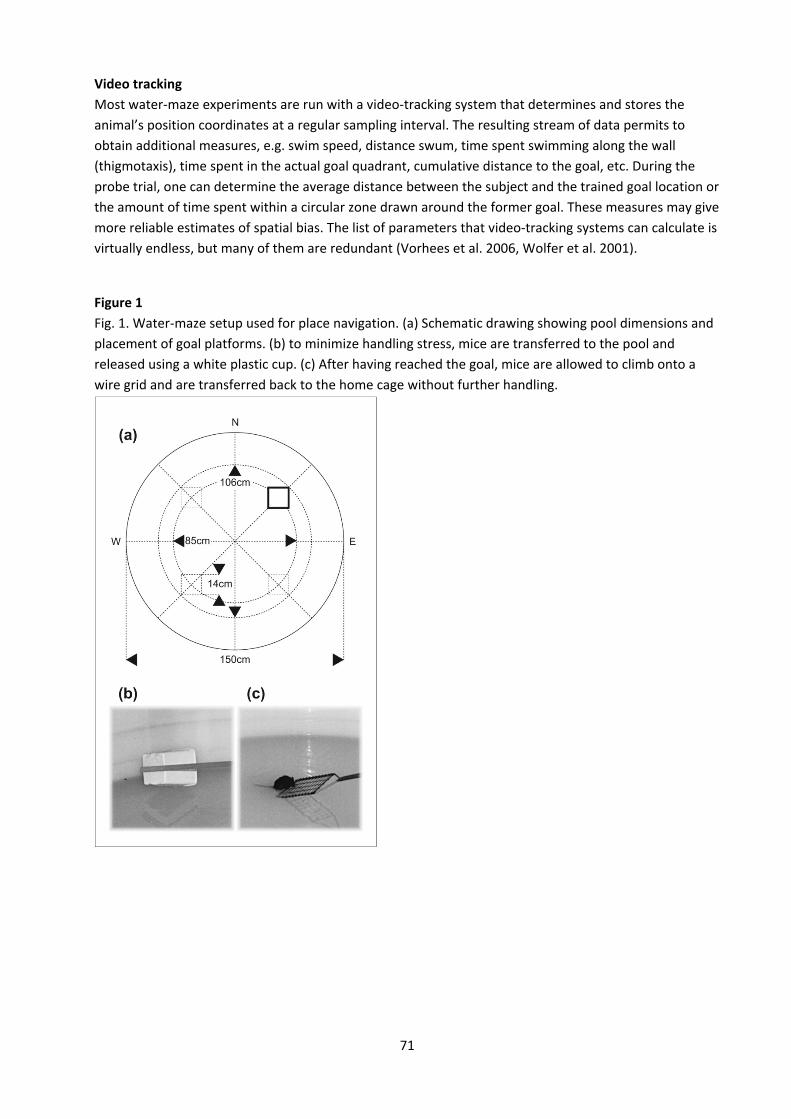
71
Video tracking Most water‐maze experiments are run with a video‐tracking system that determines and stores the animal’s position coordinates at a regular sampling interval. The resulting stream of data permits to obtain additional measures, e.g. swim speed, distance swum, time spent swimming along the wall (thigmotaxis), time spent in the actual goal quadrant, cumulative distance to the goal, etc. During the probe trial, one can determine the average distance between the subject and the trained goal location or the amount of time spent within a circular zone drawn around the former goal. These measures may give more reliable estimates of spatial bias. The list of parameters that video‐tracking systems can calculate is virtually endless, but many of them are redundant (Vorhees et al. 2006, Wolfer et al. 2001).
Figure 1 Fig. 1. Water‐maze setup used for place navigation. (a) Schematic drawing showing pool dimensions and placement of goal platforms. (b) to minimize handling stress, mice are transferred to the pool and released using a white plastic cup. (c) After having reached the goal, mice are allowed to climb onto a wire grid and are transferred back to the home cage without further handling.

72
Table 1 version 1 version 2 version 3 version 4 trial start goal start goal start goal start goal 1 NW SW SW SE SE NE NE NW 2 SE SW NE SE NW NE SW NW 3 W SW S SE E NE N NW 4 E SW N SE W NE S NW 5 NE SW NW SE SW NE SE NW 6 S SW W SE N NE W NW 7 SE SW NE SE NW NE SW NW 8 NW SW SW SE SE NE NE NW 9 S SW E SE N NE W NW 10 N SW W SE S NE E NW 11 W SW S SE E NE N NW 12 E SW N SE W NE S NW 13 NE SW NE SE SW NE SE NW 14 W SW S SE E NE N NW 15 N SW W SE S NE E NW 16 S SW E SE N NE W NW 17 SE SW NE SE NW NE SW NW 18 NW SW SW SE SE NE NE NW 19 S NE S NW N SW N SE 20 W NE E NW E SW W SE 21 E NE N NW W SW S SE 22 SW NE SE NW NE SW NW SE 23 NW NE SW NW SE SW NE SE 24 SE NE NE NW NW SW SW SE 25 SW NE SE NW NE SW NW SE 26 N NE W NW S SW E SE 27 SE NE NE NW NW SW SW SE 28 NW NE SW NW SE SW NE SE 29 E NE N NW W SW S SE 30 SW NE SE NW NE SW NW SE Table 2 trial 1 2 3 4 5 6 7 8 9 10 11 12 start SE NE W N NW SE SW NE NW SW E S goal SW NW SE NE SW NE SE NW NE NW SW SE
73
References 1. Crawley JN, Belknap JK, Collins A, Crabbe JC, Frankel W, Henderson N, Hitzemann RJ, Maxson SC, Miner LL, Silva AJ,
Wehner JM, Wynshawboris A, Paylor R (1997) Behavioral phenotypes of inbred mouse strains: implications and recommendations for molecular studies. Psychopharmacol 132:107‐124.
2. D’Hooge R, De Deyn PP (2001) Applications of the Morris water maze in the study of learning and memory. Brain Res Rev 36:60‐90.
3. Grant SGN, O’Dell TJ, Karl K, Stein P, Soriano P, Kandel ER (1992) Impaired long‐term potentiation, spatial learning and hippocampal development in fyn mutant mice. Science 258:1903‐1910.
4. Logue SF, Paylor R, Wehner JM (1997) Hippocampal lesions cause learning deficits in inbred mice in the Morris water maze and conditioned‐fear task. Behav Neurosci 111(1):104‐113.
5. Morris RG (1981) Spatial localization does not require the presence of local cues. Learn Motiv 12:239‐260. 6. Morris RG, Garrud P, Rawlins JNP, O'Keefe J (1982) Place navigation impaired in rats with hippocampal lesions. Nature
297: 681‐683. 7. Morris RG (1984) Developments of a water‐maze procedure for studying spatial learning in the rat. J Neurosci Methods
11:47‐60. 8. Morris RG, E Anderson E, G Lynch G, M Baudry M (1986) Selective impairment of learning and blockade of long‐term
potentiation by an N‐methyl‐D‐aspartate antagonist, AP5. Nature 319:774‐776. 9. Schoepke R, Wolfer DP, Lipp HP, Leisinger‐Trigona MC (1991) Swimming navigation and structural variations of the
infrapyramidal mossy fibers in the hippocampus of the mouse. Hippocampus 1:315‐328. 10. Silva AJ, Paylor R, Wehner JM, Tonegawa S (1992) Impaired spatial learning in alpha‐calcium‐calmodulin kinase II mutant
mice. Science 257:206‐211. 11. Upchurch M, Wehner JM (1988) Differences between inbred strains of mice in Morris water maze performance. Behav
Genet 18:55‐68. 12. Vorhees CV, Williams MT (2006) Morris water maze: procedures for assessing spatial and related forms of learning and
memory. Nat Protoc 1:848‐857. 13. Wolfer DP, Müller U, Stagliar‐Bozizevic M, Lipp HP (1997) Assessing the effects of the 129/Sv genetic background on
swimming navigation learning in transgenic mutants: a study using mice with a modified beta‐amyloid precursor protein gene. Brain Res 771:1‐13.
14. Wolfer DP, Madani R, Valenti P, Lipp HP (2001) Extended analysis of path data from mutant mice using the public domain software Wintrack. Physiol Behav 73:745‐753.
15. Wolfer DP, Colacicco G, Welzl H (2011) unpublished book chapter
74
Project B
Radial‐ maze spatial working memory procedure Introduction In the classical procedure on the eight‐arm radial maze, a rat is placed on a central platform from which it collects single invisible baits placed at the end of the arms. Rats quickly adopt a win‐shift foraging strategy, avoiding double entries. Olton and Samuelson (1976) popularized the task and showed that rats rely on extramaze cues to accomplish it, rather than utilizing intramaze cues or consistent chains of responses. In order to avoid errors during this task as the number of remaining pellets decreases, it becomes increasingly important for the animal to remember whether an arm has already been visited during that particular trial or not. It must thus flexibly use spatial information which is only valid for one trial, but not for subsequent trials. This requirement classifies the task as a spatial working memory procedure. Olton et al. (1978) and other groups showed that learning of rats in this procedure critically depends on the integrity of the hippocampus and its efferent and afferent connections. Lesions of extra‐hippocampal systems, including the amygdala, neocortex, and caudate nucleus apparently did not affect learning, unless the procedure was modified. See the review by Lipp et al. (2001). More recently, activation of the hippocampal formation during a spatial working memory test on the radial maze was demonstrated by analysis of Fos production (Vann et al. 2000). The radial maze task has been adapted to mice by Reinstein et al. (1983) who compared the performance of different strains and confirmed the disruption of learning by fornix lesions. Rossi‐Arnaud et al. (1991) confirmed the negative effect of hippocampal lesions in in DBA and C57 mice, but found that performance of C57 mice was also impaired by lesions of the amygdala. Meanwhile, a number of studies had revealed a strong positive correlation between the size of the hippocampal intra‐ and infrapyramidal mossy fiber terminal fields and performance on the radial maze, provided that the procedure required processing of spatial information. See review by Schwegler and Crusio (1995). In recent years, the eight‐arm radial maze has repeatedly been used to assess learning of mice with mutations that affect the nervous system. For example, two studies found impaired radial maze learning in mouse models of Alzheimer’s disease (Chapman et al. 1999, Morgan t al 2000). Minichiello et al. (1999) found a partial impairment of radial maze learning in conditional knockout mice lacking TrkB receptors in their forebrains. Protein tyrosine phosphatase delta deficient mice performed badly on the test, despite having enhanced hippocampal LTP (Uetani et al. 2000). Double mutant mice lacking APLP2 and expressing solely the secreted ectodomain of APP were unable to learn the task (Weyer et al 2011). Enhanced learning, by contrast was described in mice overexpressing the growth associated protein GAP‐43 (Routtenberg et al. 2000). Some studies have used modified protocols in an attempt to introduce a reference memory component into the task (Schmitt et al. 2003). Equipment Eight arms (7x38 cm) extend from an octagonal center platform (diameter 18.5 cm). The distance from the platform center to the end of each arm is 47 cm. The apparatus is constructed of grey poly‐vinyl chloride, has a smooth surface. U‐shaped clear Perspex profiles can be attached to the arms to provide 4 cm high side walls (Fig. 1B) or to form a tunnel. The maze is elevated 38 cm from the floor and placed in a dimly lit room (4 40 W bulbs) which is rich in salient extramaze cues (we use the same room as for water‐maze testing). Baits are small pellets (ca. 6 mg) prepared from standard mouse chow or cereal. They are placed in small metal cups (diameter 3 cm, 1 cm deep) at the end of each arm, in such a way
75
that the mouse cannot see them without completely entering the arm. The center platform is confined by a cylinder (height 20 cm) of clear Perspex that has a rounded opening for each of the 8 arms. Many variants of the apparatus exist. Arm visits can be made more aversive by removing side walls, or less aversive by using higher walls and placing the maze directly on the floor. Opaque walls can prevent animals from using extramaze cues during control experiments with intra‐maze visual, tactile, or olfactory cues. Gates may be used to confine the mouse on the center platform (to prevent chaining responses), to prevent it from entering particular arms, or to mark arms that were already visited. Procedure Dietary restriction to 85% of free‐feeding body weight
‐ On the first day measure body weight and remove food. ‐ On the second and later days measure body weight and feed limited amount of food according
to weight loss: Body weight relative to day 1 100‐95% 95‐90% 90‐85% <85% Amount of food to be given 0.5g 1.0g 2.0g 3.0g
‐ Several days may be needed to reach a stable body weight in each mouse of the test sample. To habituate the mice to eating small food pellets, 4 of those are placed on the homecage floor on each habituation day.
‐ This dietary restriction to 85% of free‐feeding body weight is maintained during the whole experiment. The mice are weighed daily and are fed with the quantity of food needed to maintain body weight at 85%. Mice should be fed within 1h after finishing their habituation or training sessions. Water is given at libitum during the entire period.
‐ Note that some mutants may not well tolerate the food restriction and may become weak and inactive. Such animals should not be used for radial maze experiments.
Habituation session
‐ As soon as all mice in a testing sample are stabilized at 85% of free‐feeding body weight, they are habituated to collecting food on the radial‐maze apparatus.
‐ Bring the rack with the home cages to the testing room approximately 30 min before the session begins.
‐ Clean the apparatus with water and dry before each session. Place one food pellet in the middle and one in the bating cup of each arm. Release the mouse on the center platform and allow it to collect pellets until it has collected 4 of them.
‐ When all mice have completed their habituation session, clean the maze first with water, then with 70% ethanol.
76
Training sessions ‐ Bring the rack with the home cages to the testing room approximately 30 min before the session
begins. ‐ Clean the apparatus with water before each session. Place a single food pellet in each baiting
cup. ‐ Place the mouse on the center platform, allow it to explore the maze and to collect food until it
has consumed all pellets or 10 min have elapsed. ‐ A mouse is trained for 10 days with one training session per day. ‐ When all mice have completed their training session, clean the maze first with water, then with
70% ethanol. Parameters Record the arm number on a protocol sheet each time the mouse visits an arm, that is: enters it with all four paws coming from the center platform. Note whether it consumes a pellet or not. From this sequence of arm entries the following parameters are determined:
‐ Number of correct choices (= arm entries with pellet consumption) until the first error is made ‐ Working memory errors = re‐entries to arms where the pellet has already been consumed ‐ Procedural errors = arm entries without consumption of the available pellet ‐ Total number of errors = sum of working + procedural errors ‐ Number of pellets collected ‐ Average angle between arm choices ‐ Preferred angle between arm choices (e.g. 45° from arm 1 to 2, ‐90° from arm 1 to 7) ‐ Ratio of choices with preferred angle over total choices
Total time and latency to the first arm visit are recorded as well. Also take note of inappropriate behaviors such as long periods of inactivity, grooming, avoidance of or preference for particular arms. Frequent entries to baited arms without pellet consumption indicate a problem with motivation and may question the validity of the experiment as a working memory procedure.
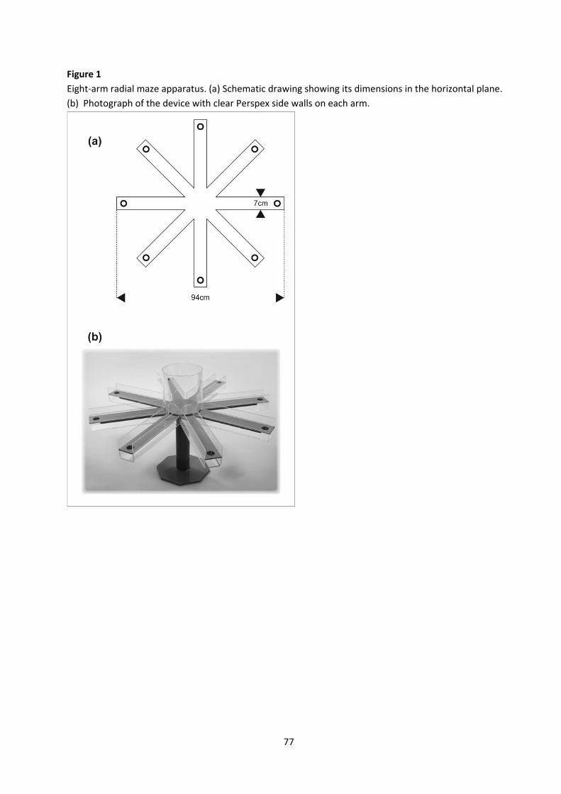
77
Figure 1 Eight‐arm radial maze apparatus. (a) Schematic drawing showing its dimensions in the horizontal plane. (b) Photograph of the device with clear Perspex side walls on each arm.

78
References 1. Chapman PF, White GL, Jones MW, Cooper‐Blacketer D, Marshall VJ, Irizarry M, Younkin L, Good MA, Bliss TVP, Hyman
BT, Younkin SG, Hsiao KK (1999) Impaired synaptic plasticity and learning in aged amyloid precursor protein transgenic mice. Nat Neurosci 2:271‐276.
2. Lipp HP, Pleskacheva MG, Gossweiler H, Ricceri L, Smirnova AA, Garin NN, Perepiolkina OP, Voronkov DN, Kuptsov PA, Dell'Omo G (2001) A large outdoor radial maze for comparative studies in birds and mammals. Neurosci Biobehav Rev 25:83‐99.
3. Minichiello L, Korte M, Wolfer DP, Kühn R, Unsicker K, Cestari V, Rossi‐Arnaud, C Lipp HP, Bonhoeffer T, Klein R (1999) Essential role for TrkB receptors in hippocampus‐mediated learning. Neuron 24:401‐414.
4. Morgan D, Diamond DM, Gottschall PE, Ugen KE, Dickey C, Hardy J, Duff K, Jantzen P, DiCarlo G, Wilcock D, Connor K, Hatcher J, Hope C, Gordon M, and Arendash GW (2000) A‐beta peptide vaccination prevents memory loss in an animal model of Alzheimer's disease. Nature 408:982‐985.
5. Olton DS, Samuelson RI (1976) Remembrance of places passed: spatial memory in rats. J Exp Psych: Animal Behav Proc 2:97‐116.
6. Olton DS, Walker JA, Gage FH (1978) Hippocampal connections and spatial discrimination. Brain Res 139:295‐308. 7. Reinstein DK, DeBoissiere T, Robinson N, Wurtman RJ (1983) Radial maze performance in three strains of mice: role of
the fimbria/fornix. Brain Res 263:172‐176. 8. Rossi‐Arnaud C, Fagioli S, Ammassari‐Teule M (1991) Spatial learning in two inbred strains of mice: genotype‐dependent
effect of amygdaloid and hippocampal lesions. Behav Brain Res 45:9‐16. 9. Routtenberg A, Cantallops I, Zaffuto S, Serrano P, Namgung U (2000) Enhanced learning after genetic overexpression of a
brain growth protein. PNAS 97:7657‐7662. 10. Schmitt WB, Deacon RM, Seeburg PH, Rawlins JN, Bannerman DM (2003) A within‐subjects, within‐task demonstration
of intact spatial reference memory and impaired spatial working memory in glutamate receptor‐a‐deficient mice. J Neurosci 23:3953‐3959.
11. Schwegler H, Crusio WE (1995) Correlations between radial‐maze learning and structural variations of septum and hippocampus in rodents. Behav Brain Res 67:29‐41.
12. Uetani N, Kato K, Ogura H, Mizuno K, Kawano K, Mikoshiba K, Yakura H, Asano M, Iwakura Y (2000) Impaired learning with enhanced hippocampal long‐term potentiation in PTPdelta‐deficient mice. Embo J 19:2775‐2785.
13. Vann SD, Brown MW, Erichsen JT, Aggleton JP (2000) Fos imaging reveals differential patterns of hippocampal and parahippocampal subfield activation in rats in response to different spatial memory tests. J Neurosci 20:2711‐2718.
14. Weyer SW, Klevanski M, Delekate A, Voikar V, Aydin D, Hick M, Filippov M, Drost N, Schaller KL, Saar M, Vogt MA, Gass P, Samanta A, Jäschke A, Korte M, Wolfer DP, Caldwell JH, Müller UC (2011) APP and APLP2 are essential at PNS and CNS synapses for transmission, spatial learning and LTP. EMBO J 30:2266‐2280.
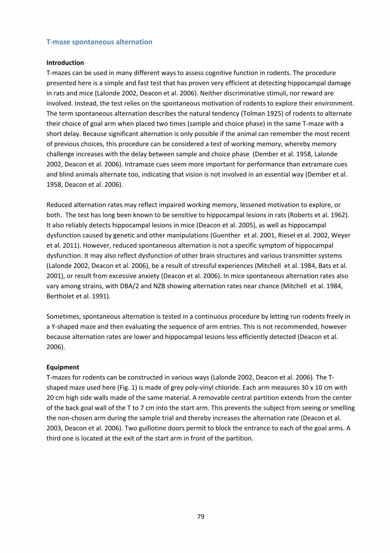
79
T‐maze spontaneous alternation Introduction T‐mazes can be used in many different ways to assess cognitive function in rodents. The procedure presented here is a simple and fast test that has proven very efficient at detecting hippocampal damage in rats and mice (Lalonde 2002, Deacon et al. 2006). Neither discriminative stimuli, nor reward are involved. Instead, the test relies on the spontaneous motivation of rodents to explore their environment. The term spontaneous alternation describes the natural tendency (Tolman 1925) of rodents to alternate their choice of goal arm when placed two times (sample and choice phase) in the same T‐maze with a short delay. Because significant alternation is only possible if the animal can remember the most recent of previous choices, this procedure can be considered a test of working memory, whereby memory challenge increases with the delay between sample and choice phase (Dember et al. 1958, Lalonde 2002, Deacon et al. 2006). Intramaze cues seem more important for performance than extramaze cues and blind animals alternate too, indicating that vision is not involved in an essential way (Dember et al. 1958, Deacon et al. 2006). Reduced alternation rates may reflect impaired working memory, lessened motivation to explore, or both. The test has long been known to be sensitive to hippocampal lesions in rats (Roberts et al. 1962). It also reliably detects hippocampal lesions in mice (Deacon et al. 2005), as well as hippocampal dysfunction caused by genetic and other manipulations (Guenther et al. 2001, Riesel et al. 2002, Weyer et al. 2011). However, reduced spontaneous alternation is not a specific symptom of hippocampal dysfunction. It may also reflect dysfunction of other brain structures and various transmitter systems (Lalonde 2002, Deacon et al. 2006), be a result of stressful experiences (Mitchell et al. 1984, Bats et al. 2001), or result from excessive anxiety (Deacon et al. 2006). In mice spontaneous alternation rates also vary among strains, with DBA/2 and NZB showing alternation rates near chance (Mitchell et al. 1984, Bertholet et al. 1991). Sometimes, spontaneous alternation is tested in a continuous procedure by letting run rodents freely in a Y‐shaped maze and then evaluating the sequence of arm entries. This is not recommended, however because alternation rates are lower and hippocampal lesions less efficiently detected (Deacon et al. 2006). Equipment T‐mazes for rodents can be constructed in various ways (Lalonde 2002, Deacon et al. 2006). The T‐shaped maze used here (Fig. 1) is made of grey poly‐vinyl chloride. Each arm measures 30 x 10 cm with 20 cm high side walls made of the same material. A removable central partition extends from the center of the back goal wall of the T to 7 cm into the start arm. This prevents the subject from seeing or smelling the non‐chosen arm during the sample trial and thereby increases the alternation rate (Deacon et al. 2003, Deacon et al. 2006). Two guillotine doors permit to block the entrance to each of the goal arms. A third one is located at the exit of the start arm in front of the partition.
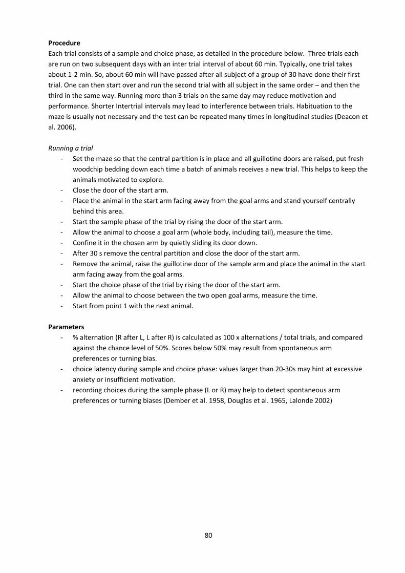
80
Procedure Each trial consists of a sample and choice phase, as detailed in the procedure below. Three trials each are run on two subsequent days with an inter trial interval of about 60 min. Typically, one trial takes about 1‐2 min. So, about 60 min will have passed after all subject of a group of 30 have done their first trial. One can then start over and run the second trial with all subject in the same order – and then the third in the same way. Running more than 3 trials on the same day may reduce motivation and performance. Shorter Intertrial intervals may lead to interference between trials. Habituation to the maze is usually not necessary and the test can be repeated many times in longitudinal studies (Deacon et al. 2006). Running a trial
‐ Set the maze so that the central partition is in place and all guillotine doors are raised, put fresh woodchip bedding down each time a batch of animals receives a new trial. This helps to keep the animals motivated to explore.
‐ Close the door of the start arm. ‐ Place the animal in the start arm facing away from the goal arms and stand yourself centrally
behind this area. ‐ Start the sample phase of the trial by rising the door of the start arm. ‐ Allow the animal to choose a goal arm (whole body, including tail), measure the time. ‐ Confine it in the chosen arm by quietly sliding its door down. ‐ After 30 s remove the central partition and close the door of the start arm. ‐ Remove the animal, raise the guillotine door of the sample arm and place the animal in the start
arm facing away from the goal arms. ‐ Start the choice phase of the trial by rising the door of the start arm. ‐ Allow the animal to choose between the two open goal arms, measure the time. ‐ Start from point 1 with the next animal.
Parameters
‐ % alternation (R after L, L after R) is calculated as 100 x alternations / total trials, and compared against the chance level of 50%. Scores below 50% may result from spontaneous arm preferences or turning bias.
‐ choice latency during sample and choice phase: values larger than 20‐30s may hint at excessive anxiety or insufficient motivation.
‐ recording choices during the sample phase (L or R) may help to detect spontaneous arm preferences or turning biases (Dember et al. 1958, Douglas et al. 1965, Lalonde 2002)
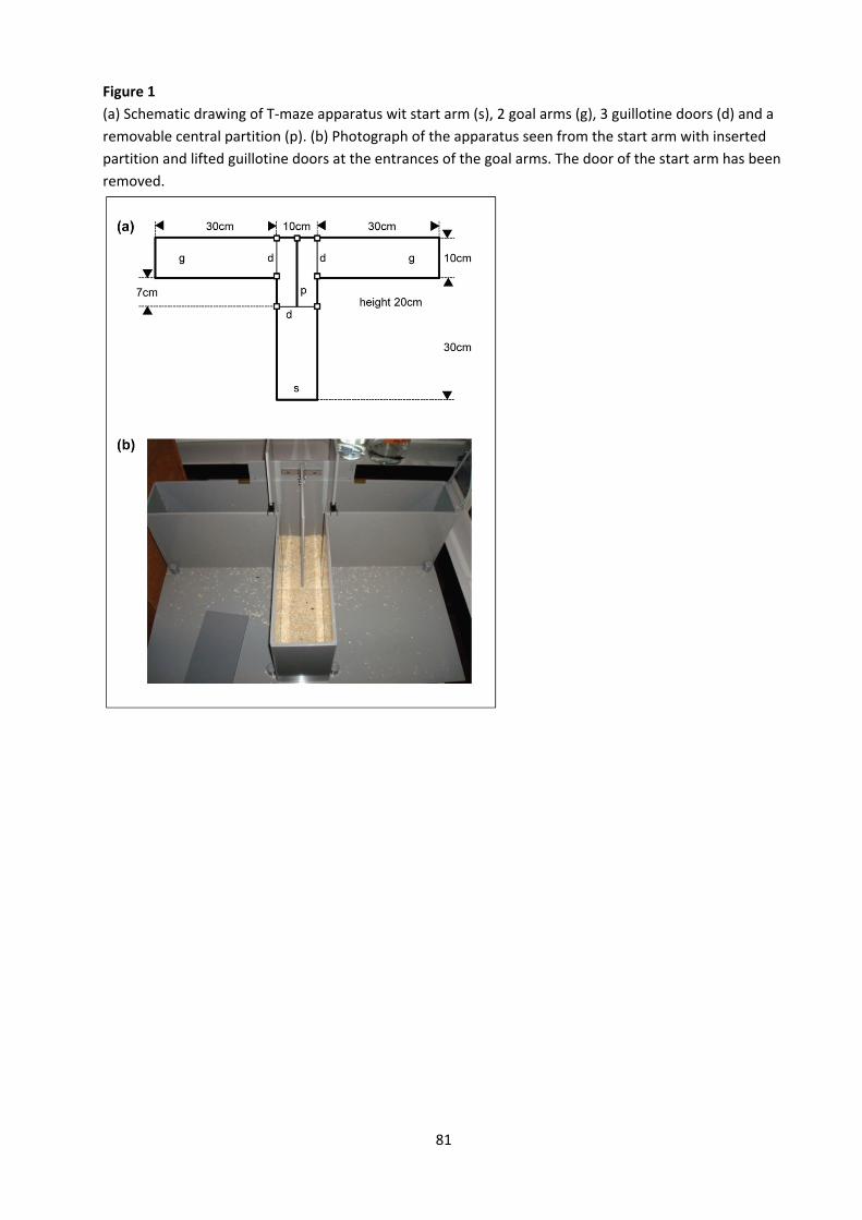
81
Figure 1 (a) Schematic drawing of T‐maze apparatus wit start arm (s), 2 goal arms (g), 3 guillotine doors (d) and a removable central partition (p). (b) Photograph of the apparatus seen from the start arm with inserted partition and lifted guillotine doors at the entrances of the goal arms. The door of the start arm has been removed.

82
References 1. Bats S, Thoumas JL, Lordi B, Tonon MC, Lalonde R, Caston J (2001) The effects of a mild stressor on spontaneous
alternation in mice. Behav Brain Res 118:11‐15. 2. Bertholet JY, Crusio WE (1991) Spatial and non‐spatial spontaneous alternation and hippocampal mossy fibre
distribution in nine inbred mouse strains. Behav Brain Res 43:197‐202. 3. Deacon RM, Penny C, Rawlins JN (2003) Effects of medial prefrontal cortex cytotoxic lesions in mice. Behav Brain Res
139:139‐155. 4. Deacon RMJ, Rawlins JNP (2005) Hippocampal lesions, species‐typical behaviors and anxiety in mice. Behav Brain Res
156:241‐249. 5. Deacon RMJ, Rawlins JNP (2006) T‐maze alternation in the rodent. Nat Protoc 1:7‐12. 6. Dember WN, Fowler H (1958) Spontaneous alternation behavior. Psychol Bull 55:412‐428. 7. Douglas RJ, Isaacson RL (1965) Homogeneity of single trial response tendencies and spontaneous alternation in the T‐
maze. Psychol Rep 16:87‐92. 8. Henderson ND (1970) A genetic analysis of spontaneous alternation in mice. Behav Genet 1:125‐132. 9. Guenther K, Deacon RM, Perry VH, Rawlins JNP (2001) Early behavioural changes in scrapie‐affected mice and the
influence of dapsone. Eur J Neurosci 14:401‐409. 10. Lalonde R (2002) The neurobiological basis of spontaneous alternation. Neurosci Biobehav Rev 26:91‐104. 11. Mitchell D, Koleszar AS, Scopatz RA (1984) Arousal and T‐maze choice behavior in mice: A convergent paradigm for
neophobia constructs and optimal arousal theory. Learn Motiv 15:287‐301. 12. Reisel D, Bannerman DM, Schmitt WB, Deacon RM, Flint J, Borchardt T, Seeburg PH, Rawlins JNP (2002) Spatial memory
dissociations in mice lacking GluR1. Nat Neurosci 5:868‐873. 13. Roberts WW, Dember WN, Brodwick M (1962) Alternation and exploration in rats with hippocampal lesions. J Comp
Physiol Psychol 55:695‐700. 14. Tolman EC (1925) Purpose and cognition: the determiners of animal learning. Psychol Rev 32: 285‐297. 15. Weyer SW, Klevanski M, Delekate A, Voikar V, Aydin D, Hick M, Filippov M, Drost N, Schaller KL, Saar M, Vogt MA, Gass P,
Samanta A, Jäschke A, Korte M, Wolfer DP, Caldwell JH, Müller UC (2011) APP and APLP2 are essential at PNS and CNS synapses for transmission, spatial learning and LTP. EMBO J 30:2266‐2280.
83
Project C
Contextual and delay (tone) fear conditioning Introduction If a dog is presented with food in the mouth (unconditioned stimulus = US), this elicits salivation (unconditioned response = UR). When Ivan Petrovich Pavlov paired the food with a tone, a neutral stimulus which by itself does not induce salivation (conditioned stimulus = CS), he observed that, through this pairing, the tone acquired itself the potency to elicit salivation (conditioned response = CR). The discovery of this form of associative learning, which is now known as classical Pavlovian conditioning, earned Pavlov the 1929 Nobel Prize. Fear conditioning is another example of classical Pavlovian conditioning. A neutral tone (CS) is paired with a brief electric foot‐shock (US) several times in a novel context. Rats and mice trained in this manner develop a fear (CR) of both the tone and training context which they now associate with the shock. This learned fear is expressed as defensive behaviour (typically freezing in rodents), as an autonomic and endocrine reaction (increased heart rate and blood pressure, stress hormone release), and as sensory‐motor alterations (analgesia, potentiated startle, potentiated eye blink response). Thus, the “CR” is a complex adaptive response that consists of multiple reactions all destined to improve the animal’s chance to survive threatening situations, such as the presence of a predator (simulated in most fear conditioning procedures by the electric shock). Pavlovian fear conditioning in rats and mice is a convenient model for the study of emotional learning and memory, but also of anxiety‐related disorders. Studies using amygdalectomised rats have shown that the amygdala is required for both the acquisition and expression of a learned fear response to explicit cues or a context. There is now considerable evidence that plastic changes in the lateral amygdala directly underlie acquisition and retention of Pavlovian fear conditioning (LeDoux 2000, Repa et al. 2001). The hippocampus is not required to associate a single cue such as a tone with foot shock (tone fear conditioning), provided that the two stimuli overlap temporally (delay fear conditioning). Hippocampal lesions do, however, cause anterograde and time‐limited retrograde amnesia for association between shock and training context (contextual fear conditioning) (LeDoux 2000, Anagnostaras et al. 2001), although anterograde amnesia may not be evident in all contextual fear conditioning procedures (Frankland et al. 1998, Anagnostaras et al. 2001). In mice, considerable strain differences exist with respect to the efficiency of context learning and the effects of lesions (Paylor et al. 1994, Logue et al. 1997, Ammassari‐Teule et al. 2000). Despite this, Pavlovian fear conditioning is frequently used to assess emotional memory in mice carrying mutations that affect the nervous system. For example, Brambilla et al. (1997) demonstrated deficient synaptic plasticity in the amygdala and impaired fear conditioning in mutant mice with a disrupted RAS signaling pathway. On the other hand, Minichiello et al. (1999) found largely intact fear conditioning despite severe learning deficits in other tasks when they investigated mice lacking TrkB receptors in their forebrains. Tang et al. demonstrated facilitated fear conditioning in transgenic mice overexpressing the NMDA receptor subunit 2B in their forebrains. In a recent study Frankland et al. (2001) found that alpha‐CaMKII‐dependent plasticity in the neocortex was necessary for long‐term stabilization of fear memories. Overall, a great deal has been learned about the behavioural characteristics and neural mechanisms of fear acquisition, whereas much less is known about the mechanisms of fear inhibition. However, increased interest has been directed at understanding the neuronal basis of fear extinction and in
84
laboratory conditions fear extinction is one of the most studied form of behavioural inhibition (Myers and Davis, 2007; Herry et al., 2010). Understanding the neuronal substrates of fear extinction would be of great clinical relevance, as extinction is the cornerstone of psychological therapy of several anxiety disorders and because the relapse of maladaptive fear and anxiety is a major clinical problem. Apparatus The main requirement for the fear conditioning equipment is a shock generator for delivering calibrated current to the grid floor (foot shock, unconditioned stimulus). In addition, it should be possible to apply auditory and/or visual cues as conditioned stimuli. The most precise way to quantify freezing behaviour is by direct observation. Freezing is defined as the absence of all movement except respiration. Freezing animals often show a characteristic crouched position. While there is general agreement that a mouse must not show any locomotion during a freezing episode, experimenters may disagree on the interpretation of small movements that do not lead to displacement of the animal. We tend to allow only small vertical movements of the head as they are often associated with increased respiration. All other movements, including head turns, grooming, rearing or leaning are qualified as non‐freezing. An experimenter may use slightly different criteria, but it is essential that the criteria be consistent throughout the entire study. Measurement of freezing behavior is done by different means. Freezing time can be measured continuously using a stop watch, or be estimated through intermittent observations. For the latter method, the animal is observed for 2 s every 5 s. If there was no movement during the entire 2 s, the observation is recorded as freezing, else as non‐freezing. To allow for rescoring of ambiguous experiments by an independent observer, it is recommended to record all sessions on videotape. Because manual scoring of freezing is time consuming and not immune against subjective bias, automated recording systems have been developed. Some systems detect freezing by digital comparison of images that are taken at regular intervals, or use piezo‐electric sensors to detect animal movements, the most common technique is to monitor locomotion using arrays of IR‐beams. The training chamber (247 x 168 mm, height 230 mm) has four opaque side walls made of grey polyvinyl chloride and a removable foot‐shocking grid floor. During the experiment, training chamber is placed in a sound‐attenuated box with a background noise level of 55 dB. A loudspeaker and a video camera overseeing all movements of the mouse are placed above the animal.

85
Procedure ‐ Bring the home cages to the testing room approximately 30 min before the experiment begins.
Session 1 (day 1): training ‐ Clean the training chamber thoroughly with water. ‐ Introduce the animal, close the lids of training chamber and sound attenuated box. ‐ Observe the animal for 60 s (baseline activity). ‐ Then run 3 training cycles:
‐ 30 s tone stimulus (CS, 15 s, 2000 Hz, 92 dB volume) ‐ 2 s foot‐shock co‐terminating with CS (US, at 0.25 mA average intensity) ‐ 30 s inter‐trial interval
‐ Return the animal to its home cage (or to transfer cage if group housed) 30 s after last CS‐US pairing.
Session 2 (day 2): context test (same context as training, no tone)
‐ Clean the training chamber thoroughly with water. ‐ Introduce the animal, close the lids of training chamber and sound attenuated box. ‐ Observe for 120 s, no tone, no shock. ‐ Return the animal to its home cage (or to transfer cage if group housed)..
Session 3 (day 2): tone test (novel context)
‐ Tone test is done 30‐90 min after the context test. ‐ Replace the grid floor with smooth sheet of plastic; add a handful of bedding material and stone
on the floor ‐ Introduce the animal, close the sound attenuated box ‐ Observe the animal for 60 s in the novel context, then activate the tone and observe for another
60 s while the tone is on. ‐ Return the animal to its home cage.
Session 4 (day 3): Context and cue re‐test (done exactly as on day 2) to test for re‐consolidation Session 5 (on day 6): Extinction test in the novel context
‐ after 70 s of free exploration the tone is activated for 30 s ‐ tone is repeated 25 times with inter‐trial interval of 5 s
Session 6 (on day 7): Repeat of sessions 5 to test for recovery of freezing behavior Parameters In the procedure described here, the learned fear response is quantified by software‐aided measuring the time during which the animal shows freezing behaviour. Percentage of the time spent freezing is determined for the following phases of the experiment:
‐ Training: 60 s baseline, during each training cycle: 30 s tone presentation, 30 s inter‐trial interval ‐ Context test: 120 s continuous observation ‐ Tone test: 60 s before tone presentation, 60 s with tone on ‐ Extinction: 70 s baseline and 25 x 30 s tone presentations

86
Freezing responses are then defined as follows:
‐ Response to context: context test minus baseline ‐ Response to tone: tone minus pre‐tone during tone test session ‐ Immediate training response: last tone presentation during training minus baseline ‐ Generalized fear response: pre‐tone period of tone test session minus baseline. A substantial
generalized response indicates that the contexts during training and tone testing are not sufficiently different. Note that animals should show no or very little freezing during baseline recording.
‐ Extinction: decrease of the freezing response across the 25 tone presentations of the extinction session
‐ Recovery: difference between the last tone presentation of the first extinction session and the first tone presentation of the second extinction session
References 1. Ammassari‐Teule M, Passino E, Restivo L, and De Marsanich B (2000) Fear conditioning in C57/BL/6 and DBA/2 mice:
variability in nucleus accumbens function according to the strain predisposition to show contextual‐ or cue‐based responding. Eur J Neurosci 12:4467‐4474.
2. Anagnostaras SG, Gale GD, Fanselow MS (2001) Hippocampus and contextual fear conditioning: recent controversies and advances. Hippocampus 11:8‐17
3. Brambilla R, Gnesutta N, Minichiello L, White G, Roylance A, Herron C, Ramsey M, Wolfer DP, 4. Cestari V, Rossi‐Arnaud C, Grant SGN, Chapman PF, Lipp HP, Sturani E, Klein R (1997) A role for the RAS signaling
pathway in synaptic transmission and long‐term memory. Nature 390:281‐286. 5. Frankland PW, Cestari V, Filipkowski RK, McDonald RJ, Silva AJ (1998) The dorsal hippocampus is essential for context
discrimination, but not for contextual conditioning. Behav Neurosci 112:863‐874. 6. Frankland PW, O'Brien C, Ohno M, Kirkwood A, Silva AJ (2001) alpha‐CaMKII‐dependent plasticity in the cortex is
required for permanent memory. Nature 411:309‐313. 7. Herry C, Ferraguti F et al. (2010) Neuronal circuits of fear extinction. Eur J Neurosci 31(4):599‐612. 8. LeDoux JE (2000) Emotion circuits in the brain. Annu Rev Neurosci 23:155‐184 9. Logue SF, Paylor R, Wehner JM (1997) Hippocampal lesions cause learning deficits in inbred mice in the Morris water
maze and conditioned‐fear task. Behav Neurosci 111:104‐113. 10. Minichiello L, Korte M, Wolfer DP, Kühn R, Unsicker K, Cestari V, Rossi‐Arnaud C, Lipp HP, Bonhoeffer T, Klein R. (1999)
Essential role for TrkB receptors in hippocampus‐mediated learning. Neuron 24:401‐414. 11. Myers KM & Davis M (2007) Mechanisms of fear extinction. Mol Psychiatry 12(2):120‐150. 12. Paylor R, Tracy R, Wehner JM, Rudy JW (1994) DBA/2 and C57BL/6 mice differ in contextual fear but not auditory fear
conditioning. Behav Neurosci 108:810‐817. 13. Repa JC, Muller J, Apergis J, Desrochers TM, Zhou Y, LeDoux JE (2001) Two different lateral amygdala cell populations
contribute to the initiation and storage of memory. Nat Neurosci 4:724‐731. 14. Tang YP, Shimizu E, Dube GR, Rampon C, Kerchner GA, Zhuo M, Liu G, Tsien JZ (1999) Genetic enhancement of learning
and memory in mice. Nature 401:63‐69.
87
Trace fear conditioning Introduction The trace fear conditioning procedure is a modified form of tone fear conditioning, see protocol “Contextual and delay (tone) fear conditioning”. In a delay conditioning procedure, there is temporal overlap between the unconditioned (US) and conditioned stimulus (CS), even if the US begins with a delay after the onset of the CS. A trace conditioning procedure, by contrast, demands the association of a CS with an US across an interval of time known as “trace interval”. In the case of trace fear conditioning, the shock (US) is only delivered 15 s after the tone (CS) has been turned off. Additional temporal processing is required because the CS and the US are separated: the animal has to retain a trace of the CS across the interval in order to associate it with the US. Unlike delay tone conditioning, the acquisition of trace fear conditioning is sensitive to hippocampal lesions (McEchron et al. 1998, Desmedt et al. 2003). Trace fear conditioning can thus be regarded as an alternative to context fear conditioning procedures, whose sensitivity to pretraining lesions of the hippocampus has been subject to debate (Frankland et al. 1998, Anagnostaras et al. 2001). Recently, genetically modified mice lacking subunit 1 of the NMDA receptor specifically in area CA1 of the hippocampus (Huerta et al. 2000), as well as mice carrying a deletion of the mental retardation gene Gdi1 (D’Adamo et al. 2002) were shown to be impaired on trace fear conditioning. Mice carrying only one functional allele of the GABAA receptor subunit 2, by contrast, showed enhanced behavioural inhibition toward natural aversive stimuli and heightened responsiveness in trace fear conditioning (Crestani, 1999). Apparatus The apparatus is essentially the same as described in the protocol “Contextual and delay (tone) fear conditioning”. Procedure The procedure is essentially the same as in “Contextual and delay (tone) fear conditioning”. However, trace conditioning requires more trials for the animals to associate the cue with the US. Bring the home cages to the testing room approximately 30 min before the experiment begins. Session 1 (day 1): training
‐ Clean the training chamber with water. ‐ Introduce the animal, close the lids of training chamber and sound attenuated box. ‐ Observe the animal for 60 s (baseline activity). ‐ Then run 5 training cycles, with variable intertrial interval (this should minimize the possibility of
the animals “expecting” a new trial: ‐ 30 s tone stimulus (CS, 15 s, 2000 Hz, 92 dB volume) ‐ 15 s trace interval ‐ 2 s foot‐shock (US, at 0.25 mA average intensity) ‐ 43‐73 s inter‐trial interval
‐ Return the animal to its home cage 30 s after last CS‐US pairing.
88
Session 2 (day 2): context test (same context as training, no tone) ‐ Clean the training chamber thoroughly with water. ‐ Introduce the animal, close the lids of training chamber and sound attenuated box. ‐ Observe for 120 s, no tone, no shock. ‐ Return the animal to its home cage (or to transfer cage if group housed)..
Session 3 (day 2): tone test (novel context)
‐ Tone test is done 30‐90 min after the context test. ‐ Replace the grid floor with smooth sheet of plastic; add a handful of bedding material and stone
on the floor ‐ Introduce the animal, close the sound attenuated box ‐ Observe the animal for 60 s in the novel context, then activate the tone and observe for another
60 s while the tone is on. ‐ Return the animal to its home cage.
Session 4 (day 3): Context and cue re‐test (done exactly as on day 2) to test for re‐consolidation Session 5 (on day 6): Extinction test in the novel context
‐ after 60 s of free exploration the tone is activated for 30 s ‐ tone is repeated 12 times with inter‐trial interval of 10 s
Session 6 (on day 7): Repeat of sessions 5 to test for recovery of freezing behavior Parameters As in the protocol “Contextual and delay (tone) fear conditioning”, the learned fear response is quantified by measuring the time during which the animal shows freezing behaviour. Percentage of the time spent freezing is determined for the following phases of the experiment:
‐ Training: 60 s baseline, during each training cycle: 30 s tone presentation, 30 s inter‐trial interval ‐ Context test: 120 s continuous observation ‐ Tone test: 60 s before tone presentation, 60 s with tone on ‐ Extinction: 12 x 30 s tone presentations ‐
Freezing responses are then defined as follows:
‐ Response to context: context test minus baseline ‐ Response to tone: tone minus pre‐tone during tone test session ‐ Immediate training response: last tone presentation during training minus baseline ‐ Generalized fear response: pre‐tone period of tone test session minus baseline. A substantial
generalized response indicates that the contexts during training and tone testing are not sufficiently different. Note that animals should show no or very little freezing during baseline recording.

89
References 1. Anagnostaras SG, Gale GD, Fanselow MS (2001) Hippocampus and contextual fear conditioning: recent controversies
and advances. Hippocampus 11:8‐17. 2. Crestani F, Lorez M, Baer K, Essrich C, Benke D, Laurent JP, Belzung C, Fritschy JM, Luscher B, Mohler H (1999) Decreased
GABAA‐receptor clustering results in enhanced anxiety and a bias for threat cues. Nat Neurosci 2:833‐839. 3. D'Adamo P, Welzl H, Papadimitriou S, Raffaele di Barletta M, Tiveron C, Tatangelo L, Pozzi L, Chapman PF, Knevett SG,
Ramsay MF, Valtorta F, Leoni C, Menegon A, Wolfer DP, Lipp HP, Toniolo D (2002) Deletion of the mental retardation gene Gdi1 impairs associative memory and alters social behavior in mice. Hum Mol Genet 11:2567‐2580.
4. Desmedt A, Marighetto A, Garcia R, Jaffard R (2003) The effects of ibotenic hippocampal lesions on discriminative fear conditioning to context in mice: impairment or facilitation depending on the associative value of a phasic explicit cue. Eur J Neurosci 17:1953‐1963.
5. Frankland PW, Cestari V, Filipkowski RK, McDonald RJ, Silva AJ (1998) The dorsal hippocampus is essential for context discrimination, but not for contextual conditioning. Behav Neurosci 112:863‐874.
6. Huerta PT, Sun LD, Wilson MA, Tonegawa S (2000) Formation of temporal memory requires NMDA receptors within CA1 pyramidal neurons. Neuron 25:473‐480.
7. McEchron MD, Bouwmeester H, Tseng W, Weiss C, Disterhoft JF (1998) Hippocampectomy disrupts auditory trace fear conditioning and contextual fear conditioning in the rat. Hippocampus 8:638‐646.
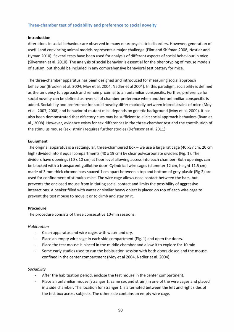
90
Three‐chamber test of sociability and preference to social novelty Introduction Alterations in social behaviour are observed in many neuropsychiatric disorders. However, generation of useful and convincing animal models represents a major challenge (Flint and Shifman 2008, Nestler and Hyman 2010). Several tests have been used for analysis of different aspects of social behaviour in mice (Silverman et al. 2010). The analysis of social behavior is essential for the phenotyping of mouse models of autism, but should be included in any comprehensive behavioral test battery for mice. The three‐chamber apparatus has been designed and introduced for measuring social approach behaviour (Brodkin et al. 2004, Moy et al. 2004, Nadler et al 2004). In this paradigm, sociability is defined as the tendency to approach and remain proximal to an unfamiliar conspecific. Further, preference for social novelty can be defined as reversal of chamber preference when another unfamiliar conspecific is added. Sociability and preference for social novelty differ markedly between inbred strains of mice (Moy et al. 2007, 2008) and behavior of mutant mice depends on genetic background (Moy et al. 2009). It has also been demonstrated that olfactory cues may be sufficient to elicit social approach behaviors (Ryan et al., 2008). However, evidence exists for sex differences in the three‐chamber test and the contribution of the stimulus mouse (sex, strain) requires further studies (Defensor et al. 2011). Equipment The original apparatus is a rectangular, three‐chambered box – we use a large rat cage (40 x57 cm, 20 cm high) divided into 3 equal compartments (40 x 19 cm) by clear polycarbonate dividers (Fig. 1). The dividers have openings (10 x 10 cm) at floor level allowing access into each chamber. Both openings can be blocked with a transparent guillotine door. Cylindrical wire cages (diameter 12 cm, height 11.5 cm) made of 3 mm thick chrome bars spaced 1 cm apart between a top and bottom of grey plastic (Fig 2) are used for confinement of stimulus mice. The wire cage allows nose contact between the bars, but prevents the enclosed mouse from initiating social contact and limits the possibility of aggressive interactions. A beaker filled with water or similar heavy object is placed on top of each wire cage to prevent the test mouse to move it or to climb and stay on it. Procedure The procedure consists of three consecutive 10‐min sessions: Habituation
‐ Clean apparatus and wire cages with water and dry. ‐ Place an empty wire cage in each side compartment (Fig. 1) and open the doors. ‐ Place the test mouse is placed in the middle chamber and allow it to explore for 10 min ‐ Some early studies used to run the habituation session with both doors closed and the mouse
confined in the center compartment (Moy et al 2004, Nadler et al. 2004). Sociability
‐ After the habituation period, enclose the test mouse in the center compartment. ‐ Place an unfamiliar mouse (stranger 1, same sex and strain) in one of the wire cages and placed
in a side chamber. The location for stranger 1 is alternated between the left and right sides of the test box across subjects. The other side contains an empty wire cage.
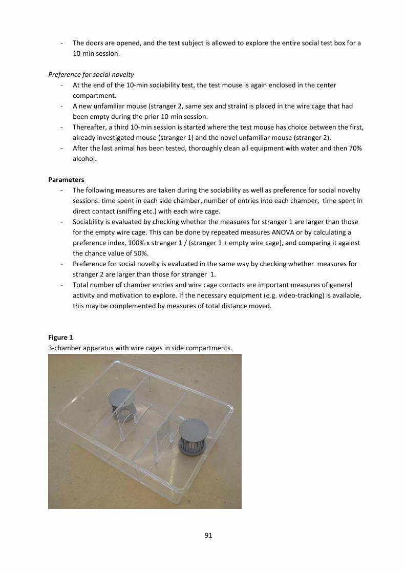
91
‐ The doors are opened, and the test subject is allowed to explore the entire social test box for a 10‐min session.
Preference for social novelty ‐ At the end of the 10‐min sociability test, the test mouse is again enclosed in the center
compartment. ‐ A new unfamiliar mouse (stranger 2, same sex and strain) is placed in the wire cage that had
been empty during the prior 10‐min session. ‐ Thereafter, a third 10‐min session is started where the test mouse has choice between the first,
already investigated mouse (stranger 1) and the novel unfamiliar mouse (stranger 2). ‐ After the last animal has been tested, thoroughly clean all equipment with water and then 70%
alcohol. Parameters
‐ The following measures are taken during the sociability as well as preference for social novelty sessions: time spent in each side chamber, number of entries into each chamber, time spent in direct contact (sniffing etc.) with each wire cage.
‐ Sociability is evaluated by checking whether the measures for stranger 1 are larger than those for the empty wire cage. This can be done by repeated measures ANOVA or by calculating a preference index, 100% x stranger 1 / (stranger 1 + empty wire cage), and comparing it against the chance value of 50%.
‐ Preference for social novelty is evaluated in the same way by checking whether measures for stranger 2 are larger than those for stranger 1.
‐ Total number of chamber entries and wire cage contacts are important measures of general activity and motivation to explore. If the necessary equipment (e.g. video‐tracking) is available, this may be complemented by measures of total distance moved.
Figure 1 3‐chamber apparatus with wire cages in side compartments.
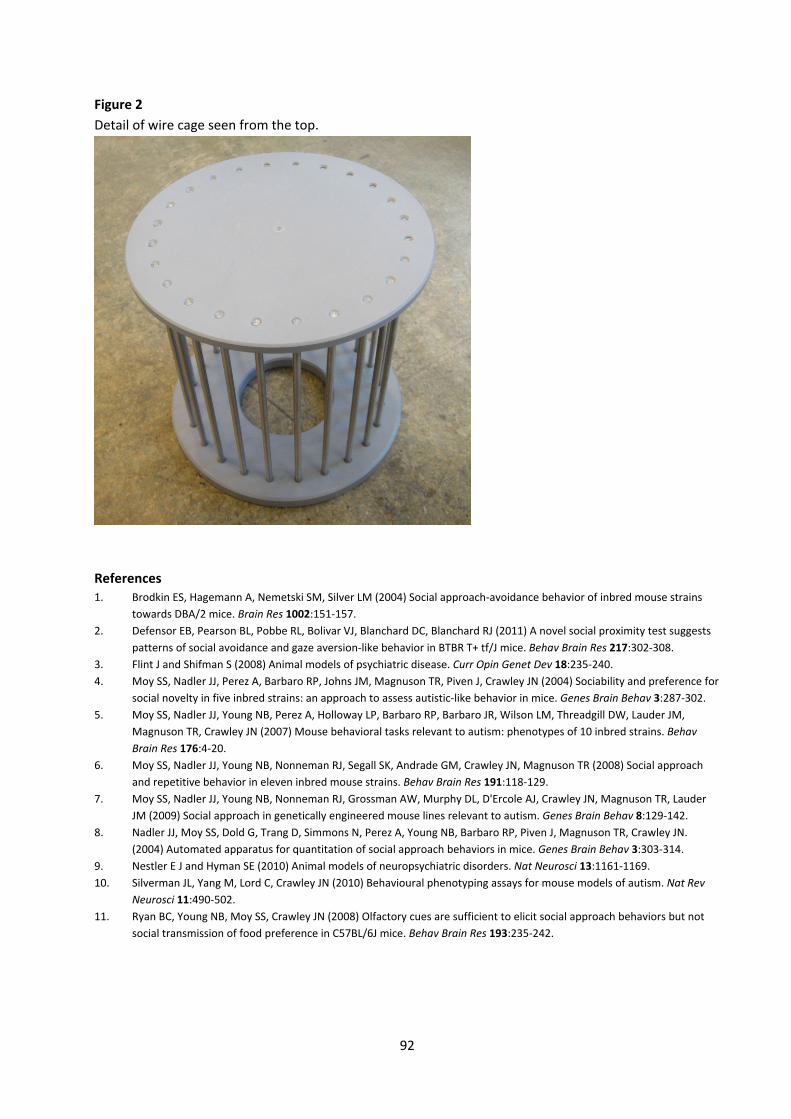
92
Figure 2 Detail of wire cage seen from the top.
References 1. Brodkin ES, Hagemann A, Nemetski SM, Silver LM (2004) Social approach‐avoidance behavior of inbred mouse strains
towards DBA/2 mice. Brain Res 1002:151‐157. 2. Defensor EB, Pearson BL, Pobbe RL, Bolivar VJ, Blanchard DC, Blanchard RJ (2011) A novel social proximity test suggests
patterns of social avoidance and gaze aversion‐like behavior in BTBR T+ tf/J mice. Behav Brain Res 217:302‐308. 3. Flint J and Shifman S (2008) Animal models of psychiatric disease. Curr Opin Genet Dev 18:235‐240. 4. Moy SS, Nadler JJ, Perez A, Barbaro RP, Johns JM, Magnuson TR, Piven J, Crawley JN (2004) Sociability and preference for
social novelty in five inbred strains: an approach to assess autistic‐like behavior in mice. Genes Brain Behav 3:287‐302. 5. Moy SS, Nadler JJ, Young NB, Perez A, Holloway LP, Barbaro RP, Barbaro JR, Wilson LM, Threadgill DW, Lauder JM,
Magnuson TR, Crawley JN (2007) Mouse behavioral tasks relevant to autism: phenotypes of 10 inbred strains. Behav Brain Res 176:4‐20.
6. Moy SS, Nadler JJ, Young NB, Nonneman RJ, Segall SK, Andrade GM, Crawley JN, Magnuson TR (2008) Social approach and repetitive behavior in eleven inbred mouse strains. Behav Brain Res 191:118‐129.
7. Moy SS, Nadler JJ, Young NB, Nonneman RJ, Grossman AW, Murphy DL, D'Ercole AJ, Crawley JN, Magnuson TR, Lauder JM (2009) Social approach in genetically engineered mouse lines relevant to autism. Genes Brain Behav 8:129‐142.
8. Nadler JJ, Moy SS, Dold G, Trang D, Simmons N, Perez A, Young NB, Barbaro RP, Piven J, Magnuson TR, Crawley JN. (2004) Automated apparatus for quantitation of social approach behaviors in mice. Genes Brain Behav 3:303‐314.
9. Nestler E J and Hyman SE (2010) Animal models of neuropsychiatric disorders. Nat Neurosci 13:1161‐1169. 10. Silverman JL, Yang M, Lord C, Crawley JN (2010) Behavioural phenotyping assays for mouse models of autism. Nat Rev
Neurosci 11:490‐502. 11. Ryan BC, Young NB, Moy SS, Crawley JN (2008) Olfactory cues are sufficient to elicit social approach behaviors but not
social transmission of food preference in C57BL/6J mice. Behav Brain Res 193:235‐242.
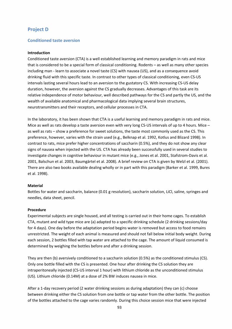
93
Project D
Conditioned taste aversion Introduction Conditioned taste aversion (CTA) is a well established learning and memory paradigm in rats and mice that is considered to be a special form of classical conditioning. Rodents – as well as many other species including man ‐ learn to associate a novel taste (CS) with nausea (US), and as a consequence avoid drinking fluid with this specific taste. In contrast to other types of classical conditioning, even CS‐US intervals lasting several hours lead to an aversion to the gustatory CS. With increasing CS‐US delay duration, however, the aversion against the CS gradually decreases. Advantages of this task are its relative independence of motor behaviour, well described pathways for the CS and partly the US, and the wealth of available anatomical and pharmacological data implying several brain structures, neurotransmitters and their receptors, and cellular processes in CTA. In the laboratory, it has been shown that CTA is a useful learning and memory paradigm in rats and mice. Mice as well as rats develop a taste aversion even with very long CS‐US intervals of up to 4 hours. Mice – as well as rats – show a preference for sweet solutions, the taste most commonly used as the CS. This preference, however, varies with the strain used (e.g., Belknap et al. 1992, Kotlus and Blizard 1998). In contrast to rats, mice prefer higher concentrations of saccharin (0.5%), and they do not show any clear signs of nausea when injected with the US. CTA has already been successfully used in several studies to investigate changes in cognitive behaviour in mutant mice (e.g., Jones et al. 2001, Stafstrom‐Davis et al. 2001, Balschun et al. 2003, Baumgärtel et al. 2008). A brief review on CTA is given by Welzl et al. (2001). There are also two books available dealing wholly or in part with this paradigm (Barker et al. 1999, Bures et al. 1998). Material Bottles for water and saccharin, balance (0.01 g resolution), saccharin solution, LiCl, saline, syringes and needles, data sheet, pencil. Procedure Experimental subjects are single housed, and all testing is carried out in their home cages. To establish CTA, mutant and wild type mice are (a) adapted to a specific drinking schedule (2 drinking sessions/day for 4 days). One day before the adaptation period begins water is removed but access to food remains unrestricted. The weight of each animal is measured and should not fall below initial body weight. During each session, 2 bottles filled with tap water are attached to the cage. The amount of liquid consumed is determined by weighing the bottles before and after a drinking session. They are then (b) aversively conditioned to a saccharin solution (0.5%) as the conditioned stimulus (CS). Only one bottle filled with the CS is presented. One hour after drinking the CS solution they are intraperitoneally injected (CS‐US interval 1 hour) with lithium chloride as the unconditioned stimulus (US). Lithium chloride (0.14M) at a dose of 2% BW induces nausea in mice. After a 1‐day recovery period (2 water drinking sessions as during adaptation) they can (c) choose between drinking either the CS solution from one bottle or tap water from the other bottle. The position of the bottles attached to the cage varies randomly. During this choice session mice that were injected

94
with the US associate the saccharin solution with nausea and drink only very little or nothing at all of the saccharin solution. Mice injected with the vehicle solution do not develop an aversion against the saccharin solution; in contrast, they greatly prefer the saccharin solution over tap water. Dependent variable The percentage of saccharin solution consumed as part from total fluid intake (saccharin + water) is taken as an aversion index (saccharin solution/total fluid) x 100. References 1. Balschun D, Wolfer DP, Gass P, Mantamadiotis T, Welzl H, Schütz G, Frey JU, Lipp H‐P (2003) Does cAMP response
element‐binding protein have a pivotal role in hippocampal synaptic plasticity and hippocampus‐dependent memory? J Neurosci 23:6304‐6314.
2. Barker LM, Best MR, Domjan M (1999) Learning Mechanisms in Food Selection. Waco:Baylor Univ Press. 3. Baumgärtel K, Genoux D, Welzl H, Tweedie‐Cullen RY, Koshibu K, Livingstone‐Zatchej M, Mamie C, Mansuy IM (2008)
Control of the establishment of aversive memory by calcineurin and Zif268. Nat Neurosci 11:572‐578. 4. Belknap JK, Crabbe JC, Plomin R, McClearn GE, Sampson KE, O’Toole LA, Goramaslak G (1992) Single‐locus control of
saccharin intake in BXD/Ty recombinant inbred (RI) mice – some methodological implications for RI strain analysis. Behav Genetics 22:81‐100.
5. Bures J, Bermudez‐Rattoni F, Yamamoto T. (Eds.) (1998) Conditioned Taste Aversion: Memory of a Special Kind. Oxford Univ Press, New York.
6. Jones MW, Errington ML, French PJ, Fine A, Bliss TVP, Garel S, Charnay P, Bozon B, Laroche S, Davis S (2001) A requirement for the immediate early gene Zif268 in the expression of late LTP and long‐term memories. Nature Neurosci 4:289‐96.
7. Kotlus BS, Blizard DA (1998) Measuring gustatory variation in mice: a short‐term fluid‐intake test. Physiol Behav 64:37‐47.
8. Stafstrom‐Davis CA, Ouimet CC, Feng J, Allen PB, Greengard P, Houpt TA (2001) Impaired conditioned taste aversion learning in spinophilin knockout mice. Learn Mem 8:272‐278.
9. Welzl H, D'Adamo P, Lipp H‐P (2001) Conditioned taste aversion as a learning and memory paradigm. Behav Brain Res 125:205‐213.
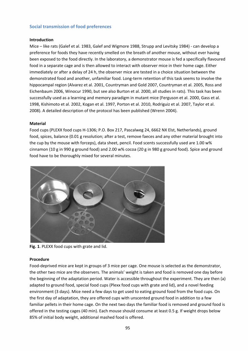
95
Social transmission of food preferences Introduction Mice – like rats (Galef et al. 1983, Galef and Wigmore 1988, Strupp and Levitsky 1984) ‐ can develop a preference for foods they have recently smelled on the breath of another mouse, without ever having been exposed to the food directly. In the laboratory, a demonstrator mouse is fed a specifically flavoured food in a separate cage and is then allowed to interact with observer mice in their home cage. Either immediately or after a delay of 24 h, the observer mice are tested in a choice situation between the demonstrated food and another, unfamiliar food. Long‐term retention of this task seems to involve the hippocampal region (Alvarez et al. 2001, Countryman and Gold 2007, Countryman et al. 2005, Ross and Eichenbaum 2006, Winocur 1990, but see also Burton et al. 2000, all studies in rats). This task has been successfully used as a learning and memory paradigm in mutant mice (Ferguson et al. 2000, Gass et al. 1998, Kishimoto et al. 2002, Kogan et al. 1997, Porton et al. 2010, Rodriguiz et al. 2007, Taylor et al. 2008). A detailed description of the protocol has been published (Wrenn 2004). Material Food cups (PLEXX food cups H‐1306; P.O. Box 217, Pascalweg 24, 6662 NX Elst, Netherlands), ground food, spices, balance (0.01 g resolution; after a test, remove faeces and any other material brought into the cup by the mouse with forceps), data sheet, pencil. Food scents successfully used are 1.00 w% cinnamon (10 g in 990 g ground food) and 2.00 w% cocoa (20 g in 980 g ground food). Spice and ground food have to be thoroughly mixed for several minutes.
Fig. 1. PLEXX food cups with grate and lid. Procedure Food‐deprived mice are kept in groups of 3 mice per cage. One mouse is selected as the demonstrator, the other two mice are the observers. The animals’ weight is taken and food is removed one day before the beginning of the adaptation period. Water is accessible throughout the experiment. They are then (a) adapted to ground food, special food cups (Plexx food cups with grate and lid), and a novel feeding environment (3 days). Mice need a few days to get used to eating ground food from the food cups. On the first day of adaptation, they are offered cups with unscented ground food in addition to a few familiar pellets in their home cage. On the next two days the familiar food is removed and ground food is offered in the testing cages (40 min). Each mouse should consume at least 0.5 g. If weight drops below 85% of initial body weight, additional mashed food is offered.
96
On day 4, (b) the demonstrator is fed a specifically flavoured food (scent A) in a separate cage (45 min) followed by an interaction period (20 min) between demonstrator and observers in the home cage. Finally, (c) the preference for food with scent A versus another flavoured food with scent B is measured either immediately or 24 hours after the interaction period. Mice prefer the food that had been consumed by the demonstrator (scent A) over the food with the unknown flavour (scent B). Dependent variables The amount of food (A) and (B) consumed during the choice test (weight difference ‘cup weight before’ – ‘cup weight after’ choice test) is measured, and the percentage of the target food (A) eaten is calculated as (target food eaten/total of food A and B eaten) x 100. In addition, ‘cup visited’ and ‘time spent inside a specific cup’ can be measured. References 1. Alvarez P, Lipton PA, Melrose R, Eichenbaum H (2001) Differential effects of damage within the hippocampal region on
memory for a natural, nonspatial Odor‐Odor Association. Learn Mem 8:79‐86. 2. Burton S, Murphy D, Qureshi U, Sutton P, O'Keefe J (2000) Combined lesions of hippocampus and subiculum Do not
produce deficits in a nonspatial social olfactory memory task. J Neurosci 20:5468‐5475. 3. Countryman RA, Gold PE (2007) Rapid forgetting of social transmission of food preferences in aged rats: relationship to
hippocampal CREB activation. Learn Mem 14:350‐358. 4. Countryman RA, Kaban NL, Colombo PJ (2005) Hippocampal c‐fos is necessary for long‐term memory of a socially
transmitted food preference. Neurobiol Learn Mem 84:175‐183. 5. Ferguson GD, Anagnostaras SG, Silva AJ, Herschman HR (2000) Deficits in memory and motor performance in
synaptotagmin IV mutant mice. PNAS 97:5598‐5603. 6. Galef BG Jr, Mason JR., Preti G, Bean NJ (1988) Carbon disulfide: a semiochemical mediating socially‐ induced diet choice
in rats. Physiol Behav 42:119‐124. 7. Galef BG Jr, Wigmore SW (1983) Transfer of information concerning distant foods: a laboratory investigation of the
'information‐centre' hypothesis. Anim Behav 31:748‐758. 8. Gass P, Wolfer DP, Balschun D, Rudolph D, Frey U, Lipp H‐P, Schütz G (1998) Deficits in memory tasks of mice with CREB
mutations depend on gene dosage. Learn Mem 5:274‐288. 9. Kishimoto Y, Fujimichi R, Araishi K, Kawahara S, Kano M, Aiba A, Kirino Y (2002) mGluR1 in cerebellar Purkinje cells is
required for normal association of temporally contiguous stimuli in classical conditioning. Eur J Neurosci 16:2416‐2424. 10. Kogan JH, Frankland PW, Blendy JA, Coblentz J, Marowitz Z, Schütz G, Silva AJ (1997) Spaced training induces normal
long‐term memory in CREB mutant mice. Curr Biol 7:1‐11. 11. Porton B, Rodriguiz RM, Phillips LE, Gilbert JW 4th, Feng J, Greengard P, Kao HT, Wetsel WC (2010) Mice lacking synapsin
III show abnormalities in explicit memory and conditioned fear. Genes Brain Behav 9:257‐268. 12. Rodriguiz RM, Gadnidze K, Ragnauth A, Dorr N, Yanagisawa M, Wetsel WC, Devi LA (2007) Animals lacking endothelin‐
converting enzyme‐2 are deficient in learning and memory. Genes Brain Behav 7:418‐426. 13. Ross RS, Eichenbaum H (2006) Dynamics of hippocampal and cortical activation during consolidation of a nonspatial
memory. J Neurosci 26:4852‐4859. 14. Strupp BJ, Levitsky DA (1984) Social transmission of food preferences in adult hooded rats (rattus norvegicus). J Comp
Psychol 98:257‐266. 15. Taylor GA, Rodriguiz RM, Greene RI, Daniell X, Henry SC, Crooks KR, Kotloski R, Tessarollo L, Phillips LE, Wetsel WC (2008)
Behavioral characterization of P311 knockout mice. Genes Brain Behav 7:786‐795. 16. Winocur G (1990) Anterograde and retrograde amnesia in rats with dorsal hippocampal or dorsomedial thalamic lesions.
Behav Brain Res 38:145‐154. 17. Wrenn CC (2004) Social Transmission of Food Preference in Mice. Curr Protocols in Neurosci S28 Chapter 8, Unit 8.5G.
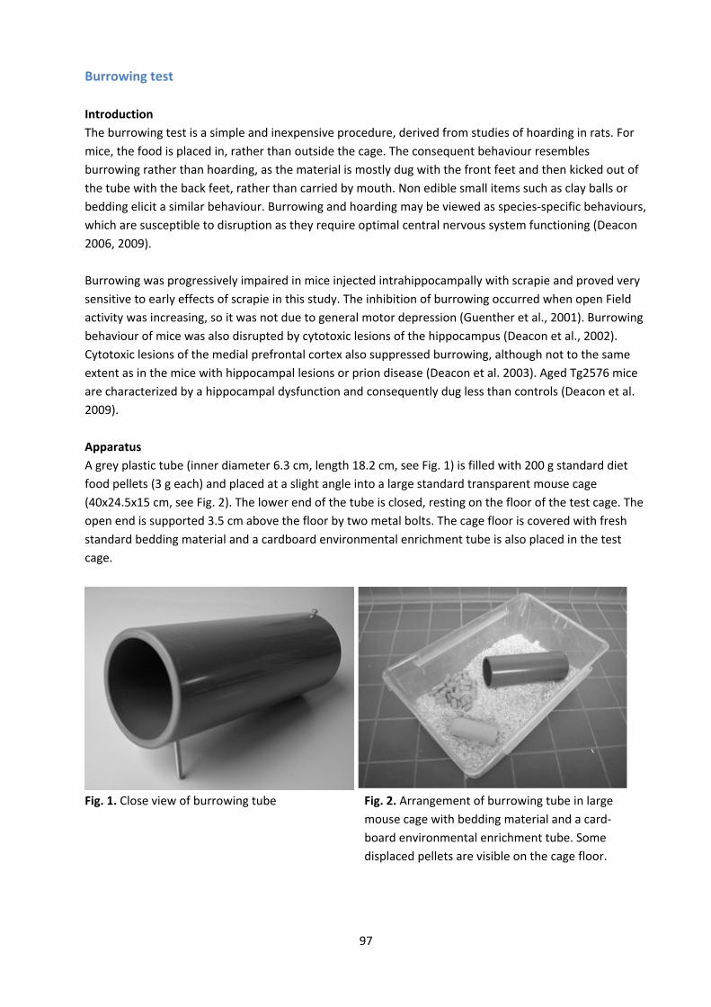
97
Burrowing test Introduction The burrowing test is a simple and inexpensive procedure, derived from studies of hoarding in rats. For mice, the food is placed in, rather than outside the cage. The consequent behaviour resembles burrowing rather than hoarding, as the material is mostly dug with the front feet and then kicked out of the tube with the back feet, rather than carried by mouth. Non edible small items such as clay balls or bedding elicit a similar behaviour. Burrowing and hoarding may be viewed as species‐specific behaviours, which are susceptible to disruption as they require optimal central nervous system functioning (Deacon 2006, 2009). Burrowing was progressively impaired in mice injected intrahippocampally with scrapie and proved very sensitive to early effects of scrapie in this study. The inhibition of burrowing occurred when open Field activity was increasing, so it was not due to general motor depression (Guenther et al., 2001). Burrowing behaviour of mice was also disrupted by cytotoxic lesions of the hippocampus (Deacon et al., 2002). Cytotoxic lesions of the medial prefrontal cortex also suppressed burrowing, although not to the same extent as in the mice with hippocampal lesions or prion disease (Deacon et al. 2003). Aged Tg2576 mice are characterized by a hippocampal dysfunction and consequently dug less than controls (Deacon et al. 2009). Apparatus A grey plastic tube (inner diameter 6.3 cm, length 18.2 cm, see Fig. 1) is filled with 200 g standard diet food pellets (3 g each) and placed at a slight angle into a large standard transparent mouse cage (40x24.5x15 cm, see Fig. 2). The lower end of the tube is closed, resting on the floor of the test cage. The open end is supported 3.5 cm above the floor by two metal bolts. The cage floor is covered with fresh standard bedding material and a cardboard environmental enrichment tube is also placed in the test cage.
Fig. 1. Close view of burrowing tube Fig. 2. Arrangement of burrowing tube in large mouse cage with bedding material and a card‐ board environmental enrichment tube. Some displaced pellets are visible on the cage floor.

98
Procedure At the beginning of their dark period, mice are placed individually in test cages and left in their familiar animal room for an observation period of 6h after which the amount of non displaced food is measured. This is followed by a second observation period of 18h. Water is available ad libitum during the entire period. Practice, i.e., giving all mice one night’s experience with a full burrow in their group home cage, improves performance. The result is a lower proportion of poor burrowers; group experience or social facilitation "kick‐starts" the natural behaviour. Parameters Weight of displaced food pellets after 6 and 18 hours. It is assumed that the amount of food eaten per mouse (~15 g/100 g/day) is a very small proportion of the 200 g available and approximately equal across the groups. References 1. Guenther K, Deacon RM, Perry VH, Rawlins JN (2001) Early behavioural changes in scrapie‐affected mice and the
influence of dapsone. Eur J Neurosci 14:401‐409. 2. Deacon RM (2006) Burrowing in rodents: a sensitive method for detecting behavioral dysfunction. Nat Protocols 1:118‐
121. 3. Deacon RM (2009) Burrowing: a sensitive behavioural assay, tested in five species of laboratory rodents. Behav Brain Res
200:128‐133. 4. Deacon RM, Croucher A, Rawlins JN (2002) Hippocampal cytotoxic lesion effects on species‐typical behaviours in mice.
Behav Brain Res 132:203‐213. 5. Deacon RM, Koros E, Bornemann KD, Rawlins JN (2009) Aged Tg2576 mice are impaired on social memory and open field
habituation tests. Behav Brain Res 197:466‐468. 6. Deacon RM, Penny C, Rawlins JN (2003) Effects of medial prefrontal cortex cytotoxic lesions in mice. Behav Brain Res
139:139‐155.
99
Nest construction test Introduction Species‐specific behaviors, such as nest building, food hoarding or burrowing, can be assessed using simple and inexpensive procedures. Despite their simplicity, such procedures can be valuable tools for the screening of mutant mice because these behaviors require optimal central nervous system functioning and are susceptible to disruption. Nest building of mice shows strain differences and is modulated by hormonal factors (e.g. Bond et al., 2002). On the other hand, it is affected by a number of experimental manipulations. For example, nest construction was significantly impaired in mice injected intrahippocampally with scrapie already before the occurrence of overt clinical signs, such as reduction in mobility, hunched posture, poor coat condition, enlarged bladder (Guenther et al., 2001). Nest building was also significantly impaired in mice with cytotoxic lesions of the hippocampus (Deacon et al., 2002). Even though medial prefrontal lesions were reported to suppress nest building in rats and hamsters, no impairment was found in mice with similar lesions (Deacon et al. 2003). Impairment of nest building has also been found in several genetically modified mouse lines, for example in lines carrying targeted point mutations of the NMDA receptor glycine binding site (Ballard et al., 2002), in vitamin D receptor mutant mice (Keisala et al., 2007), in Trpc2‐deficient lactating mice (Hasen and Gammie, 2011), in mice with a NMDA receptor NR1 subunit hypofunction (Halene et al., 2009), in an APP/PS1 mouse model of Alzheimer's disease (Filali), or in mice deficient for Dvl1, one of three mouse homologs of the Drosophila segment polarity gene Dishevelled (Lijam et al., 1997). Apparatus The procedure is done in standard mouse cages, as they are routinely used for single housing. Nesting material is provided in the form of 5x5 cm squares of pressed white cotton (‘Nestlet’, Datesand Ltd., Manchester, UK). Procedure Transfer the mice, if group housed, approximately 1 h before the dark phase to individual testing cages with sawdust bedding but no environmental enrichment items. One pressed cotton square ("Nestlet", Datesand Ltd., Manchester, UK) is placed in each cage. If housed individually, the test can be done in the home cages; remove all enrichment items and place 1 Nestlet in each cage. The mean±SEM weight of a sample of 10 Nestlets is 2.74±0.05g.

100
Parameters Nests are assessed the next morning on a 1‐5 rating scale:
1. Nestlet not noticeably touched (>90% intact) 2. Nestlet partially torn up (50‐90% remaining intact) 3. Mostly shredded but no identifiable nest site: < 50% of the Nestlet remains intact but < 90% is
within a quarter of the cage floor area, i.e. material is not gathered into a nest but spread around the cage.
4. An identifiable, but flat nest: > 90% of the Nestlet is torn up, the material is gathered into a nest within a quarter of the cage floor area, but the nest is flat, with walls < mouse body height (curled up on its side) on > 50% of its circumference.
5. A (near) perfect nest: > 90% of the Nestlet is torn up, the nest is a crater, with walls > mouse body height on > 50% of circumference.
Also any untorn Nestlet pieces are weighed (brush off loose material and sawdust). Definition untorn = > ~ 0.1 g, i.e. ~ 4% of a Nestlet. Where criteria do not agree split the difference. For example a perfect nest with an unshredded 1g piece might get a score of 4.5. In most cases the data will have a non‐normal distribution, with control mice having near‐perfect scores (ceiling effect) and none or very little of the Nestlet left untorn (floor effect). Therefore use non‐parametric statistics (Mann‐Whitney U test, or Kruskal‐Wallis ANOVA on ranks for multi‐group comparisons). References 1. Ballard TM, Pauly‐Evers M, Higgins GA, Ouagazzal AM, Mutel V, Borroni E, Kemp JA, Bluethmann H, Kew JN (2002)
Severe impairment of NMDA receptor function in mice carrying targeted point mutations in the glycine binding site results in drug‐resistant nonhabituating hyperactivity. J Neurosci 22:6713‐6723.
2. Bond TL, Neumann PE, Mathieson WB, Brown RE (2002) Nest building in nulligravid, primigravid and primiparous C57BL/6J and DBA/2J mice (Mus musculus). Physiol Behav 75:551‐555.
3. Deacon RM, Croucher A, Rawlins JN (2002) Hippocampal cytotoxic lesion effects on species‐typical behaviours in mice. Behav Brain Res 132:203‐213.
4. Deacon RM, Penny C, Rawlins JN (2003) Effects of medial prefrontal cortex cytotoxic lesions in mice. Behav Brain Res 139:139‐155.
5. Filali M, Lalonde R, Rivest S (2011) Subchronic memantine administration on spatial learning, exploratory activity, and nest‐building in an APP/PS1 mouse model of Alzheimer's disease. Neuropharmacol 60:930‐936.
6. Guenther K, Deacon RM, Perry VH, Rawlins JN (2001)Early behavioural changes in scrapie‐affected mice and the influence of dapsone. Eur J Neurosci 14:401‐409.
7. Halene TB, Ehrlichman RS, Liang Y, Christian EP, Jonak GJ, Gur TL, Blendy JA, Dow HC, Brodkin ES, Schneider F, Gur RC, Siegel SJ (2009) Assessment of NMDA receptor NR1 subunit hypofunction in mice as a model for schizophrenia. Genes Brain Behav 8:661‐675.
8. Hasen NS, Gammie SC (2011) Trpc2‐deficient lactating mice exhibit altered brain and behavioral responses to bedding stimuli. Behav Brain Res 217:347‐353.
9. Keisala T, Minasyan A, Järvelin U, Wang J, Hämäläinen T, Kalueff AV, Tuohimaa P (2007) Aberrant nest building and prolactin secretion in vitamin D receptor mutant mice. J Steroid Biochem Mol Biol 104:269‐273.
10. Lijam N, Paylor R, McDonald MP, Crawley JN, Deng CX, Herrup K, Stevens KE, Maccaferri G, McBain CJ, Sussman DJ, Wynshaw‐Boris A (1997) Social interaction and sensorimotor gating abnormalities in mice lacking Dvl1. Cell 90:895‐905.
101
IntelliCage Project Introduction Due to the fast development of molecular genetics, the number of mutant mouse lines is increasing rapidly and many of these lines need to be phenotyped at the level of behavior. However, traditional approaches to behavioral analysis cannot meet this challenge and are confronted with two major critiques. They are considered inefficient (Tecott and Nestler 2004) and poorly reproducible across labs (Chesler et al. 2002, Crabbe et al 1999). Because the human experimenter is both a limiting resource and a major source of variability in behavioral experiments, fully automated monitoring of behavior of mice in their homecage has emerged as a possible way to address both problems (De Visser et al. 2006, Spruijt and De Visser 2006). Several such systems have been developed in recent years. Among these, the IntelliCage system is unique in the sense that it permits to analyze the behavior of individual mice in a social context. A large cage for up to 16 transponder‐tagged mice is equipped with 4 learning corners with two drinking bottles each. Mice use the IntelliCage as their home‐cage and enter the corners spontaneously several times a day to perform pre‐designed tests that eventually allow accessing the water. The system continuously records all corner visits, nosepokes and licks and assigns them to individual mice. A recent multi‐lab study has demonstrated that IntelliCage produces results which are highly reproducible across labs (Krackow et al. 2010). Learning tasks in IntelliCage have been used to investigate the function of the hippocampus (Voikar et al. 2010) as well as the amygdala (Knapska et al. 2006). An increasing number of labs, including our own, use IntelliCage successfully for the phenotyping of mutant mouse lines (Kiryk et al. 2008, Jaholkowski et al. 2009, Rudenko et al. 2009, Codita et al. 2010, Konopka et al. 2010, Weyer et al. 2011). Valuable behavioral data on exploration and activity can already be collected during various phases of adaptation to the IntelliCage system. Mice are by nature curious animals and spontaneously acquire information about a novel environment by exploring it. Deviation from normal exploratory behavior may reflect neuroanatomical changes in the sizes of certain brain areas (Crusio 2001) and shall always be assessed during the characterization a specific mouse line. Many protocols have been developed to assess different cognitive functions in IntelliCage. A recent development is a delay discounting task aiming at measuring cognitive impulsivity. Time is important when assessing the value of a reward in a decision‐making situation because delayed delivery of a reward reduces its subjective value as a function of time. This phenomenon is generally referred to as delay discounting and it is measured in laboratory rodents by confronting them with a choice between a smaller reward that could be received soon and a greater but delayed reward. The delay increases with time until subjects switch their preference and choose the small and immediate reward most of the time. An exaggerated tendency to prefer immediate rewards (low tolerance to delay) is a sign of impulsivity and may indicate impaired cognitive functions and malfunctioning of the dopaminergic and glutamatergic neural circuits (Floresco et al. 2008).
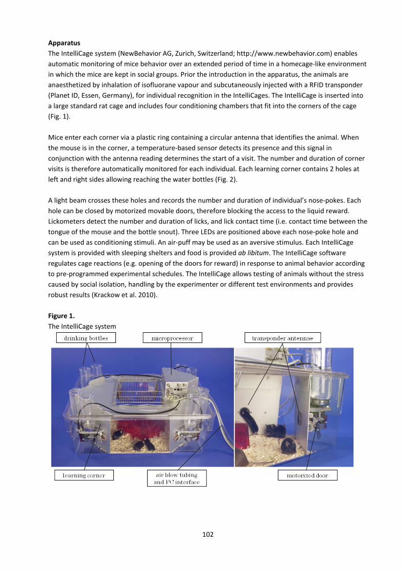
102
Apparatus The IntelliCage system (NewBehavior AG, Zurich, Switzerland; http://www.newbehavior.com) enables automatic monitoring of mice behavior over an extended period of time in a homecage‐like environment in which the mice are kept in social groups. Prior the introduction in the apparatus, the animals are anaesthetized by inhalation of isofluorane vapour and subcutaneously injected with a RFID transponder (Planet ID, Essen, Germany), for individual recognition in the IntelliCages. The IntelliCage is inserted into a large standard rat cage and includes four conditioning chambers that fit into the corners of the cage (Fig. 1). Mice enter each corner via a plastic ring containing a circular antenna that identifies the animal. When the mouse is in the corner, a temperature‐based sensor detects its presence and this signal in conjunction with the antenna reading determines the start of a visit. The number and duration of corner visits is therefore automatically monitored for each individual. Each learning corner contains 2 holes at left and right sides allowing reaching the water bottles (Fig. 2). A light beam crosses these holes and records the number and duration of individual’s nose‐pokes. Each hole can be closed by motorized movable doors, therefore blocking the access to the liquid reward. Lickometers detect the number and duration of licks, and lick contact time (i.e. contact time between the tongue of the mouse and the bottle snout). Three LEDs are positioned above each nose‐poke hole and can be used as conditioning stimuli. An air‐puff may be used as an aversive stimulus. Each IntelliCage system is provided with sleeping shelters and food is provided ab libitum. The IntelliCage software regulates cage reactions (e.g. opening of the doors for reward) in response to animal behavior according to pre‐programmed experimental schedules. The IntelliCage allows testing of animals without the stress caused by social isolation, handling by the experimenter or different test environments and provides robust results (Krackow et al. 2010). Figure 1. The IntelliCage system
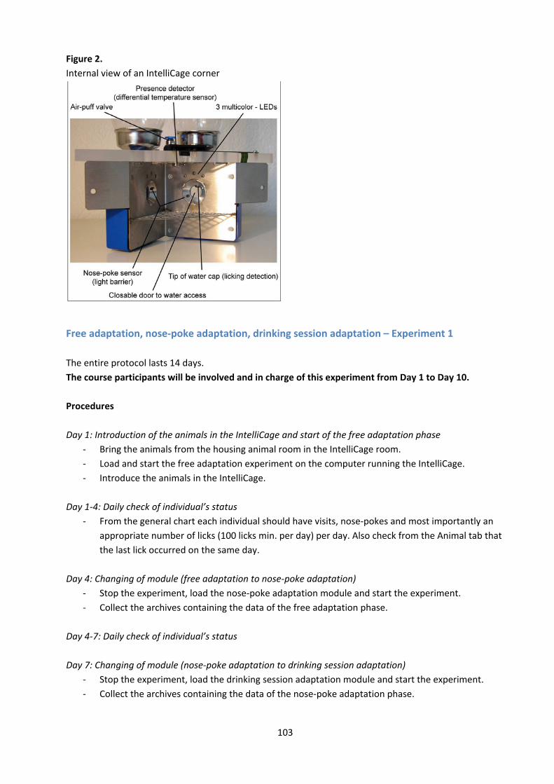
103
Figure 2. Internal view of an IntelliCage corner
Free adaptation, nose‐poke adaptation, drinking session adaptation – Experiment 1 The entire protocol lasts 14 days. The course participants will be involved and in charge of this experiment from Day 1 to Day 10. Procedures Day 1: Introduction of the animals in the IntelliCage and start of the free adaptation phase
‐ Bring the animals from the housing animal room in the IntelliCage room. ‐ Load and start the free adaptation experiment on the computer running the IntelliCage. ‐ Introduce the animals in the IntelliCage.
Day 1‐4: Daily check of individual’s status
‐ From the general chart each individual should have visits, nose‐pokes and most importantly an appropriate number of licks (100 licks min. per day) per day. Also check from the Animal tab that the last lick occurred on the same day.
Day 4: Changing of module (free adaptation to nose‐poke adaptation)
‐ Stop the experiment, load the nose‐poke adaptation module and start the experiment. ‐ Collect the archives containing the data of the free adaptation phase.
Day 4‐7: Daily check of individual’s status Day 7: Changing of module (nose‐poke adaptation to drinking session adaptation)
‐ Stop the experiment, load the drinking session adaptation module and start the experiment. ‐ Collect the archives containing the data of the nose‐poke adaptation phase.

104
Day 7‐14: Daily check of individual’s status Day 14: Stop of the experiment
‐ Stop the experiment. ‐ Collect the archives containing the data of the drinking session adaptation phase. ‐ Remove the animals from the IntelliCage and put them in normal home‐cages.
Parameters All the below variables are measured per individual and averaged and compared between groups. General parameters
‐ For assessment of initial exploration and habituation during free adaptation the following parameters are measured:
‐ Latency to first corner visit, time until all corners were visited, latency to first nose‐poke, latency to first lick, and activity (number of visits) during the first 8 hours in the IntelliCage.
General behavioural profile during free adaptation and nose‐poke adaptation is determined by:
‐ Number of visits, nose‐pokes and licks per hour during dark and light phases. ‐ The percentage of visits with nose‐pokes. ‐ The spontaneous corner preference for visits and side preference for licks.
Specific parameters for drinking session adaptation phase: The adaptation to drinking sessions can be calculated by:
‐ The ratio between the visits made during the sessions, one hour before (pre‐session) and after (post‐session) the session, out of the sessions (non‐session), and the number of visits during the whole corresponding dark phase.

105
Delay discounting task ‐ Experiment 2 The entire protocol lasts 27 days. The course participants will be involved and in charge of this experiment from Day 14 to Day 24. Day 1: Introduction of the animals in the IntelliCage and start of the free adaptation phase
‐ Bring the animals from the housing animal room in the IntelliCage room. ‐ Load and start the free adaptation experiment on the computer running the IntelliCage. ‐ Introduce the animals in the IntelliCage.
Day 1‐3: Daily check of individual’s status
‐ From the general chart each individual should have visits, nose‐pokes and most importantly an appropriate number of licks (100 licks min. per day) per day. Also check from the Animal tab that the last lick occurred on the same day.
Day 3: Changing of module (free adaptation to nose‐poke adaptation)
‐ Stop the experiment, load the nose‐poke adaptation module and start the experiment again. ‐ Collect the archives containing the data of the free adaptation phase.
Day 3‐6: Daily check of individual’s status Day 6: Changing of module (nose‐poke adaptation to DD‐adaptation with water)
‐ Stop the experiment, load the DD‐adaptation with water module and start the experiment. ‐ Collect the archives containing the data of the nose‐poke adaptation phase.
Day 6‐9: Daily check of individual’s status Day 9: Changing of module (DD‐adaptation with water to DD‐free access)
‐ Stop the experiment, load the DD‐free access module and start the experiment. ‐ Collect the archives containing the data of the DD‐adaptation with water phase.
Day 9‐13: Daily check of individual’s status Day 13: Changing of module (DD‐free access to DD‐test)*
‐ Stop the experiment. ‐ Replace 4 bottles with water with those containing a 0.5% saccharine solution in the places
defined as correct in the experimental module. ‐ Load the DD‐test module and start the experiment. ‐ Collect the archives containing the data of the DD‐free access phase.
* This module change will only occur only after all mice groups have shown a significant licking preference towards the bottles containing saccharine solution. Day 13‐24: Daily check of individual’s status Day 24: Changing of module (DD‐test to DD‐free access)*
‐ Stop the experiment, load the DD‐free access module and start the experiment.
106
‐ Collect the archives containing the data of the DD‐test phase. * This module change will only occur after all mice groups have shown a significant licking preference towards the bottles containing water (mice have switched their preference). Day 24‐27: Daily check of individual’s status Day 27: Stop of the experiment
‐ Stop the experiment. ‐ Collect the archives containing the data of the second DD‐free access phase. ‐ Remove the animals from the IntelliCage and put them in normal home‐cages.
Parameters All the below variables are measured per individual and averaged and compared between groups. General parameters
‐ For assessment of initial exploration and habituation during free adaptation the following parameters are measured:
‐ Latency to first corner visit, time until all corners were visited, latency to first nose‐poke, latency to first lick, and activity (number of visits) during the first 8 hours in the IntelliCage.
General behavioural profile during free adaptation and nose‐poke adaptation is determined by:
‐ Number of visits, nose‐pokes and licks per hour during dark and light phases. ‐ The percentage of visits with nose‐pokes. ‐ The spontaneous corner preference for visits and side preference for licks.
Specific parameters for Delay Discounting: In this experiment, the choice for the large and delayed reward (saccharine solution) compared to the small and soon available reward (water) can be assessed by:
‐ Percentage of number of licks from the sides where the bottles with saccharine solution are placed (correct sides) on the total number of licks.
107
References 1. Chesler EJ, Wilson SG, Lariviere WR, Rodriguez‐Zas SL, Mogil JS (2002) Influences of laboratory environment on behavior.
Nat Neurosci 5:1101‐1102. 2. Codita A, Gumucio A, Lannfelt L, Gellerfors P, Winblad B, Mohammed AH, Nilsson LN (2010) Impaired behavior of female
tg‐ArcSwe APP mice in the IntelliCage: A longitudinal study. Behav Brain Res 215:83‐94. 3. Crabbe JC, Wahlsten D, Dudek BC (1999) Genetics of mouse behavior: interactions with laboratory environment. Science
284:1670‐1672. 4. Crusio W (2001) Genetic dissection of mouse exploratory behaviour. Behav Brain Res 125: 127‐132. 5. De Visser L, van den Bos R, Kuurman WW, Kas MJ, Spruijt BM (2006) Novel approach to the behavioural characterization
of inbred mice: automated home cage observations. Genes Brain Behav 5:458‐466. 6. Floresco SB, Tse MT, Ghods‐Sharifi S (2008) Dopaminergic and glutamatergic regulation of effort‐ and delay‐based
decision making. Neuropsychopharmacology 33: 1966‐79. 7. Jaholkowski P, Kiryk A, Jedynak P, Ben Abdallah NM, Knapska E, Kowalczyk A, Piechal A, Blecharz‐Klin K, Figiel I, Lioudyno
V, Widy‐Tyszkiewicz E, Wilczynski GM, Lipp HP, Kaczmarek L, Filipkowski RK (2009) New hippocampal neurons are not obligatory for memory formation; cyclin D2 knockout mice with no adult brain neurogenesis show learning. Learn Mem 16:439‐451.
8. Kiryk A, Aida T, Tanaka K, Banerjee P, Wilczynski GM, Meyza K, Knapska E, Filipkowski RK, Kaczmarek L, Danysz W (2008) Behavioral characterization of GLT1 (+/‐) mice as a model of mild glutamatergic hyperfunction. Neurotox Res 13: 19‐30.
9. Knapska E, Walasek G, Nikolaev E, Neuhausser‐Wespy F, Lipp HP, Kaczmarek L, Werka T (2006) Differential involvement of the central amygdala in appetitive versus aversive learning. Learn Mem 13:192‐200.
10. Konopka W, Kiryk A, Novak M, Herwerth M, Parkitna JR, Wawrzyniak M, Kowarsch A, Michaluk P, Dzwonek J, Arnsperger T, Wilczynski G, Merkenschlager M, Theis FJ, Kohr G, Kaczmarek L, Schutz G (2010) MicroRNA Loss Enhances Learning and Memory in Mice. J Neurosci 30:14835‐1484
11. Krackow S, Vannoni E, Codita A, Mohammed AH, Cirulli F, Branchi I, Alleva E, Reichelt A, Willuweit A, Voikar V, Colacicco G, Wolfer DP, Buschmann JU, Safi K, Lipp HP (2010) Consistent behavioral phenotype differences between inbred mouse strains in the IntelliCage. Genes Brain Behav 9:722‐731.
12. Rudenko O, Tkach V, Berezin V, Bock E (2009) Detection of early behavioral markers of Huntington's disease in R6/2 mice employing an automated social home cage. Behav Brain Res 203:188‐199.
13. Spruijt BM, De Visser L (2006) Advanced behavioural screening: automated home cage ethology. Drug Discovery Today: Technologies 3:231‐237
14. Tecott LH, Nestler EJ (2004) Neurobehavioral assessment in the information age. Nat Neurosci 7:462‐466. 15. Voikar V, Colacicco G, Gruber O, Vannoni E, Lipp HP, Wolfer DP (2010) Conditioned response suppression in the
IntelliCage: assessment of mouse strain differences and effects of hippocampal and striatal lesions on acquisition and retention of memory. Behav Brain Res 213:304‐312.
16. Weyer SW, Klevanski M, Delekate A, Voikar V, Aydin D, Hick M, Filippov M, Drost N, Schaller KL, Saar M, Vogt MA, Gass P, Samanta A, Jaschke A, Korte M, Wolfer DP, Caldwell JH, Muller UC (2011) APP and APLP2 are essential at PNS and CNS synapses for transmission, spatial learning and LTP. EMBO J 30:2266‐2280
108
Demonstration Protocols
Demonstration 01 – Testing pain in mice
Models of thermal nociception: hot plate and plantar test Introduction Experimental models of nociception include tests of response thresholds to high‐intensity stimuli (acute pain tests) and of changes in spontaneous or evoked behavioral responses in animals with peripheral nerve injury or inflammation (persistent pain models). Acute thermal nociception can be assessed in the hot plate test. For pharmacological studies, the plantar test is often the preferred option, as the experimenter can direct the stimulus to an inflamed or otherwise lesioned site of the animal. Furthermore, the plantar test allows comparison of affected and non‐affected sides and repeated measurements which provide information about temporal changes. Signaling elements involved in the detection of noxious stimuli and in the propagation of nociceptive signals differ between different pain modalities. As a result, different genes or pharmacological agents may be affect pain responses in the different models differently. A basic understanding of the mechanisms involved in pain transmission is therefore important in order to choose the model most appropriate for examining the analgesic properties of the agents or genes under study. Tests of acute nociception typically use short lasting stimuli and allow the animal to withdraw from the stimulus as soon as it is felt painful. Proper experimental design, which includes the application of cut‐off times to limit the exposure to noxious stimuli, will minimize the amount of pain and distress experienced by experimental animals.
109
Hot Plate This protocol describes the use of a hot plate with an example illustrating how the potential antinociceptive effects of a compound to an acute thermal stimulus may be measured. Animals are injected with test compounds or vehicle (controls) and then placed on the hot plate one at a time. Latency to respond to the heat stimulus (the hot ground floor of the plate) is measured. The response typically is defined as flinching, licking or biting of paws. Hyperalgesic mice typically respond faster than naive mice. Compound is dissolved in saline or other appropriate vehicle and may be tested against the vehicle itself or another reference compounds, typically an agent with well established analgesic activity. The setup includes the hot plate apparatus (a metal temperature controlled plate) and a stopwatch. If drugs shall be applied, the proper needle and syringe should as well be prepared for e.g. intraperitoneal, intrathecal or per oral applications. Procedure
‐ Bring animals to test room and record their body weights ‐ Allow animals to acclimate to test room for at least 30 min ‐ Set hot plate to 55°C for mice or 52.5°C for rats ‐ Prepare doses of test compounds, vehicle (control), and reference agent (if desired) ‐ Inject animals with test compound, vehicle or reference agent. Inject animals in a “staggered”
fashion, meaning to allow a consistent time to pass between treatment and testing for each animal
‐ Randomize the order in which different treatments are administered such that each condition is distributed throughout the duration of the entire experimental procedure.
Alternatively, the treatment of the mice could be known to a second observer, while the one performing the experiment is “blinded”, i.e. the experimenter does not know what was injected until after the experiment. For example, administer vehicle, then dose 1, then dose 2, and repeat until the desired number of animals for each group is reached. The time interval used to stagger the injections will depend on the time it takes to test each animal. Testing time is generally based on the cut‐off time—the preselected time at which the test will be terminated if the animal shows no nociceptive response. Adhering to preselected cut‐off times helps minimize the tissue damage that can occur with prolonged exposure to a heated surface. For the purposes of staggering treatments, if the cut‐off time is set to 30 s, the experimenter may want to leave 45 to 60 s between the injection of the animals. The number of animals that can be treated at one time depends on the time between treatment and testing. For example, if testing is to be performed 30 min after treatment, a single experimenter, treating animals at a rate of one per minute, should treat only 30 animals. At 30 min, it will be time to test the first treated animal.
‐ When post‐treatment time (e.g. 30 min) has elapsed, begin testing animals. Place a single animal on the hot plate and immediately start a stopwatch or timer. Observe the animal until it shows a nociceptive response (e.g., licks its paw) or until the cutoff time is reached. Remove the animal from the hot plate. Record its latency to respond. For animals that do not respond prior to the cutoff time, record the cutoff time (e.g. 30 s). Repeat for all animals in the order in which they were treated.
110
Mice or rats placed on a heated surface will often lick their paws, typically the front paws first. For scoring, the latency to respond is measured as the amount of time that elapses between when the animal is placed on the hot plate and when it first licks one of its rear paws. Front paw licking is a common grooming response and may have no relation to discomfort. Therefore, rear paw licking is a more reliable measure of discomfort. The duration of action of a compound may also be determined by measuring a single animal’s response over time. For example, measurements may be taken 15, 30, and 60 min after treatment with a test compound. Of course, similar measurements must be made for vehicle‐treated animals (controls). Parameters
‐ Perform appropriate statistical analysis on the data. With multiple groups, use an ANOVA followed by post‐hoc analysis (if appropriate) to compare the treatment groups to the controls (vehicle treatment).
‐ Data from hot‐plate tests are often expressed as percent maximum possible effect (%MPE). %MPE = (test − baseline)/(cutoff − baseline) × 100, where test is the latency to respond after treatment; baseline is the latency to respond prior to treatment; and cutoff is the preset time at which the test will be ended in the absence of a response. To determine a compound’s %MPE, a baseline measure must be obtained for each animal prior to treatment with vehicle or test compound.
Reference 1. Malmberg AB, Bannon AW (1999) Models of nociception: hot‐plate, tail‐flick, and formalin tests in rodents. Curr
Protocols Neurosci S6, Unit 8.9.1‐8.9.15.
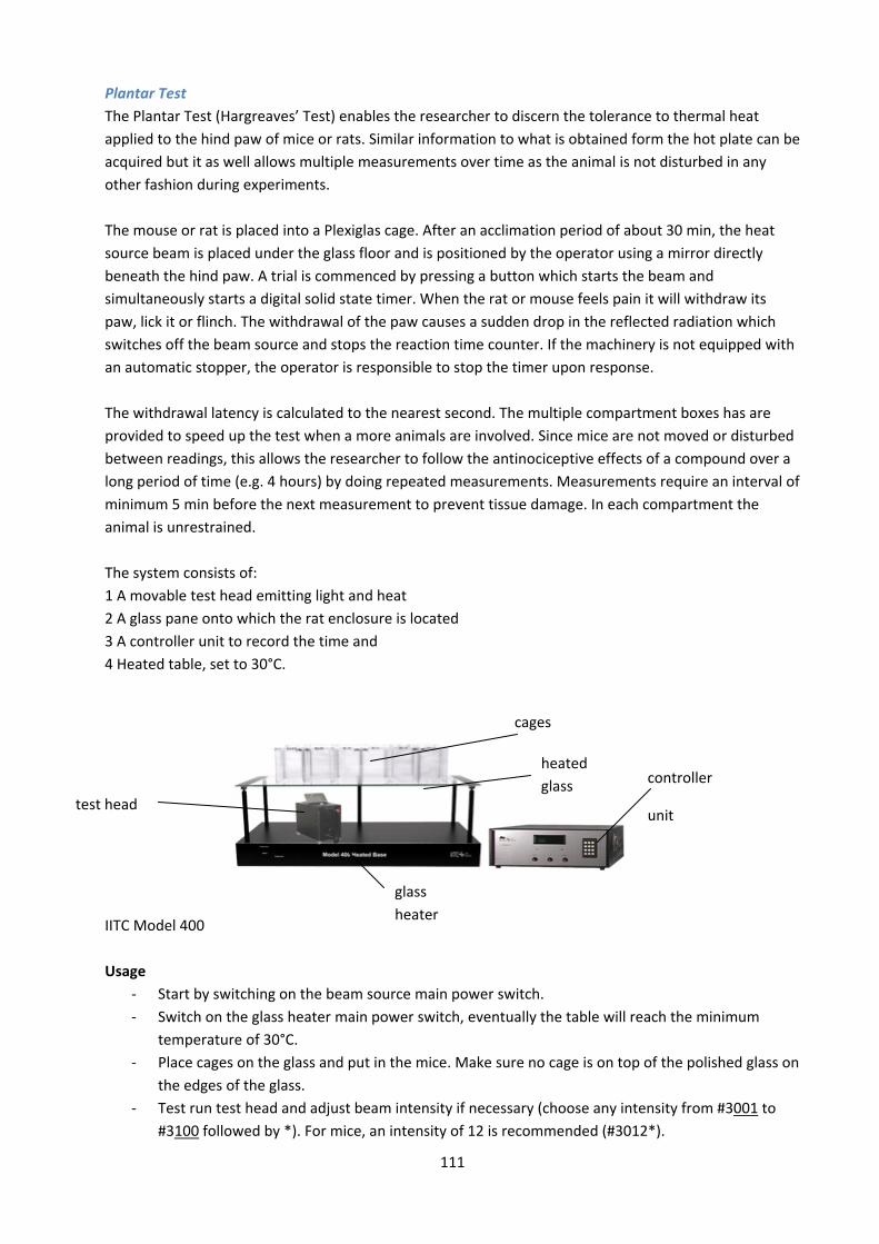
111
Plantar Test The Plantar Test (Hargreaves’ Test) enables the researcher to discern the tolerance to thermal heat applied to the hind paw of mice or rats. Similar information to what is obtained form the hot plate can be acquired but it as well allows multiple measurements over time as the animal is not disturbed in any other fashion during experiments. The mouse or rat is placed into a Plexiglas cage. After an acclimation period of about 30 min, the heat source beam is placed under the glass floor and is positioned by the operator using a mirror directly beneath the hind paw. A trial is commenced by pressing a button which starts the beam and simultaneously starts a digital solid state timer. When the rat or mouse feels pain it will withdraw its paw, lick it or flinch. The withdrawal of the paw causes a sudden drop in the reflected radiation which switches off the beam source and stops the reaction time counter. If the machinery is not equipped with an automatic stopper, the operator is responsible to stop the timer upon response. The withdrawal latency is calculated to the nearest second. The multiple compartment boxes has are provided to speed up the test when a more animals are involved. Since mice are not moved or disturbed between readings, this allows the researcher to follow the antinociceptive effects of a compound over a long period of time (e.g. 4 hours) by doing repeated measurements. Measurements require an interval of minimum 5 min before the next measurement to prevent tissue damage. In each compartment the animal is unrestrained. The system consists of: 1 A movable test head emitting light and heat 2 A glass pane onto which the rat enclosure is located 3 A controller unit to record the time and 4 Heated table, set to 30°C.
IITC Model 400 Usage
‐ Start by switching on the beam source main power switch. ‐ Switch on the glass heater main power switch, eventually the table will reach the minimum
temperature of 30°C. ‐ Place cages on the glass and put in the mice. Make sure no cage is on top of the polished glass on
the edges of the glass. ‐ Test run test head and adjust beam intensity if necessary (choose any intensity from #3001 to
#3100 followed by *). For mice, an intensity of 12 is recommended (#3012*).
test head
cages
heated glass controller
unit
glass heater
112
Further instructions can be found in the protocol from IITC in Behavior lab 1. Note The mirror of the test head might scrape against the polished glass, therefore avoid placing mice close to the edges. The center pillar supporting the heated glass may obstruct the moving of the test head and its cables. There is a risk of dropping the test head off the edge if it is pushed too far. Avoid bumping the glass as it is only fixed by being placed on the five pillars and may fall off. References 1. Hargreaves KM, Dubner R, Brown F, Flores C, Joris J (1988) A new and sensitive method for measuring thermal
nociception in cutaneous hyperalgesia. Pain 32:77‐88. 2. Hargreaves KM, R. Dubner R, Joris J (1988) Peripheral action of opiates in the blockade of carrageenan‐induced
inflammation. Pain Res Clinical Management. Vol. 3. Elsevier Science Publishers, Amsterdam: 55‐60. 3. Benneth G, Xie YK (1988) A peripheral neuropathy in rat that produces disorders of pain sensation like those seen in
man. Pain 33:87‐107. 4. Iadarola M, Draisci G (1988) Elevation of spinal cord dynorphin mRNA compared to dorsal root ganglion peptide mRNAs
during peripheral inflammation. In: The Arthritic Rat as a Model of Clinical Pain? by J. Besson and G. Guilbaud (eds.) Elsevier Press, Amsterdam: 173‐183.
5. Costello A, Hargreaves, KM (1989) Suppression of carrageenan‐induced hyperalgesia. Edema and hyperthermia by a bradykinin antagonist. Euro J Pharmacol 171:259‐263.
6. Hargreaves KM, Dubner R, Costello A (1989) Corticotropin releasing factor (CRF) has a peripheral site of action for antinociception. Euro J Pharmacol 170:275‐279.
7. Hylden J, Nahin R, Traub R, Dubner R (1989) Expansion of receptive fields of spinal lamina I protection neurons in rats with unilateral adjuvant‐induced inflammation: The contribution of central dorsal horn mechanisms. Pain 37:229‐244.
113
von Frey Filament Test Both in man and animals, pressure and pain thresholds can be assessed by applying probes of different surface areas with varying forces. The most common method was published by von Frey in his work on pressure sensitivity. At present, electronic von Frey filaments are most often used to assess responses to stimulation of slowly adapting cutaneous mechanoreceptors on the plantar side of the hindpaw of rodents. Mechanical force is exerted via application of a particular filament to the cutaneous receptive field until the filament bends. Typically, a force just sufficient to bend each filament is applied and a threshold is defined directly or indirectly by the finest filament that causes a response. This technique provides direct measurements with reasonable objectivity of the withdrawal responses. The rate at which the filament is pressed against the animal’s paw should be fast enough to avoid prolonged pressure to the tissues and interference with other stimuli or movements of the mouse. It should be slow enough to allow the investigator to apply the force in a reproducible fashion, and to minimize the effect of the animal’s withdrawal reaction time. Ideally, the pressure should increase linearly, i.e. the derivative of the applied force should be constant. Specifically, the elevated wire mesh floor supports a mouse while the filaments are applied from below, through the wire mesh, to the plantar surface of the paw. Currently available systems either use sets of calibrated von Frey filaments applied sequentially, or electronic filaments which contain a force transducer that allows the measurement of the force at which the animal withdraws its paw. Most recently, so called dynamic filaments became available which move the filament at a certain rate toward the animal’s paw. These latter instruments may provide the most accurate and objective measurements. Procedures (electronic von Frey filament)
‐ Transport the mice to the testing room in their home cages. ‐ Placed the mouse into a plexiglass cage of about 20 x 20 cm sitting on tp of a metal grid ground
floor. ‐ Let the mice accommodate to the cage for at least 30 minutes. ‐ Test sensitivity of the von Frey unit by pressing the rigid tip to a hand towel to make sure the
mechanical force is presented on the monitor. ‐ Slowly increase force to poke the hind paw with von Frey filament until a withdrawal response is
evoked and record the force from the monitor (paw withdrawal threshold). ‐ Wait for 3 min before poking the contralateral hind paw. ‐ Total of at least 4 tests on each hind paw are performed per mouse at time point. ‐ Distress of the animals is minimal because to mice can escape the unpleasant stimulus by
withdrawing their paws at any time during stimulation. At the end of the testing the animal is put back into its home cage.
‐ At the end of the experiment, clean the frame and box with water and 70% ethanol.
Applications To elucidate the role of a particular gene in the development of a pain condition (i.e. neuropathic pain) the development of mechanical hyperalgesia can be assessed with the electronic von Frey test in two different groups of mice (wild‐type and transgenic) through repetitive (i.e. daily) measurements of mechanical sensitization after nerve damage (i.e. CCI, Chronic Constriction Injury). To study the potential antihyperalgesic effect of a compound, mechanical hyperalgesia is measured in at least two different groups of mice (vehicle‐ and drug‐treated) for 4‐8 hours after the administration of the drug (or vehicle). 4–5 measurements are taken at 20‐30 minutes intervals and averaged.

114
Measurements of paw withdrawal thresholds of the injured paw and of the contralateral paw are made alternately. References 1. Möller KA, Johansson B & Berge O (1998) Assessing mechanical allodynia in the rat paw with a new electronic algometer.
J Neurosci Methods 84:41‐47. 2. Ambalavanar R et al (2007) Deep tissue inflammation upregulates neuropeptides and evokes nociceptive behaviors
which are modulated by a neuropeptide antagonist. Pain 120:53‐68. 3. Di Lio et al (2011) HZ166, a novel GABAA receptor subtype‐selective benzodiazepine site ligand, is antihyperalgesic in
mouse models of inflammatory and neuropathic pain. Neuropharmacology 60:626‐32.
115
Capsaicin model Capsaicin, the pungent component of peppers activates the transient receptor potential channel vanilloid type 1 (TRPV1), a heat‐sensitive cation channel expressed by many nociceptors. Intradermal injection of capsaicin results in dose‐dependent acute pain sensation, followed by a neurogenic inflammation consisting of allodynia, hyperalgesia, and a flare reaction, which originates from the release of vasoactive neuropeptides from peripheral nociceptor terminals. All three effects of capsaicin extend beyond the site of injection. Hyperalgesia at the site of injection (primary hyperalgesia) is thermal and mechanical while the hyperalgesia in the surrounding non‐injected area is purely mechanical. Intraplantar injection of capsaicin evokes nocifensive behavior characterized by flinching, licking and guarding of the injected paw that lasts for about 5 min. Heat hyperalgesia typically lasts for up to 45 min, whereas mechanical hyperalgesia persists longer for up to 4 h. Intracutaneous injections of capsaicin is frequently used to study changes in nociceptor activity and in the responses of spinal dorsal horn neurons accompanying primary and secondary hyperalgesia. Procedure 1) Capsaicin‐induced paw flinching
‐ Mice are placed separately in Plexiglas boxes for a 20–30 min habituation period. ‐ After the habituation, capsaicin (1, 5, or 10 μg) in 10% ethanol, 10% Tween 80 and 80% ACSF is
injected into the plantar side of the mouse left hind paw, using a 10 μl Hamilton syringe in a volume of 5 μl.
‐ Immediately after the injection, the flinching episodes or the time spent licking the paw are recorded in 1 minute bins for 5 minutes (a flinch is defined as a rapid flexor reflex of the injected paw)
‐ Control experiments need to be performed in all studies using the capsaicin vehicle. 2) Capsaicin‐induced mechanical hyperalgesia
‐ See von Frey procedure for detailed explanation. ‐ Mice are injected subcutaneously into the plantar side of the left hindpaw with a dose of 30 μg
of Capsaicin dissolved in a total volume of 10 μl in 10% ethanol, 10% Tween 80 and 80% ACSF . ‐ Immediately after the injection, mechanical sensitization is measured with von Frey filaments for
4 hours at intervals of 20‐30 minutes. Applications The capsaicin model can be used to study the effects of pharmacological and genetic manipulations on acute chemically induced pain, primary and secondary hyperalgesia and on neurogenic inflammation. To elucidate the role of a particular gene in the development of capsaicin‐induced mechanical hyperalgesia, mechanical sensitization can be assessed with the electronic von Frey test in two different groups of mice (wild‐type and transgenic) starting 15 minutes after the injection of intraplantar capsaicin at intervals of 30 minutes for a total time of 4 hours. This time frame is long enough to include the complete recovery to normal baseline values. To study the potential antihyperalgesic effect of a drug, mechanical hyperalgesia is measured in at least two different groups of mice (vehicle‐ and drug‐treated). 2 hours after the injection of capsaicin, when the mechanical sensitization is higher, the compound or the vehicle are administered. 4–5 measurements of the paw withdrawal threshold are taken for 2 hours at 20‐30 minutes intervals and

116
averaged. Measurements of mechanical sensitization of the capsaicin‐injected paw and of the contralateral paw are made alternately. References 1. Sakurada T et al. (1992) The capsaicin test in mice for evaluating Tachykinin antagonists in the spinal cord.
Neuropharmacology 31:1279‐1285. 2. Hart D et al (1996) Enhanced withdrawal responses to heat and mechanical stimuli following intraplantar injection of
capsaicin in rats. Pain 67:179‐188. 3. Pernía‐Andrade AJ et al. (2009) Spinal endocannabinoids and CB1 receptors mediate C‐fiber‐induced heterosynaptic pain
sensitization. Science 325:760‐764.
117
Demonstration 02 – Motor function
Rotarod test Introduction The rotarod test has been developed to measure motor coordination in mice and rats (Dunham and Miya 1957). A mouse has to keep its balance on a horizontal rotating rod (diameter approx. 3 cm). Rotation speed can be constant or accelerating. Usually several mice tested at the same time, separated by large disks. A trial starts when the mouse is placed on the rotating rod, and it stops when the mouse falls down. Falling down activates a switch that automatically stops a timer. The latency to fall off the rod is taken as the dependent measure. The rotarod test has been successfully used to screen motor coordination in mutant mice (e.g., Aden et al. 2003, Carter et al. 1999, D'Hooge et al. 2001, Norreel et al. 2001, Paylor et al., 1999). Performance in this test is affected by a number of variables including body weight, housing conditions and stress (personal communication, Lisa Tarantino 2000), and mouse strain tested (e.g., Crabbe et al. 1998, Homanics et al. 1999). Material Rotarod apparatus (Ugo‐Basile Model 7650), pencil, data sheet, cleaning material for the apparatus. Procedure Five mice are simultaneously placed on the rotarod apparatus with the rod rotating at 4 rpm (rotations/minute) during the first minute. Then rotation speed is increased every 30 sec by 4 rpm. A trial ends for a mouse when it falls down or when 5 min are completed. Each mouse is submitted to 5 trials on day 1 with an intertrial interval of 30 min. On day 2, all mice are tested again for 5 min at a constant speed (average of maximum speeds reached from all mice on day 1). Some mice cling to the rod and ride a full circle without falling down. Passive rides are recorded separately. Dependent variables Latency to fall off the rod, full circle rides. References 1. Aden U, Halldner L, Lagercrantz H, Dalmau I, Ledent C, Fredholm BB (2003) Aggravated brain damage after hypoxic
ischemia in immature adenosine A2A knockout mice. Stroke 34:739‐744. 2. Carter RJ, Lione LA, Humby T, Mangiarini L, Mahal A, Bates GP, Morton AJ, Dunnett SB (1999) Characterization of
progressive motor deficits in mice transgenic for the human Huntington’s disease mutation. J Neurosci 19:3248‐3257. 3. Crabbe JC, Gallaher EJ, Cross SJ, Belknap JK (1998) Genetic determinants of sensitivity to diazepam in inbred mice. Behav
Neurosci 12:668‐677. 4. D'Hooge R, Van Dam D, Franck F, Gieselmann V, De Deyn PP (2001) Hyperactivity, neuromotor defects, and impaired
learning and memory in a mouse model for metachromatic leukodystrophy. Brain Res 907: 35‐43. 5. Dunham NW, Miya TS (1957) A note on a simple apparatus for detecting neurological deficit in rats and mice. J Am
Pharm Assoc 46:208‐209. 6. Homanics GE, Quinlan JJ, Firestone LL (1999) Pharmacologic and behavioral responses of inbred C57BL/6J and strain
129/SvJ mouse lines. Pharmacol Biochem Behav 63:21‐26. 7. Norreel JC, Jamon M, Riviere G, Passage E, Fontes M, Clarac F(2001) Behavioural profiling of a murine Charcot‐Marie‐
Tooth disease type 1A model. Eur J Neurosci 13:1625‐1634. 8. Paylor R, Hirotsune S, Gambello MJ, Yuva‐Paylor L, Crawley JN, Wynshaw‐Boris A (1999) Impaired learning and motor
behavior in heterozygous Pafah 1b1 (Lis) mutant mice. Learn Mem 6:521‐537. 9. Rustay NR, Wahlsten D, Crabbe JC (2003) Influence of task parameters on rotarod performance and sensitivity to ethanol
in mice. Behav Brain Res 141:237‐249.
118
Beam walking test Introduction Deficits in limb coordination and accuracy of limb placement are examined by assessing an animal’s ability to navigate across a beam or a ladder in order to return to its home cage. The beam can be constructed in several ways, and three examples are given. (a) A simple beam (28 mm wide) leads towards an enclosed escape box. The difficulty to traverse the beam is increased by using smaller sized beams. (b) A ladder consisting of regularly spaced bars (distance between bars: approx. 1 cm) is used instead of solid beams (modification from Kunkel‐Bagden et al. 1993). Crossing this ‘horizontal ladder’ requires that animals accurately place their limbs on the bars. The difficulty of the task is increased by adding randomly assigned gaps (distances between bars ranging from 0.5 to 2.5 cm). Falling through the bars with one limb counts as an error, and errors per crossing are analysed. (c) A beam is placed as a bridge between the subject's home cage and an empty cage. Three 6x6 cm platforms are mounted on the beam, one at each end and one in the middle. The test is made more demanding by interrupting the beam surface every 2 cm by a 2 cm long groove. The difficulty of the task can be further increased by inserting grooves of irregular size and intervals. Beam walking tests have been successfully used in different mutant mice (e.g., Allen et al., 2003, Carter et al. 1999, Dluzen et al. 2001, Lipp and Wahlsten 1992, Salichon et al. 2001). Factors influencing beam walking include genetic background and anxiety (Lepicard et al. 2000). A detailed description of beam walking in rats is given by Goldstein (1993). Material Plastic beam(s), video camera, stop watch, data sheet, pencil Procedure The testing device is placed in a dimly lit room and consists of a small platform (6x6 cm) located in the middle of a wooden bar (45 cm long, 1. 6 cm wide) bridging a gap between two platforms (10 x 10 cm) which permits escape into a small Perspex cage filled with wood shavings. One bar has a smooth surface (solid bar), the other is formed by series of regularly spaced carvings of two cm length and 1. 5 cm depth. The wood is polished with sand paper in order to provide a smooth but not slippery surface. On the first day, a mouse is placed onto the middle platform of the solid bar and observed for 5 min. The variables assessed are the number of visits to the escape platforms, the number of hindpaw slips off the bar and the number of escapes into the Perspex cages. On the second day, the mouse is placed onto the middle of the carved bar, and the same measures are taken again during 5 min. Since the error score are not independent of the locomotor activity, correction procedures such as the computation of indices or multiple regression are used depending on the particular samples investigated. Mice resting immobile on the central platform (mainly BALB/c) are gently pushed in order to initiate movements. Mice that escaped into the Perspex cages are picked up and placed on the central platform again. Animals fallen off from the bar are handled similarly (Lipp and Wahlsten 1992). Dependent variables Number of failures (slipping off the beam with one paw), posture (clinging to the beam, free walking), latency to cross the beam.

119
References 1. Allen JP, Hathway GJ, Clarke NJ, Jowett MI, Topps S, Kendrick KM, Humphrey PP, Wilkinson LS, Emson PC (2003)
Somatostatin receptor 2 knockout/lacZ knockin mice show impaired motor coordination and reveal sites of somatostatin action within the striatum. Eur J Neurosci 17:1881‐1895.
2. Carter RJ, Lione LA, Humby T, Mangiarini L, Mahal A, Bates GP, Morton AJ, Dunnett SB (1999) Characterization of progressive motor deficits in mice transgenic for the human Huntington’s disease mutation. J Neurosci 19:3248‐3257.
3. Dluzen DE, Gao X, Story GM, Anderson LI, Kucera J, Walro JM (2001) Evaluation of nigrostriatal dopaminergic function in adult +/+ and +/‐ bdnf mutant mice. Exp Neurol 170:121‐128.
4. Goldstein LB (1993) Beam‐walking in rats: the measurement of motor recovery after injury to the cerebral cortex. Neurosci Prot 10:1‐13.
5. Kunkel‐Bagden E, Dai, HN, Bregman BS (1993) Methods to assess the development and recovery of locomotor function after spinal cord injury in rats. Exp Neurol 119:153–164.
6. Lepicard EM, Venault P, Perez‐Diaz F, Joubert C, Berthoz A, Chapouthier G (2000) Balance control and posture differences in the anxious BALB/cByJ mice compared to the non anxious C57BL/6J mice. Behav Brain Res 117:185‐195.
7. Lipp HP, Wahlsten D (1992) Absence of the corpus callosum. In P. Driscoll (ed): Genetically‐Defined Animal Models of Neuro‐Behavioral Dysfunction. Boston: Birkhaeuser p.217‐252.
8. Salichon N, Gaspar P, Upton AL, Picaud S, Hanoun N, Hamon M, De Maeyer E, Murphy DL, Mossner R, Lesch KP, Hen R, Seif I (2001) Excessive activation of serotonin (5‐HT) 1B receptors disrupts the formation of sensory maps in monoamine oxidase a and 5‐ht transporter knock‐out mice. J Neurosci 21: 884‐896.
120
Foot print test Introduction This tests detects gait abnormalities and ataxia suggesting changes in limb and muscle coordination (sciatic nerve/spinal) and body weight support (De Medinaceli et al. 1982). It has been successfully used in different mutant mice (e.g., Barlow et al. 1996, Carter et al. 1999, Del Bel et al., 2002, Norreel et al. 2001). A number of factors influence performance in this test including stress (Metz et al., 2001). Material Alley or tunnel (35 ‐ 80 cm long, 5 – 6 cm wide), ink [or: non‐toxic paint; mixture of icing and non‐ toxic food dye (Klapdor et al. 1997)], white paper, ruler, paper, pencil, data sheet. Procedure An animal is trained to pass through an alley or tunnel. Then its hindpaws are dipped in black ink, and it is placed at one end of the tunnel. Footprints are recorded on a sheet of paper covering the floor of the alley. This procedure is repeated three times. A series of at least 6 sequential steps was used to determine mean values of stride length, stride variability, base of support and limb rotation. Dependent variables Stride length, stride variability (= difference between longest and shortest stride length), base of support, limb rotation. Stride length is measured between two consecutive prints of one hind paw on each side. The base of support (base) is determined by measuring the core to core distance of the print representing the plantar cushion underlying the metatarso‐phalangeal (MTP) joint. Limb rotation is defined as the angle formed by the intersection of the line through the print of the third digit and the print representing the MTP joint with the line through the central pad parallel to the walking direction. References 1. Barlow C, Hirotsune S, Paylor R, Liyanage M, Eckhaus M, Collins F, Shiloh Y, Crawley JN, Ried T, Tagle D, Wynshaw‐Boris A
(1996) Atm‐Deficient Mice: A Paradigm of Ataxia Telangiectasia. Cell 86:159–171. 2. Carter RJ, Lione LA, Humby T, Mangiarini L, Mahal A, Bates GP, Morton AJ, Dunnett SB (1999) Characterization of
progressive motor deficits in mice transgenic for the human Huntington’s disease mutation. J Neurosci 19:3248‐3257. 3. Del Bel EA, Souza AS, Guimaraes FS, da‐Silva CA, Nucci‐da‐Silva LP (2002) Motor effects of acute and chronic inhibition of
nitric oxide synthesis in mice. Psychopharmacology 161:32‐37. 4. De Medinaceli L, Freed WJ, Wyatt RJ (1982) An index of the functional condition of rat sciatic nerve based on
measurements made from walking tracks. Exp Neurol 77:634‐643. 5. Klapdor K, Dulfer BG, Hamman A, Van der Stay F (1997) A low‐cost method to analyze footprint patterns. J Neurosci
Meth 75:49‐54. 6. Metz GAS, Schwab ME, Welzl H (2001) The effects of acute and chronic stress on motor and sensory performance in
male Lewis rats. Physiol Behav 72:29‐35. 7. Norreel JC, Jamon M, Riviere G, Passage E, Fontes M, Clarac F (2001) Behavioural profiling of a murine Charcot‐Marie‐
Tooth disease type 1A model. Eur J Neurosci 13:1625‐1634.
121
Grip test Introduction The grip strength test measures the strength with which an animal holds on to a grip with its forepaws, or to a grid with its hindpaws, while the experimenter pulls it gently backwards or upwards, respectively. The results give a general idea of an animal’s muscle strength. The test has been successfully used to detect impairments in muscle strength in mutant mice (Gonzalez Deniselle et al. 2002, Norreel et al. 2001). Material Newtonmeter with a grip or grid attached, pencil, data sheet. Procedure A newtonmeter with a grip bar is horizontally fixed. The mouse is held in a horizontal position and lowered towards the grip bar until the animal grasps it. Then it is gently pulled backward until it releases the grip. The force measured when the animal releases the grip is recorded. Three trials per animal and session are recorded, and the average strength is included in the analysis. The test is repeated the next day. Dependent variable Force when the grip is released. References 1. Gonzalez Deniselle MC, Lopez Costa JJ, Gonzalez SL, Labombarda F, Garay L, Guennoun R, Schumacher M, De Nicola AF
(2002) Basis of progesterone protection in spinal cord neurodegeneration. J Steroid Biochem Mol Biol 83:199‐209. 2. Norreel JC, Jamon M, Riviere G, Passage E, Fontes M, Clarac F (2001) Behavioural profiling of a murine Charcot‐Marie‐
Tooth disease type 1A model. Eur J Neurosci 13:1625‐1634.
122
Demonstration 03 – Exploratory behavior and anxiety
Open Field test Introduction A rodent that is placed in an unfamiliar empty arena will move around to explore it. This exploratory activity will gradually decrease as the arena becomes familiar (habituation). The open field test can thus reveal increased or reduced locomotor activity, as well as an altered time course of habituation. Because open space is aversive to rodents, decreased exploration of the arena centre as compared to the periphery can be an indicator of increased anxiety‐like behaviour. The open field has been an important tool in behavioural pharmacology since a long time, but is also frequently used for genetic studies in mice. A large number of genetically modified mice were found to have either increased or decreased exploratory activity in the open field. However, despite the simplicity of apparatus and protocol, open field behaviour is complex and depends on many factors that can change independently (Walsh and Cumins 1976, Wolfer et al. 2001). Therefore, changes in open field activity can usually only be interpreted in conjunction with the other tests (Stanford, 2007). Apparatus We use our round watermaze tank with a diameter of 150 cm. The tank is dried and a plastic floor is placed at the usual level of the water surface, leaving a distance of 35 cm to the top of the side wall. Illumination is by indirect diffuse room light (4 40W bulbs). Size and shape of the arena (as well as testing procedures) vary across laboratories. Many groups use smaller rectangular arenas. Procedure
‐ Transfer the rack with the home cages to the experimental room approximately 30 min before the experiment begins.
‐ Recording over longer time periods (up to 1h) allows quantification of habituation. Day 1
‐ Clean the arena with water and dry. ‐ Release the subject near the side wall and observe for 10 min.
Day 2 ‐ Clean the arena with water and dry. ‐ Release the subject near the side wall and observe for 10 min (same order of testing and time of
the day as on first day).

123
Parameters ‐ number of crosses over the lines of a regular grid drawn on the arena surface (horizontal activity) ‐ % time spent in a circular zone drawn around the arena centre (diameter 110 cm) ‐ number of rearings (vertical activity) ‐ number of faecal boli deposited (may be recorded as measure of emotionality) ‐
Activity is usually recorded electronically. Several companies sell systems that are equipped with infrared beams, permitting automatic determination of both horizontal and vertical activity. Alternatively, locomotion can be video‐tracked. This yields only indirect information of vertical activity (change of object size recorded e.g. by EthoVision), but permits finer analysis of horizontal activity (temporal patterning, geometrical parameters, measures of stereotypy). References 1. Stanford SC (2007) The Open Field Test: reinventing the wheel. J Psychopharmacol 21:134‐135. 2. Walsh RN and Cummins RA (1976) The open‐field test: a critical review. Psychol Bull 83:482‐504. 3. Wolfer DP, Madani R, Valenti P, Lipp HP (2001) Extended analysis of path data from mutant mice using the public
domain software Wintrack. Physiol Behav 73:745‐753.
124
Light‐dark transition test Introduction Exploring a novel environment is a natural behaviour of mice. On the other hand, they have the innate tendency to prefer dark and enclosed places over brightly lit open space. In this test, the mice are given a choice between a dark box and a brightly lit open chamber. They have to resolve the conflict between exploring the open chamber and avoiding it because it is brightly illuminated. Crawley and Goodwin (1980) introduced this paradigm as a model of anxiety‐like behaviour and observed that the time spent exploring the open chamber was increased by anxiolytic drugs. The paradigm was further validated and slightly modified by Costall et al. (1989) and Misslin et al. (1989). The light‐dark transition test has become one of the most frequently used paradigms to characterize anxiety‐like behaviour in mutant mice (Cryan and Holmes, 2005). Apparatus A 20x30 cm lit chamber with transparent Perspex walls (20 cm high) and open top is connected to a 20x15x20 cm polyvinyl‐chloride box. The box is dark and completely enclosed, except for the 7.5x7.5 cm opening connecting it to the lit chamber. The lit chamber is under direct room light. Misslin et al. (1990) used two 20x20x14 chambers that are connected by a 5x7x10 cm polyvinylchloride. One chamber is painted black and has an opaque cover, the other is white with a transparent cover and illuminated by a 100W desk lamp. Procedure
‐ Bring the rack with the home cages to the testing room approximately 30 min before the experiment begins.
‐ Clean the apparatus thoroughly with water and dry. ‐ Place the animal into the lit box and observe for 5 min.
Parameters
‐ Latency to escape into the dark box ‐ % time spent in the lit chamber ‐ number of transitions between lit chamber and dark box
If movements in the lit chamber are video‐tracked, parameters of general activity such as distance moved, % time active, and speed of locomotion can be calculated as well. In addition, grooming, rearings, and leanings may be recorded with a keyboard event recorder.
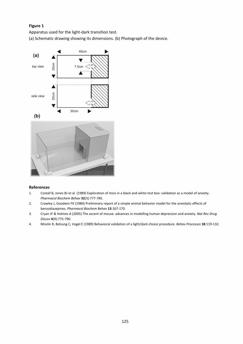
125
Figure 1 Apparatus used for the light‐dark transition test. (a) Schematic drawing showing its dimensions. (b) Photograph of the device.
References 1. Costall B, Jones BJ et al. (1989) Exploration of mice in a black and white test box: validation as a model of anxiety.
Pharmacol Biochem Behav 32(3):777‐785. 2. Crawley J, Goodwin FK (1980) Preliminary report of a simple animal behavior model for the anxiolytic effects of
benzodiazepines. Pharmacol Biochem Behav 13:167‐170. 3. Cryan JF & Holmes A (2005) The ascent of mouse: advances in modelling human depression and anxiety. Nat Rev Drug
Discov 4(9):775‐790. 4. Misslin R, Belzung C, Vogel E (1989) Behavioral validation of a light/dark choice procedure. Behav Processes 18:119‐132.
126
Elevated null‐maze Introduction As in the light‐dark transition test, the tested animal has to resolve a conflict between its natural tendency to explore a novel environment and the avoidance of a threatening situation. On an elevated maze, the exploratory drive is competing with the natural avoidance by rodents of heights and open spaces. Typically, mice prefer a zone that is protected by side walls, but after a while carefully advance onto an unprotected runway and begin to dip their head to explore the cliff. Head dips are fist made while the body is still in the protected zone (protected head dips). Then the animals stretch their body toward the open zone but often retreat before leaving the protected sector with their hindlimbs (stretch attend posture). Finally they leave the protected zone completely to explore the cliff extensively (unprotected head dips). This concept was first validated pharmacologically by Pellow et al. (1985) who observed that anxiolytic drugs made rats less reluctant to explore the open arms of a plus shaped elevated maze with two protected and two unprotected arms. The usefulness of the device to investigate both anxiolytic and anxiogenic agents in the mouse was subsequently demonstrated by Lister (1987). Shepherd et al. (1994) introduced and validated pharmacologically in the rat an alternative implementation of the test, which is now known as the elevated null‐ or zero‐maze. Their goal was to remove any ambiguity in interpretation of time spent on the central square of the traditional plus‐maze and to allow uninterrupted exploration. Konig et al. (1996) used the elevated null‐maze to demonstrate increased anxiety‐like behaviour in mutant mice which lacked the pre‐proenkephalin gene. The elevated maze is still used in both configurations and is the most frequently used model of anxiety‐like behaviour for drug studies. It is also often used to characterise the emotional behaviour of mutant mice (Cryan and Holmes, 2005), for example in a set of studies in mice carrying point mutations in GABAA receptor subunits (Rudolph et al. 1999, Low et al. 2000) or a deletion of the urocortin gene (Vetter et al. 2002). Importance of measuring the ethologically derived parameters in addition to spatiotemporal pattern of behavior has been emphasized by Rodgers et al. (1997).
127
Apparatus The maze is made of grey plastic and has an annular runway (diameter 46 cm, width 5.5 cm). Two opposing 90° sectors of the runway are protected by an inner and outer wall of grey polyvinyl‐ chloride (height 16 cm). The two remaining sectors are unprotected. The apparatus is mounted 40cm above the floor and exposed to indirect and diffuse room light (4 40W bulbs). Procedure
‐ Bring the home cages to the testing room approximately 30 min before the experiment begins. ‐ Clean the apparatus thoroughly with water and dry. ‐ Place the animal into a closed sector and observe for 10 min.
Parameters
‐ % time spent on open sectors, number of entries to open sectors ‐ Number of head dips (protected and unprotected) ‐ Number of stretch attend postures ‐ Number of faecal boli deposited on the device
If movements are video‐tracked, parameters of general activity such as distance moved, % time active, and speed of locomotion can be calculated as well. In addition, head dips may be recorded with a keyboard event recorder. Video‐tracking also permits to refine zone analysis by defining intermediate zones at the sector transitions (where animals spend most of their time).
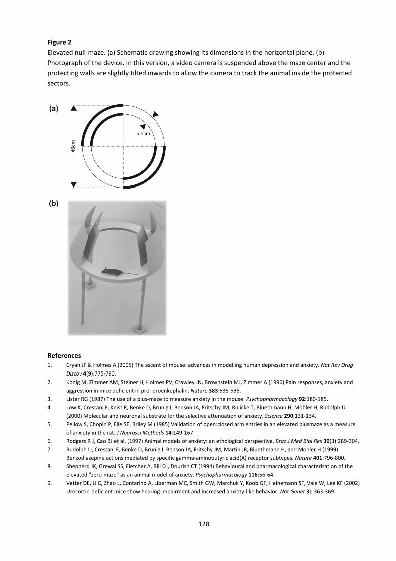
128
Figure 2 Elevated null‐maze. (a) Schematic drawing showing its dimensions in the horizontal plane. (b) Photograph of the device. In this version, a video camera is suspended above the maze center and the protecting walls are slightly tilted inwards to allow the camera to track the animal inside the protected sectors.
References 1. Cryan JF & Holmes A (2005) The ascent of mouse: advances in modelling human depression and anxiety. Nat Rev Drug
Discov 4(9):775‐790. 2. Konig M, Zimmer AM, Steiner H, Holmes PV, Crawley JN, Brownstein MJ, Zimmer A (1996) Pain responses, anxiety and
aggression in mice deficient in pre‐ proenkephalin. Nature 383:535‐538. 3. Lister RG (1987) The use of a plus‐maze to measure anxiety in the mouse. Psychopharmacology 92:180‐185. 4. Low K, Crestani F, Keist R, Benke D, Brunig I, Benson JA, Fritschy JM, Rulicke T, Bluethmann H, Mohler H, Rudolph U
(2000) Molecular and neuronal substrate for the selective attenuation of anxiety. Science 290:131‐134. 5. Pellow S, Chopin P, File SE, Briley M (1985) Validation of open:closed arm entries in an elevated plusmaze as a measure
of anxiety in the rat. J Neurosci Methods 14:149‐167. 6. Rodgers R J, Cao BJ et al. (1997) Animal models of anxiety: an ethological perspective. Braz J Med Biol Res 30(3):289‐304. 7. Rudolph U, Crestani F, Benke D, Brunig I, Benson JA, Fritschy JM, Martin JR, Bluethmann H, and Mohler H (1999)
Benzodiazepine actions mediated by specific gamma‐aminobutyric acid(A) receptor subtypes. Nature 401:796‐800. 8. Shepherd JK, Grewal SS, Fletcher A, Bill DJ, Dourish CT (1994) Behavioural and pharmacological characterisation of the
elevated "zero‐maze" as an animal model of anxiety. Psychopharmacology 116:56‐64. 9. Vetter DE, Li C, Zhao L, Contarino A, Liberman MC, Smith GW, Marchuk Y, Koob GF, Heinemann SF, Vale W, Lee KF (2002)
Urocortin‐deficient mice show hearing impairment and increased anxiety‐like behavior. Nat Genet 31:363‐369.
129
Emergence and novel object tests Introduction In contrast to the open field test which offers no possibility of escape, the emergence and novelty test are regarded as free exploration paradigms which confront the animal with a novel stimulus while offering the option to retreat into a familiar, safe area (Welker, 1957, Belzung 1992). The novel stimulus creates a conflict by concurrently evoking both approach and avoidance behaviours, reflecting an animal’s tendency to explore novel stimuli or environments and its fear of novelty, respectively. The two tests provide different degrees of stimulus novelty and escape potential within the same environmental context. In combination with an openfield test, comparisons of effect sizes observed across the tests can provide converging evidence of a phenotypic difference in either approach or avoidance responses to novelty (Dulawa et al. 1999). Dulawa et al. (1999) used these three tests to determine whether the dopamine D4 receptor primarily influences the exploratory or the anxious component of responses to approach‐avoidance conflicts. D4R‐knockout mice were significantly less behaviourally responsive to novelty than wild‐type mice in all three tests, but the largest phenotypic differences were observed in the novel object test, which maximizes approach behaviour. These observations led the authors to conclude that D4R‐knockout mice exhibit reductions in behavioural responses to novelty, reflecting a decrease in novelty‐related exploration. Impaired novelty‐related exploration occurs also in mice with altered expression of the neural protease inhibitor neuroserpin (Madani et al. 2003). By contrast, mice which lack the BDNF receptor trkB in their forebrains show enhanced approach behaviour toward a novel object (Zörner et al. 2003). This change was evident in the novel object test, but not in the emergence and open field tests. Spreng et al (2001) observed enhanced reactivity to novelty as well when testing in a similar combination of tests in mice lacking the alpha‐1b adrenergic receptor. Apparatus Walls made of non‐reflective white plastic (37 cm high) are placed on a white plastic surface to confine a square 50x50 cm arena. Illumination is by diffuse indirect room light (4 40W bulbs). Nest boxes are made of plastic and measure 12x8x4 cm with an opening of 8x4 cm (we use molecular biology ART tips boxes). The nest box is positioned in a corner, at 5 cm from the nearest walls, with the opening facing away from the wall. The novel object is a 12x4 cm semi‐transparent tube (we use a 50 ml falcon tube) and is positioned vertically in the centre of the arena.
130
Procedure Preparation
‐ If possible, single‐house animals. Never keep more than three in the same cage. ‐ 24h before testing, place a thoroughly washed nest box into the home cage of each mouse, to
allow it to adapt to the box and to introduce nesting material. ‐ Transfer the rack with the home cages to the experimental room approximately 30 min before
the experiment begins. Day 1, emergence test
‐ Wipe the arena clean with water. ‐ Transfer the nest box to the arena and secure it with adhesive tape. ‐ As soon as this is done, place the mouse into the arena, next to the nest box. ‐ Observe the mouse for 30 min, then transfer it back to its home cage.
Day 2, novelty test
‐ Wipe the arena clean with water. ‐ Introduce the mouse again into arena, without nest box. ‐ Allow the mouse to habituate for 30 min. ‐ While the mouse is still in the arena, place the novel object in the middle of the arena and secure
it with adhesive tape. Introduce only your hand and forearm into the arena and avoid brisk movements. The object should always be introduced from the same side and with the same movements.
‐ Observe the mouse for 30 min, and then transfer it back to its home cage. Parameters Emergence test
‐ % time spent inside the nest box ‐ Number of excursions into the arena
Novel object test ‐ % time spent exploring the object ‐ Number of approaches to the object
Video‐tracking of movements allows for simultaneous measurement of general parameters of activity, such as % time active, speed of locomotion, total distance travelled, etc. Additional zones of interest can be defined such as wall versus centre of the arena, etc.

131
Figure 3 Apparatus used for the emergence and novel object tests. (a) Dimensions of the arena and placement of the nest box during the emergence test. (b) Photograph of four arenas arranged next to each other for simultaneous recording using a videotracking system.
References 1. Belzung C (1992) Hippocampal mossy fibres: implication in novelty reactions or in anxiety behaviours? Behav Brain Res
51:149‐155. 2. Dulawa SC, Grandy DK, Low MJ, Paulus MP, Geyer MA (1999) Dopamine D4 receptor‐knock‐out mice exhibit reduced
exploration of novel stimuli. J Neurosci 19:9550‐9556. 3. Spreng M, Cotecchia S, Schenk F (2001) A behavioral study of alpha‐1b adrenergic receptor knockout mice: increased
reaction to novelty and selectively reduced learning capacities. Neurobiol Learn Mem 75:214‐229. 4. Welker WI (1957) "Free" versus "forced" exploration of a novel situation by rats. Psychol Rep 3:95‐108. 5. Madani R, Kozlov S, Akhmedov A, Cinelli P, Kinter J, Lipp HP, Sonderegger P, Wolfer DP (2003) Impaired explorative
behavior and neophobia in genetically modified mice lacking or overexpressing the extracellular serine protease inhibitor neuroserpin. Mol Cell Neurosci 23:473‐494.
6. Zörner B, Wolfer DP, Brandis D, Kretz O, Zacher C, Madani R, Grunwald I, Lipp HP, Klein R, Henn FA, Gass P (2003) Forebrain‐specific trkB‐receptor knock‐out mice: behaviorally more hyperactive than "depressive". Mol Psychiatry 54:972‐982.
132
Demonstration 04 – EEG monitoring of the living brain
No protocols
Demonstration 05 – Two‐photon microscopy of the living brain
No protocols
Demonstration 06 – Stereology: Phenotyping structural correlates of functional changes
No protocols


![Gene Panels: Promise, Progress, and Limitations JoAnne Zujewski [zujewskj@mail.nih.gov] Musa Mayer [musa@echonyc.com] Jane Perlmutter [janep@gemini-grp.com]](https://img.pdfslide.us/doc/110x75/56649d145503460f949e8cb8/gene-panels-promise-progress-and-limitations-joanne-zujewski-zujewskjmailnihgov.jpg)