Embed Size (px)
Citation preview

Motor Evoked Potential Intraoperative Monitoring
Ronald G. Emerson, MD
Cornell UniversityHospital for Special Surgery
New York

SEP Monitoring of Cord Function
• Uses dorsal column function as a surrogate for global cord function
• Works because:� cord compression � blunt trauma � ischemia
usually affect both sensory and motor systems

SEP Monitoring can “Fail”
• SEPs not monitorable at baseline
• Technical – instrumental
• Inherent limitation of test:
SEPs inadequate surrogate

SEP
MEP
MEP + SEP � Parallel Redundancy

SEPs not monitorable14 year old with Friedreich’s Ataxia, scoliosis correction
Absent SEPs at Baseline
Normal, Easily Monitored MEPs

SEPs not monitorable18 y/o Spinal Fracture - Fusion / Instrumentation
LE Sensory deficit / Motor 4/5

SEPs become unmonitorableInitial dorsal myelotomy may disturb dorsal column function sufficiently to make SEPs unmonitorable.
time

Caval tear � prolonged MAP 40-45Transient loss of SEP, Paraplegia
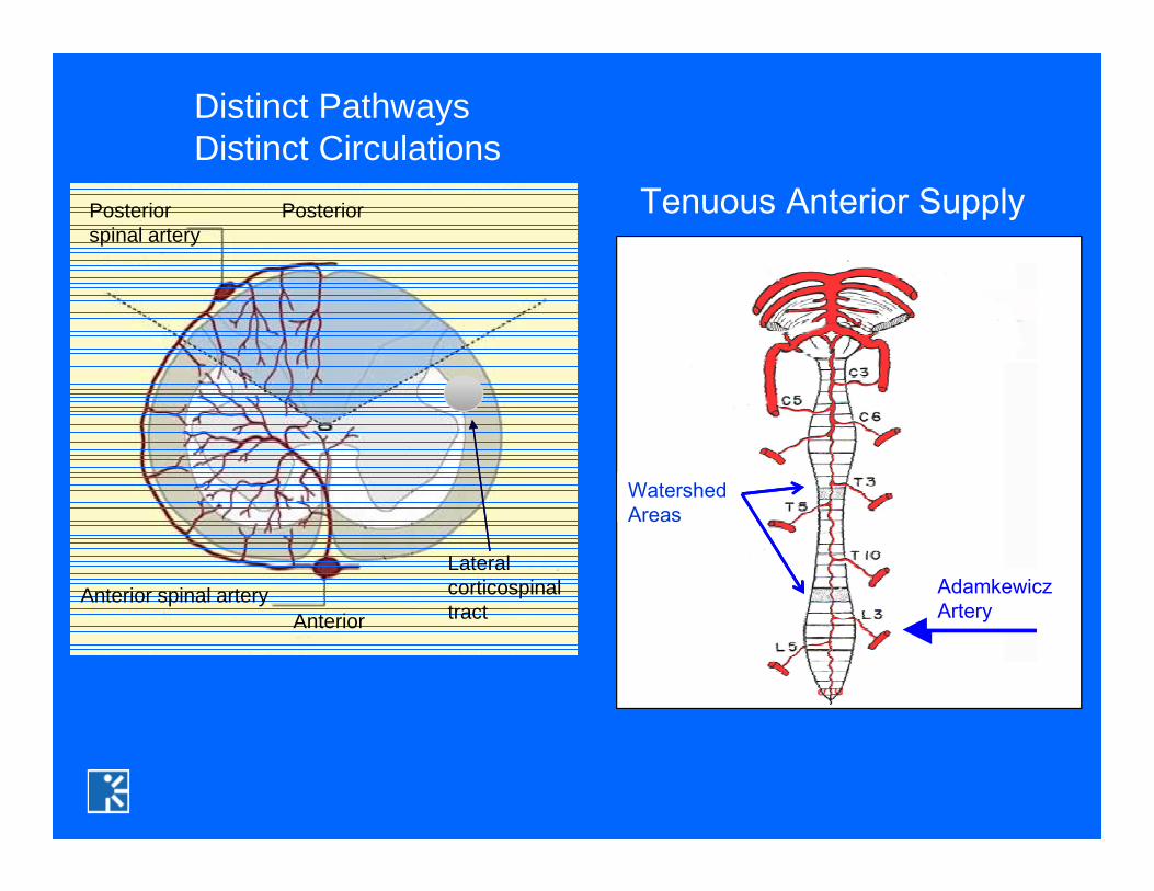
Tenuous Anterior Supply
AdamkewiczArtery
Watershed Areas
Distinct Pathways Distinct Circulations
Lateral corticospinal tract
Posterior
AnteriorAnterior spinal artery
Posterior spinal artery

Spinal deformity correctionDerotation may injure major radicular artery
Machida, M. et al. Spine. 13:1988 1119-1124. P 1122. Fig 6

Thoracoabdominal aortic aneurysm surgeryCross clamping aorta Sacrifice of significant segmental arteries
Embolization of vascular malformationsAbnormal vasculatureTerritory perfused unpredictableModality lost on test injection unpredictable

False Start: Direct Spinal Cord Stimulation
Sciatic n. record ~ scoliosis surgery
Largely Antidromic Sensory Signals
Record form peripheral nerve: Neurogenic “MEPs”
NOT Motor EPs!
Pereon Y. Electroencephalogr Clin Neurophysiol, 108:17, 1988 Fig. 6

Neurogenic “MEP”
Two false negative cases:
Unchanged neurogenic “MEP” + motor deficitMinahan et al, J Clin NP 2001.
However
Are quicker and easier than SEPs
Do provide instrumental redundancy

Intervening synapses prevent simultaneous activation of spinal cord sensory pathways

α
MEP
Muscle
Can Monitor

D-Wave I-Waves

Spinal Cord D-Wave Monitoring
Courtesy Eva Ritzl Morota N. Neurosurgery 1997;41:1327-1336 Fig. 1

D-wave Monitoring
� Compatible with complete NM blockade� Compatible with volatile anesthetics � May permit more aggressive intramedullary resection
D wave > 50% + Muscle MEP Lost � only transient deficit (Kothbauer 1998)
� Unilateral lesions hard to detect� Can’t monitor lower cord, cauda, or roots� Can’t detect spinal motor neuron ischemia� Technically difficult to position recording electrodes

(18% had >20% but <50% ↓ )
Cord Position Change � Spurious D-wave Change
Derotation �change in proximity of motor tract to epidural electrode
5% false positive (> 50% ↓) in scoliosis surgery
Ulkantan S. Clinical Neurophysiology 2006;117: 2093–2101 Fig. 5

0.2 msecThreshold
Stimulus
Train
After Phillips and Porter, Prog Brain Res ’64Baboon α Motor Neuron
αMN
∑
For Muscle MEP Monitoring, Must Fire α Motor Neuron

Stimulus Trains For Muscle MEPs
Propofol / opoid anesthesiaMacDonald J Clin Neurophysiol 2002;19:416–429
ThresholdFluctuates
Stimulus train
MEPs Exhibit Trial to Trial VariabilitySpontaneous Fluctuation of Motor Pool Excitability
After Phillips and Porter, Prog Brain Res ’64
Spinal Cordα Motor Neuron
Infrequent Testing �

20 msec
TTrain MEP MEP
Dual Train FacilitationSegmental & Suprasegmental
20 msec
Stimulus
Train

Journee HL Clinical Neurophysiology 2007;37:423-430. Fig 3
Each train:4 stimsISI 2msec100 usec125 V
Record ADM
Absolute Inhibition 35 –130 msec
Facilitation Depends on Inter-train Interval

Frei FJ Spine 2007;32:911-917
Spatial Facilitation: Medial Plantar Arch
Stim Medial Plantar Arch
10 pulses 20-60 mA0.5 msec ISI
Transcranial Electrical Stimulation
60 msec
Record MEP AH

Transcranial Electrical MEPs
Anodal stimulation
C1 “anterior” – C2 “anterior”Target LEsMay be less effective
C3 “anterior” – C4 “anterior”May be more effective for both UE and LE

Transcranial Electrical Simulation
Special purpose constant voltage, capacitive coupled stimulator
OrStandard, constant current, SEP type stimulator
Both work well; special purpose stimulators provide more rapid charge delivery and lower total charge.

Include Distal MusclesAH / APB ADM

Muscle MEP MonitoringAnesthetic Effects
• Cortex�I-wave suppression�D-waves spared
• Depress spinal motor neuron

Muscle MEP MonitoringAnesthetics
• Inhalational agents (N20, halogenated) attenuate MEPs most
• Intravenous anesthesia (propofol, dex, opioid) attenuate MEPs less
• Ketamine may be help, especially < 6 y/o (Frei, Spine 2007)

12 y/o AIS Ketamine, Propofol ISO 0.6%, No NMBObese, Labile BP
Right Tibial SSEPC4’-C3’ Cz’-C3’ Fpz-nc Cz’-nc
Right MEPAH ABP
Dual Train Adds Resiliance To Anesthetic Effects

12 y/o AIS Ketamine, Propofol ISO 0.6%, No NMBObese, Labile BP
Dual Train Adds Resiliance To Anesthetic Effects
Right MEPAH ABP

MEPs More Difficult in Young Children
Lieberman JA Anesth Analg 2006;103:316-321. Fig 5

Lyon J Neurosurg Anesthiol 2005;17:13-19 Fig. 1
“Anesthetic Fade”Threshold increases with duration of anesthesia
Independent of dose dependent depressant effectsLipid soluble and insoluble agents
Desflorane / PropofolDesflorane / N20

Neuromuscular blockade
� Attenuates MEPsMost centers avoid NMB ( 4 of 25 peds centers use, Sloan 2010)
� Improve SEPs, especially brainstem� Reduce patient movement� Reduces tongue bite incidence / severity
� Constant controlled infusion� 3-4 twitches TOF� Avoid Boluses!

Vecuronium Infusion3/4 Twitches
R MSEP L MSEP MEP
No NMB
SEPs ImprovedMEPs Still Robust
R MSEP L MSEP MEP
R TSEP L TSEP R TSEP L TSEP

12:30
14:00
14 y/o AIS PSF Instrumentation T2-L2
PropofolKetamineLidocaineIsoflurane 0.4%
Vecuronium1 mg/hr
3 / 4 twitches
Partial NMB Does Not Interfere with MEP Monitoring
C3’- C4’ Cz’- C4’ Fpz – SC5 Cz’- SC5
L Tibial SEP 3 uV/div 10 msec/div 500 uV/div 2000 uV 2000 uV/Div10 msec/div
L TA L AH L APB

13 y/o AIS PSF T4 – L1
PropofolKetamineFentanylN20 50%ISO 45%
8:35 6 MG VEC Induction
BOLUS1 MG VEC
RLGRTA RMG RAH
200 uV, 10 msec / div
4/4 Twitches
Small Vecuronium Bolus Transiently Abolishes MEP

Interpretative Criteria

Identical stimuli produce variable Muscle MEPs!
Calancie B J Neurosurg 1998;88:457-470. Fig 3
N20, narcotic, propofol

Muscle MEPs are Quite Sensitive Indicators of Cord FunctionMEP Lost @ 33% D-wave Decrease
Ependymoma T6-T6
Modified from Sala Clin Neurophysiol 2008;119:248-264

Deletis and Sala , Clin Neurophysiol 2008;119:248-264
Relationship Between D-Wave, Muscle MEP and Outcome

Proposed “Alarm Criteria”
• Complete loss• % amplitude decrease (75 – 90)• Stimulus threshold criteria• Waveform complexity criteria• Combination (Amplitude x Area x Duration x Phase)/Latency
Notify surgeon when baseline variability is exceeded.

Baseline 40 sec 8 min
Skinner SA et al. J Clin Monit Comput 2013;27:195-201
Transient MEP enhancement & EMG injury activityPorcine Thermal Cord Injury Model
Injury may open “leakage conductance” channels

Tce-MEP Safety
15,000 published & unpublished cases McDonald JCN 2002
27 Bite injuries (lip/tongue) – Bite Block1 Mandibular fracture5 Seizures (? Coincidental)5 Arrhythmias (?? Coincidental)
(we observed one additional related bradyarrhythmia)
1 Intraoperative Awareness
Charge density10,000,000 * < histological damage1000 * < used for cortical functional localization

A couple cases ……..

Left SEP Right SEP MEP
©RGE
12 y/o Neuromuscular Scoliosis TIVA / Partial NMB

Left SEP Right SEP MEP
©RGE

Left SEP Right SEP MEP
©RGE

Left SEP Right SEP MEP
©RGE

Left SEP Right SEP MEP
©RGE

Left SEP Right SEP MEP
©RGE

Left SEP Right SEP MEP
©RGE

Left SEP Right SEP MEP
©RGE

Left SEP Right SEP MEP
©RGE

Left SEP Right SEP MEP
©RGE

Post Op Exam: No Deficits
Left SEP Right SEP MEP
©RGE

Return of MEPs and SSEPs with increased blood pressure

14:3712 y/o AIS Posterior Fusion
Screws Placed Propofol/Narcotic MAP 65 mmHg
Left SEP Right SEP MEP
10 ms
0.5 uV 50 uV
©RGE
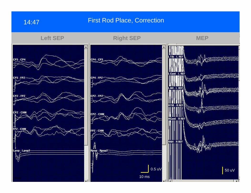
14:47 First Rod Place, Correction
Left SEP Right SEP MEP
10 ms
0.5 uV 50 uV
©RGE

MEP Loss14:52
Left SEP Right SEP MEP
10 ms
0.5 uV 50 uV
©RGE

MAP ↑ 95 mmHg14:55
Left SEP Right SEP MEP
10 ms
0.5 uV 50 uV
©RGE
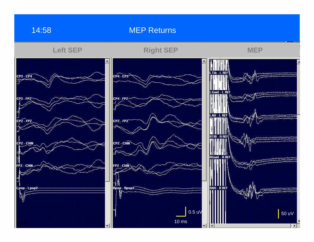
MEP Returns14:58
Left SEP Right SEP MEP
10 ms
0.5 uV 50 uV
©RGE

Second Rod Placed15:09
Left SEP Right SEP MEP
10 ms
0.5 uV 50 uV
©RGE

Nuwer 1995
Forbes1991
MacDonald2007
Schwartz2007
Multicenter RetrospectiveSurvey
Royal Ortho HospitalConsec Retro
King Faisal HRiyadhConsec Retro
CHOP, RWJSt. ChrisConsec Retro
Mixed spinal instrumentation
Mixed spinalinstrumentation
Mixed spinalInstrumentation
AIS Only
No. Cases 51,263 1168 207 1121
False Negative 0.13% 0% 1.4%* 0%True Positive 0.42% 3.6% 2.8% 0.8
“False Positive” 1.5% 6.5% 4.3% 5.2%
Motor Deficits Detected by SEP100% 100% 50% 43%
Motor Deficits Detected MEP n/a n/a 100% (4) 100% (7)
* 2 radiculopathies, 1 delayed paraparasis
What happened to SEPs between ’95 and ’07?
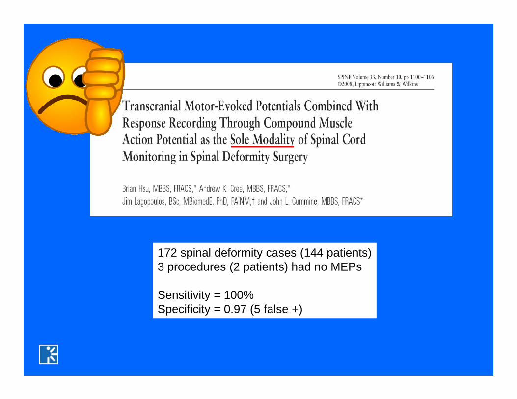
172 spinal deformity cases (144 patients)3 procedures (2 patients) had no MEPs
Sensitivity = 100%Specificity = 0.97 (5 false +)

Concurrent use of SSEPs & MEPs
Parallel Redundancy
Each is a surrogate for global cord function
Complimentary
MEPs – intermittent, but fast and very sensitive
SEPs – continuous, but respond slower and less sensitive

MEPs to Detect Root Injury??

After Mok JM Spine 2008;33:E465-E473 Table 1
Root Muscle MeanReduction
95% ConfidenceInterval
L3 Rectus Femoris 48% 23 – 73%
L4 Vastus Lateralis 40% 24 – 56%
L5 Tibialis Anterior 67% 57 – 78%
Reduction of tcMEP Amplitude After Root Ligation in Pig
Despite Multi-segmental InnervationThere’s Usually a Dominant Root

L4-5 TLIF
Pre-injury
Post-injury
↑ 26 v
↑ 54 v
Both Cases Foot Drop Post OP
Lyon J Clin Mon Comput 2010
Pre-injury
Post-injury
↑ 20 v
↑ 50 v
L5 pedicle subtraction osteotomy

Effect of increase stimulation voltage on TA amplitudeFollowing ligation of dominant root (L4, 5, or 6) in pig.
Lyon R J Clin Mon Comput 2010;24:411-448 Fig 3