Embed Size (px)
Citation preview
MOTOR AREAS EXTRAPYRAMIDAL SYSTEM
Definition:
Tracts other than corticospinal tract are
Known as extrapyramidal tracts.
The word extrapyramidal is slowly being
replaced by Corticonuclear &
corticobulbar tracts.
Components of extrapyramidal system
1. Basal Ganglia
2. Midbrain giving rise to following bulbospinal tracts:
A. Rubrospinal tract.B. Vestibulospinal Tract.C. Reticulospinal TractD. Tectspinal Tract.E. Olivospinal Tract.
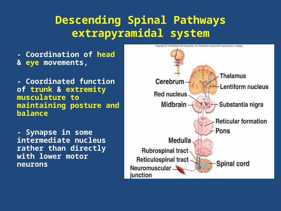
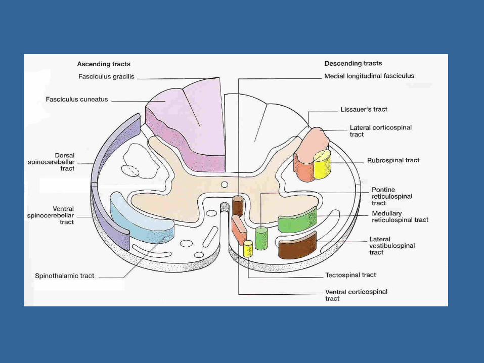
Descending Spinal Pathwaysextrapyramidal system
- Coordination of head & eye movements,
- Coordinated function of trunk & extremity musculature to maintaining posture and balance
- Synapse in some intermediate nucleus rather than directly with lower motor neurons
Reticulospinal TractThe reticular formation makes up a central corethrough much of the brainstem. It contains manydifferent nuclear groups.
Pontine and medullary nuclei projects to the anteriorhorn of the spinal cord.
Functions: influence motor functions as for examplevoluntary & reflex movement and is also responsiblefor the muscle tone.
Olivospinal Tract
It arises in the cells of inferior olive of the
medulla and is found only in the cervical region
of the spinal cord.
Function is unknown.
Role of the brain stem
Support of the Body Against Gravity.
Roles of the Reticular and Vestibular nuclei.
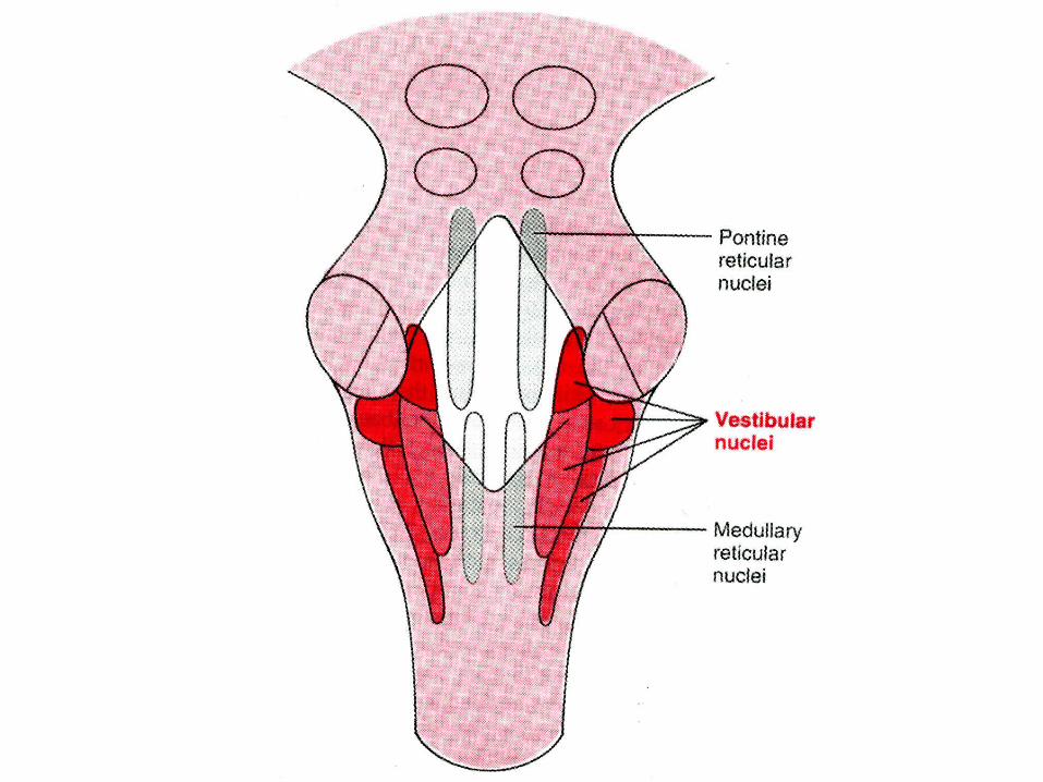
1- The vestibular nuclei:Selectively control the excitatory signals to the different antigravity muscles (the muscles of vertebral column and the extensor muscles of the limbs). to maintain equilibrium in response to signals from the vestibular apparatus.
2- The medullary reticular system:Transmit inhibitory signals to the same antigravity anterior motor neurons (medullary reticulospinal tract).
So that under normal conditions, the body muscles are normally tense.
Functions of the CerebellumAnterior and posterior lobes govern subconscious aspects of skeletal muscle movements.
Flocculondular lobe on inferior surface contributes to equilibrium and balance.
Main functions:Cerebellum evaluates movements
• smoothes movements• corrects errors• coordinates sequence• regulates posture and balance• makes possible all skilled muscular activities
11
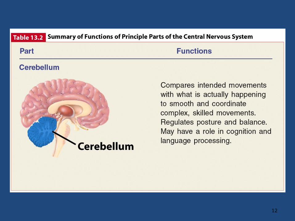
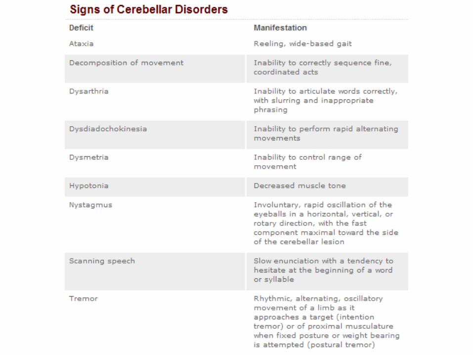
Table 13.2 part 3
12
BASAL GANGLIA AND CONTROL OF MOTOR FUNCTIONS
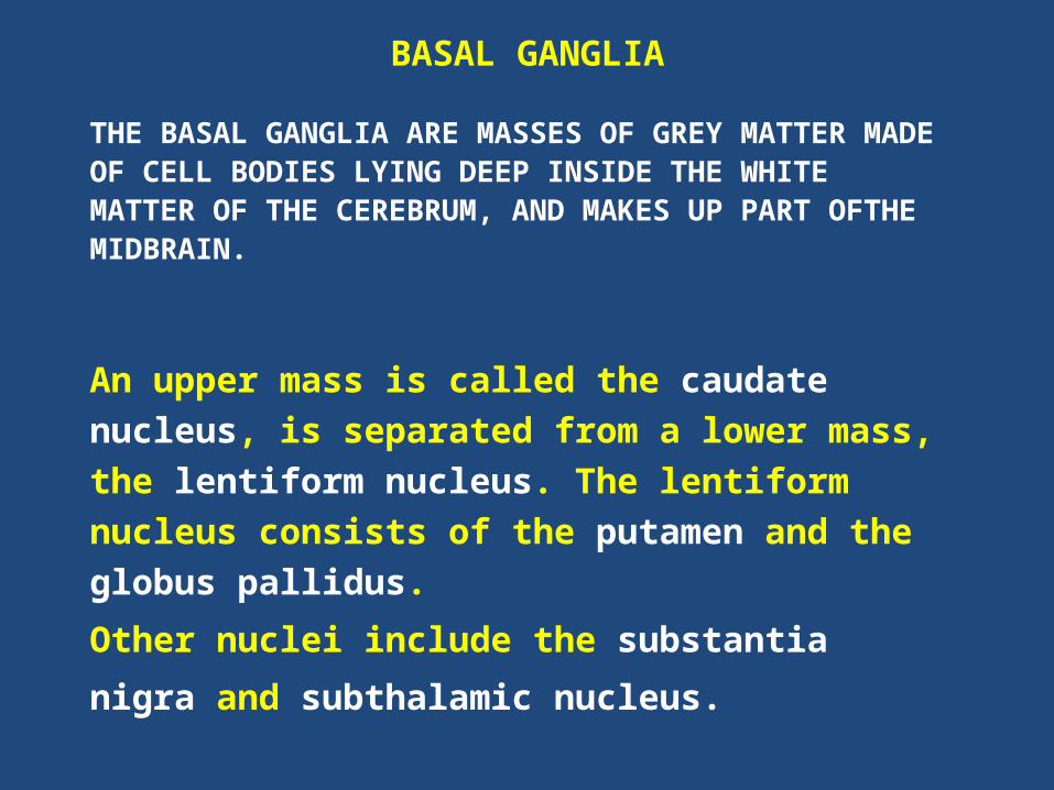
BASAL GANGLIA
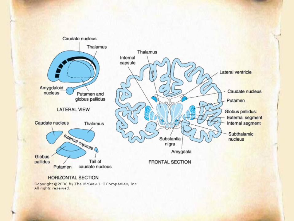
THE BASAL GANGLIA ARE MASSES OF GREY MATTER MADE OF CELL BODIES LYING DEEP INSIDE THE WHITE MATTER OF THE CEREBRUM, AND MAKES UP PART OFTHE MIDBRAIN.
An upper mass is called the caudate nucleus, is
separated from a lower mass, the lentiform
nucleus. The lentiform nucleus consists of the
putamen and the globus pallidus.
Other nuclei include the substantia
nigra and subthalamic nucleus.
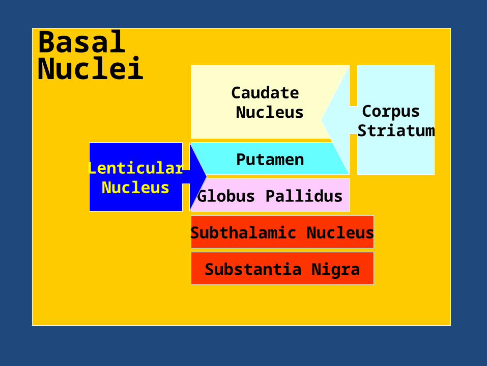
Basal Nuclei
Subthalamic Nucleus
Caudate Nucleus
Putamen
Globus Pallidus
Substantia Nigra
LenticularNucleus
Corpus Striatum
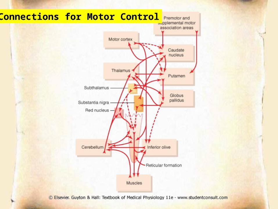
Connections for Motor Control
Functions of Basal Ganglia
• Control of movements
• Planning and programming of movements
• Cognition
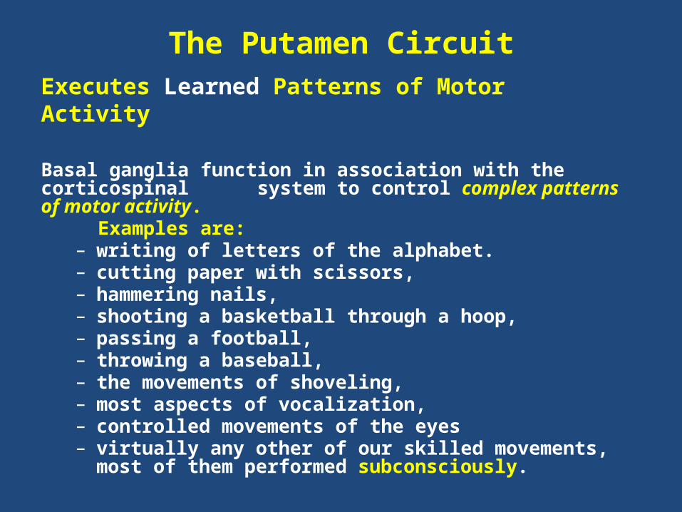
The Putamen CircuitExecutes Learned Patterns of Motor Activity
Basal ganglia function in association with the corticospinal system to control complex patterns of motor activity. Examples are:
– writing of letters of the alphabet. – cutting paper with scissors, – hammering nails, – shooting a basketball through a hoop, – passing a football, – throwing a baseball, – the movements of shoveling, – most aspects of vocalization, – controlled movements of the eyes – virtually any other of our skilled movements, most of them
performed subconsciously.
The Caudate Circuit
Cognitive Control of Sequences of Motor Patterns:
Cognition means the thinking processes of the brain, using both sensory input to the brain plus information already stored in memory. Thoughts are generated in the mind by a process called cognitive control of motor activity.
Example:A person seeing a lion approach and then responding instantaneously and automatically by:
(1) turning away from the lion, (2) beginning to run, and (3) even attempting to climb a tree.
Thus, cognitive control of motor activity determines subconsciously, and within seconds, which patterns of movement will be used together to achieve a complex goal.
The Caudate Circuit
Change the Timing and to Scale the Intensity of Movements.
Two important capabilities of the brain in controlling movement are:
(1) to determine how rapidly the movement is to be performed and
(2) to control how large the movement will be.
For instance, a person may write the letter "a" slowly or rapidly. Also, he or she may write a small "a" on a piece of paper or a large "a" on a chalkboard. Regardless of the choice, the proportional characteristics of the letter remain nearly the same.
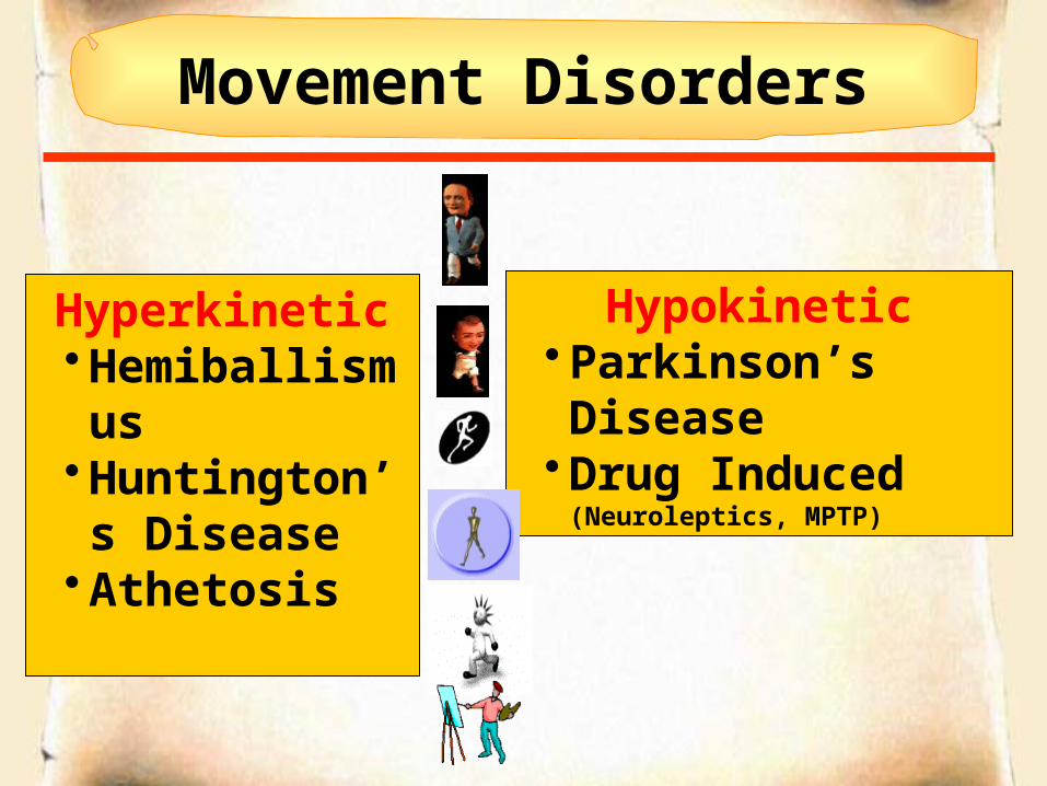
Movement Disorders
Hyperkinetic•Hemiballismus
•Huntington’s Disease
•Athetosis
Hypokinetic•Parkinson’s Disease
•Drug Induced (Neuroleptics, MPTP)
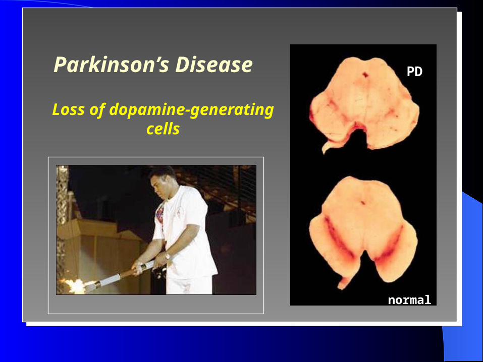
Parkinson’s Disease
Loss of dopamine-generating cells
PD
normal
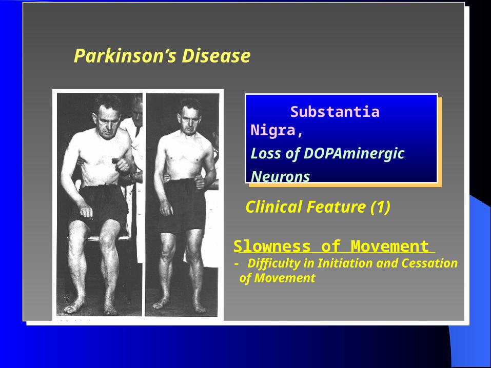
Substantia Nigra,
Loss of DOPAminergic
Neurons
Slowness of Movement- Difficulty in Initiation and Cessation of Movement
Clinical Feature (1)
Parkinson’s Disease
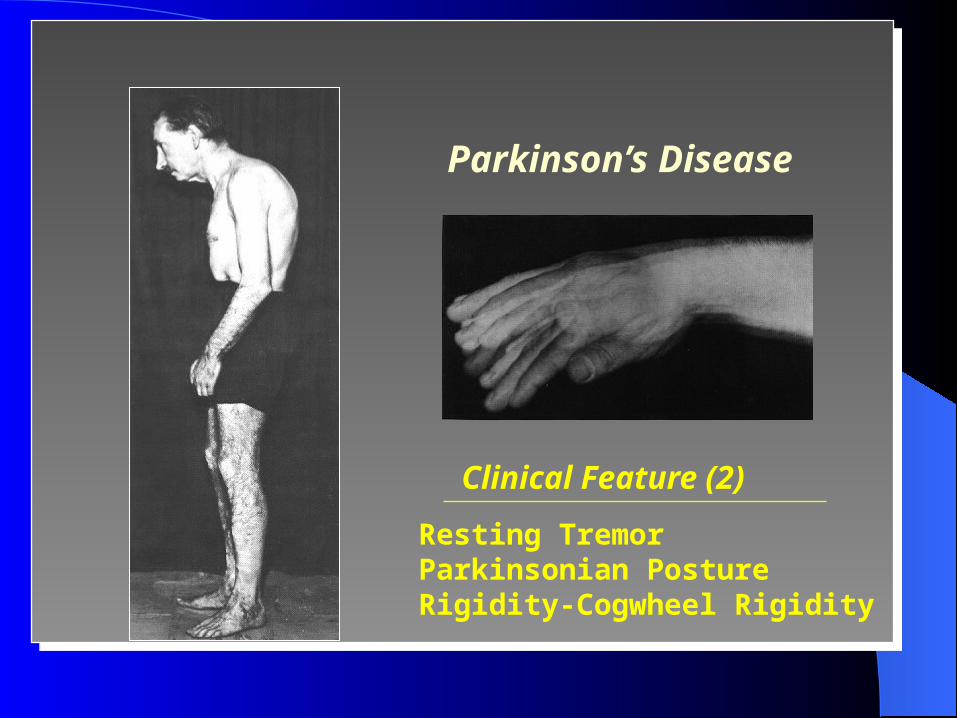
Clinical Feature (2)
Resting TremorParkinsonian PostureRigidity-Cogwheel Rigidity
Parkinson’s Disease
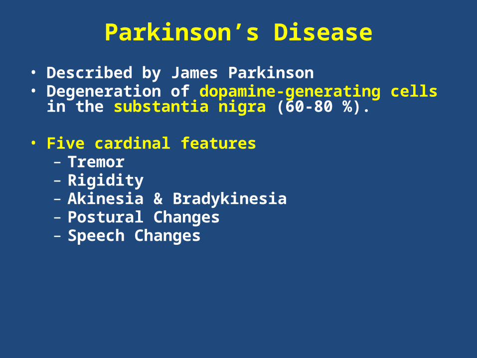
Parkinson’s Disease• Described by James Parkinson • Degeneration of dopamine-generating cells in the
substantia nigra (60-80 %).
• Five cardinal features– Tremor– Rigidity– Akinesia & Bradykinesia– Postural Changes– Speech Changes
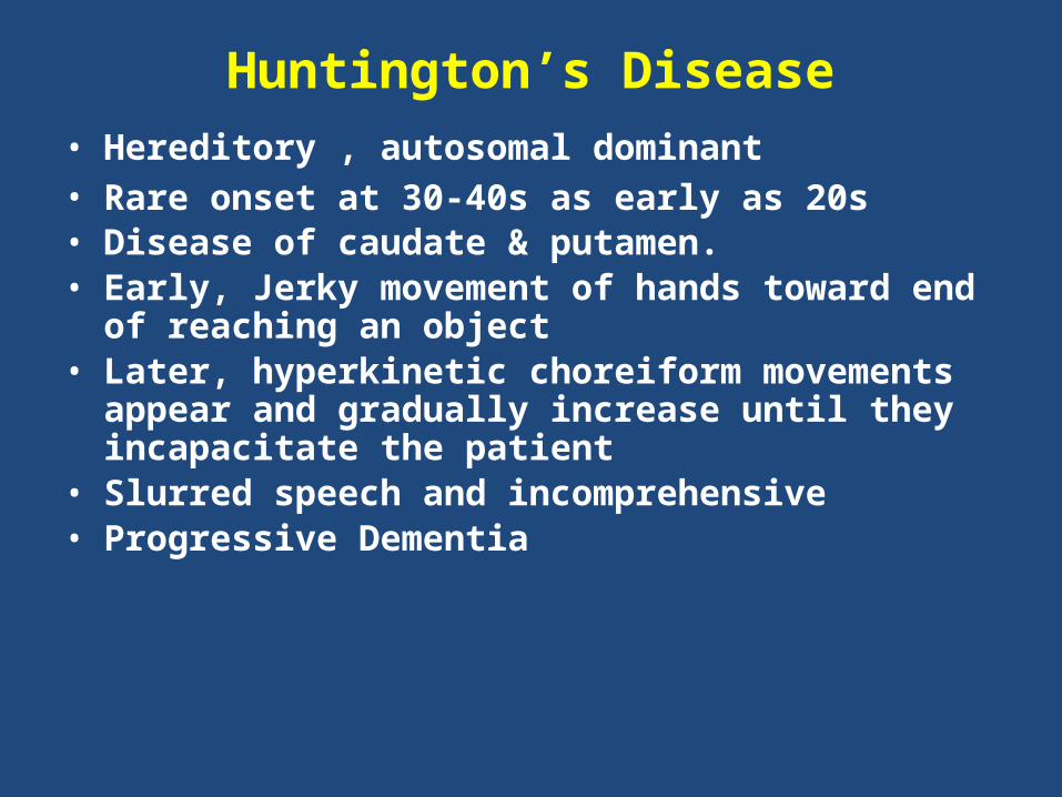
Huntington’s Disease• Hereditory , autosomal dominant• Rare onset at 30-40s as early as 20s • Disease of caudate & putamen.• Early, Jerky movement of hands toward end of
reaching an object• Later, hyperkinetic choreiform movements appear
and gradually increase until they incapacitate the patient
• Slurred speech and incomprehensive• Progressive Dementia