Embed Size (px)
Citation preview

MALE SLIDESIMPORTANT FEMALE SLIDES LECTURER’S NOTESEXTRA
LECTURE V: Physiology of the Small Intestine: Motility and Secretion
EDITING FILE

Lecture Five OBJECTIVES
1 PHYSIOLOGY OF THE SMALL INTESTINE: MOTILITY AND SECRETION
● Motility in the small intestine.● Control of intestinal motility.● Secretions of the small intestine.● Digestion of carbohydrates, proteins and fats. ● Basic principles of gastrointestinal absorption of carbohydrates, proteins and fats.● Absorption and secretion of electrolytes and water.
The movements of the small intestine can be divided into five main types of movement:
Motility in the small intestine
● Stimulus distention .● activated by enteric nervous system (ENS). ● It's a localized contraction of circular smooth
muscles that constricts (divide) the intestine into spaced segments, last for fraction of min.
● chain of sausages appearance (As one of segmentation contractions relaxes, a new often begins at points between the previous ones).
● The segmentation contractions become weak when the excitatory activity of ENS is blocked by the drug atropine1.
● Stimulus distention .A contraction ring appears, moves forwardoccur in any part of the small intestine, at velocity of 0.5 to 2.0 cm/sec, it's faster in proximal intestine slower in the terminal intestine. very weak after traveling only 3 to 5 cm, the net movement along the small intestine normally averages only 1 cm/min .● 3 - 5 hours are required for passage of chyme
from the pylorus to the ileocecal valve.
● Myenteric plexus is important for these movements
● blocked by atropine
● Its divide into:
★ Receiving segment that contract longitudinal and relax circular muscles.
★ Propulsive segment that contract circular and relax longitudinal Muscles.
Propulsive / Peristalsis contractions
They’re bursts of depolarization accompanied by peristaltic contraction that begins in empty stomach during interdigestive period (after absorption occurs)
★ travels along whole length of small intestine to reach ileocaecal valve after 1.5-2 h. Where it disappears then a new wave starts.
★ Its activity terminates as soon as food is ingested.
★ Its function is to propel/sweep any remnants in stomach & small intestine into colon during the interdigestive period( in between meals).
★ Regulated by autonomic nerves and by release of hormone motilin.
Migrating motor complex (MMC)
A wave of contraction in the alimentary canal that passes in an oral direction(i.e. upward or backwards) and propel the chyme in the opposite direction2.
Occurs between:-★ Stomach and duodenum to allow
more time for neutralization of chyme.
★ Ileum and caecum to allow more time for absorption.
★ Mostly physiological
Antiperistalsis
Segmenting / Mixing contractions
Powerful rapid peristalsis due to intense irritation of intestinal mucosa (eg: infectious diarrhea).
★ Initiated mainly by extrinsic nervous reflexes to brain stem and back to gut.
★ Sweeps the contents of intestine into the colon and thereby relieving the small intestine of irritative chyme or excessive distension.
★ Pathological
Peristaltic rush
Figure 5-1
Figure 5-2
Mixing V.S Peristalsis
Figure 5-3
FootnotesFOOTNOTES1. Parasympatholytic (sympathomimetic).2. Antiperistalsis is the opposite to peristalsis in which the bolus moves forward or in anal direction. These actions happen physiologically in the mentioned cases, but antiperistasis can occur pathologically in
cases such as: vomiting.
The significance of segmentation contraction:
★ Blend different juices with the chyme.★ Bring products in contact with absorptive
surfaces.
The significance of peristaltic contractions:
★ Organize propulsion of material over variable distances within the intestinal lumen.

Lecture Five 2 PHYSIOLOGY OF THE SMALL INTESTINE: MOTILITY AND SECRETION
CONTROL OF INTESTINAL MOTILITY
NEURONAL● Vagal(parasympathetic):
excitation increases intestinal and villous movements.
● Sympathetic:excitation decreases intestinal and villous movements.
Gastroileal reflex:Initiated by gastric distension mediated by vagus nerve. Impulses are conducted through myenteric plexus to initiate a fast peristaltic wave passing to the ileum. The ileocaecal valve relaxes allowing chyme to pass into cecum.
Source Of Small Intestinal Secretions
HORMONAL● Gastrin, CCK, insulin and serotonin
stimulate intestinal motility. ● Gastrin and CCK2 relax ileocaecal
sphincter.● Secretin2 and glucagon inhibits
intestinal motility and contract ileocaecal sphincter.
● Motilin secreted from duodenum stimulates intestinal motility and regulate MMC.
*Remember that Villikinin stimulates movement of the villi.
The villous movement consists of fast shortening and slow lengthening as well as side to side movements.★ Villous contractions are initiated by local nervous reflexes in response to chyme in small intestine. ★ They are stimulated by villikinin1 hormone released by intestinal mucosa when it comes in contact with
digestive products.★ They facilitate absorption and lymph flow from central lacteals into lymphatic system.
Movement Of Villi
Brunner’s Glands Crypts of Lieberkühn★ Located in the wall of the first few
centimeters of the duodenum.
★ Secrete large amounts of alkaline mucus to protect the mucosa, which contains a large amount of bicarbonate ions.
stimulated by :● secretin, tactile (chyme contacts brushborder) and vagal
stimulation, irritating stimuli on the duodenal mucosa.inhibited by:● sympathetic stimulation.
★ Located in small pits which lie between intestinal villi★ Secrete Intestinal juices (Succus Entericus).The surfaces crypts and villi are covered by an epithelium composed of 2 types of cells: ➔ goblet cell secrete mucus.➔ enterocytes secrete large quantities of H2O and electrolytes. And
reabsorb H2O & end-products of digestion over the surfaces of adjacent villi.
★ The enterocytes of the mucosa contain the following digestive enzymes:
➔ Aminopeptidases, Oligopeptidases, Intracellular di / tri peptidases for splitting small peptides into amino acids.
➔ sucrase, maltase, isomaltase, lactase ,a-dextrinase for splitting disaccharides into monosaccharides.
➔ Small amounts of intestinal lipase for splitting neutral fats into glycerol and fatty acids.
➔ Nucleotidases for splitting nucleotides into purine and pyrimidine bases, phosphoric acid and pentose sugar.
★ Intestinal juice participates in the neutralization of acid chyme delivered from stomach.
At a volume of 1800 ml/day (Composition: 0.6 % organic (enzymes & mucus), 1 % inorganic (electrolytes) substance and a pH: 7.5-8 .★ Most of the enzymes are found either in the brush border or in the
cytoplasm of the enterocytes.★ enteropeptidase and amylase secreted into the lumen.
stimulated by :● Distension, tactile and irritating stimuli.● Hormones as gastrin, secretin, CCK & glucagons & enterocrinin(produced by small
intestine).Inhibited by: sympathetic stimulation.
FOOTNOTES1. Suffix kinin = stimulator.2. Recall that both Secretin and CCK increase pancreatic secretions, yet it’s notable here that they’re opposing in action.

➔ Digestion of CarbohydrateIn the Mouth and Stomach:
★ The ptyalin (an α-amylase) enzyme in saliva hydrolyzes starch into the disaccharide maltose and other small polymers of glucose.
★ The starch digestion sometimes continues in the fundus and body of the stomach for 1 hour before the food becomes mixed with the stomach secretions.
In the Small Intestine (by Pancreatic Amylase):
★ Pancreatic secretion has α-amylase (identical in its function with the α-amylase of saliva but is several times as powerful).
★ within 15 to 30 minutes after the chyme empties into the duodenum and mixes with pancreatic juice, carbohydrates will have become digested.
★ The carbohydrates are almost converted into maltose and/or other very small glucose polymers before passing beyond the duodenum or upper jejunum.
The enterocytes lining the villi contain 4 enzymes (lactase, sucrase, maltase, and a-dextrinase , isomaltase), which are capable of splitting the disaccharides lactose, sucrose, and maltose plus other small glucose polymers, into their constituent monosaccharides.
These enzymes are located in the enterocytes covering the intestinal microvilli brush border, so disaccharides are digested as they come in contact with these enterocytes.
➔ Absorption of Carbohydrate
Lecture Five 3 PHYSIOLOGY OF THE SMALL INTESTINE: MOTILITY AND SECRETION
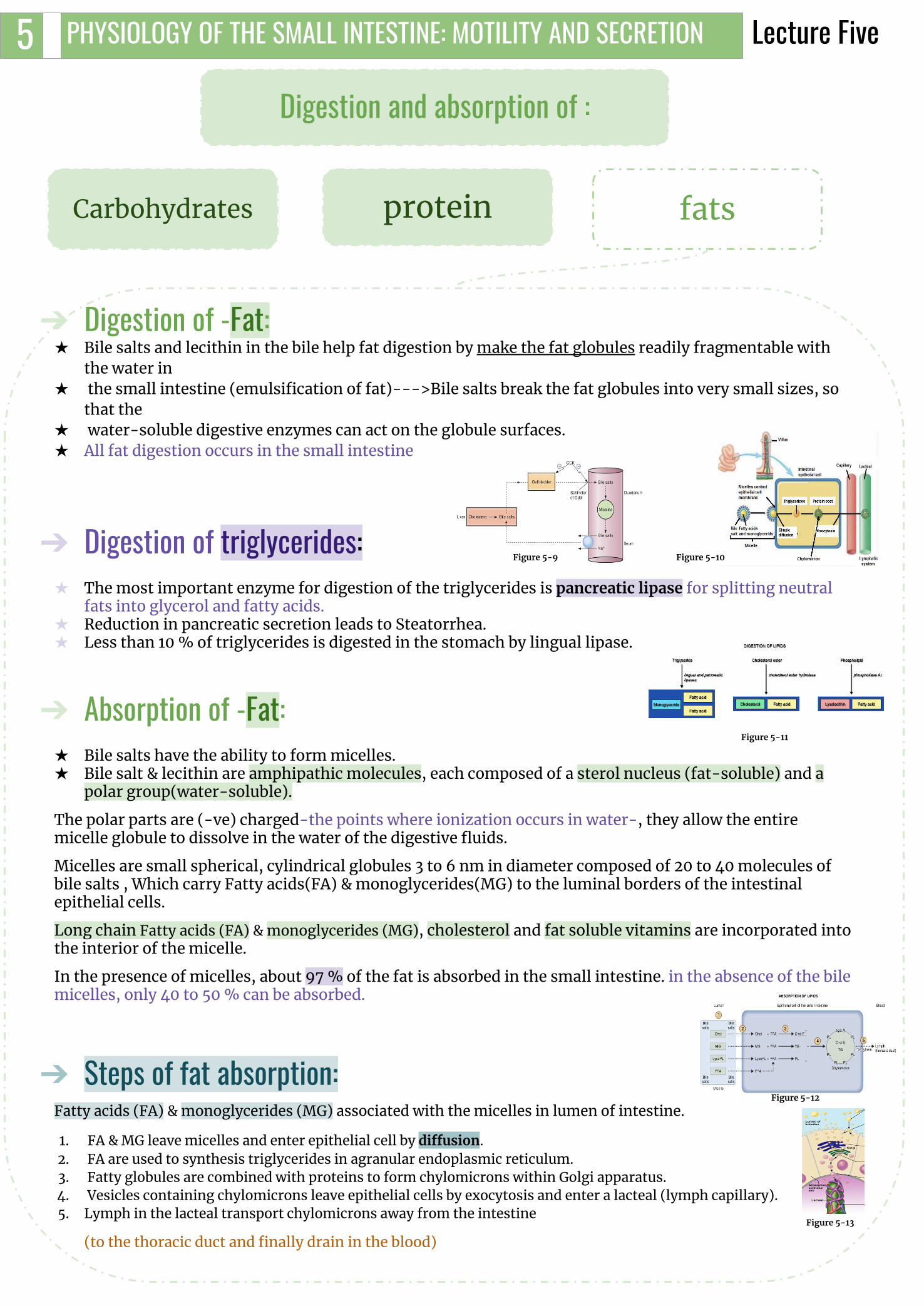
Digestion and absorption of :
Carbohydrates Protein Fats
Figure 5-5
CLINICAL RELEVANCE● Glucose enters the cell with Na+ on the
SGLT symporter and exits on GLUT2. ● Fructose enters on GLUT5 and exits on
GLUT2.Both of which can be blocked to produce therapeutic action2.
Figure 5-6
FOOTNOTES1. Meat and other products contain nucleic acids (pentose + nitrogen ring+ phosphate group), these nucleotides can be broken by pancreatic nuclease into nucleosides (pentose +
nitrogen ring), nucleosides are further hydrolysed by intestinal nucleosidase into a free nitrogen ring and a pentose sugar (ribose), this pentose sugar can then be absorbed. 2. glucose excretion can be induced by blocking the activity of the renal sodium-glucose cotransporter 2 (SGLT-2) which corrects hyperglycemia independently of
insulin.
All the carbohydrates in the food are absorbed in the form of monosaccharides only a small fraction are absorbed as disaccharides.
● Glucose and galactose absorption occurs in a co-transport mode with active transport of Na+ (2ry active transport) (fastest).
● Fructose is independent on Na+ but it transports in luminal membrane via facilitated diffusion.
● Pentose is transported by passive diffusion1 (slowest)Figure 5-4

Lecture Five 4 PHYSIOLOGY OF THE SMALL INTESTINE: MOTILITY AND SECRETION
Digestion of Proteins:Digestion of protein in the stomach :
★ Pepsin is the important peptic enzyme of the stomach (active at a pH:2-3, inactive at a pH above about 5.0). Recall that the pH of the stomach averages around 2.0 - 3.0.
★ pepsin have the ability to digest the protein collagen.★ Collagen is a major constituent of the intercellular connective tissue of meats therefore, for the
digestive enzymes of the digestive tract to penetrate meats and digest the other meat proteins, it is first necessary that the collagen fibers be digested.
★ Pepsin initiates the process of protein digestion, usually providing 10 to 20 % of the total protein digestion.
Digestion of protein in the intestines:A small percentage of proteins are digested to amino acids(AA) by the pancreatic juices.
Both trypsin and chymotrypsin split protein molecules into small polypeptides; carboxypolypeptidase then cleaves individual AA from the carboxyl ends of the polypeptides.
Proelastase is converted into elastase, which then digests elastin fibers that partially hold meats together.
Most remain as dipeptides and tripeptides to be digested by Peptidases (di/tri peptidases) in the Enterocytes mainly in the duodenum and jejunum(intracellularly).
Most protein digestion occurs in the duodenum and jejunum by aminopeptidases, oligopeptidases and Di/tri peptidases .
➔ Absorption of Proteins:● Proteins are absorbed in the form of dipeptides, tripeptides, and a few free amino acids.● D- AA1 are transported by passive diffusion.● L- AA 1 are transported by 2ry active transport.● Di and tripeptides cross the brush border by active transport protein carrier. Then they're hydrolyzed by
brush border and cytoplasmic oligopeptidases.● AA leaves the cell at the basolateral membrane by facilitated transport.
Digestion and absorption of :
Carbohydrates Protein Fats
FOOTNOTES1. Only L-amino acids are manufactured in cells and incorporated into proteins. Some D-amino acids are found in the cell walls of bacteria, but not in bacterial proteins
Figure 5-7
Figure 5-8
Lecture Five 5 PHYSIOLOGY OF THE SMALL INTESTINE: MOTILITY AND SECRETION
➔ Digestion of -Fat:★ Bile salts and lecithin in the bile help fat digestion by make the fat globules readily fragmentable with
the water in★ the small intestine (emulsification of fat)--->Bile salts break the fat globules into very small sizes, so
that the★ water-soluble digestive enzymes can act on the globule surfaces.★ All fat digestion occurs in the small intestine
➔ Digestion of triglycerides:★ The most important enzyme for digestion of the triglycerides is pancreatic lipase for splitting neutral
fats into glycerol and fatty acids. ★ Reduction in pancreatic secretion leads to Steatorrhea.★ Less than 10 % of triglycerides is digested in the stomach by lingual lipase.
➔ Absorption of -Fat:★ Bile salts have the ability to form micelles.★ Bile salt & lecithin are amphipathic molecules, each composed of a sterol nucleus (fat-soluble) and a
polar group(water-soluble).
The polar parts are (-ve) charged-the points where ionization occurs in water-, they allow the entire micelle globule to dissolve in the water of the digestive fluids.
Micelles are small spherical, cylindrical globules 3 to 6 nm in diameter composed of 20 to 40 molecules of bile salts , Which carry Fatty acids(FA) & monoglycerides(MG) to the luminal borders of the intestinal epithelial cells.
Long chain Fatty acids (FA) & monoglycerides (MG), cholesterol and fat soluble vitamins are incorporated into the interior of the micelle.
In the presence of micelles, about 97 % of the fat is absorbed in the small intestine. in the absence of the bile micelles, only 40 to 50 % can be absorbed.
➔ Steps of fat absorption:Fatty acids (FA) & monoglycerides (MG) associated with the micelles in lumen of intestine.
1. FA & MG leave micelles and enter epithelial cell by diffusion.2. FA are used to synthesis triglycerides in agranular endoplasmic reticulum.3. Fatty globules are combined with proteins to form chylomicrons within Golgi apparatus.4. Vesicles containing chylomicrons leave epithelial cells by exocytosis and enter a lacteal (lymph capillary).5. Lymph in the lacteal transport chylomicrons away from the intestine
(to the thoracic duct and finally drain in the blood)
Digestion and absorption of :
Carbohydrates protein fats
Figure 5-9 Figure 5-10
Figure 5-11
Figure 5-12
Figure 5-13

Lecture Five 6 PHYSIOLOGY OF THE SMALL INTESTINE: MOTILITY AND SECRETION
● Fat-soluble vitamins (A, D, E, & K) are incorporated into micelles and absorbed along with other lipids.
● water-soluble vitamins (C, B1, B2, B6, and folic acid) most are absorbed by Na+-dependent cotransport mechanisms.○ Vitamin B12 is absorbed in the terminal part of ileum and requires
intrinsic factor. So in the following cases deficiency occurs.
- Ileal resection → vitamin B12 deficiency1.
-Gastrectomy → loss of intrinsic factor → pernicious anemia.
Electrolytes and H2O cross intestinal epithelial cells by either transcellular or paracellular route( fig 5-13)
The permeability of the tight junctions varies with the type of epithelium.
● Leaky epithelia are in the small intestine and gallbladder.● A tight epithelium is in the colon.
Vitamins absorption
Figure 5-15
Water And Electrolytes Secretion & Absorption
Figure 5-16
Figure 5-17Figure 5-18, Total Secreted + Ingested = 9000 ml/day
Total Absorbed = 8900 ml/day (8500ml from S-intestine, 400ml from colon), so only 100 ml/day gets excreted.
● The absorptive surface of the small intestinal mucosa shows many folds called valvulae conniventes, well developed in the duodenum and jejunum. They increase the surface area of the absorptive mucosa X 3-fold .
● The presence of villi on the mucosal surface enhances X 10-fold . ● The epithelial cell on each villus is characterized by a brush border,(Provides
the surface area equivalent to a tennis court) consisting of as many as 1000 microvilli (X 20- fold).
All these increase the intestinal surface 600x
Absorptive surface
Figure 5-14, Longitudinal section of the small intestine, showing the valvulae conniventes covered by villi
FOOTNOTES1. Recall from the CNS block, that the absorption of vitamin B12 occurs in the small intestines. While the production occurs in the large intestines and that’s why we
need to supplement it in the diet

Na+ absorption Cl- absorptionNa+ moves into the intestinal cells by the following mechanisms:
★ Passive diffusion.★ Na+ - glucose or Na+ - amino acid
co-transport.★ Na+ - Cl- exchange.★ Na+ - H+ exchange1.
The next step is osmosis of water into the paracellular spaces; because a large osmotic gradient has been created by the elevated concentration of ions in the paracellular space.
Aldosterone Enhances Na+ Absorption.
This effect is important in the colon because it allows virtually no loss of NaCl and water.
Cl- absorption accompanies Na+ absorption by the following mechanisms:
★ Passive diffusion★ Na+ - Cl- cotransport★ Cl- - HCO- exchange
Ca++ absorptionLow plasma Ca++ → Elevated Parathyroid hormone activates Vitamin D:-
25-hydroxy-vitamin D3 → 1,25-dihydroxy-vitamin D3
Which stimulates synthesis of Calcium binding protein and Calcium- ATPase in enterocytes
absorption and secretion of K+ Secretion of HCO3 in the ileum• K+ is absorbed in the small intestine by passive diffusion.• K+ secretion in the colon is stimulated by aldosterone.• Excessive loss of K+ in diarrheal fluids causes hypokalemia.
The epithelial cells on the surfaces of the villi in the ileum and large intestine have a special capability of secreting bicarbonate ions in exchange for absorption of Cl-.
So it provides alkaline bicarbonate ions that neutralize acid products formed by bacteria in the large intestine.
Lecture Five 7 PHYSIOLOGY OF THE SMALL INTESTINE: MOTILITY AND SECRETION
★ Glucocorticoid = absorption of H2O & ions (small & large intestine).
★ Somatostatin = absorption of H2O & ions (ileum & colon).
★ Epinephrine = absorption of NaCl (ileum).
★ Aldosterone = synthesis of Na+ channels (colon).
Hormonal control of absorption & secretion
FOOTNOTES1. The sodium-hydrogen exchanger/antiporter is primarily responsible for maintaining the balance of sodium. It is also indirectly linked to buffering of blood pH by
means of absorbing Na in exchange with H which binds with HCO3- to make up H2CO3 which then dissociates into water and CO2 and finally enter the ducal cell to form HCO3- that gets secreted into the lumen, hence the buffering action. (And yes, HCO3- can’t simply pass).

1. The migrating motor complex is triggered by the release of which of the following?A. Motilin B. NOC. CCK
2. An example of a pathological Antiperistaltic movement:
A. Duodenum —> StomachB. Stomach—> esophagus C. Caecum—> Ileum
3. The propulsive contractions are:A. Slower in proximal intestines and stops at terminal intestines.B. Slower in the terminal and faster at the proximal intestines.C. Speed is equal at both ends.
4. Amino acids leave the cell at the basolateral border via:A. Facilitated diffusionB. Secondary active transport C. Primary active transport
5. Which if the following stimulate intestinal motility and relax the ileocecal sphincter?A. CCKB. SecretinC. Gastrin
6. Most protein digestion occur in:A. Large intestines B. MouthC. Duodenum
SHORT ANSWER QUESTIONSQ1: What are the mechanisms of Na+ absorption?Q2: What are the functions of the following enzymes: Maltase, Sucrase, lactase?
1. Passive diffusion, Na+-glucose or Na+-amino acid co-transport, Na+-Cl- exchange & Na+-H+ exchange.
2.- Maltase: a brush border enzyme that splits maltose into 2 glucose.- Sucrase: a brush border enzyme that splits sucrose into glucose &
fructose.- Lactase: a brush border enzyme that splits lactose into glucose &
galactose.
ANSWER KEY: A, B, B, A, A&C, C

FEMALE PHYSIOLOGY CO-LEADERS Maha Alnahdi, Taif Alshammari
MALE PHYSIOLOGY CO-LEADERS Nayef Alsaber, Hameed M. Humaid
REFERENCES- Guyton and Hall Textbook of Medical Physiology
- Ganong’s Review of Medical Physiology
Nujud Alabdullatif


![Small Intestine Prof. K. Sivapalan.. 2013Small Intestine2 Small Intestinal Motility Segmental movement [mixing 7/min in ileum] Peristalsis [propagation](https://img.pdfslide.us/doc/110x75/56649c915503460f9494c7b5/small-intestine-prof-k-sivapalan-2013small-intestine2-small-intestinal.jpg)