Embed Size (px)
Citation preview
Received June 1, 2015, accepted July 15, 2015, date of publication September 16, 2015, date of current version December 4, 2015.
Digital Object Identifier 10.1109/ACCESS.2015.2478701
Children Absorb Higher Doses of RadioFrequency Electromagnetic RadiationFrom Mobile Phones Than AdultsROBERT D. MORRIS1, L. LLOYD MORGAN2, AND DEVRA DAVIS11Environmental Health Trust, Teton Village, WY 83025, USA2Environmental Trust, Berkeley, CA 94709, USA
Corresponding author: Robert D. Morris ([email protected])
This work was supported by the Community Foundation of Jackson Hole.
ABSTRACT The greater vulnerability of children to the effects of environmental hazards has raised concernsabout their exposure to and the resultant absorption of mobile phone radiation. Foster and Chou (2014)reviewed published studies that used computer models of radio-frequency electromagnetic fields to estimateand compare the tissue dose rate in the heads of children and adults using mobile phones. Their reviewconfuses exposure with absorption, and the study results conclude erroneously that children are not moreexposed than adults. We show that their review was not executed systematically. There are discrepanciesbetween text summaries and the graphed ratios of child: adult peak special specific absorption rate, inline with the author’s hypothesis that children have the same or lower tissue dose than adults. Even theunderlying precept of their review is flawed, as the results of deterministic models are treated as randomvariables. In fact, model results are entirely determined by the underlying assumptions and the structure ofthe model. Models are included in their unsystematic review that do not consider differences in dielectricconstants among different tissues, or across ages, while other models that consider such differences are notincluded. In this paper, we discuss the differences between exposure and tissue absorption and re-examinethe results presented by Foster and Chou. Based upon our review, we suggest an alternative interpretation ofthe published literature. In an Appendix, we discuss modeling of tissue dose in the context of governmentalsafety certification processes.
INDEX TERMS Blood-brain-barrier (BBB), certification process, children, dosimetry, exposure-limits,EMR (electromagnetic radiation), FACTS (Finite difference time domain Anatomically Correct TissueSpecific), FDTD (finite-difference, time-domain), RF (radio frequency) SAM (specific anthropomorphicmannequin), SAR (specific absorption rate), virtual family (VF), WTDs (wireless transmitting devices).
I. INTRODUCTIONIn recognition of the unique sensitivity of children toenvironmental health hazards, the U.S. Environmental Pro-tection Agency, in 1996, adopted a National Agenda toProtect Children’s Health from Environmental Threats [1],and in 1997 established an Office of Children’s Health [2]dedicated to determining how to ensure that environmentalpolicies adequately protect children. Although considerableattention has been paid to reducing chemical hazards inenvironments frequented by the young, relatively little focushas been applied to physical hazards such as those posedby radio-frequency electromagnetic radiation (RF-EMR)emitted by mobile phones and other wireless transmittingdevices (WTDs).
To the extent that RF-EMR poses a risk, is that riskuniquely elevated in children? Foster and Chou [3] argue
that children have the same exposure to the brain as adults,and face equal risks, based on their review of studies com-paring the intracranial dose rates of absorbed RF-EMR inadults and children. Others, for example Gandhi [4], contendthat children have proportionally greater intracranial peaktissue dose given their thinner skulls and the higher watercontent of their cerebral tissues. Moreover, the rapid rate ofgrowth and development, and incomplete myelination of thebrain, make children uniquely susceptible to the effects ofradiation [5], [6].
The current study considers the methods used byFoster and Chou [3] to identify and abstract data from rel-evant studies. The results of these studies, as presented byFoster and Chou, were examined in detail in an effort tounderstand why their conclusions differ from those drawn byother authors.
VOLUME 3, 20152169-3536 2015 IEEE. Translations and content mining are permitted for academic research only.
Personal use is also permitted, but republication/redistribution requires IEEE permission.See http://www.ieee.org/publications_standards/publications/rights/index.html for more information.
2379
R. D. Morris et al.: Children Absorb Higher Doses of Radio Frequency Electromagnetic Radiation
II. EXPOSURE VERSUS DOSEThe distinction between exposure and dose is fundamen-tal to environmental health research. When considering apotentially toxic substance, exposure is the amount of thatsubstance that is ingested, inhaled, or deposited on the body.In the case of radiation, such as RF-EMR, exposure is theduration and intensity of radiation that reaches the surface ofthe body. The term ‘‘tissue dose,’’ on the other hand, refersto the amount of radiant energy absorbed by a specific tissue,and the ‘‘dose rate’’ is the energy absorbed per unit time.
The Specific Absorption Rate (SAR), which is the focusof the Foster and Chou analysis, is a measure of the tissuedose rate of microwave radiation, not exposure. The doseis the specific absorption (SA), typically measured inJoules per kilogram (J/kg). The reports assembled byFoster and Chou compare estimated dose rates in the headsof adults and children using simulation models that, bydesign, have the same exposure. Thus the flaws in this paperbegin with its title, ‘‘Are Children More Exposed to RadioFrequency Energy From Mobile Phones Than Adults?’’ Thisis an important question, but the topic their paper actuallyreviews should be restated as: are peak RF-EMR doses frommobile phones higher in children than adults? Thus, thepaper’s title conflates exposure and dose.
III. REVIEW METHODOLOGYRecognizing that this is an article on tissue dose rate,the following section considers whether Foster and Chouprovide a systematic, comprehensive, meaningful, and objec-tive review consistent with current scientific practice.
A literature review, whether qualitative or quantitative,involves, at a minimum, three principal steps: 1) literaturesearch and report selection, 2) abstraction of study attributesand results, and 3) analysis of abstracted data. The use ofmeta-analysis is desirable whenever possible [7]–[9].
A. STUDY SELECTIONThe validity of a scientific review is rooted in the comprehen-sive identification of relevant research. Missing or exclud-ing potentially relevant studies opens the door to bias, butbibliographic search strings and methods used to assemblethe Foster and Chou review were not presented. Studies wereselected ‘‘that permit a direct comparison of SAR in heads ofchildren and adults from use of mobile phones . . . limitedto dosimetric issues [of] age-related differences . . . [3].’’Twenty-three studies were reviewed, all of which use finitedifference time domain (FDTD) calculation methods.
The major differences among the selected studies involvethe design of the simulation models, which have evolvedsteadily with the growth in computing power. Early modelswere relatively simplistic, using spheres [10] and cylindersas crude approximations of the human head. All of theseearly models required the simplifying assumption that humantissue was a uniform, undifferentiated substance, character-ized by a single set of dielectric constants, and child headmodels were merely scaled down adult models. As a result,
the only differences between the tissue dose in adult andchild models resulted from either the position of the phoneor the penetration into additional anatomical regions resultingfrom the smaller head size. Refinements in recent years usingthe Talairach atlas (available since 1988) allow for modelimprovements based on high-resolution characterization ofbrain tissues, including adjustments for higher water contentin younger brains, which, as a result, absorb RF-EMR moreavidly [11].
In 2005, investigators at the U.S. Food and Drug Adminis-tration working together with researchers at the Swiss IT’ISFoundation developed a set of digital human models of theentire body, not just the head, with organs and tissues inanatomically correct locations [12]. These models, whichbecame known as the Virtual Family (VF), incorporatedtissue-specific parameters for conductivity and permittivity,and a series of researchers have introduced other FDTDAnatomicallyCorrect,Tissue Specific (FACTS)models [13].
By coupling data from high-resolution MRI scans of abroad range of subjects, researchers around the world, includ-ing teams in Brazil [14] and Korea [15], have added to thelibrary of available FACTS models. Currently the VF hasmore than a dozen different models, including male andfemale children of various ages, men, women and even preg-nant women at 1, 3, 7 and 9 months gestation [13]. Additionalmodels continue to be introduced. Absorption related param-eters are derived from empirical measurements of dielectricparameters in animal tissues of various ages immediatelyafter death. Themodels andWTD antennae can be configuredin any possible position, to predict the effects of exposure oftissues of various sensitivities.
Foster and Chou acknowledge that, prior to the introduc-tion of FACTS models, simulations ‘‘were not designed toexplore the effects of human variability on SAR, which onthe basis of [36] and other studies are considerable.’’
Despite the fact that this statement seems to suggest thatthese older models would not be suited to identifying dif-ferences in tissue dose, Foster and Chou included manysuch studies. Of the 22 distinct studies (2 are companionstudies [24], [25]) in their Table 2, only ten used FACTSmodels [20]–[24], [26], [28], [29], [31], [35]. Foster and Choulumped these FACTS models together with ten older, lesssophisticated models spanning 19 years (1994-2012), whichsimply used scaled down, non-FACTS models of adult headsto model children without any consideration for the models’limitations.
B. DATA ABSTRACTIONTo summarize a series of studies concisely, reviewersmust distill the findings of any particular study into afew numbers. If the process of abstracting three or fourstatistics to characterize an entire paper is not done accordingto a clear, systematic protocol with meticulous attention todetail, a strong potential for bias is introduced.
The papers that were selected by Foster and Choureported modeling exercises that differed in important ways.
2380 VOLUME 3, 2015
R. D. Morris et al.: Children Absorb Higher Doses of Radio Frequency Electromagnetic Radiation
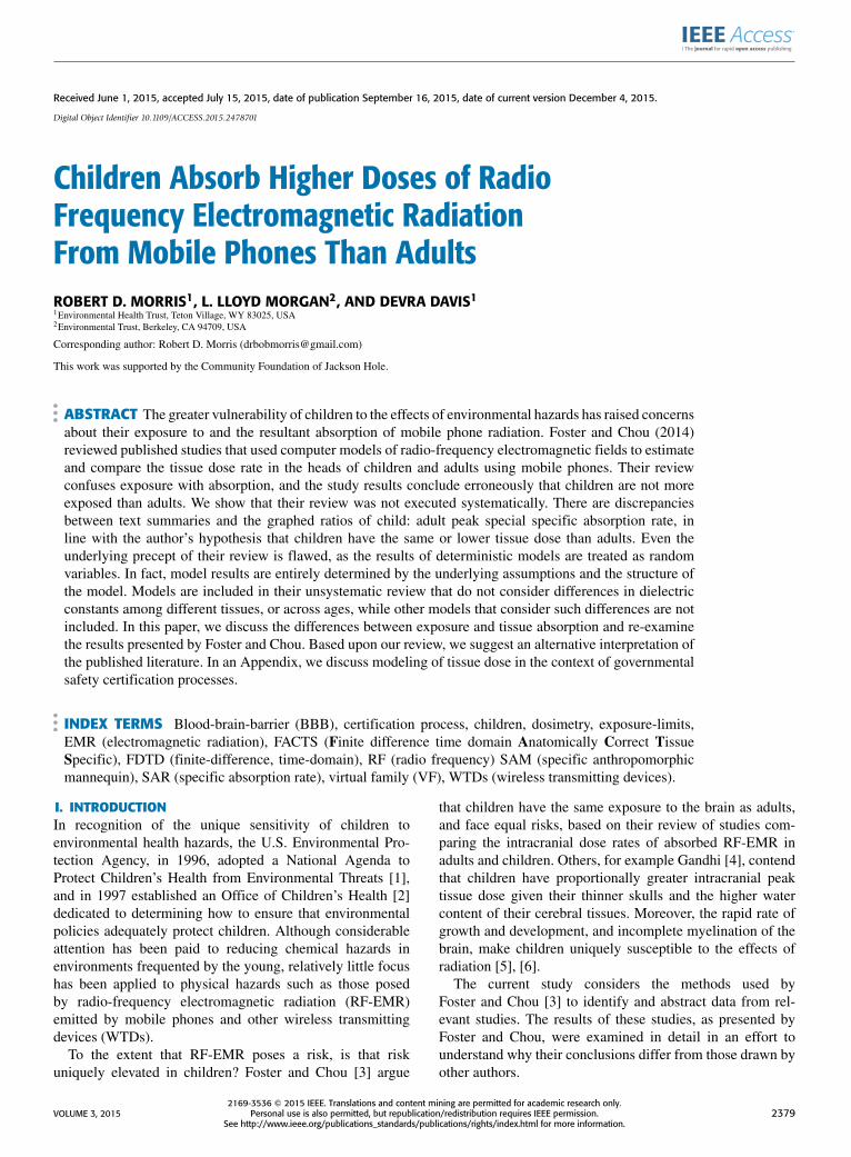
TABLE 1. Comparisons of qualitative study results from Foster and Chou [3] as summarized in their Table 2 and the quantitative results depictedin their Figure 1.
These include: the precise positioning and nature of the radi-ation source; the ages of the simulated heads; the degree towhich different tissue characteristics are considered (if at all);and most importantly, the specific choice of anatomical sim-ulation model. A table summarizing these variables for thecollection of studies would have been extremely informative.
Table 1 of the current paper summarizes the literatureselection, modeling designs and summary of results depictedFigure 1 and Table 2 of Foster and Chou [3].
C. INCONSISTENCIES BETWEEN TABLE 2 ANDFIGURE 1 IN FOSTER AND CHOUComparison of Foster and Chou’s Table 2 and Figure 1suggests a pattern of inconsistencies and errors in extract-ing information. Although their Table 2 includes almost nonumerical data, a careful reading of the text summaries allowsclassification of most studies according to which age grouphad a higher peak tissue dose rate. Based on these determi-
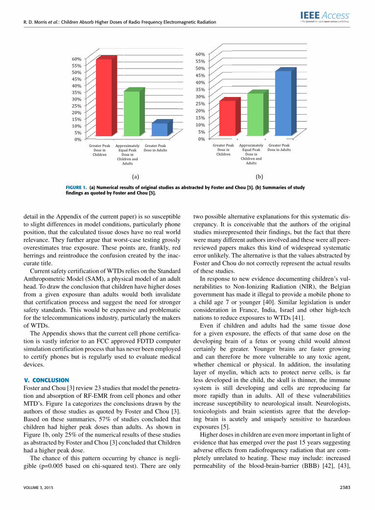
nations, as shown in Table 1 of this paper, 11 of 22 distinctstudies [10], [14], [16]–[24] concluded tissue doses werehigher in children, 7 found no difference [26]–[32] and only2 found higher doses in adults [15], [33]. In 2 cases the textsummaries were unclear [34], [35]. In other words, studiesreporting higher doses in children outnumber those reportinghigher doses in adults by a ratio of more than five to one,according to the text summaries of the study results providedby Foster and Chou in their Table 2.
Figure 1 from Foster and Chou does not accurately reflectthe information provided in their Table 2. Figure 1 from theirpaper depicts 57 ratios of child/adult psSAR as abstractedfrom 19 studies. Of these values, 14 (25%) indicate higherpeak dose in children, 17 (30%) found little or no difference(0.95− 1.05), and 26 (46%) found higher peak dose in adults.Of all the values in Figure 1 from Foster and Chou [3], 60%were greater than 1.00. Yet, according to Table 2, the per-centage of studies that concluded that psSAR was higher in
VOLUME 3, 2015 2381

R. D. Morris et al.: Children Absorb Higher Doses of Radio Frequency Electromagnetic Radiation
children was 57% while only 10% concluded that doses werehigher in adults. Figure 1 indicates psSAR ratios both aboveand below unity formany studies, yielding ambiguous results.For two studies summarized as reporting higher absorption inchildren, all of the values in their Figure 2 represent higherpeak dose in adults [17], [19]. Because the authors did notpool results quantitatively, the reader can not make conclu-sions with respect to whether or not the combined studiessuggest the ratio of peak dose for children as compared toadults is significantly different from 1.0.
Four of the studies listed in Table 2 were omittedfrom Figure 1 including two that found higher doses inchildren [22], [23] and two that concluded there were no dif-ferences between adults and children [29], [30]. The reasonsfor this omission are unclear.
Wiart et al. [22] stated that peripheral brain tissue had‘‘. . . higher exposure with children than with adults.’’Lu and Ueno [23] conclude that ‘‘[t]he induced SAR canbe significantly higher in subregions of the child’s brain.’’Both of these quotes were taken directly from Table 2 inFoster and Chou, but their Figure 1 shows results from neitherpaper.
For at least two papers [17], [19], none of the resultsin Figure 1 from Foster and Chou corresponds to thesummary of findings in their Table 2. In referring toGandhi and Kang [17], their Table 2 states that the modelof the child’s head has ‘‘peak 1 g SARs that may beup to 50-55% higher compared to the SARs for thelarger [adult] model particularly for a PCS frequency of1900 MHz [High Band].’’ In contrast, the bar graphin Figure 1 shows the ratio of Child/Adult psSAR1g values<1.0 in both the Low and High Bands.
According to Foster and Chou’s Table 2, Hadjem et al. [19]estimated that, for two child head models, the peak 10 gmSAR in the brain ‘‘is slightly more significant [higher] thanthat for the adults one.’’ Their Figure 1 implies that adultshave higher dosage rates.
In other words, four studies were described in Table 2,but omitted from Figure 1 and at least two other studies hadresults reported in Figure 1 that were not consistent withFoster and Chou’s own description of the results in Table 2.Our Table 1 suggests additional contradictions between theirTable 2 and Figure 1.
Readers who rely on the visual summary of findings inFigure 1 will infer that the majority of studies found higherpeak doses in adults. Readers diligent enough to sort throughthe dense text of Table 2, will reach the opposite conclusion.
More important to the issue at hand is that many of themodels cited by Foster and Chou do not take into account dif-ferences in the dielectric characteristics of the tissues of chil-dren, compared with adults [29], [37]. Without this, modelsonly consider children as small adults. This all but assures thatthere will be little difference in peak tissue dosage betweenchildren and adults, except to the extent that children’ssmaller heads lead to higher doses in particular anatomicalregions of the brain when compared to the larger adult head.
D. ANALYSIS OF STUDY RESULTSThere are two approaches to combining numerical resultsabstracted from a group of comparable individual studies.The first is to employ the statistical models commonly usedin meta-analysis, which pool results of experimental studiesmathematically using the standard error of the effect esti-mates. The modeling studies reviewed by Foster and Chouare not experimental, so their results cannot be pooled usingstandard meta-analytical techniques.
Results from deterministic models, such as those reviewedby Foster and Chou [3], can be systematically comparedbased on study characteristics. Steady improvements inmodel sophistication and dramatic increases in memory andprocessing speed of computers would lead one to expectmore accurate results from more recent models. Of the fivestudies using sophisticated FACTSmodels for both adults andchildren and published in the past ten years, four found higherpeak dose rates in children.
Of 22 paragraphs devoted to discussing differences amongmodels, Foster and Chou [3] devote nine to an extendeddiscussion of two models that are 14 and 20 years old. Ofthe fifteen models published in the past ten years, less thanhalf are mentioned in the discussion.
The reason Foster and Chou chose to criticize the workof a particular author is suggested by their discussion ofPenetration Depth, in which they focus almost exclusivelyon Gandhi’s 2002 Figure 3 image of RF-EMR absorptionin the brain at different ages. They assert, ‘‘A similar setof false-color figures . . . showed SAR patterns in all threedifferently sized head models that extended about the samedistance into the head.’’ This is true, as would be expected,because the child’s head is smaller (scaled down from anadult’s head). This study predated FACTS models, whichaccount for differences in dielectric properties between youngand older heads. The apparently controversial message of thisimage is that RF-EMR penetrates proportionally deeper intothe brain of a child than an adult. If, as Foster and Chou assert,absorption is the same in the pediatric and adult brains, thenthe smaller size of a child’s head will guarantee higher dosesto tissues deeper in the brain. Much of their argument relieson a paper [27], co-authored by Chou in 2005, a ten-year-old study which relies on a simple, scaled down model of theadult head.
IV. DISCUSSIONIn their Discussion, Foster and Chou state: ‘‘In summary,simple generalizations found on the Internet about ‘kidsabsorbing more RF energy than adults from cell phonesaren’t supported by available dosimetry studies.’’’ The textualsummaries of study findings, as provided by Foster and Chouin their Table 2, appear to support exactly the opposite conclu-sion. These 25 words represent the only part of the Discussionsection that refers directly to the topic of the paper—thedifferences between tissue doses in adults and children.
The remainder of their Discussion argues that none ofthis is relevant because compliance testing (as discussed in
2382 VOLUME 3, 2015
R. D. Morris et al.: Children Absorb Higher Doses of Radio Frequency Electromagnetic Radiation
FIGURE 1. (a) Numerical results of original studies as abstracted by Foster and Chou [3]. (b) Summaries of studyfindings as quoted by Foster and Chou [3].
detail in the Appendix of the current paper) is so susceptibleto slight differences in model conditions, particularly phoneposition, that the calculated tissue doses have no real worldrelevance. They further argue that worst-case testing grosslyoverestimates true exposure. These points are, frankly, redherrings and reintroduce the confusion created by the inac-curate title.
Current safety certification ofWTDs relies on the StandardAnthropometric Model (SAM), a physical model of an adulthead. To draw the conclusion that children have higher dosesfrom a given exposure than adults would both invalidatethat certification process and suggest the need for strongersafety standards. This would be expensive and problematicfor the telecommunications industry, particularly the makersof WTDs.
The Appendix shows that the current cell phone certifica-tion is vastly inferior to an FCC approved FDTD computersimulation certification process that has never been employedto certify phones but is regularly used to evaluate medicaldevices.
V. CONCLUSIONFoster and Chou [3] review 23 studies that model the penetra-tion and absorption of RF-EMR from cell phones and otherMTD’s. Figure 1a categorizes the conclusions drawn by theauthors of those studies as quoted by Foster and Chou [3].Based on these summaries, 57% of studies concluded thatchildren had higher peak doses than adults. As shown inFigure 1b, only 25% of the numerical results of these studiesas abstracted by Foster and Chou [3] concluded that Childrenhad a higher peak dose.
The chance of this pattern occurring by chance is negli-gible (p=0.005 based on chi-squared test). There are only
two possible alternative explanations for this systematic dis-crepancy. It is conceivable that the authors of the originalstudies misrepresented their findings, but the fact that therewere many different authors involved and these were all peer-reviewed papers makes this kind of widespread systematicerror unlikely. The alternative is that the values abstracted byFoster and Chou do not correctly represent the actual resultsof these studies.
In response to new evidence documenting children’s vul-nerabilities to Non-Ionizing Radiation (NIR), the Belgiangovernment has made it illegal to provide a mobile phone toa child age 7 or younger [40]. Similar legislation is underconsideration in France, India, Israel and other high-technations to reduce exposures to WTDs [41].
Even if children and adults had the same tissue dosefor a given exposure, the effects of that same dose on thedeveloping brain of a fetus or young child would almostcertainly be greater. Younger brains are faster growingand can therefore be more vulnerable to any toxic agent,whether chemical or physical. In addition, the insulatinglayer of myelin, which acts to protect nerve cells, is farless developed in the child, the skull is thinner, the immunesystem is still developing and cells are reproducing farmore rapidly than in adults. All of these vulnerabilitiesincrease susceptibility to neurological insult. Neurologists,toxicologists and brain scientists agree that the develop-ing brain is acutely and uniquely sensitive to hazardousexposures [5].
Higher doses in children are evenmore important in light ofevidence that has emerged over the past 15 years suggestingadverse effects from radiofrequency radiation that are com-pletely unrelated to heating. These may include: increasedpermeability of the blood-brain-barrier (BBB) [42], [43],
VOLUME 3, 2015 2383
R. D. Morris et al.: Children Absorb Higher Doses of Radio Frequency Electromagnetic Radiation
genotoxic effects on human cell lines [44], braincancer [45]–[47], acoustic neuroma [48]–[50], and spermdamage [51]–[53]. In 2013, the World Health Organization’sInternational Agency for Research on Cancer (IARC) classi-fied RF-EMR as a possible (2B) human carcinogen [54].
In light of explosive growth in usage rates and rapid techno-logical change in wireless devices, the American Academy ofPediatrics [55] supports ‘‘reassessment of radiation standardsfor cell phones and other wireless products and the adoptionof standards that are protective of children and reflect currentuse patterns.’’ The U.S. GAO has also recommended that theFCC reassess its exposure limits in light of new evidence [56].
In sum, the review by Foster and Chou suffers from thefollowing weaknesses.
1. There is no clear protocol specified for the identifica-tion of studies and the extraction and summary of data.
2. There are major, systematic discrepancies between thesummaries of study results in Foster and Chou’s Table 2and the data presented in their Figure 1.
3. The authors spend almost half of their discussion focus-ing on papers that are more than a decade old, butsay nothing about half of the studies published in thepast decade, most of which contradict their primaryconclusion.
APPENDIXRF-EMR EXPOSURE LIMITS AND COMPLIANCE TESTINGIn order to give some context to the concerns about com-pliance testing raised by Foster and Chou [3], we presenta brief overview of RF-EMR exposure standard-setting andcompliance assessment.
A. RF-EMR EXPOSURE LIMITSTwo RF-EMR exposure limit standards are in general use.The FCC 1996 standard [58] was substantially based onthe Institute of Electrical and Electronic Engineers (IEEE)C95.1, 1991 standardwithminor input fromNational Councilon Radiation Protection and Measurements (NCRP) ReportNo. 86. The other, standard primarily used in the EuropeanUnion (E.U.), was authored by the International Commissionon Non-Ionizing Radiation (ICNIRP) [59], [60].
For the general U.S. public the maximum permissible spe-cific absorption rate in any 1 g of tissue (SAR1g) is 1.6 W/kgaveraged over 30 minutes. In contrast, the correspondingexposure limit for the general public in the E.U. (ICNIRP) inany 10 gram cube of tissue is 2W/kg averaged over 6minutes.ThemaximumSAR increases as the tissueweight and volumedecrease [61], so the E.U. limit allows roughly 2 to 3 timesgreater exposure than the U.S. limit [21].
B. COMPLIANCE TESTING – TWO FCCAPPROVED METHODSApplicants requiring certification of wireless transmittingdevices (WTDs) by the FCC and/or those E.U. agen-cies adhering to the ICNIRP guidelines are permittedto use either a finite-difference time-domain (FDTD)
Computer Simulation Process, or the Specific Anthropomor-phic Mannequin (SAM) physical model to certify that WTDsdo not exceed the exposure limit [62].
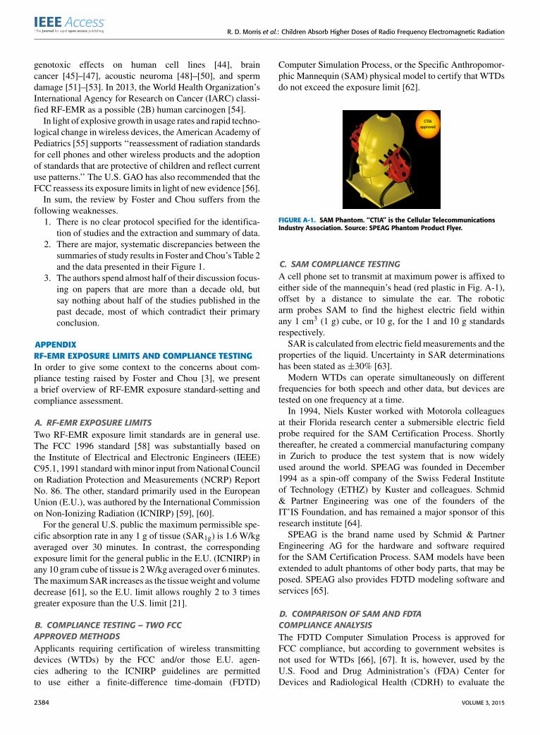
FIGURE A-1. SAM Phantom. ‘‘CTIA’’ is the Cellular TelecommunicationsIndustry Association. Source: SPEAG Phantom Product Flyer.
C. SAM COMPLIANCE TESTINGA cell phone set to transmit at maximum power is affixed toeither side of the mannequin’s head (red plastic in Fig. A-1),offset by a distance to simulate the ear. The roboticarm probes SAM to find the highest electric field withinany 1 cm3 (1 g) cube, or 10 g, for the 1 and 10 g standardsrespectively.
SAR is calculated from electric fieldmeasurements and theproperties of the liquid. Uncertainty in SAR determinationshas been stated as ±30% [63].Modern WTDs can operate simultaneously on different
frequencies for both speech and other data, but devices aretested on one frequency at a time.
In 1994, Niels Kuster worked with Motorola colleaguesat their Florida research center a submersible electric fieldprobe required for the SAM Certification Process. Shortlythereafter, he created a commercial manufacturing companyin Zurich to produce the test system that is now widelyused around the world. SPEAG was founded in December1994 as a spin-off company of the Swiss Federal Instituteof Technology (ETHZ) by Kuster and colleagues. Schmid& Partner Engineering was one of the founders of theIT’IS Foundation, and has remained a major sponsor of thisresearch institute [64].
SPEAG is the brand name used by Schmid & PartnerEngineering AG for the hardware and software requiredfor the SAM Certification Process. SAM models have beenextended to adult phantoms of other body parts, that may beposed. SPEAG also provides FDTD modeling software andservices [65].
D. COMPARISON OF SAM AND FDTACOMPLIANCE ANALYSISThe FDTD Computer Simulation Process is approved forFCC compliance, but according to government websites isnot used for WTDs [66], [67]. It is, however, used by theU.S. Food and Drug Administration’s (FDA) Center forDevices and Radiological Health (CDRH) to evaluate the
2384 VOLUME 3, 2015
R. D. Morris et al.: Children Absorb Higher Doses of Radio Frequency Electromagnetic Radiation
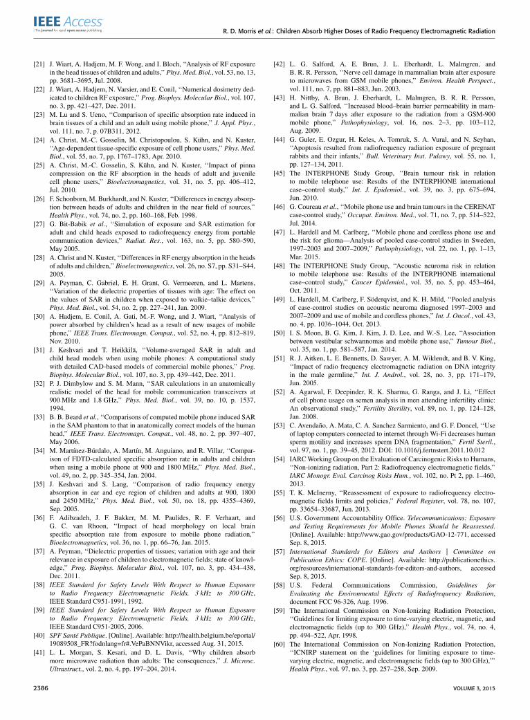
TABLE A-1. Comparison of cell phone certification processes.
safety of medical implants by relying on anatomically basedmodels for persons of varying ages and sizes [68], [69].
Compared with the homogenous fluid-filled SAM headphantom, the FDTD Computer Simulation Process usingFDTD Anatomically Correct, Tissue Specific (FACTS) mod-els provides fine-grained resolution of RF-EMR absorptionin tissues in any volume within the body, of any age orsex, with any location of the WTD (e.g., adjacent to apregnant abdomen, or in a trouser pocket in proximity toa testicle).
Table A-1 compares the attributes of the two FCC approvedcertification processes.
ACKNOWLEDGMENTThe authors wish to thank Margaret Sears, for her extensivecontributions as both writer and editor and Barbara Payne forher extraordinary copy-editing skill. Environmental HealthTrust provided support for Robert Morris, LloydMorgan, andDevra Davis during the preparation of this manuscript, asdid grants from the Community Foundation of Jackson Holeand Lucy R. Wiletzky MD.
REFERENCES[1] U.S. Environmental Protection Agency. EPA’s National Agenda to Protect
Children’s Health From Environmental Threats. [Online]. Available: http://www2.epa.gov/children/epas-national-agenda-protect-childrens-health-environmental-threats, accessed Aug. 3, 2015.
[2] U.S. Environmental Protection Agency. History of Children’s Environ-mental Health Protection at EPA. [Online]. Available: http://www2.epa.gov/children/history-childrens-environmental-health-protection-epa,accessed Aug. 3, 2015.
[3] K. R. Foster and C.-K. Chou, ‘‘Are children more exposed to radio fre-quency energy from mobile phones than adults?’’ IEEE Access, vol. 2,pp. 1497–1509, Dec. 2014.
[4] O. P. Gandhi, ‘‘Yes the children are more exposed to radiofrequency energyfrom mobile telephones than adults,’’ IEEE Access, vol. 3, pp. 985–988,Jul. 2015.
[5] D. Rice and S. Barone, Jr., ‘‘Critical periods of vulnerability for thedeveloping nervous system: Evidence from humans and animal models,’’Environ. Health Perspect., vol. 108, no. Suppl 3, pp. 511–533, Jun. 2000.
[6] B.Weiss, ‘‘Vulnerability of children and the developing brain to neurotoxichazards,’’ Environ. Health Perspect., vol. 108, no. Suppl 3, pp. 375–381,Jun. 2000.
[7] R. D. Morris, ‘‘Meta-analysis in cancer epidemiology,’’ Environ. HealthPerspect., vol. 102, no. Suppl 8, pp. 61–66, Nov. 1994.
[8] A. Blair et al., ‘‘Guidelines for application of metaanalysis in environ-mental epidemiology,’’ Regulatory Toxicol. Pharmacol., vol. 22, no. 2,pp. 189–197, 1995.
[9] A. A. Rooney, A. L. Boyles, M. S. Wolfe, J. R. Bucher, and K. A. Thayer,‘‘Systematic review and evidence integration for literature-based environ-mental health science assessments,’’ Environ. Health Perspect., vol. 122,no. 7, pp. 711–718, Jul. 2014.
[10] V. Anderson, ‘‘Comparisons of peak SAR levels in concentric sphere headmodels of children and adults for irradiation by a dipole at 900 MHz,’’Phys. Med. Biol., vol. 48, no. 20, pp. 3263–3275, Oct. 2003.
[11] J. L. Lancaster et al., ‘‘Automated Talairach atlas labels for functional brainmapping,’’ Hum. Brain Mapping, vol. 10, no. 3, pp. 120–131, Jul. 2000.
[12] IT’IS Foundation. IT’IS Foundation History. [Online]. Available:http://www.itis.ethz.ch/who-we-are/history, accessed Aug. 29, 2015.
[13] IT’IS Foundation. Virtual Population. [Online]. Available: http://www.itis.ethz.ch/virtual-population/virtual-population-cvip-vip/overview/,accessed Aug. 9, 2015.
[14] A. A. de Salles, G. Bulla, and C. E. F. Rodriguez, ‘‘Electromagneticabsorption in the head of adults and children due to mobile phone operationclose to the head,’’ Electromagn. Biol. Med., vol. 25, no. 4, pp. 349–360,2006.
[15] A.-K. Lee, H.-D. Choi, H.-S. Lee, and J.-K. Pack, ‘‘Human head sizeand SAR characteristics for handset exposure,’’ ETRI J., vol. 24, no. 2,pp. 176–180, Apr. 2002.
[16] O. P. Gandhi, G. Lazzi, and C. M. Furse, ‘‘Electromagnetic absorption inthe human head and neck for mobile telephones at 835 and 1900 MHz,’’IEEE Trans. Microw. Theory Techn., vol. 44, no. 10, pp. 1884–1897,Oct. 1996.
[17] O. P. Gandhi and G. Kang, ‘‘Some present problems and a proposedexperimental phantom for SAR compliance testing of cellular telephonesat 835 and 1900 MHz,’’ Phys. Med. Biol., vol. 47, no. 9, pp. 1501–1518,May 2002.
[18] J. Wang and O. Fujiwara, ‘‘Comparison and evaluation of electromagneticabsorption characteristics in realistic human head models of adult andchildren for 900-MHz mobile telephones,’’ IEEE Trans. Microw. TheoryTechn., vol. 51, no. 3, pp. 966–971, Mar. 2003.
[19] A. Hadjem, D. Lautru, C. Dale, M. F. Wong, V. F. Hanna, and J. Wiart,‘‘Study of specific absorption rate (SAR) induced in two child head modelsand in adult heads using mobile phones,’’ IEEE Trans. Microw. TheoryTechn., vol. 53, no. 1, pp. 4–11, Jan. 2005.
[20] J. Wiart et al., ‘‘Modeling of RF head exposure in children,’’Bioelectromagnetics, vol. 26, no. S7, pp. S19–S30, 2005.
VOLUME 3, 2015 2385
R. D. Morris et al.: Children Absorb Higher Doses of Radio Frequency Electromagnetic Radiation
[21] J. Wiart, A. Hadjem, M. F. Wong, and I. Bloch, ‘‘Analysis of RF exposurein the head tissues of children and adults,’’ Phys. Med. Biol., vol. 53, no. 13,pp. 3681–3695, Jul. 2008.
[22] J. Wiart, A. Hadjem, N. Varsier, and E. Conil, ‘‘Numerical dosimetry ded-icated to children RF exposure,’’ Prog. Biophys. Molecular Biol., vol. 107,no. 3, pp. 421–427, Dec. 2011.
[23] M. Lu and S. Ueno, ‘‘Comparison of specific absorption rate induced inbrain tissues of a child and an adult using mobile phone,’’ J. Appl. Phys.,vol. 111, no. 7, p. 07B311, 2012.
[24] A. Christ, M.-C. Gosselin, M. Christopoulou, S. Kühn, and N. Kuster,‘‘Age-dependent tissue-specific exposure of cell phone users,’’ Phys. Med.Biol., vol. 55, no. 7, pp. 1767–1783, Apr. 2010.
[25] A. Christ, M.-C. Gosselin, S. Kühn, and N. Kuster, ‘‘Impact of pinnacompression on the RF absorption in the heads of adult and juvenilecell phone users,’’ Bioelectromagnetics, vol. 31, no. 5, pp. 406–412,Jul. 2010.
[26] F. Schonborn,M. Burkhardt, andN. Kuster, ‘‘Differences in energy absorp-tion between heads of adults and children in the near field of sources,’’Health Phys., vol. 74, no. 2, pp. 160–168, Feb. 1998.
[27] G. Bit-Babik et al., ‘‘Simulation of exposure and SAR estimation foradult and child heads exposed to radiofrequency energy from portablecommunication devices,’’ Radiat. Res., vol. 163, no. 5, pp. 580–590,May 2005.
[28] A. Christ and N. Kuster, ‘‘Differences in RF energy absorption in the headsof adults and children,’’ Bioelectromagnetics, vol. 26, no. S7, pp. S31–S44,2005.
[29] A. Peyman, C. Gabriel, E. H. Grant, G. Vermeeren, and L. Martens,‘‘Variation of the dielectric properties of tissues with age: The effect onthe values of SAR in children when exposed to walkie–talkie devices,’’Phys. Med. Biol., vol. 54, no. 2, pp. 227–241, Jan. 2009.
[30] A. Hadjem, E. Conil, A. Gati, M.-F. Wong, and J. Wiart, ‘‘Analysis ofpower absorbed by children’s head as a result of new usages of mobilephone,’’ IEEE Trans. Electromagn. Compat., vol. 52, no. 4, pp. 812–819,Nov. 2010.
[31] J. Keshvari and T. Heikkilä, ‘‘Volume-averaged SAR in adult andchild head models when using mobile phones: A computational studywith detailed CAD-based models of commercial mobile phones,’’ Prog.Biophys. Molecular Biol., vol. 107, no. 3, pp. 439–442, Dec. 2011.
[32] P. J. Dimbylow and S. M. Mann, ‘‘SAR calculations in an anatomicallyrealistic model of the head for mobile communication transceivers at900 MHz and 1.8 GHz,’’ Phys. Med. Biol., vol. 39, no. 10, p. 1537,1994.
[33] B. B. Beard et al., ‘‘Comparisons of computed mobile phone induced SARin the SAM phantom to that in anatomically correct models of the humanhead,’’ IEEE Trans. Electromagn. Compat., vol. 48, no. 2, pp. 397–407,May 2006.
[34] M. Martínez-Búrdalo, A. Martín, M. Anguiano, and R. Villar, ‘‘Compar-ison of FDTD-calculated specific absorption rate in adults and childrenwhen using a mobile phone at 900 and 1800 MHz,’’ Phys. Med. Biol.,vol. 49, no. 2, pp. 345–354, Jan. 2004.
[35] J. Keshvari and S. Lang, ‘‘Comparison of radio frequency energyabsorption in ear and eye region of children and adults at 900, 1800and 2450 MHz,’’ Phys. Med. Biol., vol. 50, no. 18, pp. 4355–4369,Sep. 2005.
[36] F. Adibzadeh, J. F. Bakker, M. M. Paulides, R. F. Verhaart, andG. C. van Rhoon, ‘‘Impact of head morphology on local brainspecific absorption rate from exposure to mobile phone radiation,’’Bioelectromagnetics, vol. 36, no. 1, pp. 66–76, Jan. 2015.
[37] A. Peyman, ‘‘Dielectric properties of tissues; variation with age and theirrelevance in exposure of children to electromagnetic fields; state of knowl-edge,’’ Prog. Biophys. Molecular Biol., vol. 107, no. 3, pp. 434–438,Dec. 2011.
[38] IEEE Standard for Safety Levels With Respect to Human Exposureto Radio Frequency Electromagnetic Fields, 3 kHz to 300 GHz,IEEE Standard C951-1991, 1992.
[39] IEEE Standard for Safety Levels With Respect to Human Exposureto Radio Frequency Electromagnetic Fields, 3 kHz to 300 GHz,IEEE Standard C951-2005, 2006.
[40] SPF Santé Publique. [Online]. Available: http://health.belgium.be/eportal/19089508_FR?fodnlang=fr#.VePaBNNVikr, accessed Aug. 31, 2015.
[41] L. L. Morgan, S. Kesari, and D. L. Davis, ‘‘Why children absorbmore microwave radiation than adults: The consequences,’’ J. Microsc.Ultrastruct., vol. 2, no. 4, pp. 197–204, 2014.
[42] L. G. Salford, A. E. Brun, J. L. Eberhardt, L. Malmgren, andB. R. R. Persson, ‘‘Nerve cell damage in mammalian brain after exposureto microwaves from GSM mobile phones,’’ Environ. Health Perspect.,vol. 111, no. 7, pp. 881–883, Jun. 2003.
[43] H. Nittby, A. Brun, J. Eberhardt, L. Malmgren, B. R. R. Persson,and L. G. Salford, ‘‘Increased blood–brain barrier permeability in mam-malian brain 7 days after exposure to the radiation from a GSM-900mobile phone,’’ Pathophysiology, vol. 16, nos. 2–3, pp. 103–112,Aug. 2009.
[44] G. Guler, E. Ozgur, H. Keles, A. Tomruk, S. A. Vural, and N. Seyhan,‘‘Apoptosis resulted from radiofrequency radiation exposure of pregnantrabbits and their infants,’’ Bull. Veterinary Inst. Pulawy, vol. 55, no. 1,pp. 127–134, 2011.
[45] The INTERPHONE Study Group, ‘‘Brain tumour risk in relationto mobile telephone use: Results of the INTERPHONE internationalcase–control study,’’ Int. J. Epidemiol., vol. 39, no. 3, pp. 675–694,Jun. 2010.
[46] G. Coureau et al., ‘‘Mobile phone use and brain tumours in the CERENATcase-control study,’’ Occupat. Environ. Med., vol. 71, no. 7, pp. 514–522,Jul. 2014.
[47] L. Hardell and M. Carlberg, ‘‘Mobile phone and cordless phone use andthe risk for glioma—Analysis of pooled case-control studies in Sweden,1997–2003 and 2007–2009,’’ Pathophysiology, vol. 22, no. 1, pp. 1–13,Mar. 2015.
[48] The INTERPHONE Study Group, ‘‘Acoustic neuroma risk in relationto mobile telephone use: Results of the INTERPHONE internationalcase–control study,’’ Cancer Epidemiol., vol. 35, no. 5, pp. 453–464,Oct. 2011.
[49] L. Hardell, M. Carlberg, F. Söderqvist, and K. H. Mild, ‘‘Pooled analysisof case-control studies on acoustic neuroma diagnosed 1997–2003 and2007–2009 and use of mobile and cordless phones,’’ Int. J. Oncol., vol. 43,no. 4, pp. 1036–1044, Oct. 2013.
[50] I. S. Moon, B. G. Kim, J. Kim, J. D. Lee, and W.-S. Lee, ‘‘Associationbetween vestibular schwannomas and mobile phone use,’’ Tumour Biol.,vol. 35, no. 1, pp. 581–587, Jan. 2014.
[51] R. J. Aitken, L. E. Bennetts, D. Sawyer, A. M. Wiklendt, and B. V. King,‘‘Impact of radio frequency electromagnetic radiation on DNA integrityin the male germline,’’ Int. J. Androl., vol. 28, no. 3, pp. 171–179,Jun. 2005.
[52] A. Agarwal, F. Deepinder, R. K. Sharma, G. Ranga, and J. Li, ‘‘Effectof cell phone usage on semen analysis in men attending infertility clinic:An observational study,’’ Fertility Sterility, vol. 89, no. 1, pp. 124–128,Jan. 2008.
[53] C. Avendaño, A. Mata, C. A. Sanchez Sarmiento, and G. F. Doncel, ‘‘Useof laptop computers connected to internet through Wi-Fi decreases humansperm motility and increases sperm DNA fragmentation,’’ Fertil Steril.,vol. 97, no. 1, pp. 39–45, 2012. DOI: 10.1016/j.fertnstert.2011.10.012
[54] IARCWorking Group on the Evaluation of Carcinogenic Risks to Humans,‘‘Non-ionizing radiation, Part 2: Radiofrequency electromagnetic fields,’’IARC Monogr. Eval. Carcinog Risks Hum., vol. 102, no. Pt 2, pp. 1–460,2013.
[55] T. K. McInerny, ‘‘Reassessment of exposure to radiofrequency electro-magnetic fields limits and policies,’’ Federal Register, vol. 78, no. 107,pp. 33654–33687, Jun. 2013.
[56] U.S. Government Accountability Office. Telecommunications: Exposureand Testing Requirements for Mobile Phones Should be Reassessed.[Online]. Available: http://www.gao.gov/products/GAO-12-771, accessedSep. 8, 2015.
[57] International Standards for Editors and Authors | Committee onPublication Ethics: COPE. [Online]. Available: http://publicationethics.org/resources/international-standards-for-editors-and-authors, accessedSep. 8, 2015.
[58] U.S. Federal Communications Commission, Guidelines forEvaluating the Environmental Effects of Radiofrequency Radiation,document FCC 96-326, Aug. 1996.
[59] The International Commission on Non-Ionizing Radiation Protection,‘‘Guidelines for limiting exposure to time-varying electric, magnetic, andelectromagnetic fields (up to 300 GHz),’’ Health Phys., vol. 74, no. 4,pp. 494–522, Apr. 1998.
[60] The International Commission on Non-Ionizing Radiation Protection,‘‘ICNIRP statement on the ‘guidelines for limiting exposure to time-varying electric, magnetic, and electromagnetic fields (up to 300 GHz),’’’Health Phys., vol. 97, no. 3, pp. 257–258, Sep. 2009.
2386 VOLUME 3, 2015
R. D. Morris et al.: Children Absorb Higher Doses of Radio Frequency Electromagnetic Radiation
[61] C. Fernández, A. de Salles, and D. Davis, ‘‘Preliminary SAR simulation ishighest for smallest volumes, youngest age groups, and highest dielectricconstant,’’ presented at the Joint Meeting Bioelectromagn. Soc. Eur. Bio-Electromagn. Assoc., Tech. Program General Inf. (BioEM), Thessaloniki,Greece, Jun. 2013.
[62] R. F. Cleveland, D.M. Sylvar, and J. L. Ulcek,Evaluating ComplianceWithFCC Guidelines for Human Exposure to Radiofrequency ElectromagneticFields, document OET Bulletin 65, Aug. 1997. [Online]. Available:https://transition.fcc.gov/Bureaus/Engineering_Technology/Documents/bulletins/oet65/oet65c.pdf
[63] IEEE Recommended Practice for Determining the Peak Spatial-Average Specific Absorption Rate (SAR) in the Human HeadFrom Wireless Communications Devices: Measurement Techniques,IEEE Standard 1528TM-2003, Dec. 2003.
[64] Schmid & Partner Engineering AG. SPEAG History. [Online]. Available:http://www.speag.com/about-speag/history/, accessed Sep. 6, 2015.
[65] Schmid & Partner Engineering AG. SPEAG Products. [Online]. Available:http://www.speag.com/products/, accessed Sep. 8, 2015.
[66] U.S. Federal Communications Commission. (Mar. 12, 2014). SpecificAbsorption Rate (SAR) for Cell Phones: What it Means for You.[Online]. Available: https://www.fcc.gov/guides/specific-absorption-rate-sar-cell-phones-what-it-means-you, accessed Sep. 8, 2015.
[67] I.C. Government of Canada. (Nov. 16, 2011). SAR—Certification andEngineering Bureau. [Online]. Available: http://www.ic.gc.ca/eic/site/ceb-bhst.nsf/eng/h_tt00084.html, accessed Sep. 8, 2015.
[68] M.-C. Gosselin et al., ‘‘Development of a new generation of high-resolution anatomical models for medical device evaluation: The virtualpopulation 3.0,’’ Phys. Med. Biol., vol. 59, no. 18, p. 5287, Aug. 2014.
[69] M. I. Iacono, N. Makris, L. Mainardi, L. M. Angelone, and G. Bonmassar,‘‘MRI-based multiscale model for electromagnetic analysis in the humanhead with implanted DBS,’’ Comput. Math. Methods Med., vol. 2013,May 2013, Art. ID 694171.
ROBERT D. MORRIS received the M.D. andPh.D. degrees. He has taught with the TuftsUniversity School of Medicine, the HarvardUniversity School of Public Health, and theMedical College of Wisconsin, and has servedas an Advisor to the EPA, CDC, NIH, and thePresident’s Cancer Panel. He resides inSeattle, WA. He is currently an EnvironmentalEpidemiologist and the Senior Medical Advisor tothe Environmental Health Trust. His work has been
featured in the New York Times and the London Times, and in the DatelineNBC and the BBC. His first book, entitled The Blue Death, received theNautilus Gold Award, and was named one of the Best Consumer HealthBooks of 2007 by the American Library Association.
L. LLOYD MORGAN has been involved in thestudy of exposure to electromagnetic fields andresultant health problems since 1995. He is cur-rently an Electronic Engineer by training with38 years of industrial experience and a memberof international science organizations. He contin-ues to carry out critical analyses of epidemio-logical studies in the field and presents findingsto local expert forums in Teton County, as wellas nationally and internationally. His paper, with
Dr. Gandhi, Dr. Herberman, and Dr. Davis, on brain modeling of cell phoneswas one of the most widely cited papers in the field.
DEVRA DAVIS received the B.S. and M.A.degrees from the University of Pittsburgh, thePh.D. degree in science studies from the Uni-versity of Chicago, and the M.P.H. degree inepidemiology from Johns Hopkins University.She was the Founding Director of the Centerfor Environmental Oncology with the Universityof Pittsburgh Cancer Institute, and the FoundingDirector of the Board on Environmental Studiesand Toxicology with the U.S. National Research
Council (1983–1993), where she served as a Scholar-in-Residence, anda Professor of Epidemiology with the Graduate School of Public Health(2004–2010). She served as the Presidential Appointee to the ChemicalSafety and Hazard Investigation Board and a Senior Advisor to the AssistantSecretary for Health with the Department of Health and Human Services.She has taught with the London School of Hygiene and Tropical Medicine,the Mount Sinai School of Medicine, Oberlin College, and CarnegieMellon University. She has counseled leading officials in the United States,the United Nations, the European Environment Agency, the Pan AmericanHealth Organization, the World Health Organization, the World Bank, theU.S. National Toxicology Program, and the U.S. Centers for Disease Controland Prevention. She is currently the Founder and President of the Environ-mental Health Trust and visiting Professor at both Hebrew Univ. HadassahMedical Center and OndokuzMayis Univ. Medical School. She has authoredover 200 scientific publications, ten edited monographs, three popular books,and numerous op-eds.
VOLUME 3, 2015 2387