Embed Size (px)
Citation preview
676
Accepted by P.J. Johnson: 9 Sept. 2004; published: 8 Oct. 2004 1
ZOOTAXAISSN 1175-5326 (print edition)
ISSN 1175-5334 (online edition)Copyright © 2004 Magnolia Press
Zootaxa 676: 1–46 (2004) www.mapress.com/zootaxa/
Morphology of the prothorax and procoxa in the New World Cryptocephalini (Coleoptera: Chrysomelidae: Cryptocephalinae)
MARIA LOURDES CHAMORRO-LACAYO1 & ALEXANDER S. KONSTANTINOV2
1Department of Entomology, University of Minnesota, Saint-Paul, MN 55108, USA (email: [email protected]);2Systematic Entomology Laboratory, PSI, Agricultural Research Service, U.S. Department of Agriculture, c/o National Museum of Natural History, MRC 168, Washington, DC 20560, U.S.A. (email: akon-
Table of contents
Abstract . . . . . . . . . . . . . . . . . . . . . . . . . . . . . . . . . . . . . . . . . . . . . . . . . . . . . . . . . . . . . . . . . . . . . . 1Introduction . . . . . . . . . . . . . . . . . . . . . . . . . . . . . . . . . . . . . . . . . . . . . . . . . . . . . . . . . . . . . . . . . . . 1Material and Methods . . . . . . . . . . . . . . . . . . . . . . . . . . . . . . . . . . . . . . . . . . . . . . . . . . . . . . . . . . . 3Morphology . . . . . . . . . . . . . . . . . . . . . . . . . . . . . . . . . . . . . . . . . . . . . . . . . . . . . . . . . . . . . . . . . . . 3
I. The pronotum . . . . . . . . . . . . . . . . . . . . . . . . . . . . . . . . . . . . . . . . . . . . . . . . . . . . . . . . . . . . . 4II. The prosternum . . . . . . . . . . . . . . . . . . . . . . . . . . . . . . . . . . . . . . . . . . . . . . . . . . . . . . . . . . . 5III. The procoxal cavity . . . . . . . . . . . . . . . . . . . . . . . . . . . . . . . . . . . . . . . . . . . . . . . . . . . . . . . 6IV. The trochantin and endopleuron . . . . . . . . . . . . . . . . . . . . . . . . . . . . . . . . . . . . . . . . . . . . . 7V. The procoxa and trochanter . . . . . . . . . . . . . . . . . . . . . . . . . . . . . . . . . . . . . . . . . . . . . . . . . 8VI. Variability in prothoracic structures of New World Cryptocephalini . . . . . . . . . . . . . . . . 9 MONACHULINA . . . . . . . . . . . . . . . . . . . . . . . . . . . . . . . . . . . . . . . . . . . . . . . . . . . . . . . . . . 9 CRYPTOCEPHALINA. . . . . . . . . . . . . . . . . . . . . . . . . . . . . . . . . . . . . . . . . . . . . . . . . . . . . . . . . . . . . 9 PACHYBRACHINA . . . . . . . . . . . . . . . . . . . . . . . . . . . . . . . . . . . . . . . . . . . . . . . . . . . . . . . . . . . . . 10
Conclusions . . . . . . . . . . . . . . . . . . . . . . . . . . . . . . . . . . . . . . . . . . . . . . . . . . . . . . . . . . . . . . . . . . 11Acknowledgements . . . . . . . . . . . . . . . . . . . . . . . . . . . . . . . . . . . . . . . . . . . . . . . . . . . . . . . . . . . . 13Literature cited . . . . . . . . . . . . . . . . . . . . . . . . . . . . . . . . . . . . . . . . . . . . . . . . . . . . . . . . . . . . . . . . 13APPENDIX 1. List of Cryptocephalini for which SEM images were taken . . . . . . . . . . . . . . . . . 15APPENDIX 2. Variability in prothoracic structures of New World Cryptocephalini . . . . . . . . . . 16
Abstract
The comparative morphology of the prothorax and procoxae of New World Cryptocephalini wasstudied based on representatives of 11 of the 13 genera of the tribe. This study revealed a set ofcharacters of obvious diagnostic and possible phylogenetic value supporting the currently acceptedgeneric classification and two subtribes instead of the three currently recognized. Two general types
CHAMORRO-LACAYO & KONSTANTINOV2 © 2004 Magnolia Press
676ZOOTAXA of prothoracies were found, the first occurring in Cryptocephalina and Monachulina and the second
in Pachybrachina. Previously undescribed for Polyphaga, a monocondylic joint between the coxaand trochantin, was found in all the genera studied. Possible movement of the trochanter, includingthe transfer of advance movement into rotation, is described and illustrated.
Keywords: Cryptocephalinae, prothorax, procoxa, comparative morphology, New World
Introduction
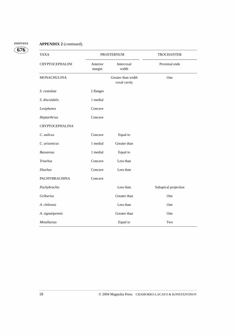
Cryptocephalini, are known as case-bearing leaf beetles, and are robust, cylindrical andcompact beetles measuring between 2–7 mm long (Fig. 1). They can easily be distin-guished from most beetles (including other leaf beetles) by the following features: headpartially or completely concealed within the prothorax when viewed dorsally (hence theirname); antennae filiform; base of the pronotum as wide as base of the elytra; seventhabdominal tergite usually visible beyond the elytra. The elytra of cryptocephalines beardistinct rows of punctures, which are sometimes useful diagnostically (White 1968). Thesexes are easily separated by a deep, large, median, setose indentation in the female’s sev-enth abdominal sternite. It is here where the egg is rotated while being coated in feces(Erber 1988).
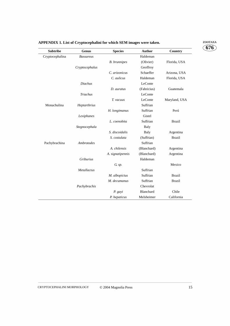
Cryptocephalinae are currently divided into three tribes, Cryptocephalini, Chlamisini,and Clytrini (Reid 1995), previously regarded as subfamilies. Thirteen valid genera inthree subtribes are presently recorded in New World Cryptocephalini: Mastacanthus Suf-frian, Sternoglossus Suffrian, Griburius Haldeman, Metallactus Suffrian, PachybrachisChevrolat, and Ambrotodes Suffrian in subtribe Pachybrachina; Heptarthrius Suffrian,Lexiphanes Gistel, and Stegnocephala Baly in subtribe Monachulina; and CryptocephalusGeoffroy, Diachus LeConte, Bassareus Haldeman, and Triachus LeConte in subtribeCryptocephalina (Seeno & Wilcox 1982). For this study, specimens of Mastacanthus andSternoglossus were not available due to their rarity in collections. Approximately 1,000species of Cryptocephalini have been recorded in the New World (Blackwelder 1944,Riley et al. 2002, Wilcox 1975, White 1968), with Cryptocephalus, Pachybrachis, Gribu-rius, Lexiphanes and Metallactus making up about 95% of the total diversity. This ismerely an estimate of Cryptocephalini species diversity, as it is based on various generalbeetle checklists (Blackwelder 1944). The Cryptocephalinae fauna of the New World,excluding North America north of Mexico, is poorly known. Many genera have not beenrevised since they were initially described and many Neotropical species await descriptionin museums and in the field.
The comparative morphology of Cryptocephalinae is not well studied. Some morpho-logical data on cryptocephalines is available in comparative studies of certain structures orattributes of Chrysomelidae as a whole (Samuelson 1996), but in such studies usually onlya few cryptocephaline genera were analyzed (Baccetti and Daccordi 1988, Suzuki 1988).
© 2004 Magnolia Press 3CRYPTOCEPHALINI MORPHOLOGY
676ZOOTAXAOther morphological data can be extracted from keys for identification (Lopatin 1977,
Riley, et al. 2002). Among the structures used as sources for diagnostic characters, the pro-thorax is often mentioned. The relative width of the prosternum, structure of the pronotalbase, and shape of the lateral margin are commonly used to separate taxa above the specieslevel in both New and Old World faunas (Lopatin 1977, Riley, et al. 2002). However, noattempts have been made to describe the diversity of prothoracic features in the Crypto-cephalini, including internal ridges of the prosternum, proendosternites, coxae, trochantersand trochantines. This paper treats the aforementioned structures in all but two (Masta-canthus and Sternoglossus, both belonging to Pachybrachina) valid New World Crypto-cephalini genera.
Material and Methods
The beetles examined in this study were obtained from the chrysomelid collection at theNational Museum of Natural History (USNM), Smithsonian Institution. We selected oneor two species representatives from each genus found in different areas of the New World(see Appendix 1 for the list of examined species and their geographic data).
Beetles were initially softened in distilled water and subsequently cleared using 10%NaOH, either by soaking them overnight, or using a heat source to speed up the process.The prothorax was then separated from the rest of the body in a dissecting dish filled withdistilled water. The head, meso- and metathoraces, and abdomen were stored in glycerinmixed with a small amount of ethanol. The remaining soft tissue in the prothorax was verycarefully removed under a Zeiss Stemi SV 11 Apo dissecting microscope, ensuring integ-rity of the structures. The left leg, along with the trochantin and coxa, were removed aftermotion of the structures had been recorded. The prothorax was then cleaned in a detergentsolution in an ultrasonic cleaner in preparation for the Scanning Electron Microscope(SEM). The prothorax, left procoxa and trochantin were then glued onto a stub and coatedwith a heavy metal. Images were taken with an AMRAY 1810 scanning electron micro-scope. In this paper we follow the morphological terminology of Snodgrass (1935), Hla-vac (1972), and Larsén (1966) and terms for some muscles were taken from Baehr (1979).Other common terms, e.g. ventral appendage, lateral projection etc., were created based onthe relative position of the morphological structures on the undissected beetle body.
Morphology
The main prothoracic differences separating the four beetle suborders occur in the pleuraand trochantin, and how these are attached to each other (Hlavac 1972). In the subordersArchostemata, Myxophaga and Adephaga the pleuron is large and rigid and makes up partof the prothoracic wall. In Polyphaga the pleuron is reduced in size, internalized and
CHAMORRO-LACAYO & KONSTANTINOV4 © 2004 Magnolia Press
676ZOOTAXA termed the endopleuron or cryptopleuron (Hlavac 1972, Larsén 1966, McHugh et al.
1997). In Archostemata the trochantin is external and movable, whereas in the Adephagathe trochantin is mobile but the pleuron and part of the sternum enclose the trochantin anda portion of the coxa. In Myxophaga and Polyphaga the trochantin and the pleura arefused. This fused structure, (trochantin-endopleuron) trochantino-cryptopleuron (Larsén1966), is moved by the tergo-pleural muscle, which in turn moves the coxa (Larsén 1966).Coxal movement is also effected by the contraction of the episterno-coxal and epimero-coxal muscles (Larsén 1966).
The prothorax of Cryptocephalini, as that of other beetles, consists of a wide dorsalsclerite, the pronotum, and a much narrower ventral sclerite, the prosternum. As in allPolyphaga (Evans 1971, Lawrence and Britton 1994), the propleuron in cryptocephalineshas become reduced, forming an endopleuron within the prothoracic cavity, which isattached to the trochantin. Anteriorly the pronotum and prosternum form a near perfectlyrounded opening (Figs. 10, 22, 32, 41, 51, 62, 72, 89, 101, 109, 119) into which the bee-tle’s head fits. The posterior opening is more complicated in shape, but always has agreater height than width (Figs. 4, 15, 23, 33, 42, 54, 63, 74, 83, 86, 92, 103, 110, 121,132).
I. The pronotum
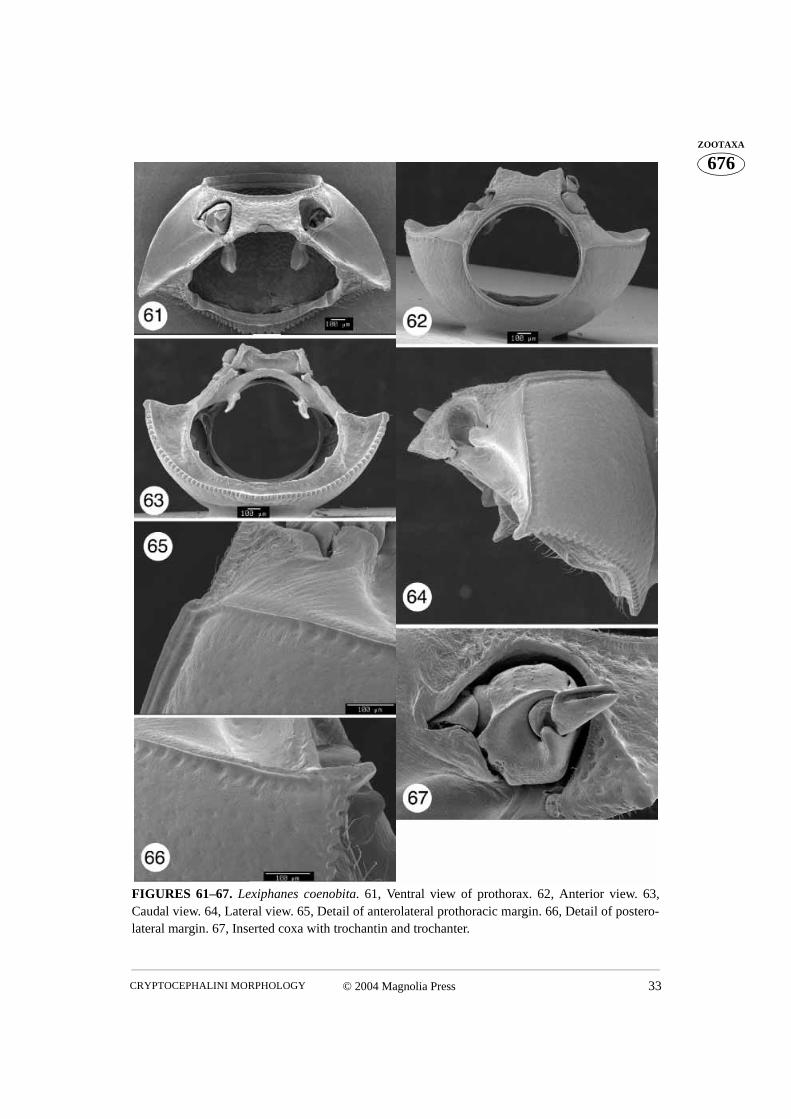
The pronotum is relatively convex, basally as wide as the elytra, and narrowing apically. Itis distinctly margined laterally, apically and basally. In the Monachulina and Cryptocepha-lina the basal margin is strongly dentate (Figs. 3, 15, 19, 21, 50, 61, 64, 71, 73, 81, 82)except for Diachus and Triachus in which the denticles are greatly reduced (Figs. 31, 34,40, 43). The basal margin is usually uniformly convex, being tallest medially, with a flatarea across the mesoscutellum. However, in some Pachybrachina the basal margin of thepronotum is sinuate, being laterally as long as medially (Figs. 85, 88, 100, 108, 131). Ven-trally from the basal margin, the pronotum forms a relatively short ridge flanking the basalopening. The border of the basal opening is margined and armed with two well separatedlongitudinal ridges in Monachulina and Cryptocephalina (Figs. 4, 15, 23, 33, 54, 63, 74,83). Most Pachybrachina lack such ridges, but in some cases the border of the basal open-ing makes a relatively abrupt ‘step’ (Fig. 121). The pronotal surface is covered with punc-tures of various sizes. The antero- and posterolateral angles sometimes protrude and carrysetiferous pores (Figs. 46, 47, 65, 66, 77, 79, 94, 95, 124, 125).
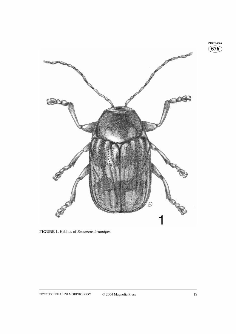
The ventral declivity of the pronotum, the hypomeron (Fig. 2), forms the lateral andpart of the ventral wall of the prothorax and is usually less strongly punctured than thepronotum. The hypomeron is triangular in shape and is connected to the prosternum alongthe notosternal suture, located anterior to the procoxa. In Cryptocephalini the suture is notwell developed. Its position can be recognized by a dramatic change of punctation of thesurface. The prosternum is more coarsely punctured than the hypomeron (Figs. 2, 50),except in some Pachybrachina. The suture is clearly visible in the lateral corner of the pro-
© 2004 Magnolia Press 5CRYPTOCEPHALINI MORPHOLOGY
676ZOOTAXAcoxal cavity (Figs. 2, 11, 68). The suture is also distinct inside the procoxal cavity,
between the lateral and medial openings (Figs. 11, 26, 68, 93, 113). Posteriorly, beyond theprocoxa, the hypomeron projects ventrally and meets the posterolateral projection of theintercoxal prosternal process. In most Cryptocephalini the apex of the hypomeron projec-tion is cylindrical and has an opening into which the tip of the posterolateral part of theintercoxal prosternal process fits (Figs. 2, 13, 20, 63, 70). However, in many Pachy-brachina the tip of the hypomeral projection and the connecting posterolateral part of theintercoxal prosternal process are dorsoventrally flattened (Figs. 86, 92, 103, 111).
Anterior to the hypomeral projection, further dorsally, deeper inside the prothoraciccavity and connected to internal (ventral) parts of the prosternum, lies a sclerotized arch(Fig. 4). It forms the ventral wall of the prothoracic cavity and bears two symmetricallyplaced appendages called apodemal apophyses (Snodgrass 1935) or furcal arms (Larsén1966). By analogy with internal metathoracic ridges they can also be called proendosterni-tes (Figs. 4, 14, 15). A portion of the sclerotized arch between the proendosternites wascalled the sternacosta by Snodgrass (1935). Whether the sternacosta, and arch as a whole(Fig. 4), is an extension of the hypomeron remains unclear. A lack of distinct sutures inthis area prevents determination of borders between the sclerites.
There are three main possibilities as to the origin of the sternacosta: it could be formedeither by the hypomeron, the prosternum, or a combination of both. Amongst these, thesecond possibility seems more justifiable, by reason of the location of the notosternalsuture at the bottom of the coxal cavity. The presence of the notosternal suture may indi-cate that the structures situated ventral to the intercoxal prosternal process belong to theprosternum. The area at the beginning of the sternacosta, dorsal to the hypomeral projec-tion, has several parallel wrinkles that may be interpreted as a connection of two differentsclerites. Snodgrass (1935) did not comment on the specific origin of this sclerite, but thefact that it is discussed in the paragraph devoted to the prosternum undoubtedly revealsSnodgrass’s thoughts on this matter. Accepting a prosternal origin would conserve existingterminology. Being attached to the sternacosta, proendosternites would have to berenamed if shown that the sternacosta or sclerotized arch is associated with thehypomeron.
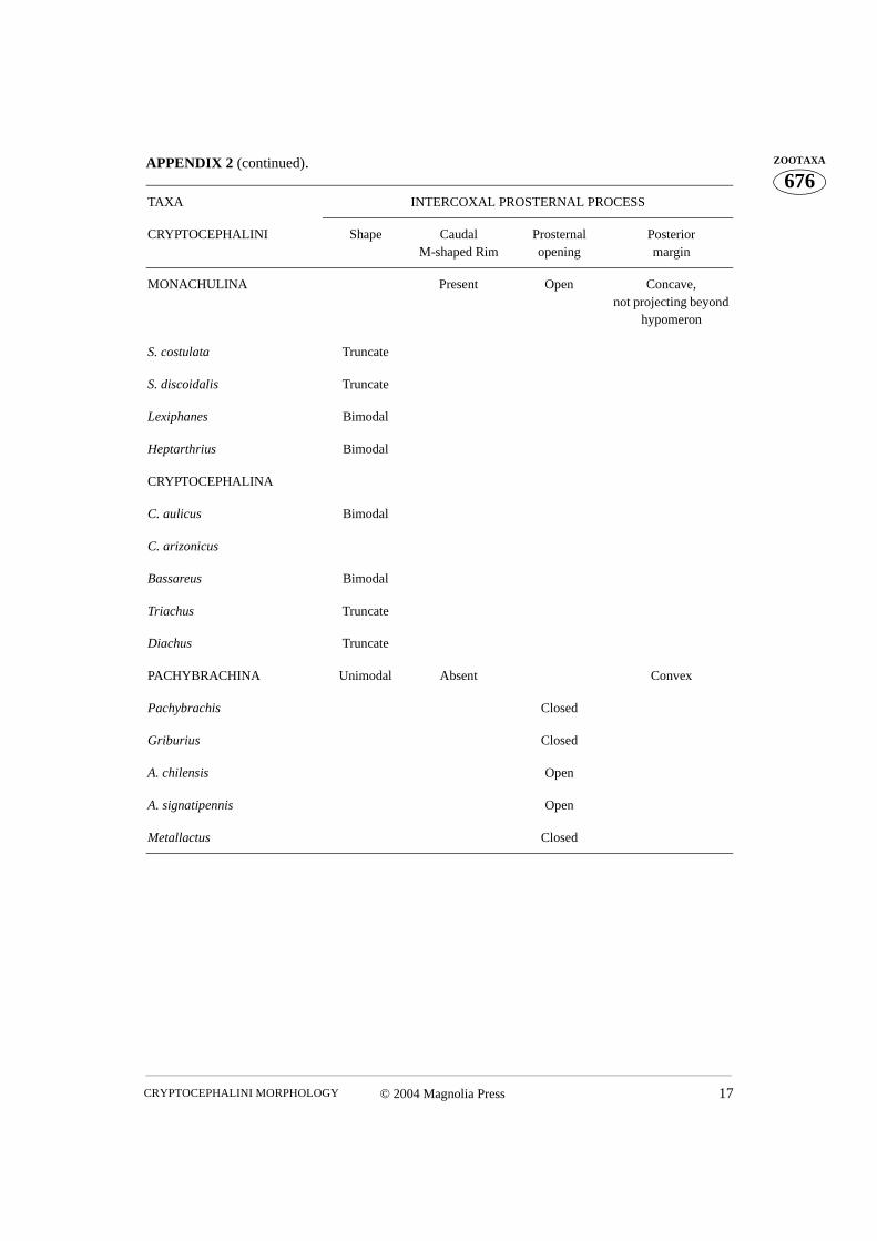
II. The prosternum
A wide and relatively short sclerite, occupying the ventral part of the prothorax, is calledthe prosternum. The section of the prosternum between the procoxal cavities is the inter-coxal prosternal process. Its shape and relative size varies greatly between Cryptocephalinitaxa and it is an important source of, mostly generic level, characters. The anterior marginof the prosternum is usually uniformly concave, however, in some cryptocephalines it isconvex medially and concave laterally, forming two elongate lateral projections (Fig. 21).The prosternal process is flat, but in males of some species it is armed with a relativelylong ‘horn’ (Figs. 21, 24). The posterior margin of the intercoxal prosternal process is usu-
CHAMORRO-LACAYO & KONSTANTINOV6 © 2004 Magnolia Press
676ZOOTAXA ally flat or concave and does not extend beyond the hypomeral projection in the Crypto-
cephalina and Monachulina (Figs. 9, 19, 21, 31, 40, 50, 61, 71, 81). The posterior marginis triangular or oval and usually extends beyond the hypomeral projection in most Pachy-brachina (Figs. 100, 108) (not that pronounced in Pachybrachis, Figs. 85, 88).
Two main kinds of internal structures of the prosternum can be recognized in Crypto-cephalini. In Cryptocephalina and Monachulina the posterior margin of the intercoxalprosternal process is bordered by a well developed internal ridge. The ridge is sometimesthickened medially (Figs. 4, 13, 20, 23, 37, 54, 70, 74, 83). The mesal part of the intercoxalprosternal process is flat with two internally directed lateral projections that are connectedto the hypomeral projections. An opening between the basal part of the sternacosta and theaforementioned projection leads into the procoxal cavity (Figs. 11, 13, 25, 26, 68). Thesternacosta is often situated just dorsal to the tip of the intercoxal prosternal process. Thedorsal surface of the prosternum has a large opening which may lead to a medial openingof the procoxal cavity.
In Pachybrachina the posterior margin of the intercoxal prosternal process has no ridgeand lies in one plane without ventrolateral projections (Figs. 86, 111, 121). Mesally theintercoxal prosternal process sometimes develops a denticle that inserts into a sharp invag-ination of the procoxal cavity, forming a medial articulation point for the coxal hinge (Fig.93). The sternacosta lies deep within the prothoracic cavity, far from the tip of the inter-coxal prosternal process (Figs. 86, 92, 103, 110, 123, 132). Relatively, the intercoxal pros-ternal process lies closer to the sternacosta in Cryptocephalina and Monachulina than itdoes in the Pachybrachina. The hypomeral projection is usually much wider in Pachy-brachina and in some genera (e. g. Griburius) completely closes any opening into the pro-coxal cavity (Fig. 111). In Ambrotodes this opening is well developed (also relativelysmaller than in the Cryptocephalina and Monachulina) (Figs. 121, 132), while in Pachy-brachis it has the shape of a narrow crack (Figs. 86, 92). In Pachybrachina an opening onthe dorsal surface of the prosternum is either small or invisible.
The proendosternites are two variously shaped appendages attached to the sternacostaof the prosternum, directed posteromedially into the prothoracic cavity. The base of eachproendosternite is narrow and thick whereas the apex is wide and thin (Figs. 4, 14). Theyare symmetrically situated, almost exactly across from the connection of the hypomeronand the intercoxal prosternal process. According to Snodgrass (1935), the proendosternitessupport the principal longitudinal muscles of the thorax called M19 by Baehr (1979). Dueto their relative simplicity, we did not find that the proendosternites provided any valuablediagnostic characters at the generic level.
III. The procoxal cavity (Figs. 11, 26, 35, 68, 93, 113, 122)
Usually the procoxal cavity in Cryptocephalini is transverse in shape (Fig. 11), but in somegenera it is nearly as wide as long (Fig. 26). It is wider medially than laterally. The antero-
© 2004 Magnolia Press 7CRYPTOCEPHALINI MORPHOLOGY
676ZOOTAXAmedial part of the bottom of the coxal cavity, the cryptosternum (Hlavac 1972), is formed
by the prosternum (Figs. 2, 68). The posterolateral wall of the cavity is formed by thehypomeron. There are two main openings at the bottom of the coxal cavity. The lateralopening is the largest and almost completely occupies the lateral corner of the cavity. It issometimes bordered anteromedially by a moderately raised ridge. The trochantin andattached endopleuron reach endolateral areas of the prothoracic cavity through this open-ing. The posterior edge of the opening, formed by the hypomeron, has a variously shapedridge (Figs. 68, 113) normally obscured by the coxa. Posteromesal to the lateral opening,separated by a narrow bridge, is a second much smaller and more rounded opening. Thisopening is closed posteriorly (Fig. 111) in cryptocephaline beetles with wide hypomeralprojections fitting exactly between the intercoxal prosternal process and the sternacosta ofthe prosternum. Our preparations did not allow us to see exactly where it leads in Gribu-rius, but an obvious assumption is that it leads into the prothoracic cavity. In Cryptocepha-lina, Monachulina, and to a lesser degree in the rest of Pachybrachina, the opening alsoleads towards the posterior, so that the coxa can be seen above the sternacosta (Figs. 4, 37,83). The bridge separating the lateral and medial opening usually has a well developednotosternal suture (Figs. 2, 68). Posteriorly the procoxal cavities are closed, or very nar-rowly open (only in the Pachybrachina), with the hypomeral projection joined to the poste-rolateral projection of the intercoxal prosternal process. IV. The trochantin and endopleuron (Figs. 5, 6, 16, 18, 28–30, 38–39, 56–58, 78, 105, 112, 117, 126)
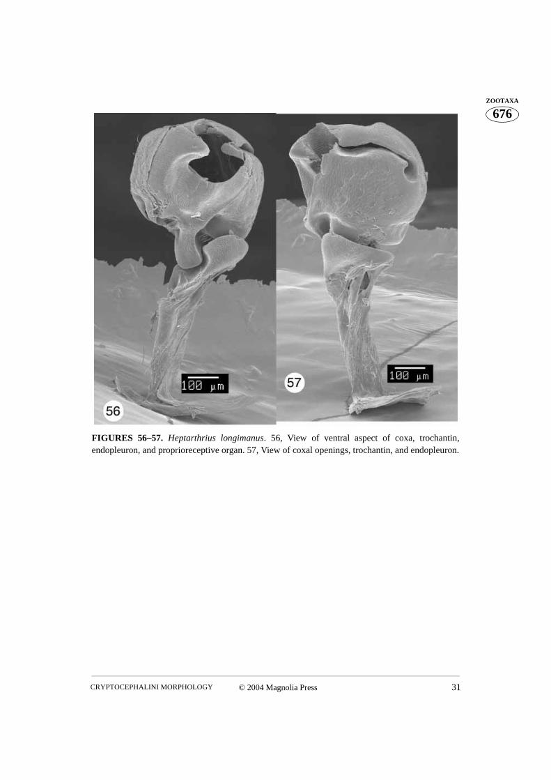
The trochantin is the smallest sclerite of the prothorax. Homologically it has been traced tothe coxopleurite, being its prearticular part in plecopterous insects (Snodgrass 1935). It istriangular in shape and the ventral (distal) edge of the trochantin rests on the lateral rim ofthe procoxa (Figs. 56–57). The posterior apex of the distal edge of the trochantin fitsbetween the lateral coxal projection and the lateral rim. A visible group of stiff setae, theproprioreceptive organ, are sensitive to movement between the trochantin and the coxa(Figs. 56, 58). The anterior apex of the trochantin borders the lateral rim of the coxa andacts as a lever (Evans 1971). A few dissections allowed us to see a tendon attached to theanteroapical, ventral (distal) edge of the trochantin (Figs. 16, 18), a potential attachmentsite for the tergo-pleural muscle (Evans 1971). The proximal apex of the trochantin isdirected dorsally along the endopleuron. The trochantin is externally visible, fitting intothe lateral corner of the procoxal cavity (Figs. 44, 67, 104). The connection between thetrochantin and the coxa is membranous, allowing for some articulation with what appearsto be a monocondylic joint (Figs. 97, 105).The endopleuron is an internal remnant of thepropleuron. It is attached to the internal surface of the trochantin and is situated in the lat-eral part of the procoxal cavity. It consists of a narrow stalk connected laterally to the tro-chantin (continuing further and apicomedially disappears into a large lateral opening in thecoxa) and a wide “base” directed dorsally (Figs. 4, 36). According to Hlavac (1972) the
CHAMORRO-LACAYO & KONSTANTINOV8 © 2004 Magnolia Press
676ZOOTAXA endopleuron provides increased surface area for muscle attachment. The endopleuron var-
ies slightly in relative length of the stalk and greatly in width of the base in different Cryp-tocephalini.
V. The procoxa and trochanter (Figs. 5–7, 16, 18, 28–30, 38–39, 48–49, 56–57, 59–60, 67, 69, 76, 78, 80, 84, 87, 96–99,106–107, 112, 114–117, 126–130, 133–136)
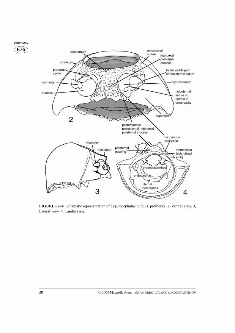
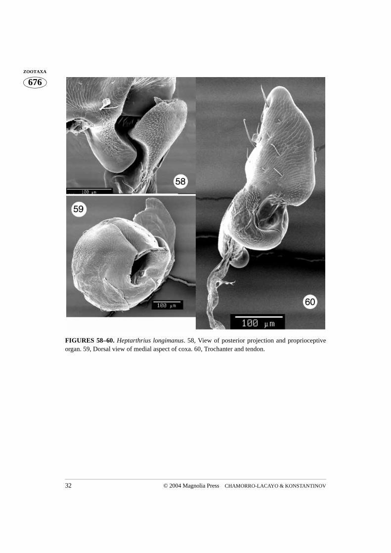
Although the coxa is a basal leg segment, it forms a functional unity together with the pro-thorax, being positioned within the procoxal cavity and attached to the trochantin. Theprocoxa is globular in shape and has three openings. The large coxal opening is ventrallysituated, inside the wide groove in which the apex of the trochanter and base of the profe-mur fit. The posterior aspect of the groove is equipped with a variously shaped projection.This projection, called the posterior projection below, fits into the indentation between thespiral ridge and the apical parts of the trochanter (Fig. 76). The coxal suture extends fromthe anterior margin of the ventral coxal opening to the much smaller dorsal opening (Figs.5, 6, 16). When the coxa is positioned within the procoxal cavity, its ventral opening fac-ing outward, the dorsal opening is hidden within the procoxal cavity (Figs. 52, 67, 76, 81).However, if the coxa is rotated towards the posterior, the dorsal opening can be observed(Figs. 27, 44). The medial aspect of the coxa is more or less uniformly convex. The lateralaspect is flattened and contains the largest opening, which can only be observed when thecoxa is removed from the coxal cavity (Fig. 97). The rimmed side of this opening (the lat-eral coxal opening) has a tendon attached to it (Figs. 97–99). Several membranes attachthe trochantin to this part of the coxa which bears a patch of stiff, short setae, termed prop-rioreceptive organs by Larsén (1966) (Fig. 17, 105). Terminal neurons of the receptorsdetect movement between the trochantin and the coxa, relaying back position, accelera-tion, and velocity information to the ganglia. Neurons in turn relay information to the mus-cles, allowing for a concerted movement of the insect (Chapman 1998). The anterioraspect of the ventral coxal groove forms a ridge which continues laterally forming a rela-tively long and curved projection (here termed lateral projection). This projection limitsposterior movement of the trochantin. The coxal “ball”, of the monocondylic “ball andsocket” joint of the trochantin and coxal articulation, is clearly visible in Figs. 49, 97, 105,117. This joint is situated anteriad of the curved lateral projection and consists of a deepgroove separating it from the lateral projection and the rim (part of it has proprioreceptivesensillae). The coxal tendon is situated nearly opposite this joint.
The trochanter has a complex shape (Figs. 7, 48, 60, 87, 106–107, 114, 129, 134). Itfits into the procoxa running the entire length of the coxal suture, its wide ventrodistalaspect protruding from the ventral coxal opening and its narrow dorsoproximal aspect vis-ible through the small dorsal opening of the procoxa (Figs. 27, 80). The profemur attachesto the wide, ventral aspect of the trochanter. An interesting feature of the trochanter is aspiral, transverse median ridge. When the trochanter is inserted into the coxa the ridge lies
© 2004 Magnolia Press 9CRYPTOCEPHALINI MORPHOLOGY
676ZOOTAXAon the coxal surface, where it slides when the trochanter rotates. The ventrally situated,
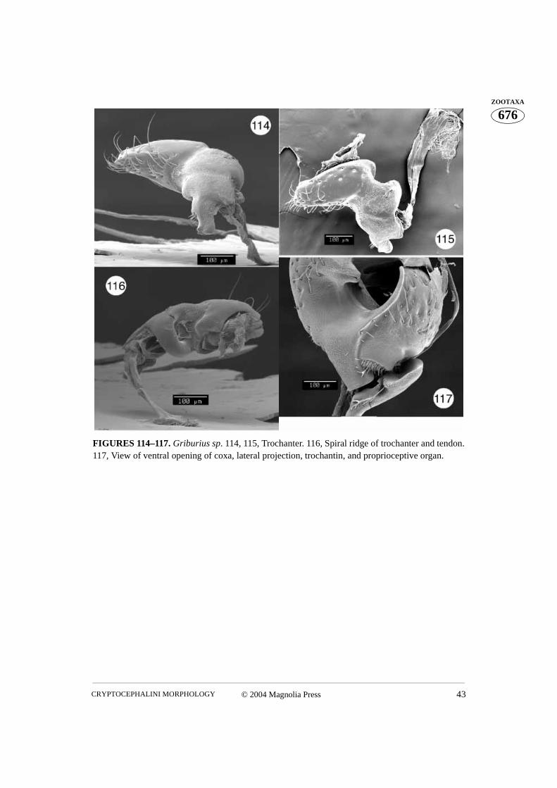
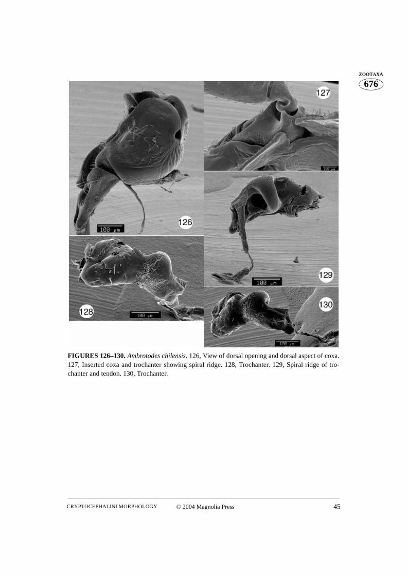
posterior projection of the coxa is inserted between the wide, ventral end and the spiralridge of the trochanter. Proprioreceptive sensillae cover the trochanteral surface betweenthe wide, ventral apex and the spiral ridge, as well as the side of the dorsal aspect. A ten-don is visible which attaches the dorsal aspect and the transverse spiral ridge of the tro-chanter (Figs. 7, 48, 60, 84, 114–116, 129–130). The trochanter of Metallactus decumanusclearly possesses a bifid dorsal aspect (Figs. 106–107). However, only one dorsal openingis present in the coxa. Pachybrachis gayi has only one dorsal aspect, similar to the otherspecies studied, but a small subapical projection is also apparent (Fig. 87).
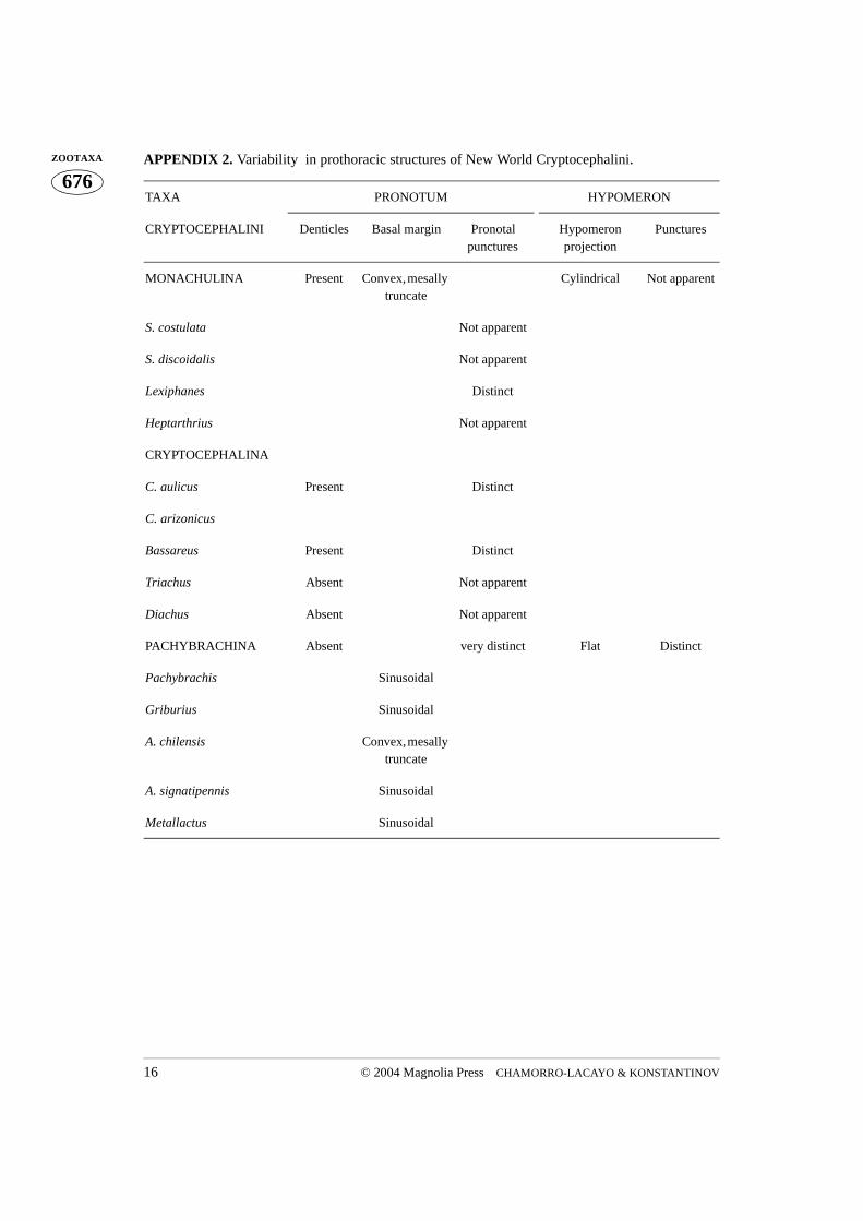
VI. Variability in prothoracic structures of New World Cryptocephalini (see Appendix 2)
Our study of New World Cryptocephalini revealed a set of characters of obvious diagnos-tic and possible phylogenetic value. Distribution of these characters is described below.
MONACHULINAPRONOTUM: Denticles present on basal margin (Figs. 54, 63, 74, 83); basal margin
convex and truncate mesally, anteriad of mesoscutellum (Figs. 53, 64, 73, 82); pronotalpunctures distinct all over or absent, strongly pronounced on all margins. HYPOMERON:Hypomeral projection cylindrical (Figs. 54, 63, 74, 83); hypomeron without apparentpunctures (Figs. 50, 61, 71, 81). PROSTERNUM: Intercoxal prosternal process truncate orbimodal; intercoxal prosternal process with caudal M-shaped rim (Figs. 54, 70, 74, 83);prosternal opening wide, when viewed caudally (Figs. 54, 70, 74, 83); posterior margin ofintercoxal prosternal process concave, never projecting beyond hypomeron (Figs. 50, 61,71, 81); anterior margin of prosternum uniformly concave, with a medial flange, or withtwo submedial flanges (Figs. 50, 61, 71, 81); intercoxal width greater than width of coxalcavity. TROCHANTER: With a single dorsoproximal end (Figs. 60, 84).
Stegnocephala: Pronotal punctures absent (Figs. 71, 81). Intercoxal prosternal processtruncate (Fig. 81); anterior margin of prosternum with a medial flange (Fig. 71), or twosubmedial flanges (Fig. 81).
Lexiphanes: Pronotal punctures distinct throughout (Fig. 64). Intercoxal prosternalprocess bimodal with small lateral projections (Fig. 62); anterior margin of prosternumuniformly concave (Fig. 61).
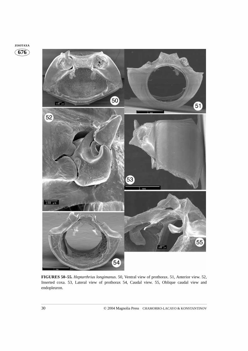
Heptarthrius: Pronotal punctures absent (Fig. 53). Intercoxal prosternal process bimo-dal (Fig. 51); anterior margin of prosternum uniformly concave (Fig. 50).
CRYPTOCEPHALINAPRONOTUM: Denticles present (Figs. 15, 19, 23) or absent on basal margin (Figs. 33,
42); basal margin convex and mesally truncate over mesoscutellum (Figs. 9, 19, 23, 33,42); pronotal punctures absent or distinct throughout, strongly pronounced on all margins.
CHAMORRO-LACAYO & KONSTANTINOV10 © 2004 Magnolia Press
676ZOOTAXA HYPOMERON: Hypomeron projection cylindrical (Fig. 13); hypomeron without distinct
punctures (Figs. 9, 12). PROSTERNUM: Intercoxal prosternal process truncate, bimodalor acutely bimodal; intercoxal prosternal process with caudal M-shaped rim (Figs. 13, 20);prosternal opening open, when viewd caudally; posterior margin of intercoxal prosternalprocess concave, never projecting beyond hypomeron (Fig. 9); anterior margin of proster-num uniformly concave or with medial flange; intercoxal width equal to, greater than, orless than width of coxal cavity.
Cryptocephalus: Denticles present on basal margin of pronotum (Figs. 15, 19); prono-tal punctures distinct throughout (Figs. 10, 12). Intercoxal prosternal process acutelybimodal (Figs. 13, 20); anterior margin of prosternum uniformly concave or with medialflange; intercoxal width equal to or greater than width of coxal cavity (Figs. 9, 19).
Bassareus: Denticles present on basal margin of pronotum (Fig. 23); pronotal punc-tures distinct throughout (Figs. 23, 24). Intercoxal prosternal process bimodal (in malesintercoxal prosternal process with medial projection) (Fig. 23); anterior margin of proster-num with medial flange (in males with sublateral indentations and lateral projections) (Fig.21); intercoxal width equal to width of coxal cavity; without pronounced ridge borderingcoxal cavity ending as small lateral projections on intercoxal prosternal process.
Triachus: Denticles absent on basal margin of pronotum (Figs. 40, 42); pronotal punc-tures absent (Fig. 43). Intercoxal prosternal process truncate (Fig. 40). Anterior margin ofprosternum uniformly concave (Fig. 40); intercoxal width less than width of coxal cavity.
Diachus: Denticles absent on basal margin of pronotum (Figs. 31, 33); pronotal punc-tures absent (Fig. 34). Intercoxal prosternal process truncate. Anterior margin of proster-num uniformly concave; intercoxal width less than width of coxal cavity (Fig. 31).
PACHYBRACHINAPRONOTUM: Denticles absent on basal margin (Figs. 85, 88, 100, 108, 118, 131);
basal margin convex and sinusoidal, mesally truncate over mesoscutellum; pronotal punc-tures distinct throughout, strongly pronounced on all margins (Figs, 90, 102, 120).HYPOMERON: Hypomeral projection flat (Figs. 86, 91, 103, 111, 123); hypomeron withdistinct punctures. PROSTERNUM: Prosternum similarly or more coarsely puncturedthan hypomeron; intercoxal prosternal process unimodal (Figs, 85, 88, 100, 108, 118,122); intercoxal prosternal process without caudal M-shaped rim (Figs. 91, 103, 111, 123);prosternal opening narrowly open or closed in caudal view; posterior margin of intercoxalprosternal process convex, projecting beyond hypomeron (Figs. 85, 88, 100, 108, 118,131); anterior margin of prosternum uniformly concave; intercoxal width equal to, greaterthan, or less than width of coxal cavity. TROCHANTER: With two dorsoproximal ends,with a subapical projection, or with only one dorsal end (Figs. 87, 106–107, 114–116,128–130, 134–135).
Pachybrachis: Basal margin of pronotum sinusoidal (Figs. 85, 88). Prosternum andhypomeron equally coarsely punctured; prosternal opening closed in caudal view; anterior
© 2004 Magnolia Press 11CRYPTOCEPHALINI MORPHOLOGY
676ZOOTAXAmargin of prosternum uniformly concave (Figs. 85, 88); intercoxal width less than width
of coxal cavity. Trochanter with subapical projection on dorsoproximal end (Fig. 87)Griburius: Basal margin of pronotum sinusoidal (Fig. 108). Prosternum more coarsely
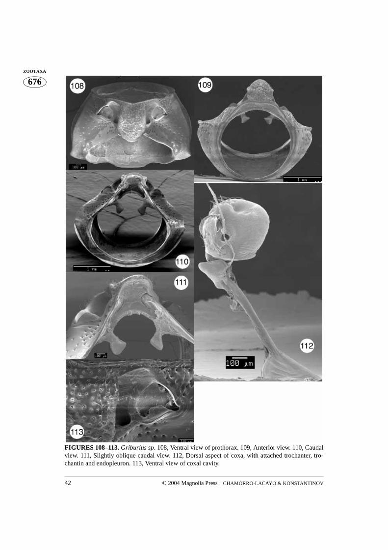
punctured than hypomeron; prosternal opening closed, in caudal view (Fig. 111); anteriormargin of prosternum uniformly concave (Fig. 108); intercoxal width greater than width ofcoxal cavity. Trochanter with one dorsal end (Figs. 114–116).
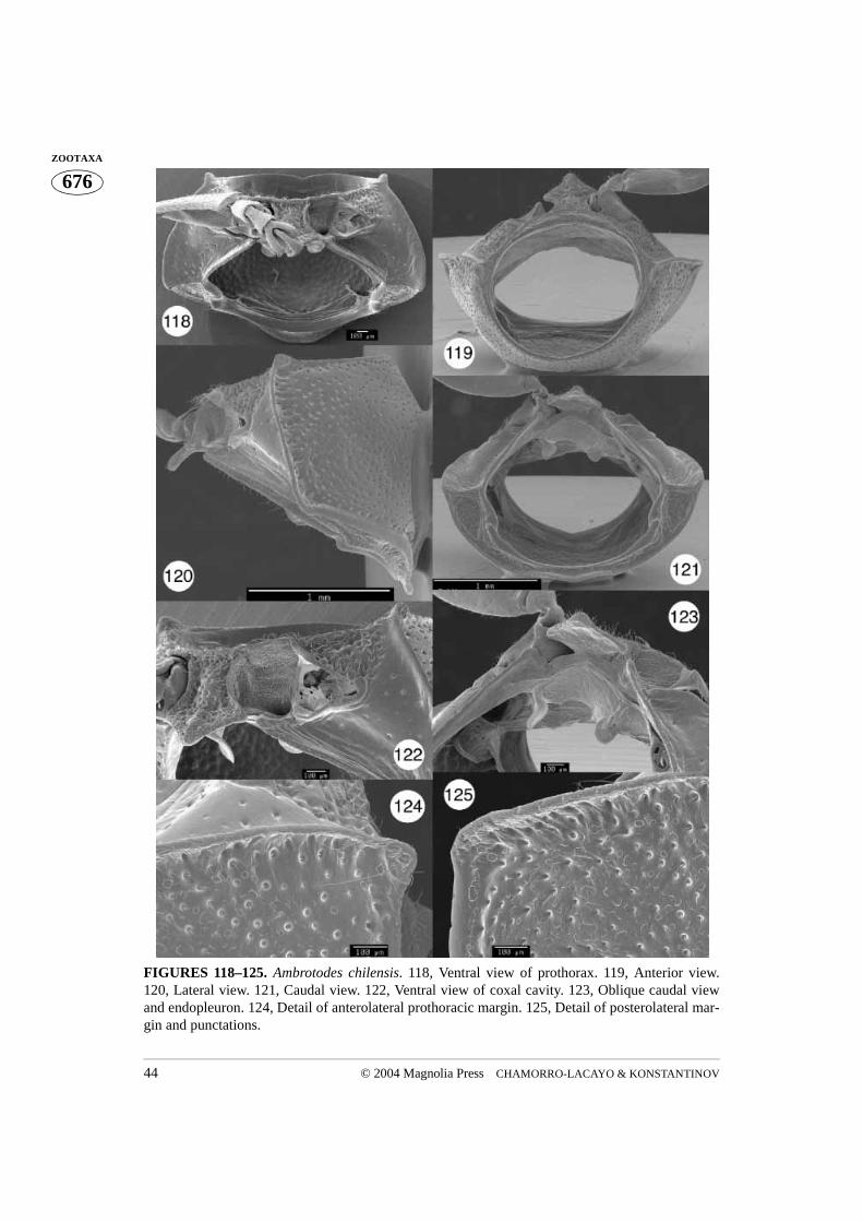
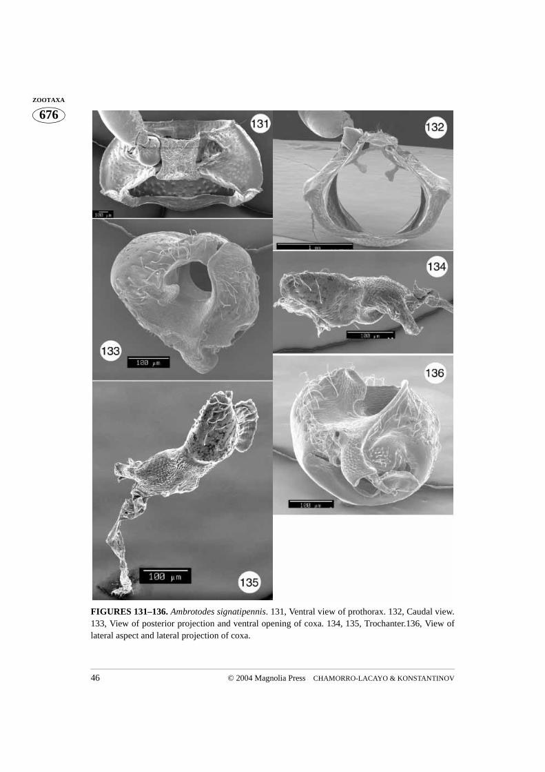
Ambrotodes: Basal margin of pronotum sinusoidal (A. signatipennis) (Fig. 131) orconvex and mesally truncate (A. chilensis) (Fig. 118). Prosternum more coarsely punc-tured than hypomeron; prosternal opening slightly open in caudal view (Figs. 121, 132);intercoxal cavity less than (A. chilensis) or greater than (A. signatipennis) width of coxalcavity. Trochanter with one dorsal end (Figs. 128–130).
Metallactus: Basal margin of pronotum sinusoidal (Fig. 100). Prosternum morecoarsely punctured than hypomeron; prosternal opening closed, in caudal view (Fig. 103);intercoxal width equal to width of coxal cavity (Fig. 100). Trochanter with two dorsoprox-imal ends (Figs. 106–107).
Conclusions
Most of the sclerites of the prothorax have joined and are therefore immobile relative toeach other, forming a structurally rigid body part. The only movable sclerites are the coxawith the attached trochantin and imbedded trochanter. Their movements determine themovement of the profemur. Due to the position of the articulation point on the intercoxalprosternal process, a horizontal hinge is formed. The procoxa has a limited ability to rotatearound an axis between this point and the lateral corner of the procoxal cavity where thetrochantin is attached. Articulation between the coxa and the trochantin is complicated.How the movement of the coxa translates into movement of the trochantin remainsunclear. One articulation point between the coxa and the trochantin is obvious. It is formedby the lateral projection, impression and rim of the procoxa, and the posterior corner of thetrochantin. This articulation point limits vertical movement of the trochantin and theattached endopleuron. Lawrence and Britton (1994) suggest that the coxa articulates withthe trochantin at two points in the Polyphaga, however, we did not find the second point inthe Cryptocephalini and therefore describe this joint as monocondylic (Figs. 49, 97, 105,117).
Movement of the trochanter consists mainly of rotation along its own axis within thecoxal cavity. This rotation allows the distal portion of the profemur and the rest of the leg awide range of movement. Rotation of the trochanter is caused by contraction of a muscleattached to the trochanter tendon. If the tendon is attached to the trochanter longitudinally,it will pull the trochanter inside the coxal cavity causing its spiral ridge to slide along thecoxal groove and the posterior projection of the coxa, hereby forcing the trochanter torotate (Fig. 8). As we did not observe these movements, our description of transferred
CHAMORRO-LACAYO & KONSTANTINOV12 © 2004 Magnolia Press
676ZOOTAXA advance movement into rotation is a hypothesis based on morphological observations. We
also failed to find a tendon, indicative of muscle attachment to pull the trochanter in theopposite direction, causing it to return to its original position. However, the coxal cuticlemay function as a spring, pushing the trochanter back after having been displaced by itsown vertical movement. If the tendon were transversely attached to the trochanter then theassociated muscle would rotate it upon contraction. The subsequent vertical movement,due to the position of the oblique spiral ridge, would transfigure the coxal cuticle, enablingto return the trochanter to its original position when the muscle relaxed. In the latter sce-nario, circular movement is transferred to advance movement. In both cases the vertical, oradvance, movement of the trochanter inside the coxa is responsible for squeezing the coxaand eventually returning the trochanter to its original position. Therefore, the trochanterand coxa represent a device which transfers one kind of movement into another. However,whether the advance movements are being transferred into rotation or the opposite,remains unclear.
This study reveals a number of useful morphological characters that may supplementphylogenetic studies of the group and further clarify relationships at the generic level andbeyond. Distribution of the main characters generally supports current generic classifica-tion of New World Cryptocephalini, but favors two family level taxa instead of the threecurrently recognized. Two main types of prothorax were found amongst the Cryptocepha-lini. The first type occurs in the Cryptocephalina and Monachulina. They share the follow-ing main character states (View Appendix 2 for table): cylindrical hypomeral projection;posterior margin of the intercoxal prosternal process not projecting beyond hypomeral pro-jection; “three dimensional” intercoxal prosternal process with totally different internalprosternal structures, described in detail above (they include the internal ridge on the mar-gin of the intercoxal prosternal process and some previously poorly described openings).They also have strongly dentate basal margin, with the exception of Diachus and Triachus,which we view as a secondary loss due to small body size. In Pachybrachina thehypomeral projection is flat; the posterior margin of the intercoxal prosternal processprojects beyond hypomeral projection; flat intercoxal prosternal process and a number ofunique features in the internal structures of prosternum; and in some genera a distinct tro-chanter. Without rigorous analysis of a variety of morphological structures we do not pro-pose any formal changes to the higher classification of the Cryptocephalini, as this wouldbe premature.
Generic level characters include the shape, details of margins, and surface structure ofthe pronotum, intercoxal prosternal process, as well as the relative width of the intercoxalprosternal process. The procoxa, trochantin, and to a lesser extent the trochanter, differ insmall details which were impossible to qualify as well separated character states. Thesestructures may prove useful at a higher level.
© 2004 Magnolia Press 13CRYPTOCEPHALINI MORPHOLOGY
676ZOOTAXAAcknowledgements
We are grateful to S. Braden and S. Whittaker (SEM Laboratory, National Museum of Nat-ural History, Smithsonian Institution) for assisting with scanning electron microscopy. Weare greatly thankful to M. Baehr (Zoologische Staatssammlung München), N. E. Woodleyand A. N. Vandenberg (Systematic Entomology Laboratory, USDA, Washington, DC) andE. Grobelaar (Plant Protection Research Institute, Pretoria, South Africa) for reviewingthis manuscript and providing valuable suggestions.
The senior author became interested in cryptocephalines while working with T. Erwinat the National Museum of Natural History, Smithsonian Institution, and finished thisproject while at the University of Minnesota as R. Holzenthal’s advisee and being fundedby NSF PEET grant number DEB-0117772, and is grateful for their support, enthusiasm,and encouragement.
Literature cited
Baccetti, B. & Daccordi, M. (1988) Sperm structure and phylogeny of the Chrysomelidae. In: Jol-ivet, P., Petitpierre, E. & Hsiao, H.H. (Eds.). Biology of Chrysomelidae. Kluwer AcademicPublishers, Dordrecht/Boston/London, pp. 357–378.
Baehr, M. (1979) Vergleichende Untersuchungen am Skelett und an der Coxalmuskulatur des Pro-thorax der Coleoptera. Ein Beitrag zur Klärung der phylogenetischen Beziehungen der Adeph-aga (Coleoptera, Insecta). Zoologica. Originalbhandlungen aus dem Gesamtgebiet derZoologie. 44. Band, 4. Lieferung, Heft 130. E. Schweizerbart’sche Verlagsbuchhandlung, Stut-tgart, 76 pp.
Blackwelder, R.E. (1944) Checklist of the Coleopterous Insects of Mexico, Central America, theWest Indies, and South America. Smithsonian Institution, United States National Museum Bul-letin, 185(1–6), 639–647.
Chapman, R.F. (1998) The Insects: Structure and Function. 4th Edition. Cambridge UniversityPress. United Kingdom, v–xvii + 770 pp.
Erber, D. (1988) Biology of Camptosomata Clytrinae, Cryptocephalinae, Chlamisinae, Lamproso-matinae, In: P. Jolivet, E. Petitpierre & Hsiao, T.H. (Eds.), Biology of Chrysomelidae. KluwerAcademic Publishers. Dordrecht, pp. 513–552.
Evans, M.E.G. (1971) Morphology and phylogeny in Coleoptera — the prothorax and procoxae.International Congress of Entomology. XIII, 1, 242–243.
Hlavac, T.F. (1972) The prothorax of Coleoptera: origin, major features of variation. Psyche, 79(3),123–149.
Larsén, O. (1966) On the Morphology and Function of the Locomotor Organs of the Gyrinidae andother Coleoptera. Opuscula Entomologica Supplement, 30, 1–242.
Lawrence, J.F. & Britton, E.B. (1994) Australian Beetles. Melbourne University Press. 192 pp.Lopatin, I.K. (1977) Leaf beetles (Chrysomelidae) of Middle Asia and Kazakhstan. “Nauka”, Len-
ingrad, 268 pp. (In Russian)McHugh, J.V., C. J. Marshall, F. L. Fawcett. 1997. A Study of Adult Morphology in Megalodacne
heros (Say) (Coleoptera: Erotylidae). Transactions of the American Entomological Society, 123(4), 167–223.
Riley, E.G., Clark, S.M., Flowers, R.W. & Gilbert, A.J. (2002) 124. Chrysomelidae Latreille 1802.In: Arnett, R. H., Thomas, M.C., Skelley, P.E. & Frank, J.H. (Eds.), American Beetles. Volume
CHAMORRO-LACAYO & KONSTANTINOV14 © 2004 Magnolia Press
676ZOOTAXA 2. CRC Press. Boca Raton, London, New York, Washington, D.C., pp. 617–691.
Reid, C.A.M. (1995) A Cladistic Analysis of Subfamilial Relationships in the Chrysomelidae sensulato (Chrysomeloidea). In: Pakaluk, J. & Slipinski, S.A. (eds.) Biology, phylogeny, and classifi-cation of Coleoptera: Papers Celebrating the 80th Birthday of Roy A. Crowson. Muzeum IInstytut Zoologii PAN, Warszawa, pp. 559–631.
Samuelson, G. A. (1996) Binding sites: elytron-to-body meshing structures of possible significancein the higher classification of Chrysomeloidea. In: Jolivet, P.H.A. & Cox, M.L. (eds.), Chry-somelidae Biology. Volume 1: The Classification, Phylogeny and Genetics. SPB AcademicPublishing bv. The Netherlands, pp. 267–290.
Seeno, T.N. & Wilcox, J.A. (1982) Leaf Beetle Genera (Coleoptera: Chrysomelidae). Entomogra-phy, 1, 1–221.
Snodgrass, R.E. (1935) Principles of Insect Morphology. McGraw-Hill Book Company, Inc. NewYork and London, 667 pp.
Suzuki, K. (1988) Comparative morphology of the internal reproductive system of the Chrysomel-idae. In: Jolivet, P., Petitpierre, E. & Hsiao, H.H. (Eds.), Biology of Chrysomelidae. KluwerAcademic Publishers, Dordrecht/Boston/London, pp. 317–355.
White, R.E. (1968) A Review of the Genus Cryptocephalus in America North of Mexico (Chry-somelidae: Coleoptera). United States National Museum Bulletin, 290, 1–124.
Wilcox, J.A. (1975) Checklist of the Chrysomelidae of Canada, United States, Mexico, CentralAmerica and the West Indies. North American Beetle Fauna Project. Red Version.BiologicalResearch Institute, NY, 166 pp.
© 2004 Magnolia Press 15CRYPTOCEPHALINI MORPHOLOGY
676ZOOTAXAAPPENDIX 1. List of Cryptocephalini for which SEM images were taken.
Subtribe Genus Species Author Country
Cryptocephalina Bassareus Haldeman
B. brunnipes (Olivier) Florida, USA
Cryptocephalus Geoffroy
C. arizonicus Schaeffer Arizona, USA
C. aulicus Haldeman Florida, USA
Diachus LeConte
D. auratus (Fabricius) Guatemala
Triachus LeConte
T. vacuus LeConte Maryland, USA
Monachulina Heptarthrius Suffrian
H. longimanus Suffrian Perú
Lexiphanes Gistel
L. coenobita Suffrian Brazil
Stegnocephala Baly
S. discoidalis Baly Argentina
S. costulata (Suffrian) Brazil
Pachybrachina Ambrotodes Suffrian
A. chilensis (Blanchard) Argentina
A. signatipennis (Blanchard) Argentina
Griburius Haldeman
G. sp. Mexico
Metallactus Suffrian
M. albopictus Suffrian Brazil
M. decumanus Suffrian Brazil
Pachybrachis Chevrolat
P. gayi Blanchard Chile
P. hepaticus Melsheimer California
CHAMORRO-LACAYO & KONSTANTINOV16 © 2004 Magnolia Press
676ZOOTAXA APPENDIX 2. Variability in prothoracic structures of New World Cryptocephalini.
TAXA PRONOTUM HYPOMERON
CRYPTOCEPHALINI Denticles Basal margin Pronotal punctures
Hypomeronprojection
Punctures
MONACHULINA Present Convex, mesally truncate
Cylindrical Not apparent
S. costulata Not apparent
S. discoidalis Not apparent
Lexiphanes Distinct
Heptarthrius Not apparent
CRYPTOCEPHALINA
C. aulicus Present Distinct
C. arizonicus
Bassareus Present Distinct
Triachus Absent Not apparent
Diachus Absent Not apparent
PACHYBRACHINA Absent very distinct Flat Distinct
Pachybrachis Sinusoidal
Griburius Sinusoidal
A. chilensis Convex, mesally truncate
A. signatipennis Sinusoidal
Metallactus Sinusoidal
© 2004 Magnolia Press 17CRYPTOCEPHALINI MORPHOLOGY
676ZOOTAXAAPPENDIX 2 (continued).
TAXA INTERCOXAL PROSTERNAL PROCESS
CRYPTOCEPHALINI Shape Caudal M-shaped Rim
Prosternal opening
Posterior margin
MONACHULINA Present Open Concave, not projecting beyond
hypomeron
S. costulata Truncate
S. discoidalis Truncate
Lexiphanes Bimodal
Heptarthrius Bimodal
CRYPTOCEPHALINA
C. aulicus Bimodal
C. arizonicus
Bassareus Bimodal
Triachus Truncate
Diachus Truncate
PACHYBRACHINA Unimodal Absent Convex
Pachybrachis Closed
Griburius Closed
A. chilensis Open
A. signatipennis Open
Metallactus Closed
CHAMORRO-LACAYO & KONSTANTINOV18 © 2004 Magnolia Press
676ZOOTAXA APPENDIX 2 (continued).
TAXA PROSTERNUM TROCHANTER
CRYPTOCEPHALINI Anterior margin
Intercoxal width
Proximal ends
MONACHULINA Greater than width coxal cavity
One
S. costulata 2 flanges
S. discoidalis 1 medial
Lexiphanes Concave
Heptarthrius Concave
CRYPTOCEPHALINA
C. aulicus Concave Equal to
C. arizonicus 1 medial Greater than
Bassareus 1 medial Equal to
Triachus Concave Less than
Diachus Concave Less than
PACHYBRACHINA Concave
Pachybrachis Less than Subapical projection
Griburius Greater than One
A. chilensis Less than One
A. signatipennis Greater than One
Metallactus Equal to Two
© 2004 Magnolia Press 19CRYPTOCEPHALINI MORPHOLOGY
676ZOOTAXA
FIGURE 1. Habitus of Bassareus brunnipes.
CHAMORRO-LACAYO & KONSTANTINOV20 © 2004 Magnolia Press
676ZOOTAXA
FIGURES 2–4. Schematic representation of Cryptocephalus aulicus. prothorax. 2, Ventral view. 3,Lateral view. 4, Caudal view.
© 2004 Magnolia Press 21CRYPTOCEPHALINI MORPHOLOGY
676ZOOTAXA
FIGURES 5–8. Schematic representation of Cryptocephalus aulicus. 5, Ventral aspect of coxa withattached trochantin and endopleuron. 6, Dorsal aspect of coxa with attached trochantin. 7, Tro-chanter. 8, Diagrammatic representation of trochanter movement within coxa.
CHAMORRO-LACAYO & KONSTANTINOV22 © 2004 Magnolia Press
676ZOOTAXA
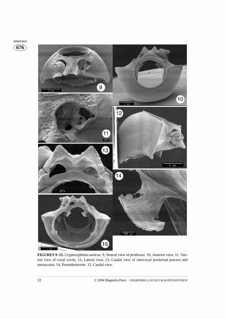
FIGURES 9–15. Cryptocephalus aulicus. 9, Ventral view of prothorax. 10, Anterior view. 11. Ven-tral view of coxal cavity. 12, Lateral view. 13, Caudal view of intercoxal prosternal process andsternacosta. 14, Proendosternite. 15, Caudal view.
© 2004 Magnolia Press 23CRYPTOCEPHALINI MORPHOLOGY
676ZOOTAXA
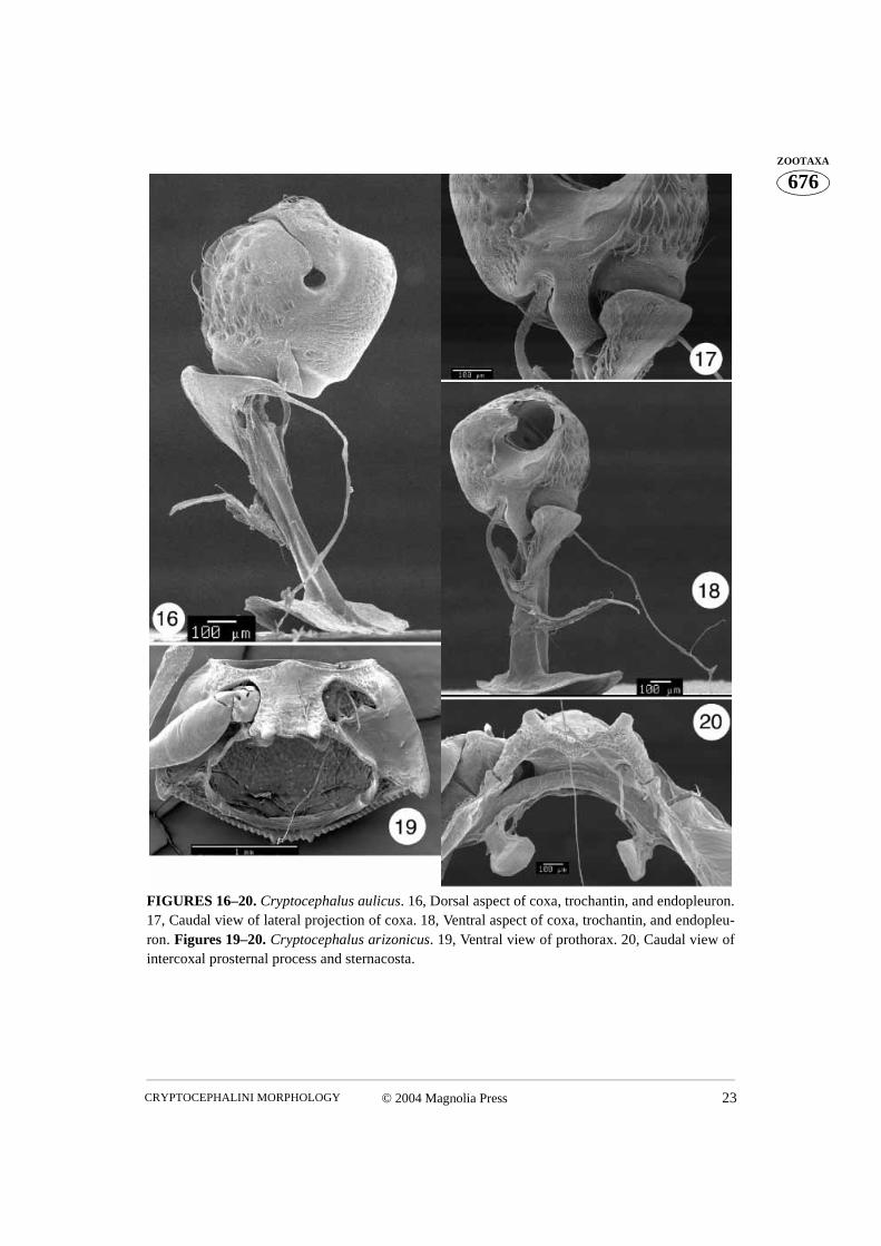
FIGURES 16–20. Cryptocephalus aulicus. 16, Dorsal aspect of coxa, trochantin, and endopleuron.17, Caudal view of lateral projection of coxa. 18, Ventral aspect of coxa, trochantin, and endopleu-ron. Figures 19–20. Cryptocephalus arizonicus. 19, Ventral view of prothorax. 20, Caudal view ofintercoxal prosternal process and sternacosta.
CHAMORRO-LACAYO & KONSTANTINOV24 © 2004 Magnolia Press
676ZOOTAXA
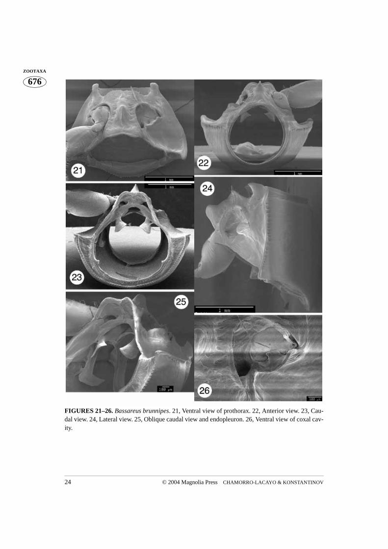
FIGURES 21–26. Bassareus brunnipes. 21, Ventral view of prothorax. 22, Anterior view. 23, Cau-dal view. 24, Lateral view. 25, Oblique caudal view and endopleuron. 26, Ventral view of coxal cav-ity.
© 2004 Magnolia Press 25CRYPTOCEPHALINI MORPHOLOGY
676ZOOTAXA
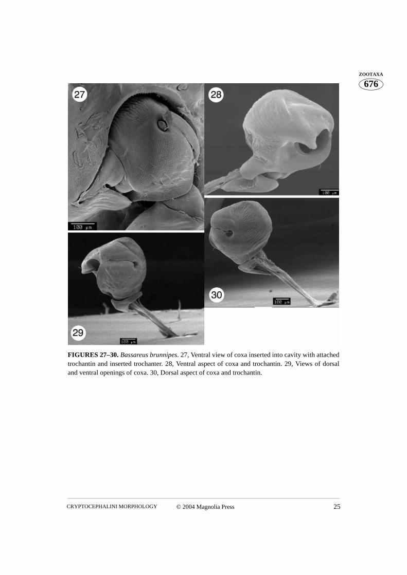
FIGURES 27–30. Bassareus brunnipes. 27, Ventral view of coxa inserted into cavity with attachedtrochantin and inserted trochanter. 28, Ventral aspect of coxa and trochantin. 29, Views of dorsaland ventral openings of coxa. 30, Dorsal aspect of coxa and trochantin.
CHAMORRO-LACAYO & KONSTANTINOV26 © 2004 Magnolia Press
676ZOOTAXA
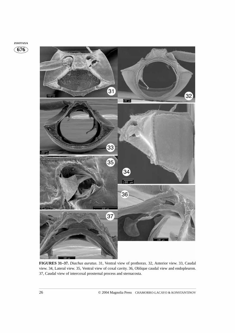
FIGURES 31–37. Diachus auratus. 31, Ventral view of prothorax. 32, Anterior view. 33, Caudalview. 34, Lateral view. 35, Ventral view of coxal cavity. 36, Oblique caudal view and endopleuron.37, Caudal view of intercoxal prosternal process and sternacosta.
© 2004 Magnolia Press 27CRYPTOCEPHALINI MORPHOLOGY
676ZOOTAXA
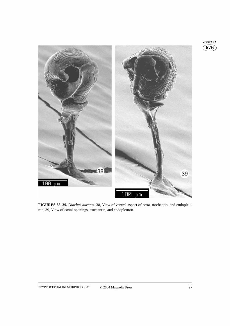
FIGURES 38–39. Diachus auratus. 38, View of ventral aspect of coxa, trochantin, and endopleu-ron. 39, View of coxal openings, trochantin, and endopleuron.
CHAMORRO-LACAYO & KONSTANTINOV28 © 2004 Magnolia Press
676ZOOTAXA
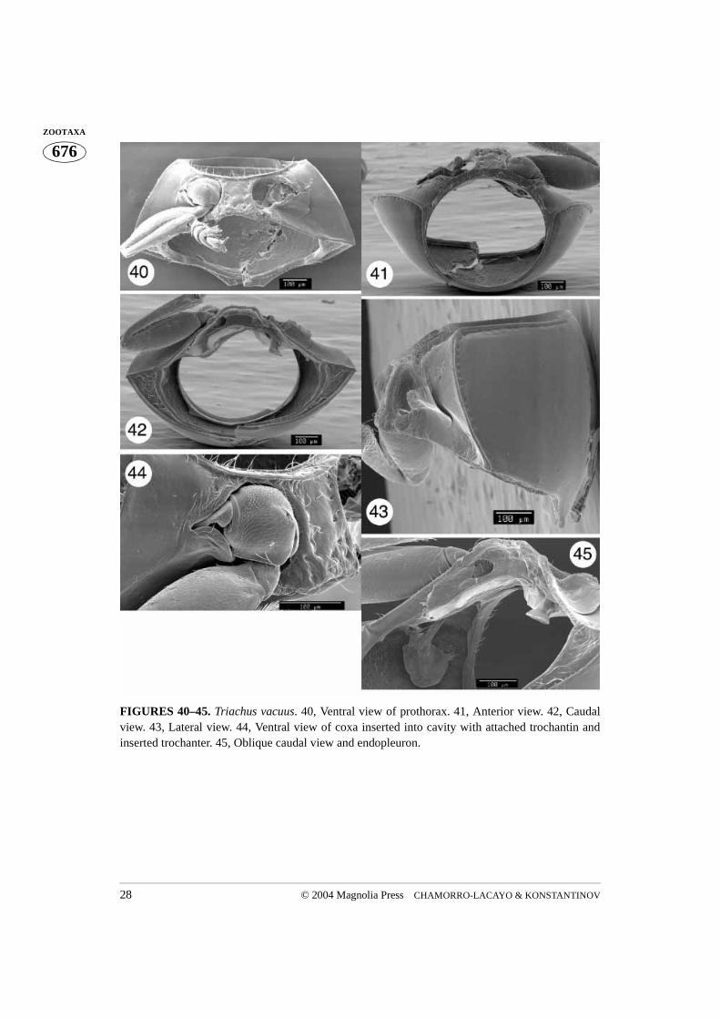
FIGURES 40–45. Triachus vacuus. 40, Ventral view of prothorax. 41, Anterior view. 42, Caudalview. 43, Lateral view. 44, Ventral view of coxa inserted into cavity with attached trochantin andinserted trochanter. 45, Oblique caudal view and endopleuron.
© 2004 Magnolia Press 29CRYPTOCEPHALINI MORPHOLOGY
676ZOOTAXA
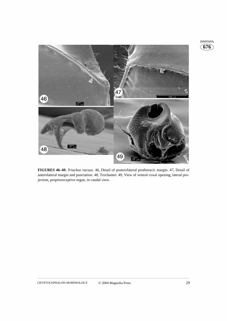
FIGURES 46–49. Triachus vacuus. 46, Detail of posterolateral prothoracic margin. 47, Detail ofanterolateral margin and punctation. 48, Trochanter. 49, View of ventral coxal opening, lateral pro-jection, proprioreceptive organ, in caudal view.
CHAMORRO-LACAYO & KONSTANTINOV30 © 2004 Magnolia Press
676ZOOTAXA
FIGURES 50–55. Heptarthrius longimanus. 50, Ventral view of prothorax. 51, Anterior view. 52,Inserted coxa. 53, Lateral view of prothorax 54, Caudal view. 55, Oblique caudal view andendopleuron.
© 2004 Magnolia Press 31CRYPTOCEPHALINI MORPHOLOGY
676ZOOTAXA
FIGURES 56–57. Heptarthrius longimanus. 56, View of ventral aspect of coxa, trochantin,endopleuron, and proprioreceptive organ. 57, View of coxal openings, trochantin, and endopleuron.
CHAMORRO-LACAYO & KONSTANTINOV32 © 2004 Magnolia Press
676ZOOTAXA
FIGURES 58–60. Heptarthrius longimanus. 58, View of posterior projection and proprioceptiveorgan. 59, Dorsal view of medial aspect of coxa. 60, Trochanter and tendon.
© 2004 Magnolia Press 33CRYPTOCEPHALINI MORPHOLOGY
676ZOOTAXA
FIGURES 61–67. Lexiphanes coenobita. 61, Ventral view of prothorax. 62, Anterior view. 63,Caudal view. 64, Lateral view. 65, Detail of anterolateral prothoracic margin. 66, Detail of postero-lateral margin. 67, Inserted coxa with trochantin and trochanter.
CHAMORRO-LACAYO & KONSTANTINOV34 © 2004 Magnolia Press
676ZOOTAXA
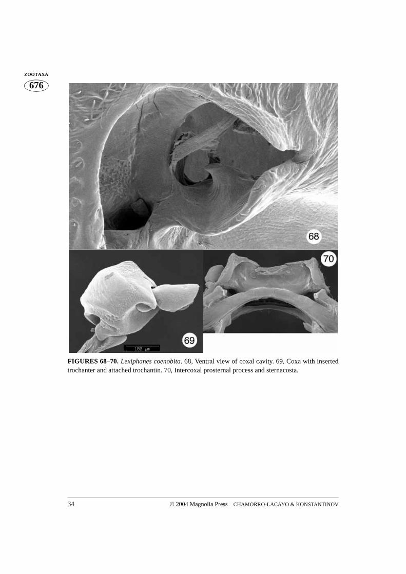
FIGURES 68–70. Lexiphanes coenobita. 68, Ventral view of coxal cavity. 69, Coxa with insertedtrochanter and attached trochantin. 70, Intercoxal prosternal process and sternacosta.
© 2004 Magnolia Press 35CRYPTOCEPHALINI MORPHOLOGY
676ZOOTAXA
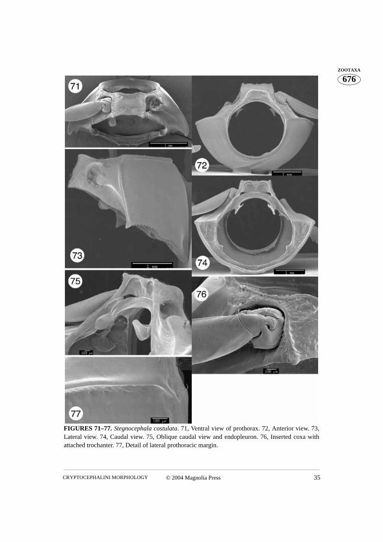
FIGURES 71–77. Stegnocephala costulata. 71, Ventral view of prothorax. 72, Anterior view. 73,Lateral view. 74, Caudal view. 75, Oblique caudal view and endopleuron. 76, Inserted coxa withattached trochanter. 77, Detail of lateral prothoracic margin.
CHAMORRO-LACAYO & KONSTANTINOV36 © 2004 Magnolia Press
676ZOOTAXA
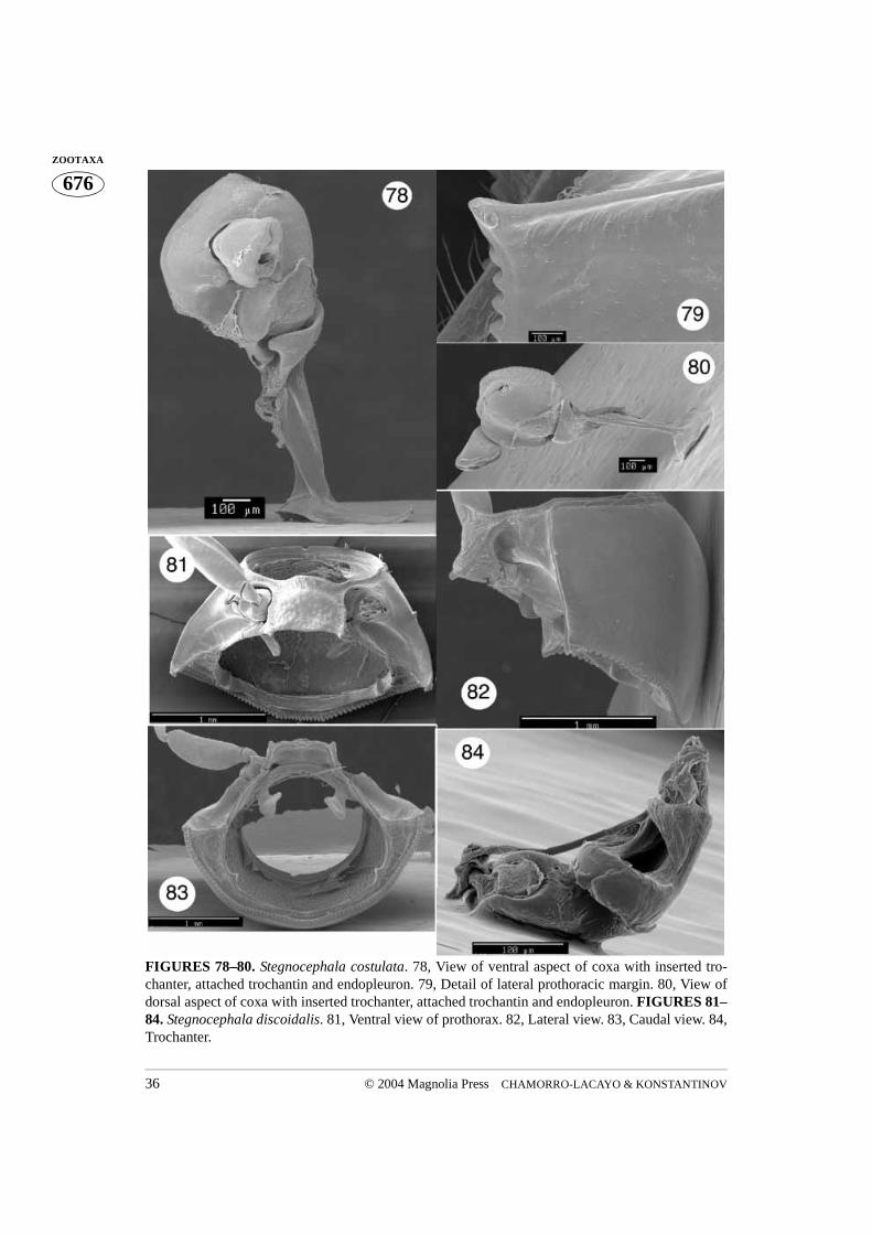
FIGURES 78–80. Stegnocephala costulata. 78, View of ventral aspect of coxa with inserted tro-chanter, attached trochantin and endopleuron. 79, Detail of lateral prothoracic margin. 80, View ofdorsal aspect of coxa with inserted trochanter, attached trochantin and endopleuron. FIGURES 81–84. Stegnocephala discoidalis. 81, Ventral view of prothorax. 82, Lateral view. 83, Caudal view. 84,Trochanter.
© 2004 Magnolia Press 37CRYPTOCEPHALINI MORPHOLOGY
676ZOOTAXA
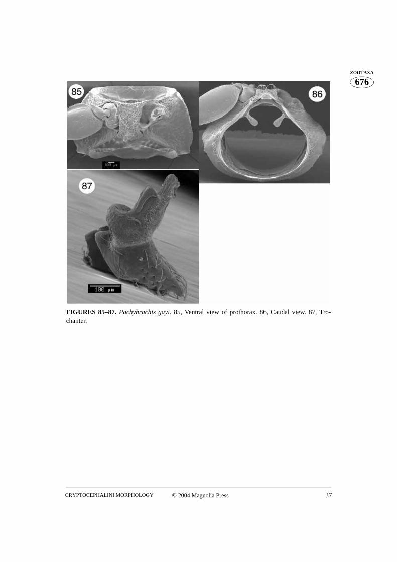
FIGURES 85–87. Pachybrachis gayi. 85, Ventral view of prothorax. 86, Caudal view. 87, Tro-chanter.
CHAMORRO-LACAYO & KONSTANTINOV38 © 2004 Magnolia Press
676ZOOTAXA
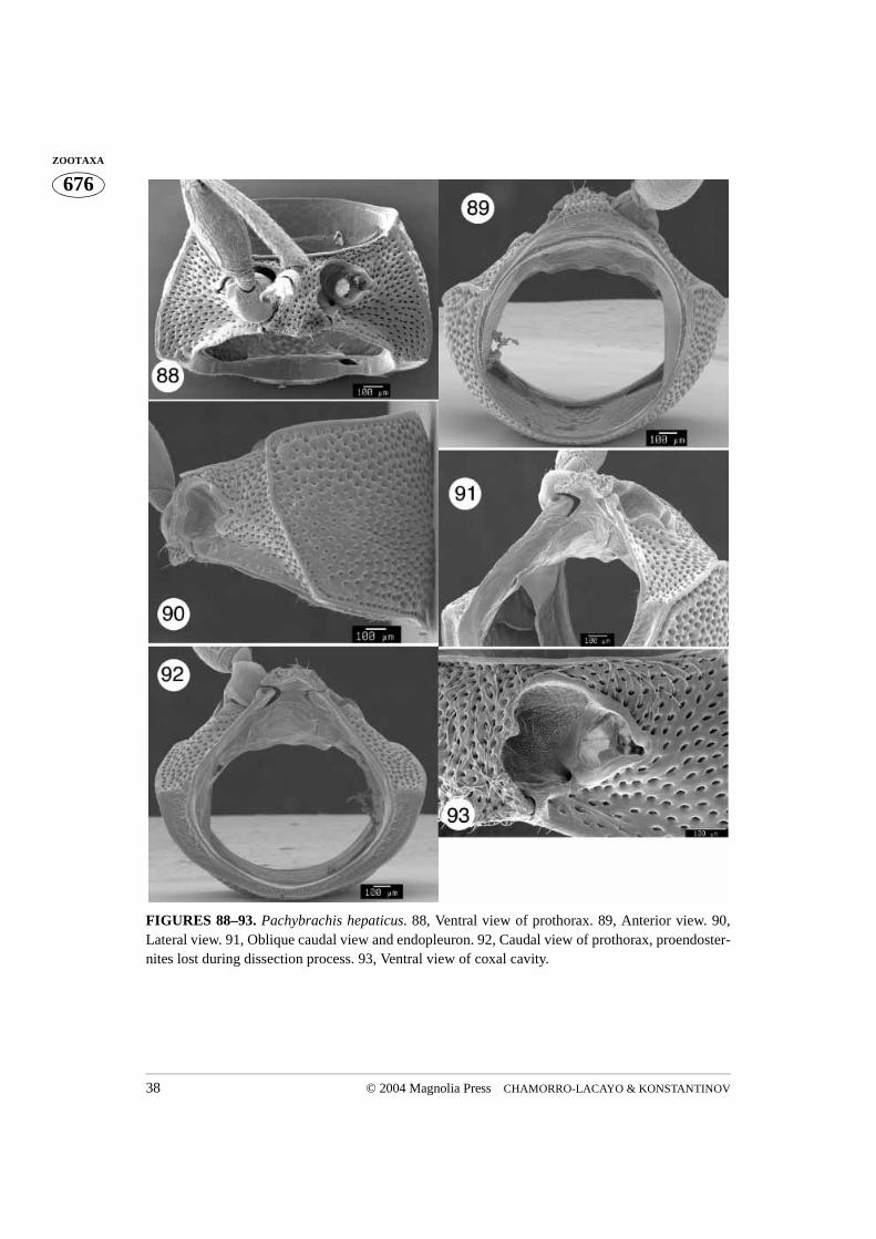
FIGURES 88–93. Pachybrachis hepaticus. 88, Ventral view of prothorax. 89, Anterior view. 90,Lateral view. 91, Oblique caudal view and endopleuron. 92, Caudal view of prothorax, proendoster-nites lost during dissection process. 93, Ventral view of coxal cavity.
© 2004 Magnolia Press 39CRYPTOCEPHALINI MORPHOLOGY
676ZOOTAXA
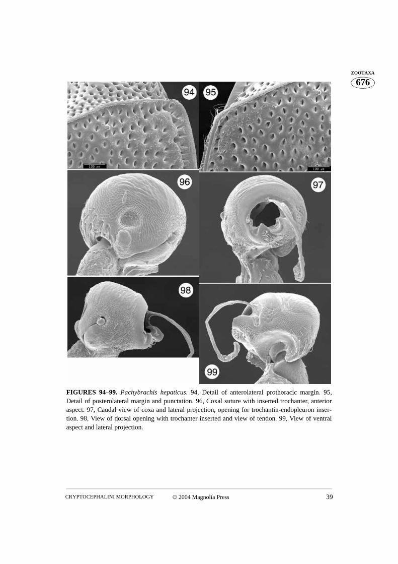
FIGURES 94–99. Pachybrachis hepaticus. 94, Detail of anterolateral prothoracic margin. 95,Detail of posterolateral margin and punctation. 96, Coxal suture with inserted trochanter, anterioraspect. 97, Caudal view of coxa and lateral projection, opening for trochantin-endopleuron inser-tion. 98, View of dorsal opening with trochanter inserted and view of tendon. 99, View of ventralaspect and lateral projection.
CHAMORRO-LACAYO & KONSTANTINOV40 © 2004 Magnolia Press
676ZOOTAXA
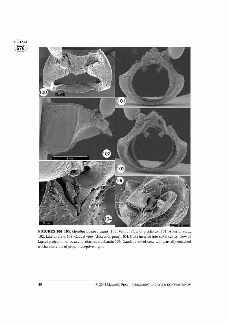
FIGURES 100–105. Metallactus decumanus. 100, Ventral view of prothorax. 101, Anterior view.102, Lateral view. 103, Caudal view (dissection poor). 104, Coxa inserted into coxal cavity, view oflateral projection of coxa and attached trochantin 105, Caudal view of coxa with partially detachedtrochantin, view of proprioreceptive organ.
© 2004 Magnolia Press 41CRYPTOCEPHALINI MORPHOLOGY
676ZOOTAXA
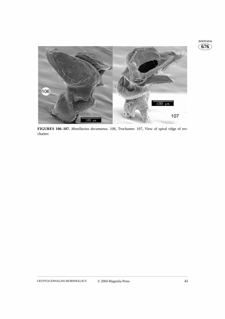
FIGURES 106–107. Metallactus decumanus. 106, Trochanter. 107, View of spiral ridge of tro-chanter.
CHAMORRO-LACAYO & KONSTANTINOV42 © 2004 Magnolia Press
676ZOOTAXA
FIGURES 108–113. Griburius sp. 108, Ventral view of prothorax. 109, Anterior view. 110, Caudalview. 111, Slightly oblique caudal view. 112, Dorsal aspect of coxa, with attached trochanter, tro-chantin and endopleuron. 113, Ventral view of coxal cavity.
© 2004 Magnolia Press 43CRYPTOCEPHALINI MORPHOLOGY
676ZOOTAXA
FIGURES 114–117. Griburius sp. 114, 115, Trochanter. 116, Spiral ridge of trochanter and tendon.117, View of ventral opening of coxa, lateral projection, trochantin, and proprioceptive organ.
CHAMORRO-LACAYO & KONSTANTINOV44 © 2004 Magnolia Press
676ZOOTAXA
FIGURES 118–125. Ambrotodes chilensis. 118, Ventral view of prothorax. 119, Anterior view.120, Lateral view. 121, Caudal view. 122, Ventral view of coxal cavity. 123, Oblique caudal viewand endopleuron. 124, Detail of anterolateral prothoracic margin. 125, Detail of posterolateral mar-gin and punctations.
© 2004 Magnolia Press 45CRYPTOCEPHALINI MORPHOLOGY
676ZOOTAXA
FIGURES 126–130. Ambrotodes chilensis. 126, View of dorsal opening and dorsal aspect of coxa.127, Inserted coxa and trochanter showing spiral ridge. 128, Trochanter. 129, Spiral ridge of tro-chanter and tendon. 130, Trochanter.
CHAMORRO-LACAYO & KONSTANTINOV46 © 2004 Magnolia Press
676ZOOTAXA
FIGURES 131–136. Ambrotodes signatipennis. 131, Ventral view of prothorax. 132, Caudal view.133, View of posterior projection and ventral opening of coxa. 134, 135, Trochanter.136, View oflateral aspect and lateral projection of coxa.

![1909] EASTON COLEOPTERA...1909] EASTON COLEOPTERA 49 A LIST OF COLEOPTERA COLLECTED WITHIN TEN MILES OF FALL RIVER, MASSACHUSETTS. BY NORMAN S. EASTON FALL …](https://img.pdfslide.us/doc/110x75/611535a3861718272235983f/1909-easton-coleoptera-1909-easton-coleoptera-49-a-list-of-coleoptera-collected.jpg)


![&'.dJ] - Centers for Disease Control and Prevention · I I prothorax well developed prothorax reduced eyes absent eyes present abd IV long abd IV short without anal spines with ana~](https://img.pdfslide.us/doc/110x75/5e805bb4717c7e64ca1a1870/dj-centers-for-disease-control-and-prevention-i-i-prothorax-well-developed.jpg)