Embed Size (px)
Citation preview
Rom J Morphol Embryol 2018, 59(3):873–877
ISSN (print) 1220–0522 ISSN (online) 2066–8279
OORRIIGGIINNAALL PPAAPPEERR
Morphological study of upper wisdom tooth
LIANA TODOR1), RUXANDRA ILINCA MATEI1), GABRIELA MUŢIU2), ANCA PORUMB1), GABRIELA CIAVOI1), EMILIA ALBINIŢA CUC1), ADRIANA ŢENŢ1), DANIELA DOMOCOŞ1), IOANA SCROBOTĂ1), SERGIU ANDREI TODOR3), MIHAELA CRISTIANA COROI4)
1)Department of Dental Medicine, Faculty of Medicine and Pharmacy, University of Oradea, Romania 2)Department of Morphological Disciplines, Faculty of Medicine and Pharmacy, University of Oradea, Romania
3)Student, Faculty of Dental Medicine, “Victor Babeş” University of Medicine and Pharmacy, Timişoara, Romania
4)Department of Surgical Disciplines, Faculty of Medicine and Pharmacy, University of Oradea, Romania
Abstract The article presents aspects of crown and root morphology of the superior wisdom teeth, aiming to several parameters: size, shape crown and occlusal surface‚ number, topography and orientation of the roots in order to determine which the most common morphological types are. For this purpose, the upper wisdom teeth were collected. According to studies, the dominant form of the dental crown is parallelepipedic, the rectangular or parallelogram shape of the occlusal surface has large mesial and distal-oriented sides. The positive occlusal forms are generally poorly demarcated; cusps are less tall and rarely individualized. In half of the cases, the root is unique, voluminous, straight or with a curved distal tip.
Keywords: morphology, upper wisdom tooth, crown, occlusal surface, root.
Introduction
Upper wisdom teeth are neither more nor less studied than other teeth by specialist researchers. Teeth have their own ontogenetic and phylogenetic development, with the most varied forms and functions, which are manifestations of adapting to the environment.
With the evolution of Homo sapiens, organs that no longer have a functional role regress, as is the case with the third molar [1]. The phylogenetic tendencies in the evolution of the human dentomaxillary system are most often noticeable in third molars [2]. In the moment of the appearance of the third row of molars are established the relations that exist between the teeth individually, to keep the harmony, the balance and perfection of the whole achieved [3]. Along with human phylogenetic evolution, maxillaries have shrunk relative to those of primates, so there is no space in the oral cavity to fully adapt to the 32 teeth [4].
The variations of the orphological traits of the third upper molar are obvious in morphological and dimen-sional variations that can go up to forms of dwarfism and homodontics. Anatomy of wisdom teeth is very varied, both coronary and especially radicular. Often endodontic treatment is required in the third molar to be used as a functional element of the dento-maxillary apparatus [5].
The natural feeding has the great advantage to insure the growth of the dental maxillary apparatus and to influence the development of its component elements [6]. The industrialization and refining of the food, caused the reduction of the jaw bones, without the need for a strong mastication [4].
Aim
The purpose of this article is to evaluate the morphology of upper wisdom tooth in the Romanian population, in the desire to add our observations to those of our predecessors, being well known that these molars exhibit the largest morphological variety.
Materials and Methods
Our personal collection consists of 114 superior wisdom teeth, 110 of which present coronary integrity. The latter we used to study the morphology of root and crown-making measurements, the rest being used only to study the root morphology.
After the extraction of each molar, the soft parts were removed, without physically and chemically altering the hard parts. They were kept for 4–5 days in hydrogen peroxide, and then washed with running water and dried, after prior immersion in chloroform. The study followed more dental parameters: size, shape occlusal surface of the crown and its number, topography and orientation of the roots in order to determine which the most common morphological types are.
The measurements were done using a caliper with an accuracy of 0.05 mm. Data processing aimed to achieve average values: the result between the minimum and maximum recorded.
The measurements proposed were: total length of the tooth; length of the crown; root length; diameter M-D (mesial-distal) up; diameter B-P (bucco-palatal) maximum.
Like all organs, teeth are also asymmetric pairs. Almost all details of relief: trenches, pits, ridges, cusps are also asymmetric. This asymmetry makes it difficult to study
R J M ERomanian Journal of
Morphology & Embryologyhttp://www.rjme.ro/
Liana Todor et al.
874
dental morphology, but on the other hand, helps us determine the exact place on one of arcade teeth.
Shape and contour of teeth results from a combination of lines and curved surfaces more or less pronounced. Studies conducted by numerous researchers have found that the form of dental crowns can be likened to different geometric forms: parallelepipeds, prisms, cubes, etc.
If we used geometric solids for the crown shape, to form occlusal surface, we used geometric figures.
From clinical experience, we know that when endo-dontic treatment is required to the higher molar teeth, there is variable number of canals. Therefore, it seemed useful to study the number of canals on molars showing a single root. That is why we performed perpendicular sections to the axis of the root in the cervical third and apical third.
Results
The values obtained after measuring total dental length, coronary or root length, and coronary diameters are represented in the Table 1.
After a careful study of each wisdom tooth higher personal collection, we grouped them into six forms of
geometric bodies to presenting the greatest similarity coronary shape.
Table 1 – The average size of the maxillary third molar (millimeters)
Lenght Maximum diameter Authors
Total Crown Root M-D B-P
Todor et al. 17.25 6.5 10.75 8.8 9.8
M-D: Mesial-distal; B-P: Bucco-palatal.
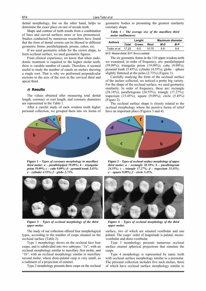
The six geometric forms in the 110 upper wisdom teeth we examined, in order of frequency, are: parallelepiped (59.09%); triangular prism (19.09%); cube (9.09%); pyramid trunk (5.45%); cylinder (4.55%); globe – sphere slightly flattened at the poles (2.73%) (Figure 1).
Carefully studying the form of the occlusal surface of the molars collected, we noticed a pretty big variety. For the shape of the occlusal surface, we used geometric similarity. In order of frequency, these are: rectangle (28.18%); parallelogram (24.55%); triangle (17.27%); trapezium (15.45%); square (9.09%); circle (5.45%) (Figure 2).
The occlusal surface shape is closely related to the occlusal morphology where the positive forms of relief have an important place (Figures 3 and 4).
Figure 1 – Types of coronary morphology in maxillary third molar: a – parallelepiped 59.09%; b – triangular prism 19.09%; c – cube 9.09%; d – pyramid trunk 5.45%; e – cylinder 4.55%; f – globe 2.73%.
Figure 2 – Types of occlusal surface morphology of upper third molar: a – rectangle 28.18%; b – parallelogram 24.55%; c – triangle 17.27%; d – trapezium 15.45%; e – square 9.09%; f – circle 5.45%.
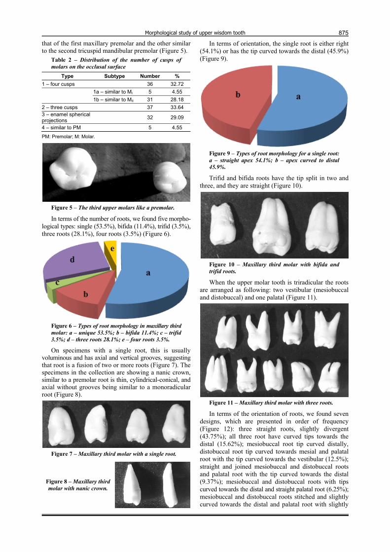
Figure 3 – Types of occlusal morphology of the third upper molar.
Figure 4 – Types of occlusal morphology of the third upper molar.
The study of our collection offered four morphological types, according to the number of cusps situated on the occlusal surface (Table 2).
Type 1 morphology shows on the occlusal face four cusps, and is subdivided into two subtypes: “1a”, with an occlusal morphology similar to maxillary first molar, and “1b”, with an occlusal morphology similar to maxillary second molar, where disto-palatal cusp is very small, as a rudiment of a projection of enamel.
Type 2 morphology presents three cusps on the occlusal
surface, two of which are situated vestibular and one palatal. The cusps’ order of magnitude is palatal, mesio-vestibular and disto-vestibular.
Type 3 morphology presents numerous occlusal surface enamel spherical projections that simulate the cusps.
Type 4 morphology is represented by nanic teeth with occlusal surface morphology similar to a premolar. The personal collection includes five such molars, three of which have occlusal surface morphology similar to
Morphological study of upper wisdom tooth
875
that of the first maxillary premolar and the other similar to the second tricuspid mandibular premolar (Figure 5).
Table 2 – Distribution of the number of cusps of molars on the occlusal surface
Type Subtype Number %
1 – four cusps 36 32.72
1a – similar to MI 5 4.55
1b – similar to MII 31 28.18
2 – three cusps 37 33.64 3 – enamel spherical projections
32 29.09
4 – similar to PM 5 4.55
PM: Premolar; M: Molar.
Figure 5 – The third upper molars like a premolar.
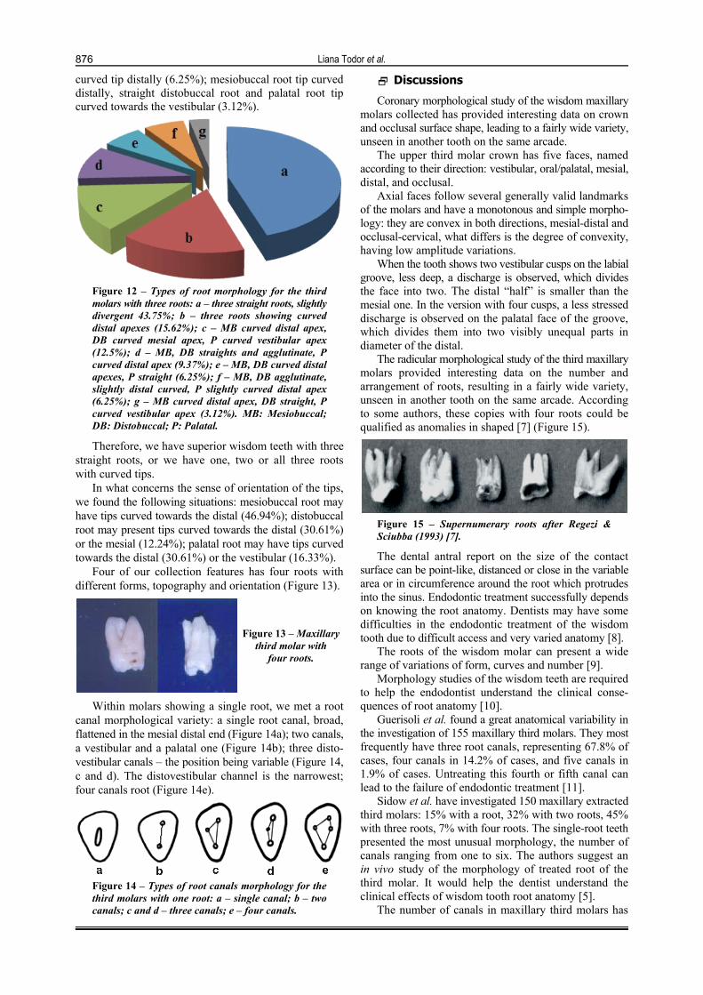
In terms of the number of roots, we found five morpho-logical types: single (53.5%), bifida (11.4%), trifid (3.5%), three roots (28.1%), four roots (3.5%) (Figure 6).
Figure 6 – Types of root morphology in maxillary third molar: a – unique 53.5%; b – bifida 11.4%; c – trifid 3.5%; d – three roots 28.1%; e – four roots 3.5%.
On specimens with a single root, this is usually voluminous and has axial and vertical grooves, suggesting that root is a fusion of two or more roots (Figure 7). The specimens in the collection are showing a nanic crown, similar to a premolar root is thin, cylindrical-conical, and axial without grooves being similar to a monoradicular root (Figure 8).
Figure 7 – Maxillary third molar with a single root.
Figure 8 – Maxillary third molar with nanic crown.
In terms of orientation, the single root is either right (54.1%) or has the tip curved towards the distal (45.9%) (Figure 9).
Figure 9 – Types of root morphology for a single root: a – straight apex 54.1%; b – apex curved to distal 45.9%.
Trifid and bifida roots have the tip split in two and three, and they are straight (Figure 10).
Figure 10 – Maxillary third molar with bifida and trifid roots.
When the upper molar tooth is triradicular the roots are arranged as following: two vestibular (mesiobuccal and distobuccal) and one palatal (Figure 11).
Figure 11 – Maxillary third molar with three roots.
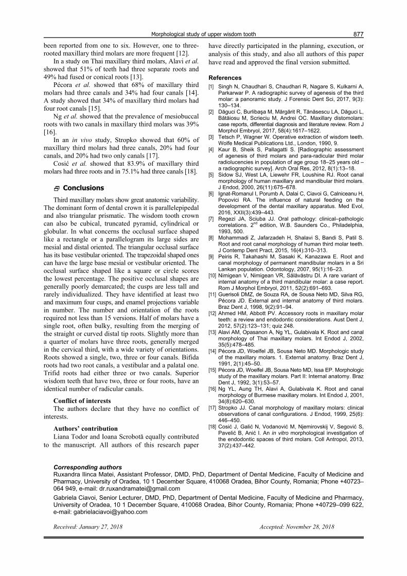
In terms of the orientation of roots, we found seven designs, which are presented in order of frequency (Figure 12): three straight roots, slightly divergent (43.75%); all three root have curved tips towards the distal (15.62%); mesiobuccal root tip curved distally, distobuccal root tip curved towards mesial and palatal root with the tip curved towards the vestibular (12.5%); straight and joined mesiobuccal and distobuccal roots and palatal root with the tip curved towards the distal (9.37%); mesiobuccal and distobuccal roots with tips curved towards the distal and straight palatal root (6.25%); mesiobuccal and distobuccal roots stitched and slightly curved towards the distal and palatal root with slightly
Liana Todor et al.
876
curved tip distally (6.25%); mesiobuccal root tip curved distally, straight distobuccal root and palatal root tip curved towards the vestibular (3.12%).
Figure 12 – Types of root morphology for the third molars with three roots: a – three straight roots, slightly divergent 43.75%; b – three roots showing curved distal apexes (15.62%); c – MB curved distal apex, DB curved mesial apex, P curved vestibular apex (12.5%); d – MB, DB straights and agglutinate, P curved distal apex (9.37%); e – MB, DB curved distal apexes, P straight (6.25%); f – MB, DB agglutinate, slightly distal curved, P slightly curved distal apex (6.25%); g – MB curved distal apex, DB straight, P curved vestibular apex (3.12%). MB: Mesiobuccal; DB: Distobuccal; P: Palatal.
Therefore, we have superior wisdom teeth with three straight roots, or we have one, two or all three roots with curved tips.
In what concerns the sense of orientation of the tips, we found the following situations: mesiobuccal root may have tips curved towards the distal (46.94%); distobuccal root may present tips curved towards the distal (30.61%) or the mesial (12.24%); palatal root may have tips curved towards the distal (30.61%) or the vestibular (16.33%).
Four of our collection features has four roots with different forms, topography and orientation (Figure 13).
Figure 13 – Maxillary third molar with
four roots.
Within molars showing a single root, we met a root canal morphological variety: a single root canal, broad, flattened in the mesial distal end (Figure 14a); two canals, a vestibular and a palatal one (Figure 14b); three disto-vestibular canals – the position being variable (Figure 14, c and d). The distovestibular channel is the narrowest; four canals root (Figure 14e).
Figure 14 – Types of root canals morphology for the third molars with one root: a – single canal; b – two canals; c and d – three canals; e – four canals.
Discussions
Coronary morphological study of the wisdom maxillary molars collected has provided interesting data on crown and occlusal surface shape, leading to a fairly wide variety, unseen in another tooth on the same arcade.
The upper third molar crown has five faces, named according to their direction: vestibular, oral/palatal, mesial, distal, and occlusal.
Axial faces follow several generally valid landmarks of the molars and have a monotonous and simple morpho-logy: they are convex in both directions, mesial-distal and occlusal-cervical, what differs is the degree of convexity, having low amplitude variations.
When the tooth shows two vestibular cusps on the labial groove, less deep, a discharge is observed, which divides the face into two. The distal “half” is smaller than the mesial one. In the version with four cusps, a less stressed discharge is observed on the palatal face of the groove, which divides them into two visibly unequal parts in diameter of the distal.
The radicular morphological study of the third maxillary molars provided interesting data on the number and arrangement of roots, resulting in a fairly wide variety, unseen in another tooth on the same arcade. According to some authors, these copies with four roots could be qualified as anomalies in shaped [7] (Figure 15).
Figure 15 – Supernumerary roots after Regezi & Sciubba (1993) [7].
The dental antral report on the size of the contact surface can be point-like, distanced or close in the variable area or in circumference around the root which protrudes into the sinus. Endodontic treatment successfully depends on knowing the root anatomy. Dentists may have some difficulties in the endodontic treatment of the wisdom tooth due to difficult access and very varied anatomy [8].
The roots of the wisdom molar can present a wide range of variations of form, curves and number [9].
Morphology studies of the wisdom teeth are required to help the endodontist understand the clinical conse-quences of root anatomy [10].
Guerisoli et al. found a great anatomical variability in the investigation of 155 maxillary third molars. They most frequently have three root canals, representing 67.8% of cases, four canals in 14.2% of cases, and five canals in 1.9% of cases. Untreating this fourth or fifth canal can lead to the failure of endodontic treatment [11].
Sidow et al. have investigated 150 maxillary extracted third molars: 15% with a root, 32% with two roots, 45% with three roots, 7% with four roots. The single-root teeth presented the most unusual morphology, the number of canals ranging from one to six. The authors suggest an in vivo study of the morphology of treated root of the third molar. It would help the dentist understand the clinical effects of wisdom tooth root anatomy [5].
The number of canals in maxillary third molars has
Morphological study of upper wisdom tooth
877
been reported from one to six. However, one to three-rooted maxillary third molars are more frequent [12].
In a study on Thai maxillary third molars, Alavi et al. showed that 51% of teeth had three separate roots and 49% had fused or conical roots [13].
Pécora et al. showed that 68% of maxillary third molars had three canals and 34% had four canals [14]. A study showed that 34% of maxillary third molars had four root canals [15].
Ng et al. showed that the prevalence of mesiobuccal roots with two canals in maxillary third molars was 39% [16].
In an in vivo study, Stropko showed that 60% of maxillary third molars had three canals, 20% had four canals, and 20% had two only canals [17].
Cosić et al. showed that 83.9% of maxillary third molars had three roots and in 75.1% had three canals [18].
Conclusions
Third maxillary molars show great anatomic variability. The dominant form of dental crown it is parallelepipedal and also triangular prismatic. The wisdom tooth crown can also be cubical, truncated pyramid, cylindrical or globular. In what concerns the occlusal surface shaped like a rectangle or a parallelogram its large sides are mesial and distal oriented. The triangular occlusal surface has its base vestibular oriented. The trapezoidal shaped ones can have the large base mesial or vestibular oriented. The occlusal surface shaped like a square or circle scores the lowest percentage. The positive occlusal shapes are generally poorly demarcated; the cusps are less tall and rarely individualized. They have identified at least two and maximum four cusps, and enamel projections variable in number. The number and orientation of the roots required not less than 15 versions. Half of molars have a single root, often bulky, resulting from the merging of the straight or curved distal tip roots. Slightly more than a quarter of molars have three roots, generally merged in the cervical third, with a wide variety of orientations. Roots showed a single, two, three or four canals. Bifida roots had two root canals, a vestibular and a palatal one. Trifid roots had either three or two canals. Superior wisdom teeth that have two, three or four roots, have an identical number of radicular canals.
Conflict of interests The authors declare that they have no conflict of
interests.
Authors’ contribution Liana Todor and Ioana Scrobotă equally contributed
to the manuscript. All authors of this research paper
have directly participated in the planning, execution, or analysis of this study, and also all authors of this paper have read and approved the final version submitted.
References [1] Singh N, Chaudhari S, Chaudhari R, Nagare S, Kulkarni A,
Parkarwar P. A radiographic survey of agenesis of the third molar: a panoramic study. J Forensic Dent Sci, 2017, 9(3): 130–134.
[2] Dăguci C, Burlibaşa M, Mărgărit R, Tănăsescu LA, Dăguci L, Bătăiosu M, Scrieciu M, Andrei OC. Maxillary distomolars: case reports, differential diagnosis and literature review. Rom J Morphol Embryol, 2017, 58(4):1617–1622.
[3] Tetsch P, Wagner W. Operative extraction of wisdom teeth. Wolfe Medical Publications Ltd., London, 1990, 9.
[4] Kaur B, Sheik S, Pallagatti S. [Radiographic assessment of agenesis of third molars and para-radicular third molar radiolucencies in population of age group 18–25 years old – a radiographic survey]. Arch Oral Res, 2012, 8(1):13–18.
[5] Sidow SJ, West LA, Liewehr FR, Loushine RJ. Root canal morphology of human maxillary and mandibular third molars. J Endod, 2000, 26(11):675–678.
[6] Ignat-Romanul I, Porumb A, Dalai C, Ciavoi G, Calniceanu H, Popovici RA. The influence of natural feeding on the development of the dental maxillary apparatus. Med Evol, 2016, XXII(3):439–443.
[7] Regezi JA, Sciuba JJ. Oral pathology: clinical–pathologic correlations. 2nd edition, W.B. Saunders Co., Philadelphia, 1993, 500.
[8] Mohammadi Z, Jafarzadeh H, Shalavi S, Bandi S, Patil S. Root and root canal morphology of human third molar teeth. J Contemp Dent Pract, 2015, 16(4):310–313.
[9] Peiris R, Takahashi M, Sasaki K, Kanazawa E. Root and canal morphology of permanent mandibular molars in a Sri Lankan population. Odontology, 2007, 95(1):16–23.
[10] Nimigean V, Nimigean VR, Sălăvăstru DI. A rare variant of internal anatomy of a third mandibular molar: a case report. Rom J Morphol Embryol, 2011, 52(2):691–693.
[11] Guerisoli DMZ, de Souza RA, de Sousa Neto MD, Silva RG, Pécora JD. External and internal anatomy of third molars. Braz Dent J, 1998, 9(2):91–94.
[12] Ahmed HM, Abbott PV. Accessory roots in maxillary molar teeth: a review and endodontic considerations. Aust Dent J, 2012, 57(2):123–131; quiz 248.
[13] Alavi AM, Opasanon A, Ng YL, Gulabivala K. Root and canal morphology of Thai maxillary molars. Int Endod J, 2002, 35(5):478–485.
[14] Pécora JD, Woelfel JB, Sousa Neto MD. Morphologic study of the maxillary molars. 1. External anatomy. Braz Dent J, 1991, 2(1):45–50.
[15] Pécora JD, Woelfel JB, Sousa Neto MD, Issa EP. Morphologic study of the maxillary molars. Part II: Internal anatomy. Braz Dent J, 1992, 3(1):53–57.
[16] Ng YL, Aung TH, Alavi A, Gulabivala K. Root and canal morphology of Burmese maxillary molars. Int Endod J, 2001, 34(8):620–630.
[17] Stropko JJ. Canal morphology of maxillary molars: clinical observations of canal configurations. J Endod, 1999, 25(6): 446–450.
[18] Cosić J, Galić N, Vodanović M, Njemirovskij V, Segović S, Pavelić B, Anić I. An in vitro morphological investigation of the endodontic spaces of third molars. Coll Antropol, 2013, 37(2):437–442.
Corresponding authors Ruxandra Ilinca Matei, Assistant Professor, DMD, PhD, Department of Dental Medicine, Faculty of Medicine and Pharmacy, University of Oradea, 10 1 December Square, 410068 Oradea, Bihor County, Romania; Phone +40723–064 949, e-mail: [email protected]
Gabriela Ciavoi, Senior Lecturer, DMD, PhD, Department of Dental Medicine, Faculty of Medicine and Pharmacy, University of Oradea, 10 1 December Square, 410068 Oradea, Bihor County, Romania; Phone +40729–099 622, e-mail: [email protected] Received: January 27, 2018 Accepted: November 28, 2018