Embed Size (px)
Citation preview

178 Acta Academiae Medicinae Wuhan 5(3):178-184, 1958
Morphological Studies on the Mechanism of
Anti-implantative and Early-pregnancy-terminative
Effect by Norethisterone Oxime in Rats
PENG Ling ( ~ ~)~), YANG Yi -d i ( ~ ) ~ , )
Family Planning Research Institute, Wuhan Medical College, Wuhan
Summary: In this study it was found that norethisterone ox ime (NorO) could produce changes in some organelles of the ovarian corpora lutea in early pregnant rats including decrease in smooth endoplasmic ret iculum, accumulation of l ipid droplets, swelling of Golgi bodies and increase in secondary lysosomes. NorO also depressed the act ivi ty of 3~]-hydroxysteroid dehydrogenase in the ovary, resulting in the obstruction of biological synthesis of steroid hormone in the ovary. I t was found that the act ivi ty of acid phosphata~e of the corFora lutea increased, which showed an increase in the membrane permeabi l i ty of lysosomes and damage to the cells. Morphological changes unfavourable to implantat ion occurred in the endometrium of the early pregnant rats, such as, decrease in the transformation of stroma cells into decidual cells, degeneration and necrosis of the decidual cells. On the 4th day of pregnancy estrogenic effect could be detected in the endometrium of the rats. Further study should be made to determine whether this is due to the depression of progesterone level by NorO and thereafter less depres- sion of the action of estrogen or due to the estrogenic ac l iv i ty of the drug itself.
Key words., norethisterone oxime, ant i implantat ion, ear ly-pregr .ancy- terminat ion
P r o g e s t e r o n e h a s esser~.tial e f fec ts on i m p l a n t a t i o n a n d e a r l y p r e g n a n c y i n h u m a n s a n d a n i m a l s . The search for d rugs t h a t can i n t e r f e r e w i t h the b i o - s y n t h e s i s o f p roges te rone in the o v a r y a n d ac t a s a c o m p e t i t i v e a n t a g o n i s t a g a i n s t p roges t e rone a t the recep tor level i s a n i m p o r t a n t sub jec t of s t u d y in the d e v e l o p m e n t of n e w a n t i i m p l a n t a t i v e a n d e a r l y - p r e g n a n c y - t e r m i n a t i v e drugs , to w h i c h m u c h a t t e n t i o n i s p a i d bo th a t home a n d a b r o a d t1'2]. N o r e t h i s t e r o n e o x i - me ( N o r O ) is a d rug s y n t h e s i z e d b y H u - b e i P h a r m a c e u t i c a l I n d u s t r i a l Research I n s t i t u t e . O u r i n s t i t u t e i n c o o p e r a t i o n w i t h W H O H u m a n R e p r o d u c t i v e P r o - g r a m m e s t u d i e d the changes caused b y a c t i o n of NorO i n the o v a r y ( a n o r g a n
t ha t secretes progesteror~.e) a~.d the u t e ru s ( t a rge t o r g a ~ a f f ec t ed b y p roges t e rone ) of e a r l y p r e g n a n t r a t s . Our s t u d y was do.ne w i t h morphc, l o g i c a l me thods i n c l u d - ing h i s t c c h e m i s t r y a n d e lec t ron m i c r o - scopy aJad is a i m e d a t u ~ d e r s t a n d i n g the m e c h a n i s m of a v t i i m p l a ~ t a t i o n a n d ea r - l y - p r e g n a n c y - t e r m i r ~ a t i o n caused b y the drug .
M A T E R I A L S A N D M E T H O D S
Fema le r a t s were caged w i t h f e r t i l e males in the a f t e r n o o n of p r c e s t ru s . The d a y cn w h i c h spe rm w a s e b s e r v e d in the v a g i n a was c e r s i d e r e d as d a y 1 of pregr~.ancy. R a t s in the con t ro l g roup were made to d r i n k aqueous b l a n k v e h i - cle c o n t a i n i n g 5 % s o d i u m c a r b o x y m e -
Acta Academiae Med~cinae Wuhan 5(3):178-184, 1985 179
thy l cellulose and 1 a~ Tween 80, those in the treated group a l iquid suspension conta in ing NorO.
1. Tests on the ovary ( 1 ) Histoehemical observation. The rats were treated wi th 4 mg /
k g / d a y NorO on the 1st to 4th day of pregnancy and were sacrificed on the 7th day. The ovaries were weighed. One ova ry was f ixed in a Bouin ' s solut ion and stair~ed wi th HE, another one was frozen and sectioned in a cryostat . They were h is tcchemica l ly stained for 38- hydroxys te ro id dehydrcgenase (3~]-HSD) by the modif ied method of Wattenberg, for acid phcsphatase ( A C P ) by the mo- dif ied method of Gomc.ri, and for lipid droplets by Red oil method.
(2 ) Electro~. microscopic observa- t ion.
The t ime for treatment and for au- topsy was the same as that for h is to- chemical observation. The control group consisted of 2 animals and the treated group of 3. Af ter autopsy the 3 - - 5 preg- nant corpora lutea were convent ional ly treated in the Department of Electron Microscopy, W u h a n Medical College.
2. Tests on the uterus ( 1 ) In the control group 5 rats
were treated wi th control vehicle on the 1st, 2nd and 3rd day of pregnancy. In the 3rd group besides NorO treatment the rats also received 15 mg progeste- rone da i ly for explor ing whether exo- genous progesterone can reverse the an t i - progesterone effect of NorO. All rats were ki l led on the 4th day. The effect of NorO on mitos is in the luminal, glandular and stromal cells of the rats was s tudied after arrest of mitosis by subcutaneous inject ion of 0.5 mg col- chic ine in. 0.5 ml saline 1 h before sa- cr i f ic ing. The middle uter i were remov- ed and f ixed in Bouin ' s solution. Transverse sect ions (5 t~) were stained wi th HE. The modif ied method of Chaud- h y r y and F inn was used. 0 Counts of mitosis in the luminal epithelium ( lumi- nal counts) are the totals for each sec- tion. ( ~ ) I n the glands and the s troma
the counts were made in two fields (400)<) located on each side of the lumew so that the inner edge of each field just overlapped the basement mem- brane of the luminal epithelium about m i d w a y between the mesometrial and an t i -mesomet r ia l apices of the lumen ( f i g . l ) . The number of cells showing mitotic ac t iv i ty was counted in 3 sec- t ions of the uter ine horns of each an i - mal. The 3 sections are 1st, 10th, 12th in 20 serial sections. The mean + SE value was noted.
"
. ~ 1 I "
Fig. i. Diagram of a transverse section of rat utercs to show the Fosition of the fields used in counting glandu- lar, luminal and stromal mitosis.
( 2 ) To observe the effect of the drug on the decidual cells, the animals were once treated wi th NorO 12 mg/kg/ day on the 7th day of pregnancy and were killed on the 9th day. Uteri were removed, such of different morphology were f ixed in Bou in ' s solut ion to be stai~.ed wi th HE and wi th Sch i f f ' s so lu- t ion for glycogen, the other uteri were stained for a lkal ine phoSphatase ( A L P ) by the modif ied method of Gomori .
RESULTS
1. As shown in table 1, no effects on ovar ian weights were n o t e d in the treated group ( P ~ 0 . 0 5 ) . Under light microscope there was no s ignif icant d i f - ference in his tological structure of the ovaries between the two groups.
2. Extensive 3B-HSD ac t iv i ty was observed in the corpora lutea of the

180
pregnant ra ts ( table 1). Decrease of 3[3- shapes in the cytoplasm. Tile external HSD ac t iv i ty was observed when NorO form of the lipid droplets was round was administered on day 1 to 4 of and varied from big to small. There pregnancy ( P ~ 0 . 0 1 ; f ig .2 ,3) . In the was no membrane. Electronic density control group the stain of 3B-HSD was was uniform. Lysosomes were of greater dark blue fo rmazan , whereas that in electronic density r o u n d in shape and the treated group was shal low, and de- black in colour. Mi tochondr ia were large creased amount of stained grains of 3~- and numerous with tubule-shaped cris- HSD was observed. In the control group tae. External form of the mi tochondr ia ac t iv i ty of A C P was rare ly seen while varied from round to concavoconvex in the t reated group extensive ac t iv i ty discs, which showed lenticular to dumb- of ACP showed in the corpora lutea of bell-shaped outline in cross-section. In pregnant rats ( P < 0 . 0 1 ; f i gA ,5 ) . In the treated group marked changes were normal pregnant corpora lutea few lipid observed ( f i g . 9 ) : SER decreased mark- droplets showed as small red grains. In edly; lipid droplets accumulated in the treated group marked increase of great quantities in the cytoplasm; and lipid droplets was observed ( P < 0 . 0 5 ; secondary lysosomes increased. In the f ig .6 ,7 ) , mi toehondr ia there were no marked chan-
3. Ul t ras t ructure of granular lutein ges. Diagrams of results previous ly cells of normal pregnant rat on day 7 obtained are shown in f ig.10. conformed wi th that of typical steroid- 4 . Results of count of mi tos is are synthes iz ing cells ( f ig .8 ) . The lutein shown in table 2. cells were character ized by an abundance 5 . Embryo and decidua of rat of smooth endoplasmic reticulum (SER) uterus on the 9th day of pregnancy of wi th tor tuous, tubular and vesicular rats treated by NorO showed pa tho!og i -
T a b l e 1 . E f f e c t of NorO on rat o v a r i a n w e i g h t and h i s t o c h e m i s t r y o n the 7 th d a y of p r e g n a n c y (mean___SE)
Ovarian weight (rag) Group Rats
Left Right
Control 6 38.66-4-5.35 38.634-6.31
Treated 6 34.334-6.21 34.50__+6.47
Histochemistry (corpora !utea)
3~-HSD ACP Lipid droplet
3.174-0.52 2.17___0.52 1.83-4-0.52
1.80-4-0.25"* 3.424-0.58** 2.91-4-0.80"
significantly different from the vehicle control value,*P< 0.05, ** P< 0.01 ; histochemically the sta- tistical values were estimated on a 4-point scale: 1.0, moderate; 2.0, good; 3.0, high; 4.0, intense
T a b l e 2 . E f f e c t of NorO on e n d o m e t r i u m ce l l d i v i s i o n in rats on d a y 4 of p r e g n a n c y ( m e a n + _ S E )
Mitotic figures Group Rats Epithelium Stroma
Luminal Glandular
Control 5 2.6_+2.1 1.8+__ i .5 120.0+__ 12.4
Treated with 5 6.2-----2.5 3.6___ 1.5 98.24- 12.0" NorO
Treated with 5 9.04- 7.0 3.2___ 3.6 107.84- 17.1 NorO + progesterone
significantly different from vehicle control value, *P<0.05

] ~
~'..
~ -_~
,~
-~,~
-- ;~.
~- .:~
; ~
..~
..~
.,~
~ ii! ̧
~i~
iiii~i~i
i!ii!i~i
~ii~ i!
° !~
i,ii~
,i
'~]]~
,~ i'ii
i i!,i
ii] ~
i~iii i i
i! !!iil i'//~
'ii'~,~,',~
i~ ¸̧, !ii
iiii'ii ¸ i!ili~ ¸~ iii!~! I i

182
cal c h a r g e s in v a r i o u s degree i n c l u d i n g d e g e n e r a t i o n , n e c r o s i s a ~ d d i s s o l u t i o n ( f i g . 1 1 , 1 2 ) . F o r c h a n g e s of g l y c o g e n a n d A L P see t ab l e 3.
D I S C U S S I O N
1. Ef fec t of N o r O on the p roges t e - rone b i o s y n t h e s i s in t h e o v a r y
( 1 ) The 3 ~ - H S D s i t u a t e d in t h e S E R of l u t e i n cel l i s a k e y e n z y m e w h i c h p l a y s a c a t a l y t i c ro l e in the p r o g e s t e r o n e b i o s y n t h e s i s ~ ] . H i s t o c h e - mica1 o b s e r v a t i o n s h o w e d t h a t t he 3~- H S D w a s decreased b y the a c t i o n of N o r O , a n d e l ec t ron m i c r o s c o p i c o b s e r v a - t i o n r e v e a l e d t h a t t he S E R w a s a l so decreased b y the s a m e d rug . F r o m t h i s i t m a y be c o n c l u d e d t h a t b i o s y n t h e s i s of p r o g e s t e r o n e w a s reduced .
( 2 ) L i p i d d r o p l e t s a r e a u n i v e r s a l c h a r a c t e r i s t i c of the s t e r o i d - h o r m o n e - s y n t h e s i z i n g cel l C~,5]. In h i s a r t i c l e e n t i t l e d " L i p i d of C e l l " , D e a n h a s n o t e d t h a t l i p i d d r o p l e t s r epresen t the p r e c u r s o r y m a t e r i a l t h a t m a y be t r a n s - f o r m e d i n t o s t e r o i d h o r m o n e w h e n s u i t - ab le s t i m u l a t i o n occurs . He, the re fo re , sugges ted t h a t the a c t i v i t y of s t e r o i d - s y n t h e t i s i z i n g cel l c o u l d be e v a l u a t e d a c c o r d i n g to the n u m b e r a n d d i a m e t e r of l i p i d d rop l e t s . W h e n i t s s e c r e t i o n i s
abu!~dant , t he l i p i d d~ople ts a r e f ew a n d v i ce v e r s a [5]. T h e l i p i d d r o p l e t s , c o n t a i n i n g l i p i d a c i d a n d choles te r01 , can t a k e p a r t i n b i o s y n t h e s i s of s t e r o i d h o r m o n e . F r o m h i s t o c h e m i c a l a n d e lec - t r on m i c r o s c o p i c o b s e r v a t i o n of the t r e a t - ed g roup i t m a y be sugges ted t h a t l i p i d d r o p l e t s in t he l u t e i n ce l l s i n - creased m a r k e d l y , s h o w i n g an o b s t r u c t i o n of the b i o s y n t h e s i s of p roge s t e rone a n d an a c c u m u l a t i o n of the p r e c u r s o r y m a t e - r i a l s . C o n s e q u e n t l y , t he f o r e g o i n g r e s u l t s g i v e c o n c l u s i v e e v i d e n c e of the p r o g e s - t e rone s y n t h e t i c o b s t r u c t i o n .
~. . ; , o o ,~:,
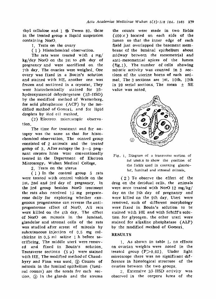
Fig. 10.Patterns of ul trastructure of lutein cells in rats during normal pregnancy and after t reatment wRh NorO. A.- dis tr ibut ion diagram; B: l ipid
droplets; C : SER, Go!gi comptex~ E': l y so -
SOffleSo
Fig . 2
Fig . 3
F ig . 4
F ig . 5
Fig . 6
F ig . 7
F ig . 8 .
Fig . 9 .
3 ~ -HSD of It:rein cells of a rat on the 7th day of normal pregnancy. Note the strong ac t iv i ty of the corpus luteum. 125×. 3~-HSD of a lutein cell of a rat in the treated group on day 7 of pregrancy. Note the decreased act iv i ty of the corpus u t eum. l125x . ACP of lutein cell of a rat on day 7 of normal pregnancy. Note the weak act iv i ty of the corpus luteum. 125 x . ACP of lutein cell of a rat in the treated group on the 7th day of pregnancy. Note the increased ac t iv i ty of ACP of corpus luteum. 125 × Rat on day 7 of normal pregnancy~ note the few l ipid droplets of corpus luteum. 125 × . Lipid droplets of a lutein cell of a rat in the treated group on day 7 of pregnancy. Note increased l ipid droplets of corpus lutecm. CL: corpus luteum.
125x . Ultrastructure of granular lutein cell of a rat on day 7 of normal pregnancy. Note the large amount of SER and mitochondria ( M ) . 6500 x . Ultrastructure of granular lutein ceils of a rat in the treated group on day 7 of pregnancy. Note the acccumulation of lipid droplets ( L ) and the decreased amount of SER, 4500 x ,

Acta Academiae Medicinae Wuha;~ 5(3):178-184, 1985 183
i ' i~"". ,~ ~ ,~., I [ ~
,~, , ,; \ : ~ ' ; ,~,~ , f , ¢ ,,~ ~ ~, ~ , , ;
' ~ ! ~ . , , . , . ~ " . . . . ~ : . ~ " . ~ ~ .
Fig. 11. Decidua and embryo of a rat in the treated group on the 9th day of pregnancy. Note that embryo lumen is filled up with blood, erythrocytes (R) and leukocytes (W), and that there is vacuolar degeneration in the decidua (D). 4000 x .
Fig. 12. The same as above. Note an abortive embryo and decidua (D) in uterus lumen. En.. endometrium. Ig5x.
Table 3. Effect of NorO on rat decidua glycogen and A L P on day 9 of pregnancy (mean__SE)
Group Rats Glycogen ALP
Control 5 3.40± 0.42 3.56+_ 0.37
Treated 7 2.88± 0.28$ 2.00± 0.55**
significantly different from vehicle control value, $P<0.05, **P<0.01
2. It ls shown tha t ACP is an 3. The mi tochondr ia supply energy essential hydrolase in the lysosomes, for cellular metabolism. There were Being norma l ly enveloped in a mere- no marked changes in mi tochondr i a in brahe, it has weak his tochemical act i- the lutein cells of the treated group. It v i ty . A few brown particles were seen is shown that NorO had no effect on in some of the cells. When cellular the metabolism of~'energy of the cells. funct ions are impaired due to toxicosis, Changes in the ovar ian weight were etc., permeabi l i ty of the membrane of seen in the -con t ro l group. However , no the lysosome wi l l increase and ACP such changes were observed in the t rea t - will be released, thereby resulting in ed group, showing no presence of autolysis of the cell. Therefore, an act i - serious a t rophy. Under l ight microscope v i t y of the ACP is considered as a there was no marked difference in h i s - sign of increased permeabi l i ty of the tological structure of the lutein cells membrane of the lysosome, e.g., in jury between the two groups. It is suggested of the cell Es~. The increased ac t i v i t y of tha t NorO affected o~.ly some of t h e ACP of lutein cells in the treated group organelles in the lutein cells, wh ich a f - demonstrated that membranous permeabi- fected, in turn , their endocrine func t ion . l i ty of lysosomes was increased and the But it produced n o marked effects on cells were injured. Under electron mi- the essential life activit ies of the cells, croscope the increase in second lysosomes for example, metabolism of energy. showed increased phagocytos is and di- 4 . Effect of NorO on the imp lan - gestion in cells, ta t ive preparat ion in the uterus

184
(1) One impor tant characteristic of endometrium in pre - impla~ta t ion is cel- lular prol iferat ion of endometrium, cha- racterized by a great deal of cellular mitosis of luminal and glandular epithe- lium cells and stromal cells. On the 1st to 3rd day of pregnancy in rats a great deal of mitosis t o ( k place in the lumi- nal and g landular epithelium, whereas ha rd ly any mitos is was noted in the stromal cells under act ion of endogenous estrogen. In rats on the 21~d or 3rd day of pregnancy the pregnant corpora lutea produced more progesterone which, on the one hand, s t imulated stromal cells to induce a great deal of mitosis, and on the other hand, could repress the act ion of eslgogen on epitheliumr~. There was, therefore, less mitosis in luminal and glar~dular epithelium, but a great deal in stromal cells on the 4th day of pregnancy. The s igni f icarce of mitosis in stromal cells lies in the fact tha t it increases the number of cells which can be t ransformed into decidual cells. In our experiment the mitcsis of stromal cells of the treated groups de- creased, suggest i rg a direct result of a decrease in progesterone. The causes of increased mitosis in luminal and glan- dular epithelium are as follows- (~)Due to a decrease in progesterone level the effects of progesterone on estrogen de- crease, result ing in the presence of es- trogenic action. (~)The drug itself, per- haps, possesses the a c t i v i t y of estrogen.
Due to a decrease in the t ransfor- ma t ion of stromal cells into decidual cells, the decidual izat ion was influenced, which , in turn, affected implantat ion of the blastula and early pregnar~cy.
(2) The specimens of conceptuses tha t were treated wi th NorO on day 7 of pregnancy and sacr i f iced on day 9 were stained w i t h HE. Results thus obta ined suggested that var ious patholo- gical changes , from degeneration, necro- sis to complete dissolut ion, took place in the conceptuses of half the animals, show- i~g interference of processes of implanta- t ion and early pregnancy by the drug.
Decidual cells conta in glycogen in abundance. The glycogen provides direct nour ishment to the blastocysts that have just made their w a y into the endome- tr ium. Chr i s t in ~7~ has shown that glyco- gen ef rat decidual cells began to appear on the 7th day of pregnancy. Its a c t i v i t y was strongest on the 9th day of preg- nancy. The glycogen diffused into blas- tocysts to supply nourishment through cellular gaps between trophoblast cells and decidual cells. The amount of glycogen of decidual cells in the treated group decreased, resul t i rg in an unfavourable influence on the growth of conceptuses.
In the decidual cells glycogen was c10sely associated wi th the ALP, which was speci f ica l ly induced in the decidual cells dur ing implan ta t ion . Its concentra- t ion was related to the synthesis and depletion of glycogen. Dur ing synthesis of glycogen the ALP diminished the permeabi l i ty of cellular membrar~e. So F inn et al pointed out that the ALP might be a d i s t ingu i sh ing characteris t ic of decidual iza t ion . ALP in the treated groups marked ly diminished. From the above it m a y be suggested that the func t ion of the decidual cells was de- pressed and synthes is and u t i l i za t ion of glycogen were obstructed.
REFERENCES
1. , ~ a ~ ~ . ~ : r - ~ t ~ a - ~ ~ a . ~-
~/~ ~ -~ ~ 1975;(4):133-42. 2. Sbroff AP, et al. Synthesis and antifer-
tility activity of some oximinoandro- stenes. J Mcd Chem 1973~16: 113-5.
3. Bjerson L. Ovarian Histochemistry. In." The Ovary. vol. I, New York: Academic Press, 1977:358-65.
4. Hoyer PE, et al. Histochemistry of 3~- hydroxysteroid dehydrogenase in rat ovary. Histochemistry 1977;51 : 167-93
5. Enders AC. Observation on the fine structure of lutein cells. J Cell Biol 1962;12: 101-13.
6. Dingle JF. Lysosomal function in the corpora lutea of the sheep. J Endocrin 1968;40: 325.
7. Christie GA. Implantation of the rat embryot glycogen and alkaline phospha- tases~ J Reprod Fert 1966;(12)- 279-92.,


![Synthesis and Antibacterial Activity of Oxime Ester Derivatives … · 2016. 7. 31. · Gram-positive bacteria only [25-28]. The oxime ester derivatives containing acrylpimaryl group](https://img.pdfslide.us/doc/110x75/6098c12840631e79f03687e7/synthesis-and-antibacterial-activity-of-oxime-ester-derivatives-2016-7-31-gram-positive.jpg)


![Pyridine and p-Nitrophenyl Oxime Esters with …...photochemistry which could also lead to DNA cleavage [51 54]; (c) pyridoyl moieties show activity as heterocyclic oxime ester conjugates](https://img.pdfslide.us/doc/110x75/5f268b8e4ac4a43d116447b6/pyridine-and-p-nitrophenyl-oxime-esters-with-photochemistry-which-could-also.jpg)