Embed Size (px)
Citation preview
75
SGerm
SG
SG
SM
V
PROOESTRUS
SC
3. MORPHOLOGICAL CHANGES DURING THE OESTROUS CYCLE
Introduction
3.1 The female reproductive tract is a highly dynamic organ system. It undergoes numerous sequential
morphological changes over the course of oestrous cycle, driven by cyclic fluctuations in several reproductive
hormones. Knowledge of the normal histological appearance of the reproductive tract at each stage of the
oestrous cycle is essential when evaluating female reproductive tissues from TG 407 studies for evidence of
endocrine disruption.
3.2 Thorough histopathological assessment and staging of the reproductive tract requires examination of
individual organs, followed by an overall assessment of the system. In practice, because the vagina undergoes
the most characteristic and consistent morphological alterations during the oestrous cycle, staging is initially
based on the appearance of this organ. The uterus and ovary are then examined for compatible, synchronous
histological changes. In essence, all parts of the reproductive tract should “tell the same story” (Li and Davies,
2007).
3.3 The histological changes occurring in the vagina do not occur uniformly along its length. Given this,
the authors prefer to examine a transverse section of the mid vagina in order to ensure consistency when
staging. Sampling from the caudal (posterior) one-third of the vagina should be avoided as the stratified
squamous epithelium in this region is permanently keratinised. Care should also be taken not to incorporate
the vulva or perineal skin in sections of the vagina (Figure 4.6, Section 4). If a longitudinal section is evaluated,
this should bisect the vagina in the horizontal (dorsal) plane.
3.4 To facilitate the assessment of female reproductive tissues from TG 407 studies, the key morphological
alterations associated with each stage of the rodent oestrous cycle are illustrated below, both at the organ
(Figures 3.1 to 3.3) and system level (Figures 3.4 to 3.7). A summary of these histological changes is provided in
Table 3.1.
Organ-specific morphological changes
A. Vagina
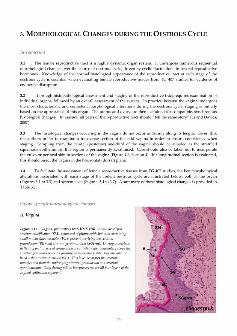
Figure 3.1a – Vagina: prooestrus (rat, H&E x20). A well developed
stratum mucification (SM), composed of plump epithelial cells containing
small mucin-filled vacuoles (V), is present overlying the stratum
granulosum (SG) and stratum germinativum (SGerm). During prooestrus,
flattening and increased eosinophilia of epithelial cells immediately above the
stratum granulosum occurs forming an amorphous, intensely eosinophilic
band – the stratum corneum (SC). This layer separates the stratum
mucification from the underlying stratum granulosum and stratum
germinativum. Only during mid to late prooestrus are all four layers of the
vaginal epithelium apparent.
SC
76
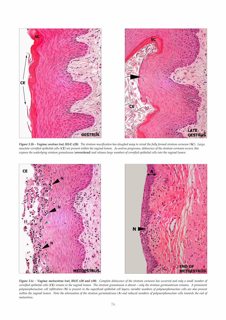
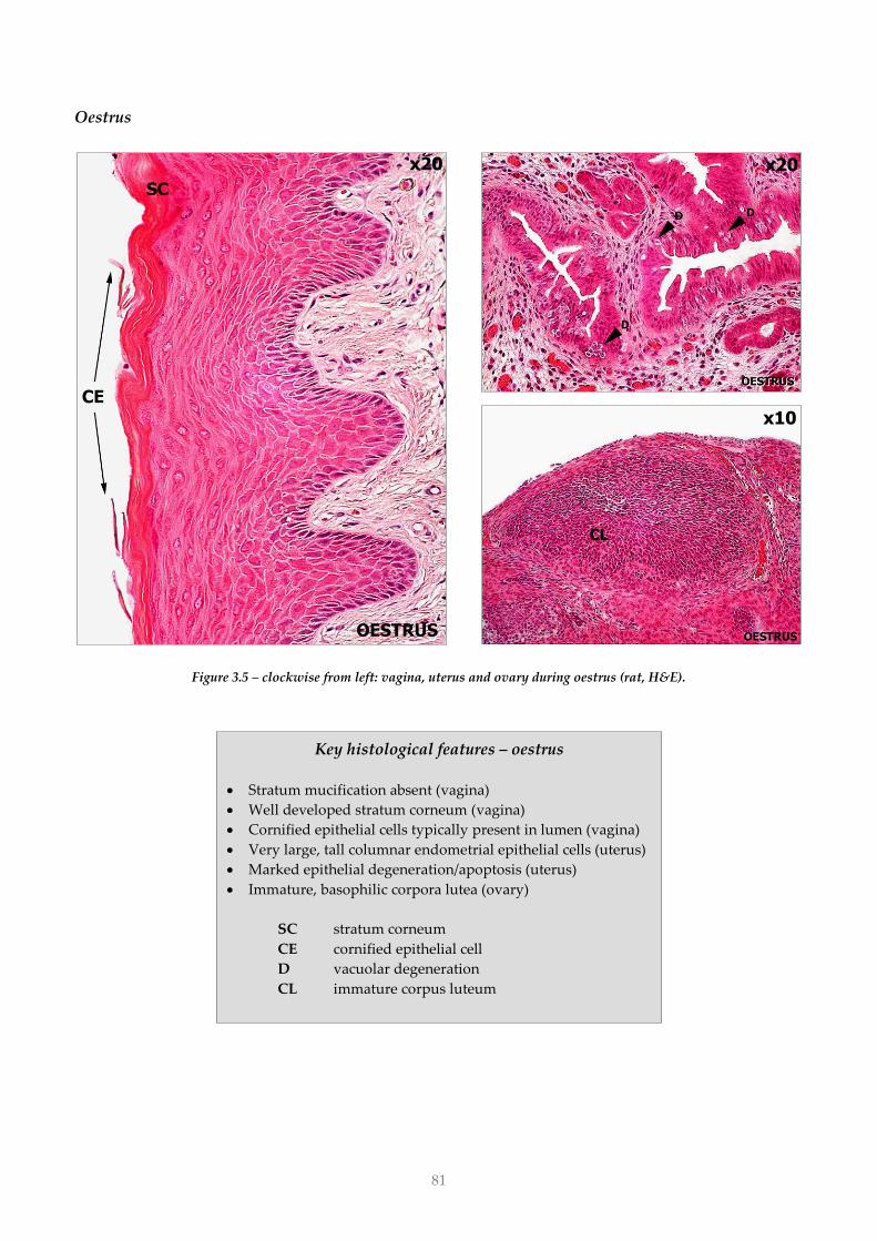
Figure 3.1b – Vagina: oestrus (rat, H&E x20). The stratum mucification has sloughed away to reveal the fully formed stratum corneum (SC). Large,
anuclear cornified epithelial cells (CE) are present within the vaginal lumen. As oestrus progresses, dehiscence of the stratum corneum occurs; this
exposes the underlying stratum granulosum (arrowhead) and releases large numbers of cornified epithelial cells into the vaginal lumen.
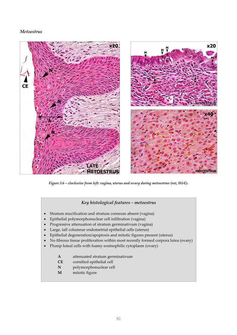
Figure 3.1c – Vagina: metoestrus (rat, H&E x20 and x10). Complete dehiscence of the stratum corneum has occurred and only a small number of
cornified epithelial cells (CE) remain in the vaginal lumen. The stratum granulosum is absent – only the stratum germinativum remains. A prominent
polymorphonuclear cell infiltration (N) is present in the superficial epithelial cell layers; variable numbers of polymorphonuclear cells are also present
within the vaginal lumen. Note the attenuation of the stratum germinativum (A) and reduced numbers of polymorphonuclear cells towards the end of
metoestrus.
END OF METOESTRUS
N
A
77
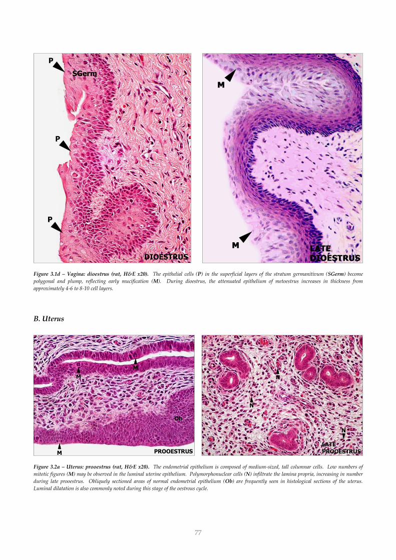
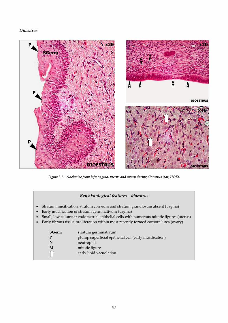
Figure 3.1d – Vagina: dioestrus (rat, H&E x20). The epithelial cells (P) in the superficial layers of the stratum germanitivum (SGerm) become
polygonal and plump, reflecting early mucification (M). During dioestrus, the attenuated epithelium of metoestrus increases in thickness from
approximately 4-6 to 8-10 cell layers.
B. Uterus
Figure 3.2a – Uterus: prooestrus (rat, H&E x20). The endometrial epithelium is composed of medium-sized, tall columnar cells. Low numbers of
mitotic figures (M) may be observed in the luminal uterine epithelium. Polymorphonuclear cells (N) infiltrate the lamina propria, increasing in number
during late prooestrus. Obliquely sectioned areas of normal endometrial epithelium (Ob) are frequently seen in histological sections of the uterus.
Luminal dilatation is also commonly noted during this stage of the oestrous cycle.
78
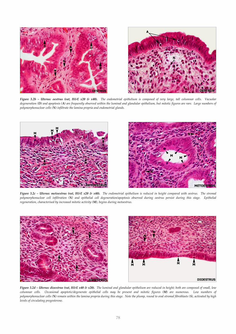
Figure 3.2b – Uterus: oestrus (rat, H&E x20 & x40). The endometrial epithelium is composed of very large, tall columnar cells. Vacuolar
degeneration (D) and apoptosis (A) are frequently observed within the luminal and glandular epithelium, but mitotic figures are rare. Large numbers of
polymorphonuclear cells (N) infiltrate the lamina propria and endometrial glands.
Figure 3.2c – Uterus: metoestrus (rat, H&E x20 & x40). The endometrial epithelium is reduced in height compared with oestrus. The stromal
polymorphonuclear cell infiltration (N) and epithelial cell degeneration/apoptosis observed during oestrus persist during this stage. Epithelial
regeneration, characterised by increased mitotic activity (M), begins during metoestrus.
Figure 3.2d – Uterus: dioestrus (rat, H&E x40 & x20). The luminal and glandular epithelium are reduced in height; both are composed of small, low
columnar cells. Occasional apoptotic/degenerate epithelial cells may be present and mitotic figures (M) are numerous. Low numbers of
polymorphonuclear cells (N) remain within the lamina propria during this stage. Note the plump, round to oval stromal fibroblasts (S), activated by high
levels of circulating progesterone.
79
C. Ovary
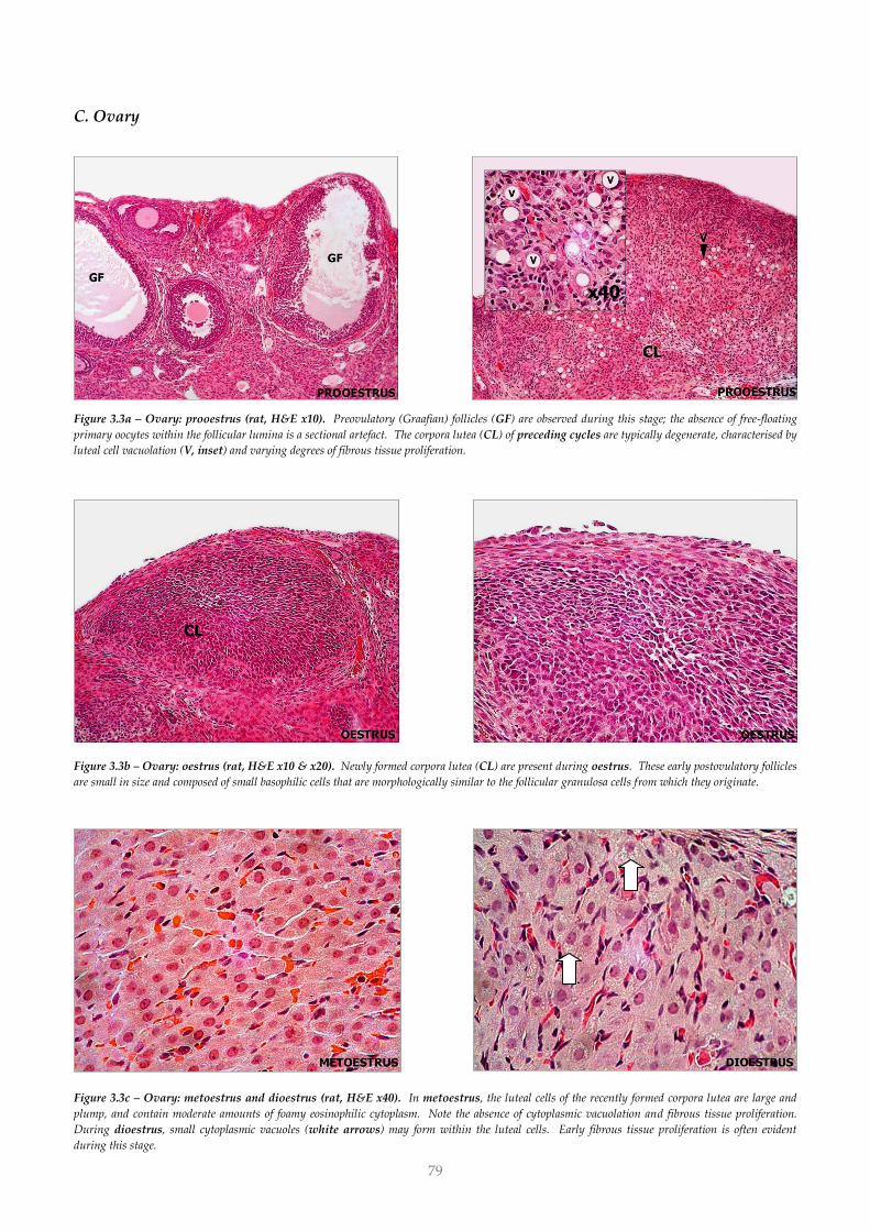
Figure 3.3a – Ovary: prooestrus (rat, H&E x10). Preovulatory (Graafian) follicles (GF) are observed during this stage; the absence of free-floating
primary oocytes within the follicular lumina is a sectional artefact. The corpora lutea (CL) of preceding cycles are typically degenerate, characterised by
luteal cell vacuolation (V, inset) and varying degrees of fibrous tissue proliferation.
Figure 3.3b – Ovary: oestrus (rat, H&E x10 & x20). Newly formed corpora lutea (CL) are present during oestrus. These early postovulatory follicles
are small in size and composed of small basophilic cells that are morphologically similar to the follicular granulosa cells from which they originate.
Figure 3.3c – Ovary: metoestrus and dioestrus (rat, H&E x40). In metoestrus, the luteal cells of the recently formed corpora lutea are large and
plump, and contain moderate amounts of foamy eosinophilic cytoplasm. Note the absence of cytoplasmic vacuolation and fibrous tissue proliferation.
During dioestrus, small cytoplasmic vacuoles (white arrows) may form within the luteal cells. Early fibrous tissue proliferation is often evident
during this stage.
x40
80
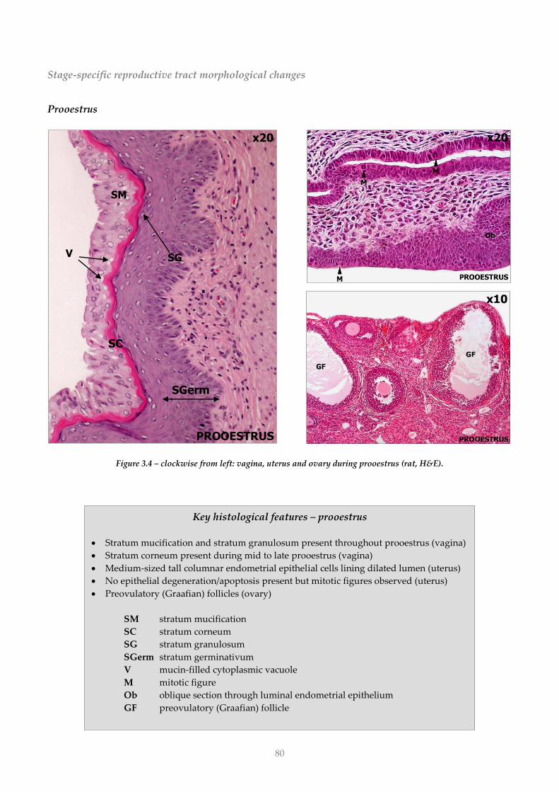
Key histological features – prooestrus
Stratum mucification and stratum granulosum present throughout prooestrus (vagina)
Stratum corneum present during mid to late prooestrus (vagina)
Medium-sized tall columnar endometrial epithelial cells lining dilated lumen (uterus)
No epithelial degeneration/apoptosis present but mitotic figures observed (uterus)
Preovulatory (Graafian) follicles (ovary)
SM stratum mucification
SC stratum corneum
SG stratum granulosum
SGerm stratum germinativum
V mucin-filled cytoplasmic vacuole
M mitotic figure
Ob oblique section through luminal endometrial epithelium
GF preovulatory (Graafian) follicle
SGerm
Stage-specific reproductive tract morphological changes
Prooestrus
Figure 3.4 – clockwise from left: vagina, uterus and ovary during prooestrus (rat, H&E).
x10 x10
x20
PROOESTRUS
SGerm
SC
SG
SM
V
x20
81
Key histological features – oestrus
Stratum mucification absent (vagina)
Well developed stratum corneum (vagina)
Cornified epithelial cells typically present in lumen (vagina)
Very large, tall columnar endometrial epithelial cells (uterus)
Marked epithelial degeneration/apoptosis (uterus)
Immature, basophilic corpora lutea (ovary)
SC stratum corneum
CE cornified epithelial cell
D vacuolar degeneration
CL immature corpus luteum
Oestrus
Figure 3.5 – clockwise from left: vagina, uterus and ovary during oestrus (rat, H&E).
x20
x10
x20
82
Key histological features – metoestrus
Stratum mucification and stratum corneum absent (vagina)
Epithelial polymorphonuclear cell infiltration (vagina)
Progressive attenuation of stratum germinativum (vagina)
Large, tall columnar endometrial epithelial cells (uterus)
Epithelial degeneration/apoptosis and mitotic figures present (uterus)
No fibrous tissue proliferation within most recently formed corpora lutea (ovary)
Plump luteal cells with foamy eosinophilic cytoplasm (ovary)
A attenuated stratum germinativum
CE cornified epithelial cell
N polymorphonuclear cell
M mitotic figure
Metoestrus
Figure 3.6 – clockwise from left: vagina, uterus and ovary during metoestrus (rat, H&E).
x40
x20 x20
x40
x20
83
Dioestrus
Figure 3.7 – clockwise from left: vagina, uterus and ovary during dioestrus (rat, H&E).
x20 x20
x40
Key histological features – dioestrus
Stratum mucification, stratum corneum and stratum granulosum absent (vagina)
Early mucification of stratum germinativum (vagina)
Small, low columnar endometrial epithelial cells with numerous mitotic figures (uterus)
Early fibrous tissue proliferation within most recently formed corpora lutea (ovary)
SGerm stratum germinativum
P plump superficial epithelial cell (early mucification)
N neutrophil
M mitotic figure
early lipid vacuolation
84
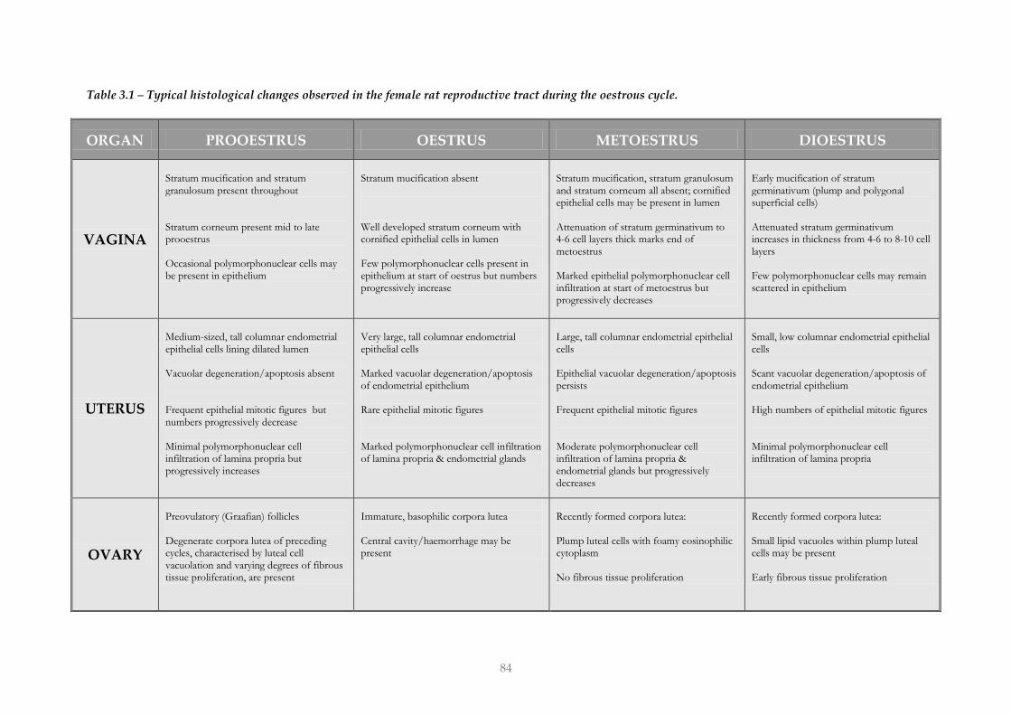
Table 3.1 – Typical histological changes observed in the female rat reproductive tract during the oestrous cycle.
ORGAN PROOESTRUS OESTRUS METOESTRUS DIOESTRUS
VAGINA
Stratum mucification and stratum granulosum present throughout Stratum corneum present mid to late prooestrus Occasional polymorphonuclear cells may be present in epithelium
Stratum mucification absent Well developed stratum corneum with cornified epithelial cells in lumen Few polymorphonuclear cells present in epithelium at start of oestrus but numbers progressively increase
Stratum mucification, stratum granulosum and stratum corneum all absent; cornified epithelial cells may be present in lumen Attenuation of stratum germinativum to 4-6 cell layers thick marks end of metoestrus Marked epithelial polymorphonuclear cell infiltration at start of metoestrus but progressively decreases
Early mucification of stratum germinativum (plump and polygonal superficial cells) Attenuated stratum germinativum increases in thickness from 4-6 to 8-10 cell layers Few polymorphonuclear cells may remain scattered in epithelium
UTERUS
Medium-sized, tall columnar endometrial epithelial cells lining dilated lumen Vacuolar degeneration/apoptosis absent Frequent epithelial mitotic figures but numbers progressively decrease Minimal polymorphonuclear cell infiltration of lamina propria but progressively increases
Very large, tall columnar endometrial epithelial cells Marked vacuolar degeneration/apoptosis of endometrial epithelium Rare epithelial mitotic figures Marked polymorphonuclear cell infiltration of lamina propria & endometrial glands
Large, tall columnar endometrial epithelial cells Epithelial vacuolar degeneration/apoptosis persists Frequent epithelial mitotic figures Moderate polymorphonuclear cell infiltration of lamina propria & endometrial glands but progressively decreases
Small, low columnar endometrial epithelial cells Scant vacuolar degeneration/apoptosis of endometrial epithelium High numbers of epithelial mitotic figures Minimal polymorphonuclear cell infiltration of lamina propria
OVARY
Preovulatory (Graafian) follicles Degenerate corpora lutea of preceding cycles, characterised by luteal cell vacuolation and varying degrees of fibrous tissue proliferation, are present
Immature, basophilic corpora lutea Central cavity/haemorrhage may be present
Recently formed corpora lutea: Plump luteal cells with foamy eosinophilic cytoplasm No fibrous tissue proliferation
Recently formed corpora lutea: Small lipid vacuoles within plump luteal cells may be present Early fibrous tissue proliferation
![CALIFORNIA [ADVANCE RELEASE] · Sh Sh MgCp SG SG SG SG SG SG SG SG SG Fe Fe Gr-s Gr-s Per CS Pum Pum Salt Salt Salt S-o S-o Zeo Dia Bent Bent Bent B B Clay Clay Dia DS DS DS DS DS](https://img.pdfslide.us/doc/110x75/5d435e0888c993ea558bc1de/california-advance-release-sh-sh-mgcp-sg-sg-sg-sg-sg-sg-sg-sg-sg-fe-fe-gr-s.jpg)