Embed Size (px)
Citation preview

Biol. Rev. (1971), 46, pp. 1-46 I
MORPHOGENESIS AND PATTERN FORMATION IN HYDROIDS
BY GERALD WEBSTER School of Biological Sciences, University of Sussex
(Received 20 March 1970)
CONTENTS I. Introduction . . . . . . . . . . . . . 11. Morphogenesis in Hydra . . . . . . . . . .
( I ) Patterns of cell division in growing Hydra . . . . . . (2) Patterns of cell movement in growing Hydra (3) Morphogenesis of the tentacles (4) Morphogenesis of the bud ( 5 ) The induction of secondary axes by grafts
(I) An outline of the problem
. . . . . . . . . . . . . .
. . . . . . . . . . . . . . . 111. The axial pattern in Hydra . . . . . . . . . .
. . . . . . . . . (2) The formation of the hypostome and the regeneration of distal structures (3) The formation of the basal disk . . . . . . . . (4) Polarity and the establishment and maintenance of the axial gradients . ( 5 ) The initiation and individuation of the bud . . . . . .
IV. The individuation of the hydranth of Tubularia . . . . . . .
(I) General principles . . . . . . . . . . . V. Theoretical models for the formation and regulation of axial patterns .
(2) ‘Positional information’ models . . . . . . . . (3) Inhibition and induction models
VI. Discussion . . . . . . . . . . . . VII. Summary . . . . . . . . . . . .
VIII. References . . . . . . . . . . . .
. . . . . . . .
IX. Addendum
. . I
. . 2 a . 3 * . 4 * * 5 . . 6 0 . 7 . . 8 . . 8
9 . 15
. . I 7 24
. . 26
. . 32
. . 32 - . 33 * f 35 * * 37 * * 39 . . 41
44
. .
. .
I. INTRODUCTION
The development of all organisms, whether from fertilized eggs, from buds or from isolated parts of adult organisms, raises two distinct, though closely related, problems. At the level of the single cell, there is the problem of cytodifferentiation, the process whereby an individual cell changes its structural and functional characteristics during the course of development. At a higher level of organization is the problem of the global and field aspects of development, the complex of processes whereby a more or less homogeneous cell population of relatively simple morphological form develops into an organism consisting of recognizably different parts arranged in a precise and reproducible spatial pattern, the whole being of a more complex morphological form. This general problem of the development of spatial organization or individuation (Waddington, 1966) has two facets in terms of the cellular activities involved. First, there is the problem of pattern formation in which we are concerned with the nature of the cellular interactions which result in the development of spatial patterns of differentiation; secondly, there is the problem of morphogenesis in which we are
I B R E 46

2 GERALD WEBSTER concerned with the nature and control of the cellular activities which produce changes in the morphological form of the cell population.
I n recent years there has been a revival of interest in both pattern formation (see Berrill, 1961 ; Lawrence, 1970; Rose, 1952; 19573; Sondhi, 1963; Ursprung, 1966; Waddington, 1962; 1966; Wolpert, 1969) and morphogenesis (see Gustafson & Wolpert, 1963; 1967; Trinkaus, 1965; 1966; Curtis, 1967).
This review is concerned with pattern formation and morphogenesis in hydroids, organisms which have for many years been favourite material for the developmental biologist interested in these problems and about which a large amount of information is now available. Hydroids have many advantages for such studies. Their develop- mental processes occur quickly and they are readily susceptible to experimental manipulation. Their spatial pattern of differentiation is relatively simple, since there is only one major, disto-proximal, developmental axis ; though it should be noted that Baird & Burnett (1967) have recently presented evidence suggesting the presence of a second, dorso-ventral, axis of symmetry in Hydra. Most important, however, hydroids have remarkable powers of regulation, so that isolated parts can reorganize and pro- duce complete miniature organisms. In such a system, the pattern of differentiation must arise as a result of cellular interactions, and hydroids thus provide ideal systems for studying these interactions.
Regulative processes occurring in the regeneration of marine hydroids, particularly Corymorpha and Tubularia, have been discussed in several earlier reviews (Huxley & De Beer, 1934; Barth, 1940; Child, 1941; Tardent, 1960, 1963, 1965; Berrill, 1961). During the last decade far more attention has been devoted to Hydra than to marine hydroids and the major part of this review is concerned with pattern regulation in this organism, A discussion of the important work of Rose (summarized, Rose, 1967b) on the individuation of the hydranth of Tubunclaria is also included, since this work can usefully be compared with that on Hydra and provides further insight into the factors responsible for the polarity of regeneration.
No consideration will be given to cytodifferentiation in hydroids; readers are referred to the review by Lentz (1966) for a summary of such literature.
11. MORF'HOGENESIS IN HYDRA
The major changes of form in Hydra which have to be explained are: (I) the development of tentacles during regeneration and budding; (2) the development of a new and complete axis during budding; (3) the development of partial secondary axes as a result of induction by grafts.
Hydra consists essentially of a hollow tube composed of two sheets of epithelial cells separated by an acellular mesogloea or basement membrane. It seems possible that, formally at least, some of the changes in form which take place could be accounted for in terms of the general mechanisms proposed by Gustafson & Wolpert (1963, 1967) and based upon their elegant analysis of sea-urchin morphogenesis. As yet, however, no systematic attempt has been made to apply these principles to the morphogenesis of Hydra, though some observations suggest that they may be applicable.

Morphogenesis and pattern formation in hydroids 3
( I ) Patterns of cell division in growing Hydra Any discussion of morphogenesis in Hydra must of necessity devote considerable
attention to the growth pattern of the animal, since the normal form (and size) is maintained in spite of continuous tissue loss and renewal, and because it has been suggested (Burnett, 1961, 1962, 1966, 1967) that growth and morphogenesis are closely related processes in Hydra and that changes in form are associated with localized growth or cell division. It has been supposed that cell proliferation occurs in a localized growth zone just below the hypostome (see Fig. I a). The existence of this zone was deduced from studies on the movement of marked tissues. In actively growing animals there is a zone of tissue in the subhypostomal region which appears to be stationary; above this region cells move into the hypostome and out along the tentacles; below it they move proximally into the bud and peduncle (Tripp, 1928; Brien & Reniers-Decoen, 1949; Burnett, 1959).
7 hypostome
digestive zone
I budding zone
peduncle
- basal disk
gonophores
proximal tentacles
hydranth base . . . . . .. . . .
. .
. .
. . . . . .
Fig. I . Diagram of Hydra showing different regions of the axis (from Webster & Wolpert, 1966). The basal disk is proximal, the tentacles distal. (b ) Diagram of hydranth of Tubuluriu showing different regions. (Redrawn after Tardent, 1963.)
In an extremely careful analysis of cell division in Hydra littoralis, Campbell ( 1 9 6 7 ~ ) found that mitotic figures were distributed along the entire length of the axis (with the exception of the tentacles) and the mitotic index was virtually identical at all levels, though somewhat lower in the peduncle. He found little difference between budding and non-budding animals as regards the distribution of cell divisions along the axis. Similar results were obtained when he examined the proportion of nuclei which incorporated tritiated thymidine.
These observations were confirmed by Clarkson & Wolpert (1967), who examined the regional mitotic indices and also the incorporation of tritiated thymidine into DNA. Webster & Hamilton (197ob) have examined the proportion of thymidine-labelled nuclei in animals that had been starved and then re-fed to initiate budding; the
1-2

4 GERALD WEBSTER proportion was approximately the same in distal, middle and proximal thirds of the axis.
Finally studies of the mitotic index in H. pseudoligactis indicate that, in this species too, cell division occurs equally in all parts of the axis (Shostak, Pate1 & Burnett, 1965 ; Campbell, 1967d).
At present therefore there is no direct evidence in favour of localized growth zones in Hydra, and good evidence against the idea in two species.
(2) Patterns of cell movement in growing Hydra Campbell (19673) has made a detailed study of patterns of cell movement in actively
growing H. littoralis using dye-marked grafts and grafts of tentacles. His results confirm and extend previous observations. As noted above, tissues continually move away from the subhypostomal region, distally into the hypostome and tentacles and proximally into the body column and bud. In a lucid and elegant analysis, Campbell showed that such a pattern of cell movement is consistent with growth occurring either in a localized region or uniformly along the axis; the different growth patterns, how- ever, should result in different rates of movement. Measurements of rates of tissue movement could not be made precisely enough to distinguish between the two growth patterns, but from the data on the distribution of mitotic activity, mentioned above, Campbell was able to calculate the expected rate of tissue movement and found that the theoretical curve agreed well with the observed curve.
Campbell was able to make estimates of the rates of loss of cells at the ends of the column and into the bud; these estimates indicate that most cells are lost into the bud. The rates of loss at the different sites are perfectly consistent with the observed position of the stationary region. This argument can also account for the fact that in starved animals, which do not bud, the position of the stationary region moves from the subhypostomal region into the middle of the axis, since in such animals the rates of cell loss at the distal and proximal ends of the axis are approximately the same; as Campbell points out, if the stationary region were a localized meristematic region such movement would be most surprising.
Thus, the essence of Campbell's argument is that the observed pattern of cell movement reflects a pattern of localized cell loss rather than one of localized cell production.
In a further study using isotopically labelled grafts. Campbell (1967~) examined the movement of different cell types in H. littoralis. The two epithelial layers, the ectoderm (including the epitheliomuscular cells and the interstitial cells) and the endoderm (including digestive and gland cells), moved as coherent cell sheets. These cell sheets moved distally and proximally from a stationary region but at different rates, and measurements indicated that the stationary regions for the two cell sheets were at different levels of the axis. Nematocytes always moved distally irrespective of their position in the axis; their movement is therefore polarized and presumably indicates that there is some vectorial property of the axis to which they respond.
Campbell (1967~) also showed that the patterns of cell movement are consistent with the histology and ultrastructure of hydroid tissues. In the endoderm the gland
Morphogenesis and pattern formation in hydroids 5 and digestive cells are in contact with the mesogloea and are bound to each other by septate desmosomes (see also Wood, 1959). The epitheliomuscular cells of the ectoderm are also in contact with the mesogloea and are bound to each other by septate desmo- somes. Interstitial cells and nematocytes are not desmosomally bound to epithelio- muscular cells in the column, but the nematocytes are so bound in the tentacles, where they end up after migration. Campbell suggests that interstitial cells are displaced passively as a result of movement of the epithelial sheets in which they reside. Nemato- cytes, on the other hand, are considered to be capable of active, independent, movement.
Shostak and his colleagues (Shostak & Globus, 1966; Shostak et al. (1965) have examined the movement of the two epithelial layers in H . eriridis. They too found that ectodermal and endodermal layers moved independently and at different rates; they suggest therefore, as does Campbell, that the mesogloea does not act as a 'cement' permanently binding the two layers together, but rather as a substrate for epithelial movement. No mechanism for this movement has been proposed.
( 3 ) Morphogenesis of the tentacles Hydra from which the hypostome and tentacles have been removed regenerate these
structures very quickly. At 26" C. tentacle rudiments appear on H. littoralis within 20-24 hr. and these elongate to produce tentacles of normal length within the next 12-18 hr. (see Webster & Wolpert, 1966).
It has been argued (Burnett, 1961, 1962, 1966) that the regeneration of tentacles and their formation in the developing bud depends on localized growth zones, though no attempt has been made to explain how this 'growth' could give rise to structures of such precise shape. As we have seen above, there is no evidence for the existence of such growth zones in Hydra, and, furthermore, several observations clearly indicate that cell division plays only a minor role in morphogenesis of the tentacles.
Clarkson (19696) observed that H. Zittmalis in which DNA synthesis had been reduced by 60 yo, by treatment with hydroxyurea, regenerated tentacle rudiments with little delay, i.e. in 24 hr.-as compared with 18-20 hr. in the controls. Under these conditions, however, the elongation of tentacles was delayed more profoundly. Web- ster & Hamilton (197oa) have also observed in the same species that the time required for tentacle morphogenesis bears no relation to mitotic activity. Hydra which have been starved for 6 days produce tentacle rudiments within 24-28 hr. compared with well-fed controls which take 20-24 hr. In starved animals the mean mitotic index is 0.12 f 0.9% as compared with well-fed controls in which it is 1.3 +0-19%. Finally, analysis of stained sections of H. pseudoligactis (Park, Ortmeyer & Blankenbaker, 1967) indicates that mitotic activity is not increased during any stage of regeneration.
It seems likely therefore that cell division is not an important mechanism in the regeneration of tentacles but cell movement is certainly involved. As pointed out above, it is known that cells move slowly into the tentacles from the column during normal growth. Webster & Hamilton (1970~) have demonstrated that during regeneration of H. littoralis morphogenesis of the tentacles is associated with rapid cell movements. Animals in which the distal part of the axis was coloured were produced by grafting stained tissues. When these animals were decapitated and allowed to regenerate, the

6 GERALD WEBSTER stained portion of the axis decreased in length as the tentacles formed and elongated. This decrease in length was temporally associated with tentacle formation and did not occur until after 18 hr.; tentacle rudiments were produced at 20-24 hr. It seems pro- bable therefore that cells move up the axis and into the new tentacles and that this movement plays an important role in the formation of the tentacles. Again the mechanism of movement is unknown. Shostak (1968) has made identical observations on the developing tentacles of buds.
In principle, it is possible to account for the formation of tentacle rudiments using the general morphogenetic mechanisms proposed by Gustafson & Wolpert (1963, 1967). A local increase in adhesion between cells at the site of a tentacle would cause a thickening of the cell sheet and a change in its curvature which would result in the formation of a small rudiment. However, there is at present no evidence that tentacle rudiments are formed in this way and our (unpublished) attempts to determine the nature of the process using fixed and stained preparations have been inconclusive.
It is improbable, however, that simple changes in adhesion can account for the further elongation of the tentacle rudiment which may involve stretching of the cell sheets, either as a result of some external force, such as hydrostatic pressure, or as a result of an autonomous stretching of individual cells.
(4) Morphogmsis of the bud In actively growing Hydra, buds are initiated in a region at the proximal end of the
digestive zone just above the junction with the peduncle. The first sign of bud formation (most easily seen in animals which have been starved and re-fed) is an increase in the optical density of the endoderm in the budding zone. This is followed (Fig. 2) by the formation of a small conical protuberance which rapidly elongates to form a tube. Tentacle rudiments appear at the distal end of the tube and, at about the same time, a construction appears at the proximal end. Eventually a small-complete Hydra detaches from the parent.
Fig. 2. The main changes in form and the approximate time course of budding in H. littoralis. (Redrawn after Clarkson & Wolpert, 1967.)
Burnett (1961, 1962, 1966) has argued that the initiation and elongation of the bud are caused by localized cell division in the budding region. There seems little doubt that this view is incorrect. As noted above, there is no evidence for any localized region of cell division in Hydra and, furthermore, under certain conditions, buds can be formed in starved animals in which cell division is at an extremely low level (Webster & Hamilton, 197ob; see Section III(5) below).
All the available evidence on budding in Hydra points towards cell movement and changes in cell contact as the primary mechanism of morphogenesis.

Morphogenesis and pattern formation in hydroids 7 Histological analysis of the early stages of bud development (Webster & Hamilton,
1970b) indicates that the increase in optical density of the budding zone mentioned above is a consequence of an increase in thickness of both ectodermal and endodermal cell layers which is particularly marked on one side of the axis. This increase in thick- ness is associated with, or quickly followed by, a change in the curvature of the cell sheets. The histology of the changes in thickness and curvature suggests that they may be caused by an increase in adhesion between the cells (see Gustafson & Wolpert, 1963, 1967). It is worth noting in this connexion that our (unpublished) observations indicate that when Hydra is placed in a medium which causes cell disaggregation (e.g. EDTA) the developing bud is always the last part to disaggregate, suggesting that cells in the bud are more strongly adhesive than those in other parts of the animal.
Although the early stages of bud formation may depend on changes in adhesion between cells, it seems unlikely that elongation could be caused by the same mechanism. Cell movement of some sort is certainly involved. As noted above, Campbell (19673) has shown that cells move into buds. Shostak & Kankel(1967) have shown, by marking experiments, that in H. oiridis the cells of the bud are derived from the column of the parent. In isolated budding zones taken from several species of Hydra it can be seen that the buds grow at the expense of parental tissue (Burnett, 1961 ; Clarkson & Wol- pert, 1967). Clarkson & Wolpert (1967) inhibited cell division in H. ZittoruZis by means of y-irradiation but found that bud elongation continued at a normal rate. They suggest that the elongation is caused by reorientation and repolarization of parental cells (see Section III(4)) and it is probable that the distal tip of the bud-which is the first part to be formed-plays an important role in this process. Li & Yao (1945) demonstrated that the tip of a very early bud when transplanted to the digestive zone of a host animal could induce a secondary axis which behaved exactly like a normal bud and eventually detached from the host.
The problem of the detachment of the bud has not been studied very fully. It is striking that the size of the bud seems to be very precisely controlled; when the budding zone is isolated, the developing bud seems to take only the cells which it would normally take, and often detaches from a very small piece of parental tissue (Burnett, 1961 ; Clarkson & Wolpert, 1967). Detachment does not seem to be depen- dent upon the developmental state of the distal tip; Shostak, Bisbee, Ashkin & Tam- mariello (1968) exchanged the tips of early and late stage buds of H. viridis and found very little effect on the time taken for detachment.
The formation of tentacles in the bud has already been discussed in the previous section.
( 5 ) Induction of secondary axes by grafts Browne (1909) showed that a partial secondary axis could be induced by transplan-
tation of small pieces of hypostome into the digestive zone of an intact host animal. This observation has since been confirmed by many workers (e.g. Yao, 1945 ; Webster, 1966a, b). The induced axis consists of a short digestive zone, a hypostome and a set of tentacles; the major part of the secondary axis consists of host tissue, the graft forming the hypostome. The mechanism of secondary axis formation probably involves re- orientation of cells and cell movement in response to influences from the graft. This is

8 GERALD WEBSTER inferred from observations of animals in which a secondary axis is induced in the proximal digestive zone and the host hypostome removed; the secondary axis then elongates at the expense of the distal part of the host axis, and the latter eventually disappears, resulting in a single axis in which the distal part is at right angles to the proximal part (Webster, 1966 a). The mechanisms involved in cellular reorientation are completely unknown, though some speculations are presented in Section I11 (4).
As mentioned above, Li & Yao (1945) showed that the distal tip of a bud of Pelmato- hydra oligactis can induce the formation of a complete secondary axis. At about the time when the bud acquires its own tentacles the inductive properties of the tip change, and it now induces a partial secondary axis identical to that induced by a hypostome. Clarkson (1967) has confirmed these observations in H. littoralis, but found that in this species the change in inductive properties occurs somewhat earlier, at about stage 3 (Fig. 2).
Browne (1909) also showed that transplantation of the basal disk resulted in the formation of a partial secondary axis consisting of a peduncle, the grafted disk per- sisting at its proximal end. Yao (1945) and J. Hicklin (personal communication) have made similar observations and have shown that the secondary axis is formed from host tissues. It must be presumed therefore that the basal disk is also capable of re- orientating cells and of causing cell movement.
All other regions, apart from the hypostome, bud-tip and basal disk, are usually assimilated after transplantation to intact host animals (Browne, 1909; Yao, 1945 ; Webster & Wolpert, 1966) though secondary axes may be formed after transplantation to decapitated hosts (see Section I11 (2)). J. Hicklin (personal communication) states that partial proximal axes with basal disks may be induced by transplantation of pieces of peduncle ; it is not known whether induction occurs directly or whether the trans- plant has first to become determined as a basal disk (see Section III(3)). Proximal parts of developing buds are also capable of inducing peduncles and basal disks after trans- plantation (Clarkson, 1967).
111. THE AXIAL PATTERN IN HYDRA
(I) An outline of the problem The body column of Hydra consists of six more or less clearly defined regions
arranged along the disto-proximal axis ; these are hypostome, tentacles, digestive zone, budding zone, peduncle and basal disk (Fig. I a). After removal or isolation of a part of the axis, regulation occurs so that the missing parts are regenerated. Under normal conditions, regulation is always polarized; that is, distal structures form at the distal end of the axis and proximal structures at the proximal end, even though any level of the axis can produce any structure. The axis of Hydra is therefore a polarized, one- dimensional, individuation field.
Two major problems are posed: (I) What is the mechanism of individuation, i.e. how is the axis subdivided into
(2) What is the basis for the polarity of regeneration? distinct regions and how are missing parts restored?
Morphogenesis and pattern formation in hydroids 9 Throughout the following discussion I shall, in general, only give formal interpre-
tations and explanations of phenomena and experimental results. At present we are almost completely ignorant of events at the cellular and biochemical levels, although, in certain situations, it is possible to speculate about the nature of these events. The concepts employed, such as ‘inhibition’ and ‘threshold’, are defined operationally and it must be borne in mind that such definitions do not carry any implication as to par- ticular mechanisms at the cellular or biochemical levels.
(2) The formation of the hypostome and the regeneration of distal structures There is no doubt that the hypostome is the dominant region of Hydra, for it
possesses all the characteristics of such a region as classically defined (Huxley & De Beer, 1934): (I) ‘it is the first region to be formed in development or regeneration’; (2) ‘its formation, once initiated, is an autonomous process independent of the formation of further dominant regions’; (3) ‘once formed it exerts an organizing influence on other regions’; (4) ‘it inhibits the formation of further dominant regions’.
The organizing ability of the hypostome was demonstrated by Browne (1909), who showed that a small piece of hypostome was capable of inducing the formation of tentacles and a short axis when transplanted to a host animal (see Section II(5)). Browne also showed that during distal regeneration the properties of the hypostome were acquired by the distal end of the axis some time before tentacle morphogenesis occurred. Webster & Wolpert (1966) confirmed this observation, and also showed that the formation of a hypostome was the first detectable event during regeneration.
The inhibitory powers of the hypostome were demonstrated by Rand, Bovard & Minnich (1926) and confirmed by Webster (1966~). The experiments showed that a grafted hypostome and tentacles could suppress distal regeneration in a host animal.
The mechanisms involved in the formation and localization of the hypostome will be considered in some detail.
(a) The axial gradient in the time required for hypostome determination Much work on various species of Hydra has shown that regeneration of tentacles
occurs more rapidly from distal than from proximal levels of the axis (Peebles, 1897; Browne, 1909; Weimer, 1928; Rulon & Child, 1937; Spangenberg & Eakin, 1961; Webster & Wolpert, 1966). Although Browne’s (1909) results suggest that tentacles develop because of the presence of a hypostome, her results also show that the time required for their formation does not necessarily reflect the time required for hypo- stome formation. We (Webster & Wolpert, 1966) have examined in H . littoralis the time required for small fragments isolated from various levels of the axis to acquire hypostomal properties, using as the criterion for this their resistance to assimilation and the induction of a secondary axis when they are transplanted into an intact host animal. The time required for determination to occur in 50% of the pieces ( T ~ o ) ranged from 4.5 hr. for the subhypostomal region to more than 50 hr. for the proximal peduncle, with intermediate values for axial levels between these extremes. There is therefore an axial gradient in the time required for determination. If the times for determination are converted into ‘rates’ ( I/T 50) and plotted against the original

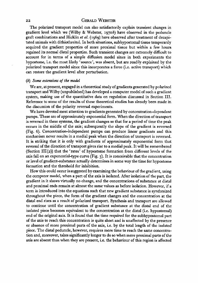
I 0 GERALD WEBSTER position of the region on the disto-proximal axis (Fig. 3), it would appear that the gradient is not linear but more or less exponential. The small number of measurements available, together with the crudity of the experimental technique, makes this conclu- sion rather tentative, though, as will be seen below (Section III(4)), there are other reasons for believing that it may be correct.
I I I I I 1 1 2 3 4
axial position
Fig. 3. ‘Rate’ of hypostome determination in isolated pieces taken from different positions on the disto-proximal axis (1-4) of H. littoralis. ‘Rates’ (open circles) are calculated as 1/T5o for hypostome determination. The curve is a theoretical one showing the axial distribution of a substance produced by a polarized-transport-back-diffusion mechanism in which transport is concentration-dependent. (Data on hypostome determination from Webster & Wolpert (1966); curve calculated by 0. K. Wilby.)
It is worth noting that the T 50 for hypostome determination in the subhypostomal region is not measurably affected by the total length of the isolated piece. In the case of the distal peduncle, however, the T5o is reduced from 28 hr. to about 16 hr. when proximal regions are present, i.e. when the piece is longer. A possible explanation for this observation will be given below (Section III(4)).
I t is important to note that the delay between hypostome determination and the onset of tentacle formation is about the same at both distal and proximal levels of the axis; i.e. the ‘rate’ determining step in distal regeneration is the formation of the hypostome. Similar conclusions can be drawn for hydranth regeneration in marine hydroids (Webster & Wolpert, 1966).
It is not known why different levels of the axis require different times to form a hypostome. The observations do not necessarily indicate that there is an axial gradient

Morphogenesis and pattern formation in hydroids I1
in rate of development or differentiation, although this is a possibility. Differences in time for determination could also arise as a consequence of axial differences in the time at which hypostome formation is initiated after decapitation, i.e. a variable lag period, or as a result of axial differences in the ‘position’ of the region on some ‘ developmental pathway’ or chreode (Waddington, 1966) leading to hypostome for- mation.
The T 50 for hypostome determination in tissue from the subhypostomal region is virtually unaffected by severe inhibition of DNA synthesis by means of hydroxyurea. However, inhibition of protein synthesis by chloramphenicol or RNA synthesis by 5-fluorouracil substantially increases the T 50 (Clarkson, 1969 b). These observations provide clear evidence that determination does not involve cell division, but that the process may involve qualitative or quantitative changes in protein or RNA com- position.
(b) The inhibition of hypostome formation Rand et al. (1926) presented some evidence that the presence of a hypostome and
tentacles prevented the regeneration of a further set of distal structures and I (Webster, 1966 a) have confirmed and extended these observations. The experiment consisted simply in transplanting a subhypostomal region to the digestive zone of a host animal in the presence or absence of the host hypostome. In its presence, the transplant was assimilated; in its absence, the transplant developed into a hypostome and a secondary axis was produced. A formal interpretation of this experiment is that when the hypo- stome is removed, the level of inhibition falls, the transplanted region is released from inhibition and hypostome determination occurs (Fig. 4). It must be emphasized that, since this is a formal explanation of the results, the experiment does not necessarily imply the presence of some inhibitory substance, although this may be the case. The results can equally well be accounted for in terms of a formally identical inductive model (see Wigglesworth, 1940). Thus, some inducer is required for hypostome formation and maintenance; the extant hypostome absorbs this and keeps it at a low level; removal of the hypostome results in the level rising and hence in new hypostome formation. Throughout this discussion, results will be considered in terms of inhibition. It should be realized that alternative, and rather different, formal explanations can also be given (see Section V).
It is clear that if hypostome formation occurs as a consequence of release from inhibition, this mode of control implies that hypostome formation is a spontaneous and autonomous process, a conclusion in accord with the classical definition of a dominant region (Huxley & De Beer, 1934).
A problem of some interest is how the inhibitory influence is transmitted from the hypostome throughout the animal. An understanding of this might be expected to shed some light on the mechanism of inhibition. We (Webster, 1966a; Wilby & Webster, 197oa) have been particularly concerned with whether inhibition ‘moves’ or acts in a polarized fashion, as postulated by Rose (1957a; b) for the hydranth of Tubularia (Section IV), and have investigated this in Hydra using transplantation techniques.

I 2 GERALD WEBSTER A hypostome transplanted to the proximal end of the digestive zone can inhibit
hypostome formation at the distal end in a significant number of animals without any change in the regeneration polarity of the inhibited axis. This experiment shows fairly conclusively that inhibition in Hydra is not polarized and that inhibitory iduences can ‘move’ or be transmitted in both a disto-proximal and a proximo-distal direction. The experiment also shows that hypostome inhibition is determined by factors quite distinct from those that determine polarity.
Fig. 4. Diagram, in terms of Lawrence’s ‘sand model’, of the results of transplanting a sub- hypostomal region into a host with and without a hypostome. The gradients are drawn as linear for simplicity. Top row : transplantation into an intact host in which the level of inhibi- tion (I) remains constant. The level of the threshold gradient (T) in the transplant falls as a result of diffusion of gradient-substance from transplant to host; the residual ‘hump’ in the gradient is eliminated as a consequence of diffusion and polarized transport. Bottom row : transplantation into a host from which the hypostome has been removed, thereby resulting in a fall in the level of inhibition (I). Diffusion of gradient-substance (T) occurs from trans- plant to host, as above, but the transplant is released from inhibition because I falls more rapidly than T. The transplant becomes determined as a hypostome, as does the distal end of the host axis, and the levels of I and T rise again.
It seems probable that the transmission of inhibition is dependent upon cell contact. A hypostome placed in the enteron of an animal is not inhibitory, nor is a hypostome isolated in a microdrop together with regenerating regions (Webster, unpublished). These observations suggest that if inhibition is due to a substance, then it is not freely diffusible. We (Wilby & Webster, 1970~) have observed that the ability of a digestive zone to transmit inhibition from a proximal hypostome graft is impaired by pre- treating the tissue with media containing reduced concentrations of calcium and magnesium ions and culturing the graft combination in the same medium. Such treat-

Morphogenesis and pattern formation in hydroids I 3 ment appears to have no effect on the inhibitory properties of the hypostome nor upon the response of the tissue to inhibitory influences. Under these conditions the adhesion between cells does not appear to be affected and we have suggested that the depletion of calcium and magnesium ions interferes with intercellular communication, possibly by affecting functional coupling between cells (Loewenstein, 1966, 1967). At present there is no direct evidence that the cells of Hydra are functionally coupled, but the above observations are consistent with this being the case, as is the presence of septate desmosomes linking both the ectodermal and the endodermal cells (Wood, 1959; Campbell, 19674 since these structures have been suggested as a possible site for coupling (Bullivant & Loewenstein, 1968).
The time required for the establishment of inhibition at the distal end of the digestive zone by a proximal hypostome graft is about 4-7 hr. from the time of trans- plantation. This is of the order of magnitude that would be expected (on the data available from other systems in which actual measurements have been made; see Loewenstein, 1966; Loewenstein & Penn, 1967) if inhibition by the graft is dependent upon the establishment of functional coupling between graft and host and the diffusion through the coupled cell system of some substance of fairly low molecular weight
The suggestion that functional coupling may be important in the transmission of inhibition must be regarded as extremely tentative. I t is also important to note that in the hydranth of Tubuluria, inhibitory interactions can occur under certain conditions in the absence of cell contact (Rose, 1 9 6 7 ~ ; Section IV below).
There is evidence (Webster, 1966a) that the intensity of inhibition is not the same at distal and proximal levels of the axis. Subhypostomal regions transplanted to the basal disk usually form secondary axes and hypostomes irrespective of whether the host hypostome is present or absent. This result should be compared with that ob- tained by transplanting subhypostomal regions to the digestive zones of intact hosts where the region is always assimilated. These results suggest that there is a disto- proximal gradient of inhibition.
The experiment cited above, in which a subhypostomal region transplanted to the digestive zone in the absence of the host hypostome itself developed into a hypostome, was interpreted as indicating that the level of inhibition falls when the hypostome is removed. By transplanting subhypostomal regions to host animals at various times after the removal of the hypostome I have shown (Webster, 1966b) that the level of inhibition rises again during regeneration. About 5-10 hr. after removal of the hypo- stome the inhibitory situation is essentially normal, which indicates that the level of inhibition rises again when the new hypostome is determined at 4-5 hr.
Finally, it may be noted that regeneration of distal structures can be inhibited by both buds and peduncle grafts. Lenhoff (1957) and Burnett (1961) have observed that distal regeneration does not occur if an animal is cut in such a way that a developing bud is adjacent to the distal end of the axis. It is not known whether it is the formation of the hypostome or the development of tentacles which is inhibited. Although a bud- tip has some of the characteristic features of a hypostome (Sections II(4) and III(5)) it is not known whether it is capable of inhibiting hypostome formation and it seems
(c 1000).

I 4 GERALD WEBSTER possible that the inhibition is of tentacle morphogenesis since it is known (Section II(3)) that this is dependent upon cell movement; the proximity of a bud into which cells are moving might inhibit this process simply by removing cells.
Wilby & Webster (197ob) observed that regeneration of distal structures was drastically slowed down as a result of grafting a peduncle on to a subhypostomal region. This seems to be due not to inhibition of hypostome formation per se but to a change in the ‘potential’ for hypostome formation (time and threshold properties) of the sub-hypostomal region so that it acquires a ‘potential’ characteristic of more proximal regions (Section III(4)).
(c) Threshold fw inhibition The experiments noted above (Webster, 1966a), in which distal regions were
transplanted to the basal disk and which suggest an axial gradient in level of inhibition, also indicate (in this terminology) that the threshold for inhibition is different in distal and proximal parts of the axis. Subhypostomal regions transplanted to this region usually produced hypostomes ; proximal digestive zones, however, under the same conditions only once produced a hypostome. If the level of inhibition in the basal disk can be regarded as constant, these results indicate that the proximal digestive zone is more easily inhibited than the subhypostomal region. This suggests an axial gradient in threshold for inhibition.
I have studied (Webster, 19663) the threshold of different regions during regenera- tion, and also after transplantation and the induction of a new axis, and have found that it is a labile property.
During the regeneration of the isolated peduncle the threshold for inhibition rises in the distal tip and this region acquires the threshold properties characteristic of the subhypostomal region after about 12 hr. of regeneration. The T 50 for hypostome determination in the distal peduncle is 16.5 hr. and that for the subhypostomal region is 4 5 hr. I t seems therefore that the threshold properties of a region are closely con- nected with those factors that determine its time for hypostome determination since after 12 hr. regeneration, the T 50 for the peduncle is 4-5 hr. the same as the T 50 for the subhypostomal region. A possible explanation of this will be given below (Section III(4)). No changes in threshold in parts of the axes other than the presumptive hypostome could be detected until well after the determination of the hypostome but, since the experimental technique was not entirely satisfactory, it cannot be concluded that changes in threshold do not take place.
The threshold properties of a region are also determined by its position on the disto-proximal axis. Thus, when a secondary axis was induced by a hypostome, the subhypostomal region of the new axis, which was derived from cells of the mid- digestive zone, possessed characteristic subhypostomal properties, i.e. they had acquired more distal properties as a result of the proximity of a hypostome. Con- versely, when subhypostomal tissue was transplanted to the digestive zone of an intact host, the characteristic properties of the region were lost and the tissue was assimilated.
The T5o for this loss of distal characteristics (and presumably acquisition of proximal characteristics) was about 4 hr.

Morphogenesis and pattern formation in hydroids 15 These transplantation experiments all show the lability of the threshold properties
of a region and also those properties which control the time for hypostome determina- tion. By means of the same tests it is also possible to show that, under other conditions, these properties are extremely stable. For example, when a hypostome is transplanted to the proximal end of the digestive zone (Wilby & Webster, 1970 b) the distal end of the digestive zone (from which the hypostome has been removed) retains its charac- teristic properties for at least 48 hr.
These rather contradictory observations that threshold can be both labile and very stable can be explained, but the explanation is better deferred until we have con- sidered how polarity is controlled (Section III(4)).
( d ) The cause of inhibition At present the mechanism of inhibition is not known. Although some observations
are consistent with inhibition being caused by an inhibitory substance, inhibitory phenomena can be accounted for without postulating such a substance, e.g. in posi- tional information models (Section V).
Few attempts have been made to isolate inhibitory substances from the tissues of Hydra. Lenicque & Lundblad (1966) isolated high-molecular-weight materials which inhibited distal regeneration, but no evidence for specificity was presented nor any reasons given for regarding this material as a genuine regulatory substance. Davis (1967) has shown that the inhibitory activity of homogenates of several species of Hydra (and of Corymorpha) is due to the presence of nematocyst toxins, in particular tetramethylammonium. Wilby (unpublished) has shown that homogenates of H. littoralis, prepared in such a way that they are probably free of nematocyst toxins, can inhibit both distal and proximal regeneration. Fractionation of these extracts yielded at least five inhibitory fractions of different molecular weight, none of which showed any specificity. There is no evidence at present to suggest that any of these fractions contains a genuine regulatory substance.
These observations demonstrate very clearly that the simple isolation of some in- hibitory substance is quite inadequate evidence that inhibitory phenomena are caused by such a substance. In addition to the obvious requirement of specificity, and the identification of the isolated substance and its mode of action, it is also necessary to demonstrate that the substance shows temporal and regional differences in concentra- tion consistent with the results of the transplantation experiments. As yet none of these requirements has been met.
It must be mentioned that Davis (1966) has isolated from crowded cultures of Hydra a substance of high molecular weight which may be secreted by the animals. I t can inhibit regeneration and reduce the growth rate of treated animals, but whether it plays any role in the normal developmental processes is unknown.
( 3 ) The formation of the basal disk There is good circumstantial evidence that the basal disk is formed in relation to the
same gradients as are involved in hypostome formation (i.e. gradients in time and threshold for inhibition). The basal disk is produced at the physiologically proximal
16 GERALD WEBSTER end of an axis which may or may not be the morphologically proximal end; that is, it forms at low levels of the gradient. This can be inferred from situations where two hypostomes are present, one at each end of the axis; whether these are produced by grafting (e.g. King, 1901 ; Goetsch, 1929; Wilby & Webster, 197ob) or by chemical treatment (Webster, 1967) is immaterial; the basal disk tends to form midway between the two. When distal parts of the axis are inserted with reversed polarity (Webster & Hamilton, 197ob), or when a hypostome is grafted to the proximal end of a digestive zone (Hicklin, Hornbruch & Wolpert, 1969; Wilby & Webster, 197ob), the basal disk often forms at the original proximal end of the reversed piece or of the digestive zone, even though this may now be immediately adjacent to a hypostome.
More direct evidence in favour of this idea comes from our studies (Wilby & Webster, 197ob) on the reversal of polarity by grafts. When a hypostome is grafted to the proximal end of an isolated digestive zone, the distal end of this region often produces a basal disk. When tested for characteristic regional properties with respect to hypostome formation (i.e. time and threshold properties) by transplantation, it is evident that the distal end of the digestive zone goes through a sequence of changes in which it first loses its characteristic distal properties and then acquires the properties of more proximal regions ultimately forming a basal disk. We interpret these results as a fall in gradient level from a high distal level to a low proximal level (this will be discussed in more detail below). This point is also brought out by some striking experiments in which, as a result of chemical treatment, the distal end of an axis (minus hypostome and tentacles) is made to regenerate a peduncle and basal disk instead of hypostome and tentacles. This can be accomplished either by treatment with dithiothreitol (DTT) (Hicklin et al. 1969) or colchicine (Corff & Burnett, 1969); with the latter substance, the existing hypostome and tentacles of an intact Hydra could occasionally be converted directly into peduncle and basal disk. Using DTT, Hicklin et al. showed that after 6 hr. treatment of animals from which the hypostome and tentacles had been removed, the distal end of the digestive zone (the subhypostomal region) tended to induce proximal structures rather than distal structures when transplanted (the new hypostome is usually determined in this region after 4-5 hr.). I have observed (Webster, 1967) that the substance colcemide (a relative of colchicine) changes the transplantation properties of the subhypostomal region to those charac- teristic or more proximal regions.
In the light of all these observations it seems reasonable to suppose that the localiza- tion of the basal disk is controlled by the same gradients as are involved in the forma- tion and localization of the hypostome. These gradients therefore are responsible for the polarity of regeneration in Hydra, the hypostome forming at high levels and the basal disk at low levels.
With regard to the actual mechanism of basal disk formation, the only experimental evidence available is that of MacWilliams & Kafatos (1968) on H. airidis. By trans- plantation experiments they showed that the presence of a basal disk can inhibit the regeneration of another disk from the mid-peduncle. The T5o for determination of the basal disk (after which it was insensitive to inhibition) was found to be 2 hr., compared with 24-48 hr. for the formation of a morphologically recognizable disk.

Morphogenesis and pattern formation in hydroids I 7 This time is very similar to that required for the determination of a hypostome (Sec- tion III(2)). These workers also observed that for a period of 5 hr. after the removal of proximal regions the peduncle was capable of forming a basal disk if released from inhibition (by removal of the grafted disk), but that after that time it lost the capacity. This seems to indicate that rapid changes occur immediately after cutting, and, in the absence of inhibition, these lead to basal-disk determination within 2 hr. ; in the presence of inhibition these changes are reversed and the region loses its capacity for basal-disk formation within 5 hr. This experiment is not unlike that in which I (Web- ster, 19663) studied the loss of characteristic subhypostomal properties after trans- plantation (Section I11 (2)) and, again, the times involved are very similar.
(4) Polarity and the establishment and maintenance of the axial gradients I have argued above that the direction of slope of the axial gradients in ‘time’ and
‘ threshold ’ properties determines the polarity of regeneration. It is therefore important to understand the mechanisms involved in the establishment and maintenance of these gradients. I t seems probable that, whatever the cellular activity or state which is subject to axial gradation, there must be a gradient in concentration of some substance or metabolite that is responsible for the variation in activity or state, I n the segments of insects there is good evidence that the gradient which determines polarity behaves as a gradient of substance (Lawrence, 1966,1970; Stumpf, 1967) and Lawrence (1966) has presented an elegant model (the ‘sand model’) which can account for the behaviour of such a substance gradient when subjected to experimental manipulation. I shall present evidence below which suggests that the axial gradients in Hydra behave as substance gradients and can be explained in terms of the ‘sand model’ developed for insect systems.
(a) Polarity reversal and a model for gradient formation Investigations of changes in the polarity of regeneration might be expected to
provide insight into the nature of the axial gradients and the mechanisms involved in their formation and maintenance. Though there is an extensive literature concerning changes in polarity brought about by grafting (e.g. Peebles, 1900; King, 1901 ; Morgan, 1901; Browne, 1909; Goetsch, 1929; Webster, 1966a, b), these changes were never investigated in detail. We (Wilby & Webster, 197ob) have carried out such an investi- gation which has proved most informative.
Experiments, first reported by Goetsch (1929) and which we have repeated and extended, provide evidence that the axial gradients behave as gradients of substance. Peduncles were grafted with opposite polarity to the distal ends of host axes from which the hypostome and tentacles had been removed. I n the absence of any inter- action, hypostome formation would be expected to occur from the host subhypostomal region, but in a significant number of the graft combinations the site of hypostome formation was displaced some way along the host axis resulting in the formation of a medial hypostome. The presence of this medial hypostome indicates that the gradient peak has been displaced from the distal (subhypostomal) end of the host axis to a point near the middle of this axis. Such a movement could occur if the gradient was of some
2 B R E A 6

18 GERALD WEBSTER substance which could difise within the tissues. In the graft combination, regions with different concentrations of gradient substance are opposed and a discontinuity in the gradient is initially present at the graft-host junction; substance would therefore tend to diffuse from high to low concentration, i.e. from host to graft. The situation can be visualized (Fig. 5) in terms of Lawrence’s (1966) ‘sand model’. The result of
Fig. 5. Diagram in terms of Lawrence’s ‘sand model’ of the results of grafting a peduncle to the distal end of a host digestive zone. The arrows along the axes (which point towards the distal end) indicate both the direction of transport of gradient-substance and the polarity of regeneration. The gradients are drawn as linear for simplicity. (0) The initial form of the gradients immediately after grafting; gradient-substance diffuses from host to graft (arrow). (b) As a result of diffusion, the peakof gradient-substance has beendisplaced along the host axis; the gradient level at the distal end of the host axis has fallen considerably, while the level at the distal end of the peduncle has risen slightly. (c) The displaced peak is unstable in its new position; polarized transport of substance moves the peak back to the junction between host and graft, which is the stable position, the gradient levels at the distal ends rising and becoming identical. (From Wilby & Webster, 19706.)
isolating host and graft after a period in combination and examining changes in trans- plantation properties are completely consistent with this interpretation. As shown in Fig. 5 , the gradient level in the host subhypostomal region will fall considerably as a result of ‘flow’ of substance into the graft and the experiments showed that such changes did occur, the host subhypostomal region acquiring properties characteristic

Morphogenesis and pattern formation in hydroids '9 of more proximal regions (i.e. lower gradient levels). Fig. 5 suggests that the initial changes in gradient level in the grafted peduncle are relatively small, and in fact, none could be detected experimentally.
If this interpretation of the experimental result is correct, it suggests that diffusion of gradient substance can occur relatively quickly since the transplantation experi- ments showed changes in regional properties after only 46 hr. in graft combina- tion.
In the light of this interpretation it is instructive to reconsider the experiment dis- cussed above (Section III(2)) in which a subhypostomal region transplanted to the digestive zone of an intact host was assimilated and lost its characteristic distal properties (Webster, 1966b). This result can also be explained in terms of the 'sand model' since the experiment involves placing a region with a high concentration of gradient substance into a region with a lower concentration. 'Flow' of substance should occur from transplant to host, so reducing the gradient level in the former and hence changing its regional properties (Fig. 4). The time required for the change in regional properties (in 50% of the animals) is 4-5 hr., i.e. identical to the time required for changes to occur in the peduncle grafts. Thus two experimental results can be inter- preted in terms of a gradient of substance and both suggest that this substance can diffuse relatively quickly.
Although these experiments provide grounds for believing that the axial gradient may be of some substance, they give no insight into the mechanism whereby the gradient is established and maintained. There are only two simple mechanisms for generating stable gradients of substance :
( I ) A simple da@sion mechanism involving continuous production of a substance by a source and continuous removal by a sink. Either the sink or the source must be localized at one end of the axis. The form of the gradient would then depend upon the rate of diffusion of the substance, the rates of its production and removal, and the sizes of the source and sink.
A modification of this basic model involves fixing the boundary conditions at each end of the axis (Wolpert, 1968). Effectively this can be done by having sinks at each end (the remainder of the axis acting as a source) which maintain fixed concentrations of substance. In this case a linear concentration gradient will be established between the sinks (Stumpf, 1967; Wolpert, 1969).
It is clear that any sink and source mechanism requires that some differentiation is present, i.e. source and sink cells are qualitatively different.
(2) A mechanism involving polarized (unidirectional) transport of substance, which is produced by all cells, coupled with back di$m'on down the concentration gradient. The form of the gradient would be dependent upon the rate constants for transport and diffusion. Two mechanisms are possible for transport of substance. (a) Polarized cells are all oriented in the same direction and are capable of undirectional active transport of the gradient-substance (this is the mechanism suggested by Lawrence (1966) as operative in the insect segment). (b) By electrophoresis ; an electrical potential gradient along the axis (see Barth 1934a, b), perhaps generated by electrically polarized cells connected in series (see Jaffe, 1968), might result in polarized movements of charged
2-2

20 GERALD WEBSTER molecules ; clearly, in this situation, two gradients of oppositely charged molecules and with opposite slopes would be established.
A polarized transport model does not depend on the system being differentiated since all cells are identical and all can produce and destroy the gradient substance.
Our experimental results on polarity reversal (Wilby & Webster, 197ob) give some insight into the sort of mechanism which may be involved in producing and main- taining the axial gradient in Hydra.
A hypostome was grafted to the proximal end of the digestive zone of a host whose hypostome had been removed. Those graft combinations in which inhibition occurred at the distal end of the axis were left for various periods of time before the grafted hypostome was removed and the regeneration of the isolated digestive zone observed. During the first 48 hr. there were no changes in regeneration polarity, indicating that, under these conditions, the gradient is extremely stable. After 72 hr. changes in polarity occurred and some animals developed a peduncle and basal disk at each end of the axis and a hypostome in the middle (a medial hypostome), indicating a partial reversal of polarity. The old distal end of the axis had acquired proximal characteristics, i.e. the gradient level had fallen, but there had been no change at the old proximal end of the axis, that adjacent to the grafted hypostome. The gradient peak had shifted from the old distal end of the axis to the middle. Further changes in polarity occurred if the graft combinations were left for longer periods of time, and by 120 hr. all animals showed complete reversal of polarity, i.e. a hypostome formed at the old proximal end of the axis, and a peduncle and basal disk at the old distal end. A similar sequence of changes could also be produced, though more rapidly, by using a peduncle (as described above) together with a hypostome in the graft combination.
I t is very difficult to explain these observations in terms of a simple diffusion model for gradient formation. Thus if the hypostome is the source of some gradient-sub- stance one would expect changes in gradient level, and hence nature of regeneration, to occur first at a point nearest to the new source (hypostome); this is not observed and the first detectable change in properties occurs at the opposite end of the axis. Furthermore, it seems impossible, with a source and sink mechanism, to account for the production of a gradient peak (and hence a hypostome) in the middle of the axis. Finally, the stability of polarity is not consistent with a simple diffusion model since the transplantation and grafting experiments discussed above indicate that diffusion (‘flow’) occurs relatively quickly (4-5 hr.).
We have therefore suggested as a working hypothesis that the gradient in Hydra is produced by some sort of polarized transport, and have made use of Lawrence’s (1966, 1970) ideas and, in particular, his simple and elegant ‘sand model’. The cells of Hydra are supposed to be polarized, all oriented towards the distal end of the axis and capable of active transport of the gradient substance. The unidirectional transport, together with back diffusion, results in the establishment of a stable axial gradient of substance. Reversal of polarity is supposed to occur as a consequence of a repolariza- tion of the cells under the influence of the hypostome. It will be remembered (Section III(z)) that the hypostome seems to establish a gradient in inhibition; this gradient could provide a vector for cellular repolarization (Sections II(4) and (5)). (It is clear

Morphogenesis and pattern formation in hydroids 21
that the electrophoretic mechanism outlined above could equally well be used, but we have preferred to consider an active transport mechanism in order that our results can be compared with those obtained with insects.)
This hypothesis is consistent with all the experimental observations. Thus, with a polarized transport system, the gradient would be expected to be completely stable until cellular repolarization occurred. As a result of such repolarization, changes in the gradient should occur such that the gradient level at the old distal end of the axis
axial position
Fig. 6. Effect of reversing the direction of transport on a gradient of substance produced by a polarized-transport-back-diffusion mechanism. The solid arrow indicates the original direction of transport and the solid curve (0) the initial, stable, gradient. The dashed arrow indicates the reversed direction of transport and the dashed curves the gradient profiles at different in- tervals of time after reversal. After a time interval of IZ units, the peak of gradient-substance is approximately in the middle of the axis. (From Wilby & Webster, 197ob.)
should fall and the gradient peak be shifted along the axis as a result of the reversed direction of transport (Fig. 6). This movement of the peak could account for the partial reversal of polarity (medial hypostome formation) seen in animals left in graft combination for intermediate periods of time. Eventually the peak would reach the old proximal end of the axis, resulting in the establishment of a new gradient of reversed slope and hence a complete reversal of regeneration polarity. The changes in gradient level at distal and proximal levels of the axis shown in Fig. 6 are completely consistent with the results of transplantation experiments carried out at various times during polarity reversal.
22 GERALD WEBSTER The polarized transport model can also satisfactorily explain transient changes in
gradient level which we (Wilby & Webster, 197ob) have observed in the peduncle graft combinations and Hicklin et al. (1969) have observed after treatment of decapi- tated animals with dithiothreitol. In both situations, subhypostomal tissue temporarily acquired the gradient properties of more proximal tissue but within a few hours regained its normal distal properties. Such transient changes are extremely difficult to account for in terms of a simple diffusion model since in both experiments the hypostome, i.e. the most likely ‘source’, was absent, but are readily explained by the polarized transport model since this incorporates a force (i.e. active transport) which can restore the gradient level after perturbation.
(b) Some extensions of the model We are, at present, engaged in a theoretical study of gradients generated by polarized
transport and Wilby (unpublished) has developed a computer model of such a gradient system, making use of the quantitative data on regulation discussed in Section 111. Reference to some of the results of these theoretical studies has already been made in the discussion of the polarity reversal experiments.
We have devoted most attention to gradients generated by concentration-dependent pumps. These are of approximately exponential form. When the direction of transport is reversed in these systems, the gradient changes so that for a period of time the peak occurs in the middle of the axis; subsequently the slope of the gradient is reversed (Fig. 6). Concentration-independent pumps can produce linear gradients and this mechanism never results in a medial peak when the direction of transport is reversed. It is striking that it is only with gradients of approximately exponential form that reversal of the direction of transport gives rise to a medial peak. It will be remembered (Section III(2)) that the ‘rates’ of hypostome formation from different levels of the axis fall on an exponential-type curve (Fig. 3). It is conceivable that the concentration or level of gradient-substance actually determines in some way the time for hypostome formation and the threshold for inhibition.
How this could occur is suggested by examining the behaviour of the gradient, using the computer model, when a part of the axis is isolated. After isolation of the part, the gradient in it shows virtually no change, and the concentrations of substance at distal and proximal ends remain at almost the same values as before isolation. However, if a term is introduced into the equations such that new gradient substance is synthesized throughout the piece, the form of the gradient changes and the concentration at the distal end rises as a result of polarized transport. Synthesis and transport are allowed to continue until the concentration of gradient substance at the distal end of the isolated piece becomes equivalent to the concentration at the distal (i.e. hypostomal) end of the original axis. It is found that the time required for the subhypostomal part of the axis to reach this concentration is quite short and is unaffected by the presence or absence of more proximal parts of the axis, i.e. by the total length of the isolated piece. The distal peduncle, however, requires more time to reach the same concentra- tion and, moreover, takes significantly longer to do so when more proximal parts of the axis are absent than when they are present, i.e. the behaviour of this region is affected

Morphogmesis and pattern formation in hydroids 23 by the total length of the isolated piece. It will be remembered from Section III(2) that the time taken for hypostome determination in the sub-hypostomal region is unaffected by the length of the isolated piece, whereas the distal peduncle takes con- siderably longer to form a hypostome when the isolated piece is short than when it is long. I t is therefore possible that the time taken for hypostome determination may reflect the time required for the presumptive hypostome to attain a certain concentra- tion of gradient-substance as a result of synthesis and transport. This model predicts that, during hypostome formation, a region would temporarily possess the concentra- tion of substance characteristic of the subhypostomal region; it will be remembered (Section I11 (2)) that the distal peduncle does temporarily acquire subhypostomal properties during regeneration.
Fig. 7. The induction of secondary axes by transplanted tissues in terms of cell polarity, polarized transport and a gradient of substance. The long arrow indicates the original polarity or orientation of cells and therefore the direction of transport of substance which gives rise to the axial gradient (drawn as linear for simplicity) at the bottom of the diagram; this gradient determines the polarity of regeneration. The small arrows indicate localized changes in cell polarity or orientation and therefore direction of transport, caused by the transplant (dotted). Left hand : a transplanted hypostome causes neighbouring cells to ‘point’ towards itself (small arrows). As a result of the localized change in direction of transport the gradient level in the vicinity of the transplant is raised and a new physiological axis is formed. Right hand: a trans- planted basal disk causes neighbouring cells to ‘point’ away from itself and the change in direction of transport results in a fall of gradient level.
Thus the polarized transport model, when coupled with synthesis of gradient- substance, can account very satisfactorily for several observations on hypostome forma- tion which had previously been rather puzzling. It is perhaps significant that Clarkson (1969~) has observed a dramatic increase in RNA synthesis after hypostome removal and it is tempting to associate this with the postulated synthesis of new gradient- substance.
It is worth while to examine briefly the induction of secondary axes from the point of view advanced above. Induction by a hypostome produces a secondary distal axis (i.e. a high gradient level); by a basal disk, a secondary proximal axis (i.e. a low gradient level). If, as argued above, a hypostome can reorientate or repolarize cells so that they ‘point towards’ it, then a new distal end is produced and polarized trans- port towards this end results in elevation of the gradient level. The same principles

24 GERALD WEBSTER apply to induction by a basal disk but cells are then reorganized to ‘ point-away ’ from the graft, and hence the gradient level falls as a result of transport (Fig. 7). This view has certain advantages over that of Wolpert (1969), who postulates that induction involves changes of polarity in relation to the new local gradients which are formed as a result of ‘flow’ of substance from the graft (see Fig. 4). Thus Wolpert regards induc- tion by both a hypostome and a subhypostomal region as primarily a consequence of the difference between graft and host in the concentration of gradient-substance. However, this ‘flow’ should occur irrespective of the presence or absence of the host hypostome and we know that in the presence of the host hypostome a transplanted subhypostomal region is assimilated. I would suggest that, in the absence of the host hypostome, the graft itself develops into a hypostome and subsequently repolarizes the cells in its vicinity, thus resulting in the establishment of a secondary gradient as outlined above. In the presence of the host hypostome, inhibitory influences prevent hypostome determination in the graft.
( c ) The cellular and biochemical basis of polarity With regard to further investigations on the cellular basis of polarity, some observa-
tions on the alteration of polarity by chemical means suggest a line of approach. Polarity changes can be produced by colcemide (Webster, 1967), colchicine (Corff & Burnett, 1969), and dithiothreitol (Hicklin et al. 1969). As Hicklin et al. have pointed out, all these agents produce disorganization of microtubules. It would be of con- siderable interest to know whether these organelles are orientated in relation to the disto-proximal axis.
With regard to the biochemical basis of the axial gradients, the only work of signifi- cance is that of Clarkson (1969a), who found an axial gradient in rate of RNA synthesis. It is not yet clear whether this gradient can be equated with those involved in the determination of polarity, or whether it simply reflects an axial variation in the com- position of the cell population. The further observation of Clarkson (mentioned above) that there is a massive increase in RNA synthesis after removal of the hypostome is very suggestive, and it is to be hoped that this work will be confirmed and extended.
Lesh & Burnett (1964,1966) have isolated a substance from homogenates of Hydra which they claim is ‘responsible for polarity’. The substance when applied to isolated regions is capable of altering polarity and producing animals with multiple distal structures. Lentz (1965) has made a similar observation, but, in view of the fact that the same changes can be produced by inhibitory and toxic agents such as colcemide, the significance of these observations must remain doubtful until more detailed in- vestigations have been made.
( 5 ) The initiation and individuation of the bud Budding is one of the most obscure developmental processes in Hydru and although
a considerable amount of experimental work has been devoted to it there are few observations of significance.
Buds are normally initiated from a region at the proximal end of the digestive zone.
Morphogenesis and pattern formation in hydroids 25
A problem of some importance is whether this is another specialized region, compar- able to the hypostome or the basal disc, as implied for example by Burnett (1961), the position of which is determined by the same factors as are responsible for the axial organization of the animal and for polarity. Little evidence is available on this point, but we have observed (Webster, 1 9 6 6 ~ ; Wilby & Webster, 1970) that, when a hypo- stome is grafted to the proximal end of the digestive zone and the host hypostome removed, new buds are initiated at the old distal end of the axis; this shift in the position of the budding zone occurs before any changes in regeneration polarity can be detected. This suggests that the budding zone is localized by factors quite inde- pendent of those responsible for axial organization and polarity.
The observation further suggests that buds are initiated at a certain distance from the hypostome and implies some sort of inhibition by the hypostome of bud initiation. This idea is supported by Burnett’s (1961) observation that bud initiation can be suppressed if the hypostome is moved nearer to the budding zone by removing part of the digestive zone.
The idea that there is some sort of inhibition by the hypostome is also consistent with the observations that budding does not occur in starving animals and that a certain amount of growth and cell division is necessary for bud initiation (Burnett, 1961 ; Li & Lenhoff, 1961 ; Shostak, 1968; Shostak et al. 1968; Webster & Hamilton, 197ob). Webster & Hamilton (197ob) have found that increase in size rather than cell division per se is the important factor. Starving animals, in which there is virtually no cell division, can be induced to form buds if their length is increased by inserting an extra piece of digestive zone. Buds are usually initiated just below the proximal graft- host junction. Interpretation of this observation is complicated by the fact that the operation is usually only successful if the graft is inserted with inverted polarity. This might be expected to have some effect on the inhibitory situation in proximal regions (see Section III(2)) and, in fact, many of the animals which do not produce buds develop hypostomes and tentacles at the proximal graft-host junction. It is important to note that simple grafting of small pieces of tissue from distal parts of the axis (e.g. the subhypostomal region) into a more proximal site does not result in bud initiation; the significant factor, therefore, seems to be increase in length.
The idea that bud initiation is subject to inhibition by the hypostome is not particu- larly surprising. It will be remembered that the distal tip of the bud, which is the first part to be formed, possesses organizing properties similar to, but not identical with, those of the hypostome and can reorientate or repolarize cells and resist assimilation (Li & Yao, 1945; Clarkson, 1967; Sections II(4) and (5 ) ) .
It is likely therefore that bud initiation involves the formation of a secondary organizing centre localized as near to the hypostome as possible.
This brings us to the problem of bud individuation, about which very little is known. It is, however, possible to make some reasonable speculations. If bud initiation involves the formation of an organizing centre which can reorientate or repolarize cells in rela- tion to itself (Clarkson & Wolpert, 1967) then there is the possibility of a new axial gradient being formed by polarized transport as discussed above; the situation is thus analogous to induction by a hypostome. Once the gradient is produced, individuation

26 GERALD WEBSTER can occur in relation to it and it is known that developing buds are capable of regula- tion (see, for example, Burnett, 1961).
At a certain stage in bud development (stage 3 in H. littoralis, Fig. 2) the distal tip acquires typical hypostomal properties and induces tentacles and a short distal axis on transplantation (Li & Yao, 1945; Clarkson, 1967). This observation suggests a second sort of ‘ inhibition’ in that complete hypostomal properties, including differentiation of tentacles, are only acquired when the distal tip of the bud has ‘grown’ farther away from the hypostome. It is clear that the development of hypostomal properties must be under some sort of dynamic control, rather than being simply deterministic, since the distal tip can be prevented from acquiring hypostomal properties by simply transplanting it back into the digestive zone of a host animal (Li & Yao, 1945; Clarkson, 1967).
In terms of the hypothesis for bud individuation presented above, the size of the bud is determined primarily by the distance over which the organizing centre can reorientate or repolarize cells, since it is only cells which are reorganized in this way which will form the new axis. The size of buds seems to be very precisely controlled and, even if the budding zone is isolated, the bud usually detaches in a normal fashion, often leaving behind only a tiny fragment of parental tissue (Burnett, 1961 ; Clarkson & Wolpert, 1967).
It is clear that this rather unsatisfactory account of budding ignores many important questions. Most important, perhaps, is why the outgrowth forms a complete new axis which detaches from the parent, rather than a partial axis which remains attached, as in the case of induction by a hypostome, if what is‘ involved in both cases is cellular repolarization and the formation of a new gradient. Such questions cannot be answered until we have more data on the process of bud individuation.
IV. THE INDIVIDUATION OF THE HYDRANTH OF TUBULARIA
The hydranth of Tubulariu is polarized and consists of several easily distinguishable regions arranged along the axis in a disto-proximal sequence; these are: oral cone (or hypostome), distal tentacles, gonophores, proximal tentacles and hydranth base (Fig. I b). During regeneration the part of the axis which becomes a hydranth behaves as a polarized individuation field and provides an ideal system for studying the inter- actions that occur in such fields.
Almost all our information about the interactions that occur during the individua- tion of the hydranth comes from the extensive work of Rose (1955, 1957u, b, 1963, 1966, 19674 b; Rose & Powers, 1966). The most significant series of experiments consisted of combining the parts of developing hydranths in various ways (Rose, 1957~) and are of such importance that they will be considered in some detail.
Graft combinations were made by transplanting distal or proximal tentacle pri- mordia to the distal or proximal ends of whole primordia with the same or opposite polarity. In describing the results I shall follow the notation used by Rose: D = distal primordium, P = proximal primordium, DP = whole primordium. Polarity is represented by arrows and by the orientation of the symbol representing the graft.

Morphogenesis and pattern formation in hydroids 27 Thus D-DP represents a distal primordium grafted with the same polarity on to the distal end of a whole primordium; DP-9 represents a proximal primordium grafted with opposite polarity on to the proximal end of a whole primordium. Rose obtained the following results.
C t
td
Combinations with the same polarity (I) D-DP (Fig. 8a) . Provided that the grafted D is more advanced that the host, it
develops normally but the host D and P disappear and are replaced by P and S (hydranth base) respectively.
( 2 ) P-DP (Fig. 8b). An unstable combination which often loses all organization, and may reorganize to produce a single complete hydranth. When organization is retained both graft and host develop independently. Frequently there are signs of polarity reversal in the graft tissue and a second set of proximal tentacles may develop oriented relative to the host.
(3) DP-D (Fig. 8c). When graft and host are of the same developmental age, there are no inhibitory interactions and both develop independently. There is often polarity reversal in the proximal region of the host and secondary proximal tentacles are produced which are oriented relative to the graft.
When the grafted D is older than the host, organization may spread from each D to the centre and two rows of gonophores develop which are oriented relative to each D, i.e. there is a polarity reversal in the host.
(4) DP-P (Fig. 8 d ) . The proximal P usually regresses and a single hydranth with no duplicated parts develops.
tt
t t
c c
tc
Combinations with opposite polarity +t
( 5 ) a-DP (Fig. 8 4 . No interactions occur and graft and host develop independently. (6) DP-(I (Figs. Sf, g). Both graft and host develop independently. There is often
polarity reversal in parts of the host and secondary proximal tentacles develop oriented relative to the graft.
(7) DP-9 (Fig. 8h). The graft P often begins to regenerate distal structures; this involves some reorganization and the graft may lose all organization and regress so that a single hydranth develops. Sometimes the graft succeeds in regenerating distal parts and two complete hydranths ' back-to-back' are produced.
(8) 4-DP. (Figs. 8i, j ) . A very unstable combination; 50% lose all organization. Several modes of development are exhibited including: (a) graft P develops normally and shares distal regions of host; (b) graft P regenerates distal structures and two complete hydranths ' head-to-head' are produced; (c) organization is lost in both graft and host; thereafter two sets of distal tentacles arise ' head-to-head' on the host-side of the graft-host junction. Gonophores develop at the distal and proximal ends of the piece, but no proximal tentacles are formed. This result clearly indicates polarity reversal in the host tissues.
t 4
t +
-*c

28 GERALD WEBSTER From these important, but somewhat complex, experiments the following con-
clusions can be drawn : ( I ) When two similar regions are placed next to each other with the same polarity
an interaction occurs such that the development of the more proximal of the two is suppressed. If the regions are combined with opposite polarity there is usually no interaction and both develop independently. (2) No interaction occurs when two similar regions are separated by a dissimilar
region irrespective of the polarity relations. (3) When a new D-P relationship is established there may be polarity changes in
either graft or host and a secondary set of structures may develop with orientation opposite to those originally present or developing.
D D P L r m
E m 3
c c
D P D (m) a 3 E D c +
D.P D
CiXizEl CBLLl
4 - 4 -
D P P
Fig. 8. Graft combinations of parts of the hydranth of TuhZa~ia made by Rose in order to study the interactions which occur during individuation. D and P refer to distal and proximal parts of hydranth primordia. The arrows indicate polarity. See text for details. (Reproduced from Rose ( 1 9 5 7 ~ ) by permission of the author and the Wistar Institute Press.)
Rose (1957a, b) argues that these experiments favour his theory of specific inhibi- tion (Section V(I)). He suggests that an isolated piece tends to produce a distal structure first (see Child, 1941; Davidson & Berrill, 1948; Rose, 1963) and that distal parts inhibit the development of other distal parts; similarly, proximal inhibit proxi- mal. He therefore formulates the rule that ‘a part may form anything in the disto- proximal sequence not already forming distal to it’. Most important, he suggests that the axis is polarized and that inhibitory ‘information’ can only flow or move in a disto- proximal direction: thus in G D P the more proximal D is not inhibited because information cannot reach it from the distal D since the polarities are different. In D-DP, on the other hand, the polarities are like and ‘inhibition’ can move from graft
3c
c c

Morphogenesis and pattern formation in hydroids 29 D to host D. In certain situations in which polarity changes occur, duplicate struc- tures are 'permitted' to develop since the direction of flow of inhibitory information is changed. Thus in DP-a or DP-D a secondary set of proximal tentacles develop in host tissue because changes in polarity prevent the region in question from receiving inhibitory information from the host P; instead, information is received from graft D, which contains no inhibitory information for proximal tentacles and they are therefore 'permitted' to develop in accordance with the rule given above.
c + c c
h, .... .... ...
4
I D P I D I
[\1 i'.
.... :
f ............ >- (4
4 -..-... "..* (4
Fig. 9. Interpretation of the grafts shown in Fig. 8 in terms of gradients of diffusible substance (solid lines and drawn as linear for simplicity). D and P refer to distal and proximal parts of primordia. Solid arrows indicate polarity and the direction of polarized transport. Dotted lines show the gradient profiles altered as a result of 'flow' and dotted arrows indicate the consequent local reversals of polarity. See text for details, and compare with Figs. 4 and 5 .
Although Rose's interpretation of these experiments may well be correct, I believe it is possible to give an alternative and perhaps more satisfactory interpretation in the same terms as were used to interpret polarity changes in Hydra, i.e. in terms of the 'sand model' (Section III(4)). This model has the advantage that it can account for the polarity changes for which Rose offers no explanation.
Let us assume that in the hydranth primordium there is a gradient of some sub- stance which is high distally and low proximally, the gradient being produced as a result of unidirectional transport coupled with back-diffusion. The level of the gradient in some way determines which structures form and also their orientation ; hence distal tentacles form at the higher levels, gonophores at somewhat lower levels, proximal tentacles at still lower levels and hydranth base at even lower levels. The gradient therefore determines the polarity of the primordium. It is not necessary at the moment to consider how the particular structures are produced in relation to the

30 GERALD WEBSTER different gradient levels since all the developmental changes can be accounted for in terms of changes in gradient level. Any of the models outlined in Section V could be applied to this situation. I t should be noted that Lawrence (1970) and Wolpert (1969) have also pointed out that the ‘sand model’ can be used to account for polarity changes in Tubularia.
I will only consider three of Rose’s experimental combinations in detail but, as will become clear from consideration of the general principles and careful study of Figs. 8 and 9, the same explanation can be used for them all. The arguments used will be exactly comparable to that used for the hydra experiments (Section III(4)).
( I ) D-DP (Fig. 9a) . At the junction between the graft and host tissues a dis- continuity in the gradient is present. ‘Flow’ occurs from the host to the graft, thus reducing the height of the gradient peak in the host and displacing it to the right. The residual ‘bump’ in the gradient will be eliminated since both components of the combination are transporting gradient-substance to the left. The end result will be a single field in which one gradient extends through graft and host tissues. The gradient peak at the left-hand end (graft distal end) will be higher than that present before interaction, while the gradient level at the old distal end of the host will be lower than before. Since the relative gradient level is supposed to determine, in some way, the nature of the structures produced, the distal structures of the graft will continue to develop while those of the host will be replaced by the more proximal structures (proximal tentacles) which are appropriate to the new, lower, gradient level. The proximal tentacles of the host will in turn be replaced by a more proximal structure- that is, the hydranth base. This experimental situation is comparable with that of transplanting a subhypostomal region of Hydra to the digestive zone of an intact host (Section III(4)).
It is clear that a very similar explanation can be applied to the combination DP-P. (2) G D P (Fig. 9b). There is no interaction between graft and host. The opposite
directions of transport of gradient-substance tend to maintain the gradient peaks of host and graft at the junction between the two and there are no changes in gradient level. Both components therefore develop exactly as they would in isolation.
A similar explanation can be applied to DP-9 in which there is also no interaction. (3) DP-D (Fig. 9c). Again a discontinuity is present at the graft-host junction and
‘flow’ occurs as shown in the diagram. A new, reversed gradient is produced, part of which extends into the host. This results in a localized reversal of polarity in the most proximal part of the host together with a rise in gradient level. The presence of this new gradient can account for the development of secondary proximal tentacles (Fig. 8c) oriented relative to the graft D. One might expect this situation to be some- what unstable since the slope of the new gradient is opposite to the direction of transport. It is possible that cellular repolarization occurs in relation to the grafted D, as postulated for Hydra (Section III(4))’ in which case the situation would be stabilized. Rose notes that, in two cases, the graft D regressed and was assimilated; this is consistent with the fact that the direction of transport is the same in graft and host since the gradient peak in the graft D would tend to be eliminated, as in the
cc
c t
--++
- -. t t

Morphogenesis and pattern fmmation in hydroids 3' D-DP situation discussed above. The reversal of the direction of transport postulated above would prevent this occurring, and stabilize the gradient level in the graft D, thus allowing distal structures to develop.
It is clear that the situation in the combinations DP-(I and P-DP (Fig. 9d) is similar, and comparable changes will occur, including polarity reversal.
I t will be clear from the above analysis that the polarized individuation of the hydranth of Tubuluria can be explained in exactly the same terms as those employed to discuss polarized regulation in Hydra and these experiments support the model for polarity presented in Section III(4). It has not been necessary to invoke polarized flow of inhibition to account for the interactions and it will be remembered that in Hydra no evidence could be found for polarized inhibition (Section III(2)).
Pursuing the idea of specific inhibition, Rose (1963) adopted the hypothesis that the natural bioelectric field of the organism (see Barth, 1934a, b) determines the direction of movement of specific inhibitors. This hypothesis was tested by exposing the combination D-DP to an electric field. When the grafted D faced the positive pole, normal interaction occurred and the host D regressed; when it faced the negative pole no interaction occurred and graft and host developed independently. Rose concluded that the inhibitory interaction is caused by positively charged particles which can be prevented from moving from graft to host by an electric field of suitable orientation.
In an extensive series of investigations Rose (1963, 1966, 1967a; Rose & Powers, 1966) examined the effects of homogenates of whole and partial primordia on re- generating hydranth primordia. In addition to simply immersing primordia in the homogenate, in many experiments the homogenate was incorporated in an agar or starch-gel plug which was then inserted into the hydranth primordium. The whole was then subjected to an electric field with the object of electrophoresing the in- hibitory substance out of the gel. Homogenates of proximal regions of adult hydranths were usually toxic (nematocyst toxin?) and those of proximal parts of primordia had little effect. Extracts of distal regions of either hydranths or primordia, however, contained some factor which seemed to maintain the organization of proximal struc- tures while preventing the formation of distal structures.
The homogenates were fractionated by electrophoresis and the material tested by the inserted plug method. Negatively charged material had little effect. Positively charged material from proximal parts of primordia prevented the development of both proxi- mal and distal structures but allowed hydranth constriction to occur. Positively charged material from distal regions prevented the formation of distal tentacles but allowed the development of proximal structures. Stem extract had no inhibitory effect whatsoever-a surprising result since, on the basis of the specific inhibition theory, one would expect the stem to contain inhibitors for all distal structures. Inhibitory effects could be abolished by treating the homogenate with trypsin.
In an elegant series of experiments, Rose (19674 showed that inhibitory interactions could occur without cell contact provided that the graft combination was placed in an electric field. Grafts and hosts were connected by means of plain agar plugs but remained separated by about 0.1 mm. In graft combinations of distal regions
tc
- 3 c c
C t

32 GERALD WEBSTER connected to less advanced whole primordia, graft and host developed independently when facing the negative pole. When facing the positive pole the graft produced distal tentacles and the host proximal tentacles and hydranth base, i.e. the two behaved as though functionally connected. Grafts of mid-regions of primordia when facing the positive pole suppressed all development in the host apart from the hydranth base. Grafts of proximal regions of primordia suppressed all development in the host when facing the positive pole but had no effect when facing the negative pole.
I believe that Rose’s evidence for regionally specific inhibitors in the Tubulariu hydranth is very suggestive, but by no means conclusive. It is disturbing that no inhibitory activity can be found in the stem. Also disturbing is the fact that the extracts of proximal primordia suppress virtually all development, although this would be expected on the basis of Rose’s theory. Proximal tentacles are both longer and more numerous than distal tentacles and extracts of this region would be expected to contain more inhibitory nematocyst toxin (see Section I11 (2)) than extracts of distal regions. A conclusive demonstration of regionally specific inhibitory substances must await the identification of the factors in the homogenates and a study of the dynamics of the substances during individuation.
Although there is no doubt that imposed electric fields can effect inhibitory inter- actions, it has not been satisfactorily established that the intrinsic bioelectric field acts in the way that Rose suggests, and it remains to be demonstrated that substances can actually be electrophoresed through tissues, or even that current normally flows.
V. THEORETICAL MODELS FOR THE FORMATION AND REGULATION OF AXIAL PATTERNS
( I ) General principles We have seen that there is overwhelming evidence for the presence and importance
of gradients of some sort along the disto-proximal axis of hydroids, and further, that the spatial pattern of differentiation arises in relation to these gradients. A major problem, therefore, is to understand the causal relation between the gradients and the pattern.
It has often been argued that the gradients, which can be detected by various means, are not primary causal factors in pattern formation but simply a secondary expression of the pattern. This seems unlikely. As Spiegelman (1945) has pointed out, gradients or differences of some sort are a necessary prerequisite for pattern formation in regulative systems. His argument is very simple: the fact that two regions are capable of a particular differentiation and yet only one succeeds in differentiating means that there must be some difference between them. In a system such as Hydra, where any level of the axis can form a hypostome but only the distal end does so, there must be a continuous difference in properties along the axis; in other words, a gradient. Spiegel- man further points out that, although the presence of a gradient is a necessary condi- tion for pattern formation, it is not sufficient. In addition to the gradient it is necessary to have what Spiegelman calls ‘ a principle of limited realization’ ; that is, a mechanism which ensures that a particular differentiation is restricted to only one part of the

Mmphogmsis and pattern formation in hydroids 33 system even though it can occur anywhere. The models which will be discussed below essentially differ in the nature of the principle of limited realization which is postulated.
I t is clear that pattern formation in a system which is capable of regulation must involve cellular interactions. The models which have been proposed seem to be of two basic types, the difference between them being in the nature of the cellular interactions which are supposed to occur.
Fig. 10. Diagram illustrating the principle of ‘positional information’ models for regulation. A linear gradient ( I ) exists whose form is controlled so that the values of the graded factor are the same at the same relative position in all systems irrespective of size, e.g. Il and I, at the ends of the axis. Differentiation into regions A, B and C occurs at particular values of the gradient, e.g. when I > TA region A forms; when TA > I > TB region B forms; when I < TB region C forms. (Redrawn after Wolpert, 1968.)
( 2 ) ‘Positional information’ models In models of the first type, exemplified by those of Wolpert (1968, 1969) and
Goodwin & Cohen (1969), the interactions are concerned with establishing a gradient of a definite form and a particular part of the pattern develops at a particular level of the gradient (Fig. 10). These models are therefore essentially extensions and elabora- tions of Child’s (1941) old ideas (which were never developed into a formal model) coupled with Driesch’s (see Oppenheimer, 1965) classic argument that the differentia- tion of a cell is a function of its position.
Wolpert (1969) has developed these ideas into a formal set of concepts which provides a language in which pattern formation in many different systems can be dis- cussed and compared from this point of view. He argues that cells have their positions specified with respect to reference points in the system, i.e. a set of coordinates is established ; this specification of position is ‘positional information’, which is a gradient of some sort, and cells differentiate, with respect to their genome and developmental history, in relation to the positional information they receive. Wolpert (1968, 1969) has suggested two models, using the concepts of positional information, which can account in a moderately satisfactory fashion for many of the experimental observations. The major defect of these models is that they are of a very formal nature and make no attempt to suggest the kinds of cellular activities that might be involved.
3 B R E 46

34 GERALD WEBSTER Goodwin & Cohen (1969) have presented a particularly lucid analysis of the
problems of pattern formation from the point of view of positional information. They point out that the establishment of a developmental axis in which cells differentiate according to their position and in which the pattern is independent of the size of the system requires: (I) ‘a reference point specifying the origin of the axis; the point from which the measurement begins’; (2) ‘a direction for measurement along the axis’; (3) ‘ a scale adjustment mechanism which can alter the units of measurement along the axis so that the total number of units on the axis remains invariant to changes of absolute axial length’.
Goodwin & Cohen have put forward a very sophisticated model for regulative development based upon the recent demonstrations that the oscillatory behaviour of cellular control processes can result in the temporal organization of cells. Essentially, this temporal organization takes the form of particular phase relationships between different oscillatory (periodic) metabolic events. The Goodwin-Cohen model makes use of intercellular coupling and an organization centre which acts as a pacemaker to ensure that the temporal organization of different cells in the tissue varies in a well- defined manner with their position in the tissue. Thus, the gradient which is established as a result of cellular interaction is one of phase relationship between particular metabolic events. The model has been developed in considerable detail at the bio- chemical level and is capable of accounting satisfactorily for a wide variety of experi- mental observations in many systems, including hydroids.
Formally, the ideas involved in the phase-shift model have great similarity to the experimentally derived concepts discussed in Section I11 (2) involving gradients in threshold and inhibition. The novelty lies in the mechanisms which are proposed at the cellular level, involving differences (gradients) in cell state which are due to tran- sient and temporal, rather than permanent, variations in biochemical composition. This, of course, is a somewhat speculative idea as regards developmental systems but it has the great virtue that it suggests new experimental approaches. Thus, one can look for periodic metabolic events or signals in developing systems or attempt to modify regulation by introducing such signals. Goodwin (1970) has carried out some preliminary experiments along these lines on Hydra which have produced interesting results. A periodic electrical signal is introduced into the proximal end of an isolated digestive zone for about 24 hr. This produces a severe disturbance of regulation, and in many animals the site of hypostome formation is shifted to the middle of the axis and the original distal end produces a basal disk; these modifications are virtually identical to those produced by temporarily grafting a hypostome to the proximal end of the digestive zone (Section I11 (4)). The response is strongly frequency-dependent. Stimulation at the distal end of the digestive zone has little effect, apart from some disorganization of tentacle pattern. The result is certainly very suggestive in general terms, though the precise nature of the disturbance produced is not easily explained in terms of the Goodwin-Cohen model as currently formulated.

Morphogenesis and pattern formation in hydroids 35
( 3 ) Inhibition and induction models The second type of model which has been proposed is rather different from those
discussed above. In this type, the pattern is supposed to consist of qualitatively distinct regions such as distal tentacles, gonophores, proximal tentacles, etc. The cellular interactions consist of inductive and inhibitory interactions between these regions which occur during the differentiation of the regions. This sort of model is essentially based on ideas derived from studies of inductive interactions in embryonic develop- ment. The gradient plays no direct role in differentiation but merely provides dif- ferences which ensure that a particular differentiation occurs in only one part of the axis. Models of this type have been proposed by Rose (1952) and Webster (1964, 1966b).
Wolpert (1969) has pointed out that this way of looking at pattern formation may be misleading. He argues that it may be wrong to regard the parts of the hydranth of Tubuluriu, for example, as qualitatively different regions since they may simply be the morphological expression of an essentially quantitative spatial variation in a few cellular activities such as adhesion. This is a very important point and it certainly seems probable that the same cellular activities are involved in the morphogenesis of both the distal and the proximal tentacles of Tubuluria, for example. However, it seems equally probable that there must be qualitative differences between parts as well as quantitative ones; gonophores, for example, are rather specialized organs with a particular function and, although their morphogenesis may involve cellular activities identical to those involved in tentacle morphogenesis, there must be differences in the nature of the cellular differentiation involved. Even if there are differences, however, Wolpert’s argument may still be valid, and gonophores, for example, may not be a ‘region’ which is a unit of developmental interaction in the sense used by Rose (1952) and Webster (1964, 1966b).
The specific inhibition model of Rose (1952) proposes that the development of pattern is a consequence of the presence of an hierarchy of self-limiting reactions and the spread of specific-inhibitory information from one differentiating region to another (see Section IV). Gradients in rate of differentiation are assumed to exist. Rose’s original model has been critized by Webster (1964) and the model in a more generalized form by Wolpert (1969). The major defect is that inhibition is not a dynamic process so that removal of any region after the completion of differentiation would have no effect on the inhibitory situation. Therefore, the system would not ‘know’ that a part was missing and, thus, regulation would not occur.
The threshold-gradient model (Webster, 1964, 1966 b) has some similarity to the model of Rose (1952) in that it makes use of specific inhibitory (or inductive) sub- stances; the major difference is that control is dynamic and involves continuous intercellular communication, thereby permitting regulation to occur at any time. The model, which is capable of perfect, size-independent regulation, is based upon the control system which seems to be operative in Hydra. An axial gradient in threshold for response to an inhibitory or inductive substance (I) is assumed. Cells can reversibly transform from one type, A, which produces the substance, to a second type, B, which
3-2

36 GERALD WEBSTER destroys the substance, when the substance exceeds a threshold concentration. Transformation ceases when the rate of production of the substance equals the rate of its destruction, and at this time the system, which consists of the two cell types, A and B, is in dynamic equilibrium. Removal of any part of the system disturbs this equilibrium by affecting the rates of production or destruction of the substance and hence its concentration. This results in transformation of cells to restore the equili- brium, and hence in regulation to restore the missing part. The presence of a gradient in threshold ensures that regulation is polarized (Fig. I I).
Fig. I I. Diagram illustrating the threshold-gradient model for regulation. (0) The intact system: all parts of region B (unshaded) are prevented from transforming into A (shaded) since the level of I is above the threshold T. (b ) Region A has been removed and I has fallen below the threshold at the left-hand side of region B, thereby permitting part of B to trans- form into A (shaded). (c) A new equilibrium has been established and transformation of B into A has ceased: proportionality has been re-established at a lower level of I. (d) Threshold has risen and has been accompanied by an increase in the level of I to that of the original system. (From Webster, 19666.)
On the same principles, the system can be subdivided into any number of regions by using different inhibitory and inductive substances. The model therefore predicts that specific inhibitory or inductive substances are permanently present in regulative systems such as hydroids. At present the evidence for such substances, although sugges- tive, is not conclusive (Sections II(2), IV).

Morphogenesis and p a t t m formataon an hydroids 37 The threshold gradient model can account for most features of regulation in hy-
droids, including the results of transplantation experiments, although it is certainly not applicable to other systems. Its major defect lies in the fact that it regulates per- fectly and thus makes it difficult to account for the failures of regulation which are so common in short pieces of Tubularia, for example, in which the ‘scale of organization’ is altered so that distal structures of normal size are produced but proximal structures are small or absent (Child, 1931 ; Section IV).
VI. DISCUSSION
Work during the last few years has resulted in significant advances in our under- standing of morphogenetic mechanisms in Hydra. It is now clear that cell division and growth processes do not play a major role in morphogenesis, and the available evidence points towards cell movement and, possibly, changes in cell contact relations as the primary factors involved in changes of form. Although we are still some way from understanding the morphogenetic processes in detail, these preliminary conclusions bring Hydra into line with other systems which have been studied in much greater detail, such as the sea-urchin embryo (Gustafson & Wolpert, 1963, 1967).
A most important feature of morphogenesis which deserves much greater attention than it has received so far is the problem of the cellular reorientation which seems to be involved in the formation of new axes during budding and as a consequence of induc- tion by grafts. This oriented cell movement seems to take place under the influence of an organization centre which may be a hypostome, the tip of a bud, or the basal disk. The mechanism is completely unknown. It is worth bearing in mind that, in many developing systems, organization centres or inductively active regions are capable of causing cellular reorientation or oriented morphogenetic movements, although this aspect of induction has usually been neglected. Examples are: the head-ganglia of planaria (Brmdsted, 1969); the apical ectodermal ridge or cap of the developing or regenerating vertebrate limb (Zwilling, 1961 ; Thornton, 1960); the micromeres of the sea-urchin embryo (Horstadius, 1939); the intersegmental membranes of the insect segment (Lawrence, 1966, 1970); the anterior tip of the slime mould grex and its precursor, the aggregation centre (Shaffer, 1962); the dorsal lip of the blastopore of the amphibian embryo (Spemann, 1938). The only system in which detailed studies of oriented morphogenetic movements have been made is the aggregation stage of the cellular slime mould where chemotaxis is involved (Shaffer, 1962).
In the case of pattern formation, the most significant advance in recent years has been in ideas concerning the nature of polarity which have come from Lawrence’s (1966) work on the insect segment. The extension of these ideas to hydroids has been made independently by Lawrence (1970), Wolpert (1969) and Wilby & Webster (197ob). The idea that polarity can be regarded as a gradient of substance is consistent with experimental observations on both Hydra and Tubularia. At present it is not clear how the gradient in substance is produced or maintained, though some experimental results with Hydra point towards polarized transport as a possible mechanism. We (Wilby & Webster, 1970) have suggested that the situation in hydroids may be similar to that which has been postulated to exist in the insect segment (Lawrence, 1966).

38 GERALD WEBSTER Polarized transport is supposed to occur as a consequence of active transport through polarized cells which are all orientated in the same direction and ‘point’ to the distal end of the axis. It must be emphasized that this is nothing more than an extremely tentative working hypothesis and there is, at present, no direct evidence in hydroids either for this sort of cellular polarity or for polarized transport along the axis. It is worth remembering, however, that the suggestion is not biologically unreasonable, and that there is good evidence for the existence of a comparable process in plants (see Leopold & Hall, 1966). In fact, some of the processes which we have postulated, such as the displacement of a gradient peak along an axis (Fig. 6), have actually been experimentally demonstrated in plants (see Goldsmith, 1967).
The idea that axial polarity may be an expression of an underlying cellular polarity is also consistent, to some extent at least, with the fact that the former is often asso- ciated with an electrical polarity and can be altered by imposed electric fields (Barth, 1g34u, b). There is now good evidence that the functional polarity of at least some single cells is associated with an electrical polarity (Jaffe, 1968 ; Bingley & Thompson, 1962).
The key features of this hypothesis are the ideas of cell polarity and cell orientation and I have suggested that both of these may be influenced by organization centres such as the hypostome and bud-tip. It is therefore important to understand how organization centres act (for example, during the formation of a new axis in budding), not only in relation to the problems of morphogenesis, but also in relation to pattern formation. The two processes are intimately related, and it is conceivable that the mechanism which is involved in the formation of a new morphological axis is also responsible for the formation of a new physiological axis. Pushing this argument further, it is possible that the primary role of an organization centre in ‘induction’ is to bring about cellular orientation or polarization and thereby the formation of a polarized developmental axis (Webster, 1964).
With regard to the problem of how the axis is subdivided into distinct regions and how these are replaced during regeneration, progress has been less marked. Although we have fairly detailed descriptions of the interactions which occur during pattern formation in both Hydra and Tubuluria, these descriptions are in purely operational terms and the nature of the cellular activities involved remains obscure. The regulatory process about which most is known is that which occurs during hypostome formation in Hydra. This seems to involve an interaction between two factors, the ‘potential’ for hypostome formation (which is bound up with polarity) and inhibition of hypostome formation. The situation in Hydra is therefore broadly similar to that in insects in which pattern formation involves an interaction between ‘ pre-pattern ’ and ‘ com- petence’ (see Wolpert, 1969).
One direct line of approach which may be expected to shed some light on the problem at the cellular level is a study of the mechanisms involved in intercellular communication, since this is an area in which there have been considerable advances, both conceptual and technological, in recent years (Loewenstein, 1966, 1967). Preliminary investigations on Hydra along these lines have already produced some interesting results (Section I11 (2)).
Morphogenesis and pattern formation in hydroids 39 The publication of several different theoretical models for pattern formation is a
significant advance. From a conceptual point of view, Wolpert’s (19%) ideas on positional information and the models which have been provoked by them suggest that some older ideas and interpretations were, perhaps, rather naive. It is no longer possible, for example, to assume that the operational demonstration of inhibition necessarily implies the existence of some inhibitory substance, although this remains a possibility and, in Tubulariu at any rate, there is some, not altogether satisfactory, evidence in support of the idea. Attempts to isolate such substance should certainly not be abandoned.
New theoretical models are also useful because they suggest new experimental approaches to the problem. From this point of view, the phase-shift model of Goodwin & Cohen (1969) is particularly welcome since the ideas which it employs, such as the temporal organization of cells and periodic signalling, suggest completely new sorts of experiments. The preliminary results obtained by Goodwin (1970) are very en- couraging.
The investigation of pattern formation at the cellular level in hydroids certainly presents formidable difficulties, particularly in view of the heterogeneous nature of the cell population. Until this is accomplished, however, there seems little hope of asking meaningful questions at the biochemical level.
VII. SUMMARY
I . This review is concerned with the mechanisms of morphogenesis and pattern formation which are involved in the regeneration and asexual reproduction of Hydra and in the regeneration of the hydranth of Tubular~ia.
2. There is no evidence that the processes of growth and morphogenesis in Hydra are closely linked. The available evidence points towards cell movement and changes in cell contact as being the important factors in morphogenesis. The formation of secondary axes during budding or as a result of induction may involve reorientation or repolarization of cells as a result of influences from organization centres.
3. Pattern formation in hydroids raises two distinct, though related, problems : (Q) the mechanism whereby the axis is subdivided into different regions and missing parts are restored; (b) the basis of regeneration polarity. At present only formal explanations can be given and the concepts employed can only be defined operation-
4. Measurable factors in the formation and localization of the hypostome in H y d ~ a include: (u) the time required for hypostome determination; (b) inhibition of hypo- stome determination by an extant or pre-existing hypostome; (c) threshold for in- hibition. The ‘value’ for each of these factors is graded along the axis. The time factor is unaffected by inhibition of DNA synthesis but is affected by inhibition of RNA and protein synthesis. There is no satisfactory evidence in favour of the idea that inhibition is caused by an inhibitory substance. There is no evidence for polarized transmission of inhibition. Contact-mediated intercellular communication is probably involved in the transmission of inhibition. The nature of threshold is unknown. Both inhibition
ally.

40 GERALD WEBSTER and threshold are labile and can be altered by transplantation and grafting. There is evidence for an inhibitory mechanism involved in the formation of the basal disk.
5 . Circumstantial evidence suggests that the basal disk is formed in relation to the axial gradients which are involved in hypostome formation. It is supposed that re- generation polarity is determined by the direction of slope of these gradients.
6. The gradients responsible for polarity behave as gradients of substance. Experiments on reversal of polarity suggest that the gradients may be produced by a mechanism involving polarized transport ; the evidence is not consistent with a simple diffusion model of gradient formation. A model for polarized transport is suggested involving polarized cells, all oriented in the same direction and capable of active transport. Cellular orientation and polarization are supposed to be effected by the hypostome and possibly the basal disk. The model allows explanations to be offered for several experimental results.
7. Asexual reproduction of Hydra raises two problems: (u) the localization of the budding zone in relation to polarity; (b) the mechanism of bud initiation and individua- tion. There is evidence both against the idea that the site of the budding zone is determined by polarity and in favour of the idea that buds are initiated at a certain distance from the hypostome. Initiation probably involves the formation of a secondary organization centre which may cause the formation of a new polarized axis as a result of reorientation of polarized cells.
8. The experiments of Rose on the mechanisms involved in the individuation of the hydranth of Tubuluriu are considered in detail. Grafting experiments indicate that there are: (u) inhibitory interactions which are dependent upon polarity relationships ; (b) interactions which cause polarity changes. All of the experimental results can be accounted for in terms of the model for polarity developed for Hydra, involving a gradient of substance produced by polarized transport. I t is not necessary to postulate polarized transmission of inhibition. There is evidence that inhibitory interactions can be affected by imposed electric fields but the mechanisms involved are unclear. There is some, not entirely satisfactory, evidence in favour of specific inhibitory substances.
9. Some theoretical models for the formation of patterns in relation to axial gradients are considered. The models differ in the mechanism proposed to account for the restriction of a particular differentiation to one part of the system even though it can occur anywhere. Two basic types of model have been suggested: (4) ‘positional information’ models in which interactions are concerned with establishing a gradient of a definite form and in which parts of the pattern form at particular gradient levels; (b) models in which interactions are directly concerned in the differentiation of qualitatively distinct regions and in which the gradient merely provides differences between parts of the system. Each of the models has certain advantages and, at present, it is not possible to accept or reject any of them completely.
10. It is concluded that there are close links between the processes of morpho- genesis and pattern formation in hydroids. Important problems bearing on both pro- cesses are : (u) the mechanism and control of oriented cell movements ; (b) the cellular basis of polarity; (c) the mechanism of intercellular communication and the nature of the ‘ information’ communicated.

Morphogenesis and pattern fmmation in hydroids 41 I am grateful to Professor L. Wolpert, Dr B. C. Goodwin and Mr 0. K. Wilby for valuable diecus-
sions on problems of pattern formation. I should like to thank Miss Susan Hamilton for assistance with the preparation of the figures.
VIII. REFERENCES BAIRD, R. V. & BURNETT, A. L. (1967). Observations on the discovery of a dorso-ventral axis in Hydra.
BARTH, L. G. ( 1 9 3 4 ~ ) . The effect of constant electric current on the regeneration of certain hydroids.
BARTH, L. G. (19346). The direction and magnitude of potential differences in certain hydroids.
BARTH, L. G. (1940). The process of regeneration in hydroids. Biol. Rev. 15, 405-20. BERRILL, N. J. (1961). Growth, Dewelopment and Pattern. San Francisco: Freeman. BINGLEY, M. S. & THOMPSON, C. M. (1962). Bioelectric potentials in relation to movement in amoebae.
BRIEN, P. & RENIERS-DECOEN, M. (1949). La croissance, la blastogbnkse, I’ovogbnkse chez Hydra fusca
BR~NDSTEI), H. v. (1969). Pkanariun Regeneration. Oxford: Pergamon Press. BROWNE, E. N. (1909). The production of new hydranths in Hydra by the insertion of small grafts.
BULLIVANT, S. & LOEWENSTEIN, W. R. (1968). Structure of coupled and uncoupled cell junctions.
BURN^, A. L. (1959). Histophysiology of growth in hydra. J. exp. Zool. 140, 1-23. BURNETT, A. L. (1961). The growth process in hydra. J. exp. Zool. 146, 21-84. BURNETT, A. L. (1962). The maintenance of form in hydra. In Regeneration (ed. D. Rudnick), pp. 27-52.
BURNETT, A. L. (1966). A model of growth and differentiation in hydra. Am. Nut. 100, 165-90. BURNETT, A. L. (1967). Control of polarity and cell differentiation through autoinhibition. In Morpho-
logical and Biochemical Aspects of Cytodijferentiution (ed. E. Hagen, W. Wechster and P. Zilliken), pp. 125-40 Basel: S. Karger.
CAMPBELL, R. D. ( 1 9 6 7 ~ ) . Tissue dynamics of steady state growth in Hydra littoralis. I. Patterns of cell division. Devl Biol. 15, 487-502.
CAMPBELL, R. D. (1967b). Tissue dynamics of steady state growth in Hydra littoralis. 11. Patterns of tissue movement. J. Morph. IZI, 19-28.
CAMPBELL, R. D. ( 1 9 6 7 ~ ) . Tissue dynamics of steady state growth in H y d m littoralis. 111. Behaviour o f specific cell types during tissue movements. J. exp. Zool. 164, 37-2.
CAMPBELL, R. D. (1967d). Hydra growth pattern: distribution of mitotic cells in H. pseudoligactis. Trans. Am. microsc. SOC. 86, 16973.
CHILD, C. M. (1931). Experimental modification of the scale of organization in the reconstitution of Tubularia. Physiol. 2001. 4 , 165-88.
CHILD, C. M. (1941). Patterns and Problems of Development. University of Chicago Press. CLARKSON, S. G. (1967). Studies on r ucleic acid and protein synthesis in relation to pattern regulation in
hydra. Ph.D. thesis, University of London. CLARKSON, S. G. ( 1 9 6 9 ~ ) . Nucleic acid and protein synthesis and pattern regulation in hydra. I.
Regional patterns of syntheais and changes in synthesis during hypostome formation. J. Embryol. exp. Morph. ZI, 33-54.
CLARKSON, S. G. (1969b). Nucleic acid and protein synthesie and pattern regulation in hydra. 11. Effects of inhibition of nucleic acid and protein synthesis on hypostome formation. J. Embryol. ex$. Morph. 21, 55-70.
J. Embryol. exp. Morph. 17, 35-81.
Physiol. Zool. 7, 340-64.
Physiol. Zool. 7, 365-99.
J. theoret. Biol. z, 16-32.
(Pallas). Bull. biol. Fr. Belg. 82, 293-386.
J. exp. Zool. 7, 1-23.
J. Cell Biol. 37, 621-32.
New York : Ronald Press.
CLARKSON, S. G. & WOLPERT, L. (1967). Bud morphogenesis in hydra. Nature, Lond. 214, 780-3. CORFF, S. C. & BURNETT, A. L. (1969). Morphogenesis in hydra. I. Peduncle and basal disc formation
at the distal end of regenerating hydra after exposure to co1chicine.J. Embryol. exp. Morph. 21,417-43. CURTIS, A. S. G. (1967). The Cell Surface: Its tnoleculur role in morphogenesis. London: Logos Preas. DAVIDSON, M. E. & BERRILL, N. J. (1948). Regeneration of primordia and developing hydranths of
DAVIS, L. V. (1966). Inhibition of growth and regeneration in hydra by crowded culture water. Nature,
DAVIS, L. V. (1967). The source and identity of a regeneration-inhibiting factor in hydroid polyps.
Tubu1aria.J. exp. Zool. 107,46478.
L m d . 2x2, 1215-17.
J. exp. Zool. 164, 187-4.

42 GERALD WEBSTER GOBT~CH, W. (I 929). Das Regenerationsmaterial und seine experimentelle Beeinflusung. Wilhelm R o w
GOLDSMITH, M. H. M. (1967). Separation of transit of auxin from uptake: Average velocity and
GOODWIN, B. C. (1970). In preparation. GOODWIN, B. C. & C o w , M. H. (1969). A phase-shift model for the spatial and temporal organization
GUSTAFSON, T. & WOLPBRT, L. (1963). The cellular basis of morphogenesie and sea urchin development.
GUSTAFSON, T. & WOLPERT, L. (1967). Cellular movement and contact in sea urchin morphogenesis.
HICKLIN, J., HORNBRUCH, A. & WOLPERT, L. (1969). Inhibition of hypostome formation and polarity
HdRSTmIUs, S. (1939). The mechanics of sea urchin development, studied by operative methods. Biol.
HUXLEY, J. S. & DE BEER, G. (1934). The Elements of Experimental Embryology. Cambridge University
JAFFE, L. F. (1968). Localization in the developing Funu egg and the general role of localizing currents.
KING, H. D. (1901). Observations and experiments on regeneration in Hydra vfridir. Wilhelm R m Arch.
LAWRENCE, P. A. (1966). Gradients in the insect segment: the orientation of hairs in the milkweed bug
LAWRENCE, P. A. (1970). Adu. Insect Physiol. 7, 197-265. LENHOFF, H. M. (1957). The induction of a new center of polarity in regenerating Hydra littoralis
LENICQWE, P. & LUNDBLAD, M. (1966). Promotors and inhibitors of development during regeneration of
LENTZ, T. L. (1965). Induction of supernumerary heads by isolated neurosecretory granules. Science,
LENTZ, T. L. (1966). The Cell Biology of Hydra. Amsterdam: North-Holland Publishing Co. LEOPOLD, A. C. & HALL, 0. F. (1966). Mathematical model of polar auxin transport. PI. Physiol. 41,
LESH, G. E. & BURNETT, A. L. (1964). Some biological and biochemical properties of the polarizing
LESH, G. E. & BURNRIT, A. L. (1966). The chemical control of polarized form in hydra. J. exp. Zool.
LI, H. P. & YAO, T. (1945). Studies on the organiser problem in PeZmatohydra oliguctis. 111. Bud induc- tion by the developing hypostome. J. exp. Biol. 21, 15j-60.
LI, Y. F. & LENHOFF, H. M. (1961). Nucleic acid and protein changes in budding Hydra littoralis. In The Biology of Hydm, ed. W. F. Loomis and H. M. Lenhoff. Coral Gables, Florida: University of Miami Press.
LOEWENSTEIN, W. R. (1966). Permeability of membrane junctions. Ann. N.Y. Acad. Sci. 137,44172. LOEWENSTEIN, W. R. (1967). On the genesis of cellular communication. Devl Biol. 15, 503-20. LOBWENSTEIN, W. R. & PR", R. D. (1967). Intercellular communication and tissue growth. 11. Tissue
MACWILLIAMS, H. K. & KAFATOS, F. C. (1968). Hydra viridis: inhibition by the basal disc of basal disc
MORGAN, T. H. (1901). Regeneration. New York: Macmillan Co. OPPENHEIMER, J. M. (1965). Questions posed by classical descriptive and experimental embryology. In
PARK, H. D., ORTMBYHR, A. & BLANKENBAKER, D. (1967). Mitotic activity in Hydra pseudoliguctis during
PEEBLB~, F. (1897). Experimental atudies on Hydra. Wilhelm R m Arch. EntwMech. Org. 5, 794-819, PE~BLES, F. (1900). Experiments in regeneration and in grafting of Hydrozoa. W i h l m Rowe Arch.
RAND, H. W., BOVARD, J. F. & MINNICH, D. E. (1926). Localisation of formative agencies in Hydra.
ROSE, S. M. (1952). A hierarchy of self-limiting reactions an the basis of cellular differentiation and
ROSE, S. M. (1955). Specific inhibition during differentiation. Ann. N.Y. Acad. Sci. 60, 1136-59.
Arch. EntwMech. Org. 117, 211-311.
reversible inhibition by anaerobic conditions. Science, N . Y . 156, 661-3.
of developing systems. J. theoret. Biol. 15, 49-107.
Int. Rev. Cytol. 15, 13-14.
Biol. Rev. 4, 442-98.
reversal in Hydra. Nature, Lond. 221, 126871.
Rev. 14, 13279.
Press.
Adu. Motphogm. 7,295-326.
EntwMech. Org. 13, 13578.
Oncopeltus fusciatus. J. exp. Bwl. 4, 607-ao.
Anat. Rec. 127, 325.
the hypostome and tentacles of Hydra littoralis. Actu 2001. 47, 1-1 I.
N.Y. 150, 633-5.
1476-80.
factor in hydra. Nature, Lond. ~ a ( , 492-3.
163*5578.
regeneration. J. Cell Biol. 33, 235-42.
differentiation. Science, N. Y . 159, 12467.
Ideas in Modern Biology: hot. XVI Int. Cow. Zool. vol. 6 (ed. J. A. Moore), pp. 205-28.
regeneration of apical atructures. Am. Zoologist 7, 750-1 (Abstr.).
EntwMech. Org. 10,43548.
Proc. natn. Acad. Sci. U.S.A. 12, 56570.
growth control. Am. Not. 86,337-54.

Morphogenesis and pattern formation in hydroids 43 ROSE, S. M. ( 1 9 5 7 ~ ) . Polarised inhibitory effects during regeneration in TubulmiO. J. Morph. 100,
ROSE, S. M. (19576). Cellular interaction during differentiation. Biol. Rev. 32, 351-8a. ROSE, S. M. (1963). Polarised control of regional structure in Tubulmia. Devl Biol. 7, 488-501. ROSE, S. M. (1966). Polarised inhibitory control of regional differentiation during regeneration in
Tubularia. 11. Separation of active materials by electrophoresis. Gowth 30, 429-47. ROSE, S. M. ( 1 9 6 7 ~ ) . Polarised inhibitory control of regional differentiation during regeneration in
Tubuluria. 111. The effects of grafts across sea water-agar bridges in electric fields. G o w t h 31, 14p64. ROSE, S. M. (19676). The aging of the system for the transmission of information controlling differentia-
tion. J. Geront. 22, 28-41. ROSE, S. M. & POWERS, J. A. (1966). Polarised inhibitory control of regional differentiation during
regeneration in Tubuluria. I. The effect of extracts from distal and proximal regions. Growth 30,
RULON, 0. & CHILD, C. M. (1937). Observations and experiments on developmental pattern in Pel-
SHAFFER, B. M. (1962). The Acrasina. Adv. Morphogm. 2, ~og-82. SHOSTAK, S. (1968). Growth in Hydra viridis. J. exp. Zool. 169, 431-46. SHOSTAK, S., BISBE, J. W., ASHKIN, C. & TAMMARIELLO, R. V. (1968). Budding in Hydra viridis. 3. exp.
SnosTAK, S. & GLOBUS, M. (1966). Migration of epithelio-muscular cells in Hydra. Nature, Lund. 210,
SHOSTAK, S. & KANKEL, D. R. (1967). Morphogenetic movements during budding in Hydra. Devl Biol.
SHOSTAK, S., PATEL, N. G. & BURNETT, A. L. (1965). The role of the mesogloea in mass cell movement
SONDHI, K. C. (1963). The biological foundations of animal patterns. Q. Rev. Biol. 38, 289-327. SPANGENBERG, D. B. & EAKIN, R. E. (1961). A study of the variation in the regeneration capacity of
SPJMA", H. (1938). Embryonic Development and Induction. New Haven: Yale University Press. SPIEGELMAN, S. (1945). Physiological competition as a regulatory mechanism in morphogenesis. Q. Rev.
187-205.
419-27.
matohydra oligactis. Physiol. Zool. 10, 1-13.
Z O O l . 169, 423-30.
218-19.
15,45143.
in Hydra. Devl Biol. 12, 434-50.
Hydra. J. exp. Zool. 147, 25910.
Bwl. w, 121-46. STUMPF. H. F. (1067). uber den Verlauf einer schumenorientierenden Geftilles bei Gullena mellonella. . . ,I
Wilheim Rouv Arch. EntwMech. Org. 158, 315-36:
Systems and their Control (ed. D. Rudnik), pp. 21-43. New York: Ronald Press Co. TARDENT, P. (1960). Principles governing the process of regeneration in hydroids. In Developing Cell
TARDENT, P. (1963). Regeneration in the hydrozoa. Biol. Rev. 38, 293-333. TARDENT, P. (1965). Developmental aspects of regeneration in Coelenterates. In Regenmution in Animals
and Related Problems (eds. V. Kiortis and H. A. L. Trampusch), pp. 71-88. Amsterdam: North- Holland Publishing Co.
THORNTON, C. S. (1960). Influence of an eccentric epidermal cap on limb regeneration in Ambystoma larvae. Devl Bwl. 2, 5 5 1 9 .
TRINKAUS, J. P. (1965). Mechanisms of morphogenetic movements. In Orgunogenesis (eds. R. L. DeHaan and H. Ursprung), pp. 55-104. New York: Holt, Rinehart and Winston.
TRINKAUS, J. P. (1966). Morphogenetic cell movements. In Mujor Problems in Developmentul Biology (ed. M. Locke), pp. 12576. New York: Academic Press.
TRIPP, K. (1928). Regeneratiomfahigkeit von Hydren in den verschiedenen Korperregionen nach Regenerations und Transplantationsversuchen. 2. wiss. 2001. 132, 476-525.
URSPRUNG, H. (1966). The formation of patterns in development. In Mu3or Problems in Developmev.td Biology (ed. M. Locke), pp. 177-216. New York: Academic Press.
WADDINCTON, C. H. (1962). New Puttents m Genetics und Development. New York: Columbia University Press.
WADDINGTON, C. H. (1966). Fields and gradients. In Mqor Problems in DeveIopmentuI Biology (ed. M. Locke), pp. xog-zq. New York: Academic Press.
WEBSTER, G. (1964). Studies on pattern regulation in Hydra. Ph.D. thesis, University of London. WEESTER, G. ( 1 9 6 6 ~ ) . Studies on pattern regulation in Hydra. 11. Factors controlling hypostome forma-
WBBSTER, G. (1966b). Studies on pattern regulation in Hydra. 111. Dynamic aspects offactors controlling
WEBSTBR, G. (1967). Studies on pattern regulation in Hydra. IV. The effect of colcemide and puromycin
WBBSTER, G. & HAMILTON, S. ( 1 9 7 0 ~ ) . Tentacle morphogenesis in Hydra. (Submitted for publication.)
tion. J. Embryol. exp. Morph. 16, 1 0 5 1 2 .
hypostome formation. J. E-I. exp. M e h . 16, 123-41.
on polarity and regulation. J. Embryol. ezp. Morph. 18, 181-97.

44 GERALD WEBSTER WEBS=, G. & HAMILTON, S. (19706). Bud initiation in Hydra: the role of cell division and cell move-
ment. (In preparation.) WEBSTER, G. & WOLPERT, L. (1966). Studies on pattern regulation in Hydra. I. Regional differences in
the time required for hypostome formation. J. Embryd. exp. Morph. 16, 91-104. WEIMER, B. R. (1928). The physiological gradients of Hydra. I. Reconstitution and budding in relation
to length of piece and body level in Pelmatohydra oligactis. Physiol. Zool. I, 183-230. WICGLESWORTH, V. B. (1940). Local and general factors in the development of pattern in Rhodnius
prolims (Hemiptera). J. exp. Biol. 17, 180-200. WILBY, 0. K. & WEBSTER, G. ( 1 9 7 0 ~ ) . The inhibition of hypostome formation in Hydra: mechanisms
involved in the transmission of inhibition. J. Embryol. ex$. Morphol. (In the Press.) WILBY, 0. K. & WEBBTW, G . (1970b). Experimental studies on axial polarity in Hydra. J. E-ol. exp.
Mwphol. (In the Press.) WOLPERT, L. (1968). The French flag problem: a contribution to the discussion on pattern development
and regulation. In Towards a Theoretical Biology. I . Prolegomena (ed. C . H. Waddington), pp. 125-33. Edinburgh University Press.
WOLPERT, L. (1969). Positional information and the spatial pattern of cellular differentiation. J. theoret. Biol. 25, 1-47.
WOOD, R. L. (1959). Intercellular attachment in the epithelium of Hydra as revealed by electron microscopy. J. bwphys. biochem. Cytol. 6, 343-52.
YAO, T. (1945). Studies on the organiser problem in Pelmatohydra oligactis. I. The induction potency of the implant and the nature of the induced hydranth. J. exp. Biol. 21, 147-50.
ZWILLING, E. (1961). Limb morphogenesis. Adw. Morphogen. I, 301-28.
IX. ADDENDUM
Campbell (1970) has published a bibliography on hydroids listing papers published up to 1969.
Josephson & Macklin (1969) have discussed the electrical properties of the body wall of Hydra in relation to the mechanisms involved in the propagation of the large spontaneous voltage pulses known as contractile pulses. They argue that the con- duction of the pulses along the axis is probably due to current flow between adjacent epithelial cells. This implies that the epithelial cells are functionally coupled, at least as regards the movement of ions, and lends support to the argument that the transmis- sion of inhibitory influences is dependent upon functional coupling between cells (Section I11 (2)).
Macklin (1970) has shown that the axis of Hydra is electrically polarized, with the distal end approximately - 18 mV relative to the proximal end. I t is possible, there- fore, that the axial gradient which determines the polarity of regeneration is an electri- cal potential gradient, since this could behave like a gradient of substance and obey the rules of the ‘sand-model’ (Section I11 (4)). However, Macklin was unable to affect either regeneration rate or polarity by subjecting animals to external, constant, electric fields for periods up to 8 hr. In view of the considerable time required to change polarity in graft combinations, it is possible that an electric field might have to be applied for a longer period in order to be effective.
Rose (1970) has provided evidence that, in Tubulariu, the presence of the intrinsic bioelectric field is necessary for regeneration. Ligatured stems do not regenerate and their intrinsic electrical potential is lower than normal. The normal potential can be restored by the application of an external, constant, electric field, and under such conditions many ligatured stems regenerate. Further, the sign of the intrinsic electrical polarity and the polarity of regeneration can be reversed by an external field. Rose suggests that the most negative part of the stem becomes an organization centre.
Morphogenesis and pattern formation in hydroids 45 Corff & Burnett (1970) have shown that after exposure to low temperature (8" C)
the distal end of decapitated Hydra oligactis may regenerate a peduncle and basal disk, i.e. low temperature has the same effect as colchicine or dithiothreitol (Section I11 (3)). This observation is consistent with polarity being determined by an actively maintained gradient of diffusible substance (Section I11 (4)), since a low temperature would be expected to impair transport processes but have no effect on diffusion. The gradient level at the distal end of the axis would, therefore, fall to that characteristic of more proximal regions.
Compelling evidence that, in Hydra, the hypostome does not act as the source of the gradient of substance which determines polarity comes from experiments reported by Wolpert, Hicklin & Hornbruch (1970) which we (Wilby & Webster, unpublished) have confirmed. When a hypostome is grafted to the distal end of the proximal half of the digestive zone, the gradient level at this end remains stable for at least 72 hr. Conversely, when the animal is simply cut to remove the hypostome and the distal half of the digestive zone, the gradient level at the distal end of the proximal half rises to subhypostomal level within about 20 hr. Thus the hypostome cannot act as a source of gradient substance, but on the contrary actually stabilizes the gradient and prevents the level rising. This observation is completely consistent with the computer-derived model for gradient regulation, presented in Section I11 (4), in which changes in the stable gradient only occur when more gradient substance is synthesized-a process which is postulated to be inhibited by the hypostome.
Lenhoff, Rutherford & Heath (1969) have described interesting polarity changes in relation to budding in Hydra. A mutant of Hydra is also described which does not reproduce by budding and whose size is ten times that of normal animals. During regeneration, multipolar forms are often produced. Grafts can be made between normal and mutant animals and the authors describe interesting interactions between normal and mutant cells. This mutant seems potentially very useful for investigations on the mechanisms involved in budding. The authors also describe the results of microbiochemical enzyme assay on different parts of the axis of normal and mutant forms; although enzyme distribution is not uniform along the axis there is no indication of an axial gradient.
Crick (1970) has shown by simple order of magnitude calculations that simple diffusion processes are capable of establishing gradients relatively quickly in develop- mental fields. Although the gradient which determines polarity is unlikely to be a diffusion gradient (Section I11 (4)), that which is responsible for the inhibition of hypostome formation might be. Crick's discussion of the kinetics of diffusion pro- cesses, as compared with processes involving propagation of a signal (e.g. the phase- shift model), implies that it should be possible to distinguish between suggested mechanisms for the transmission of inhibition by relatively simple experiments.

46 GERALD WEBSTER
REFERENCES CAMPBELL, R. D. (1970). A Computer-Organized Bibliography of Hydra. California: Irvine. CORFF, S. C. & BURNETT, A. L. (1970). Morphogenesis in hydra. 11. Peduncle and basal disc formation
at the distal end of regenerating hydra after exposure to low temperatures. J. Embryol. exp. Morphol. 24, 21-32.
CRICK, F. (1970). Diffusion in embryogenesis. Nature Lond. 225,420-2. JOSEPHSON, R. K. & MACKLIN M. (1969). Electrical properties of the body wall of Hydra.3. gen. Physiol.
53,63845. LBNHOFP, H. M., RUTHERFORD, C. & Wm, H. D. (1969). Anomalies of growth and form in hydra:
polarity, gradients, and a neoplasia analog. In Neoplasms and Related Disorders of Inuertebrates and Lower Vertebrate Animals. Nut. Cancer Inst. Monogr. 31,709-37.
IMACKLw, M. (1970). The absence of electrically demonstrable polarizing facto rain hydra. J. Cell Physiol. (in the press).
ROSE, S. M. (1970). Restoration of regenerative ability in ligated stems of Tubularia in an electric field Bid. Bull. w. biol. Lab., Woods Hole 138, 344-53.
WOLPBRT, L., HICKLIN, J. & HORNBRUCH, A. (1970). Positional information and pattern regulation in the regeneration of hydra. Symp. SOC. exp. B id . 25, (in the press).