Embed Size (px)
Citation preview
www.elsevier.com/locate/pain
Pain 132 (2007) 154–168
Morphine treatment accelerates sarcoma-induced bone pain, boneloss, and spontaneous fracture in a murine model of bone cancer
Tamara King a,c, Anna Vardanyan a, Lisa Majuta a, Ohannes Melemedjian a,Ray Nagle c, Anne E. Cress b,c, Todd W. Vanderah a, Josephine Lai a, Frank Porreca a,c,*
a Department of Pharmacology, College of Medicine, University of Arizona HSC, 1501 N. Campbell Avenue, Tucson, AZ 85724, USAb Department of Cell Biology and Anatomy, University of Arizona, Tucson, AZ 85724, USA
c University of Arizona Cancer Center, University of Arizona, Tucson, AZ 85724, USA
Received 14 July 2006; received in revised form 29 March 2007; accepted 20 June 2007
Abstract
Metastatic bone cancer causes severe pain that is primarily treated with opioids. A model of bone cancer pain in which the pro-gression of cancer pain and bone destruction is tightly controlled was used to evaluate the effects of sustained morphine treatment.In cancer-treated mice, morphine enhanced, rather than diminished, spontaneous, and evoked pain; these effects were dose-depen-dent and naloxone-sensitive. SP and CGRP positive DRG cells did not differ between sarcoma or control mice, but were increasedfollowing morphine in both groups. Morphine increased ATF-3 expression only in DRG cells of sarcoma mice. Morphine did notalter tumor growth in vitro or tumor burden in vivo but accelerated sarcoma-induced bone destruction and doubled the incidence ofspontaneous fracture in a dose- and naloxone-sensitive manner. Morphine increased osteoclast activity and upregulated IL-1bwithin the femurs of sarcoma-treated mice suggesting enhancement of sarcoma-induced osteolysis. These results indicate that sus-tained morphine increases pain, osteolysis, bone loss, and spontaneous fracture, as well as markers of neuronal damage in DRGcells and expression of pro-inflammatory cytokines. Morphine treatment may result in ‘‘add-on’’ mechanisms of pain beyond thoseengaged by sarcoma alone. While it is not known whether the present findings in this model of osteolytic sarcoma will generalize toother cancers or opioids, the data suggest a need for increased understanding of neurobiological consequences of prolonged opioidexposure which may allow improvements in the use of opiates in the effective management of cancer pain.� 2007 Published by Elsevier B.V. on behalf of International Association for the Study of Pain.
Keywords: Cancer pain; Opioid hyperalgesia; Cancer; Osteoclast; IL-1; Opiate receptors
1. Introduction
Common cancers, such as breast and prostate, metas-tasize to bone eliciting osteolytic and osteoblastic reac-tions associated with incapacitating bone pain andfracture (Coleman, 1997, 2001; Luger et al., 2001).Advanced cancer pain is described as ‘‘moderate to
0304-3959/$32.00 � 2007 Published by Elsevier B.V. on behalf of Internatio
doi:10.1016/j.pain.2007.06.026
* Corresponding author. Address: Department of Pharmacology,College of Medicine, University of Arizona HSC, 1501 N. CampbellAvenue, Tucson, AZ 85724, USA. Tel.: +1 520 626 7421; fax: +1 520626 4182.
E-mail address: [email protected] (F. Porreca).
severe’’ in approximately 40–50% and as ‘‘very severeor excruciating’’ in 25–30% of the patients (Ripamontiand Dickerson, 2001). Bone cancer pain is continuousand can be exacerbated by episodes of breakthroughpain (Clohisy and Mantyh, 2003). Cancer pain manage-ment follows the guidelines outlined in the WHO Lad-der Approach for Relief of Cancer Pain that suggestadjusting the strength of the prescribed analgesicsaccording to pain intensity (WHO, 1986; Blum et al.,2003; Mercadante and Fulfaro, 2005).
Opioids are recommended for treatment of moderateto severe cancer pain, with morphine most frequentlyused (Mercadante et al., 2005). In animal models of
nal Association for the Study of Pain.
T. King et al. / Pain 132 (2007) 154–168 155
bone cancer, acute morphine administration alleviatespain (Luger et al., 2002; Menendez et al., 2003a,b,2005; Baamonde et al., 2005; Urch et al., 2005). How-ever, the chronic nature of cancer pain often requiresprolonged opioid administration through controlledrelease tablets, repeated bolus injections, or transdermalpatches (Heiskanen and Kalso, 1997; Warfield, 1998;Mercadante, 1999a,b; Allan et al., 2001; Hanks et al.,2001). Moreover, many (but not all) cancer patientsrequire opioid dose escalation to maintain adequatepain relief due to the diminished analgesia with repeatedopioid administration (i.e., ‘‘analgesic tolerance’’), theadvancement of the disease resulting in greater painand therefore requiring more opioid, or both (Merca-dante and Portenoy, 2001; Blum et al., 2003).
Clinical studies have reported that opioids adminis-tered through different routes of administration (trans-dermal, oral, i.th., i.v.) can unexpectedly producehyperalgesia and allodynia, particularly during rapidopioid dose escalation (De Conno et al., 1991; Sjogrenet al., 1993, 1994; Jacobsen et al., 1995; Mercadanteet al., 2003; Chu et al., 2006), a phenomenon describedas an Emerging Iatrogenic Syndrome (Mercadanteet al., 2003). Preclinical studies have unexpectedly dem-onstrated that opioids can paradoxically enhance pain(Gardell et al., 2002; Ossipov et al., 2004; King et al.,2005a,b). Structurally distinct opioids including non-peptidic agonists (e.g. morphine, oxymorphone, fenta-nyl) as well as peptides acting at l-opioid receptors(e.g. DAMGO) have been shown to produce hyperalge-sia (Laulin et al., 1998, 1999; Vanderah et al., 2000; Gar-dell et al., 2002, 2006; Celerier et al., 2006; Pud et al.,2006). It is presently unknown how opioid-inducedpronociceptive changes might influence pain or diseaseprogression in bone cancer.
In the present study, osteolytic sarcoma cells wereinjected and sealed into mouse femurs for evaluationof possible effects of sustained morphine infusion onpain, tumor burden, bone loss and spontaneous fractureunder tightly controlled conditions. Sustained morphineadministration elicited pronociceptive neuroplasticadaptations and enhanced, rather than reduced, sar-coma-induced pain in a dose-dependent and naloxone-sensitive manner. Surprisingly, morphine infusion alsoaccelerated sarcoma-induced bone loss and enhancedspontaneous fracture in a dose-dependent and nalox-one-sensitive manner.
2. Methods
2.1. Surgical procedures and drug treatment
2.1.1. Strain of mouse
Male adult C3H/HeJ normal mice, weighing 20–25 g (Jack-son Laboratories, Bar Harbor, ME), were chosen for histo-compatability with the NCTC 2472 tumor line [American
Type Culture Collection (ATCC), Rockville, MD], previouslyshown to form lytic lesions in bone after intramedullary injec-tion (Clohisy et al., 1995, 1996; Schwei et al., 1999; Luger et al.,2001).
2.1.2. Murine CCL-11 cells
Murine CCL-11 (NCTC clone 2472) sarcoma cells weremaintained in NCTC 135 media containing 10% horse sera,passaged every 4 days, and harvested between 2 and 12passages.
2.1.3. Surgery
Baseline spontaneous and evoked pain behaviors (describedbelow), and radiograph images of the femur were obtained.Animals were anesthetized with ketamine/xylazine and anarthrotomy was performed exposing the condyles of the distalfemur. A hole was drilled into the femur for the injection nee-dle. Radiograph images (Faxitron) were taken on two planesto verify needle placement inside the intramedullary space ofthe femur. Sarcoma cells, 106 in 5 ll of alpha minimal essentialmedium (a MEM) containing 1% bovine serum albumin (BSA)or 5 ll of alpha MEM containing 1% BSA alone (control),were injected into the intramedullary space of the mouse femurof the right leg and the injection site sealed with dentalamalgam.
2.1.4. Drug treatment
Mice were anesthetized with 0.5% halothane in O2 andprimed osmotic minipumps (Alzet # 1007D; 0.5 ll/hour across7 days) filled with morphine sulfate (50, 25, 7.5, 2.5 mg/ml) dis-solved in saline or with saline were implanted (s.c.) 7 days follow-ing injection of CCL-11 cells into the femur. The concentrationsof morphine sulfate resulted in daily doses of 20, 10, 3, or 1 mg/kg/day. These doses were chosen from, and were within, theranges used for bolus and infusion protocols in mice (Lugeret al., 2002; Juni et al., 2006) and did not produce overt signsof sedation or disruption of motor function. Blood morphinelevels for the 10 and 20 mg/kg/day doses of morphine deliveredthrough minipumps have been reported at 48-h post-pumpimplantation (Feng et al., 2006). Morphine sulfate(C34H40N2O10S; molecular weight 668.75) was obtained as agenerous gift from National Institute on Drug Abuse Drug Sup-ply Program (RTI Batch No. 8981-52; Ref. No. 10678).
Naloxone hydrochloride dihydrate (Sigma) was dissolved insaline (25 mg/ml) and administered (s.c.) via osmotic mini-pumps. Mice receiving both naloxone and morphine had twoseparate minipumps, one per compound, implanted. Pilotexperiments demonstrated no effect of implantation of a sec-ond minipump delivering saline on morphine activity and thesecond minipump did not disrupt animal behavior or weightgain or produce overt signs of discomfort.
2.2. Behavioral pain measures
Each mouse was tested for movement-evoked pain, sponta-neous pain behaviors (flinching and guarding), and tactilehypersensitivity, in that order, prior to treatment (BL) and 6,10, and 12 days following surgery. For the control–saline, con-trol–morphine, sarcoma–saline, and sarcoma–morphinegroups, we used 10–12 mice per group. For the naloxone
156 T. King et al. / Pain 132 (2007) 154–168
studies, 8–10 animals were used for each group (sarcoma–sal-ine, sarcoma–morphine, sarcoma–naloxone, and sarcoma–morphine/naloxone). All pain measures were conducted bythe same experimenter who was blinded to the treatments.
2.2.1. Thermal antinociception
Nociceptive testing was performed by utilizing the 52 �Chot plate (HP) test. HP test was performed by placing themouse on a heated surface and determining the latency untila nociceptive response, demonstrated by licking or withdraw-ing of a hindpaw or attempts to jump out of the enclosure,was evident. The HP latencies were determined once beforepump implantation, on day 6 after the surgery, and 24 h afterthe implantation of osmotic minipumps. A cut-off latency of30 s was used to prevent tissue damage.
2.2.2. Spontaneous pain
Mice were placed in raised plexiglass chambers with a wiregrid floor and allowed to acclimate to the chamber for 1 h.Guarding and flinching behaviors were measured during a2 min observation period. The number of flinches was counted,and the time spent guarding the foot (the foot is lifted off of thefloor) was measured.
2.2.3. Movement-evoked pain
Limb use was assessed as previously described (Luger et al.,2001). The mouse was placed in an empty mouse pan andobserved while walking across the pan in a continuous motion.Limping and/or guarding behavior of the right (sarcoma trea-ted) hindlimb was rated on the following scale: 0 = completelack of use, 1 = partial non-use, 2 = limping and guarding,3 = limping, 4 = normal walking.
2.2.4. Tactile hypersensitivity
Paw withdrawal thresholds in response to probing with cal-ibrated von Frey filaments were determined in the mannerdescribed by Chaplan et al. (1994). Mice were kept in sus-pended cages with wire mesh floors and the von Frey filamentapplied perpendicularly to the plantar surface of the ipsilateralpaw until it buckled slightly and was held for 3–6 s. A positiveresponse was indicated by a sharp withdrawal of the paw. Aninitial probe equivalent to 2 g was applied and if the responsewas negative, the stimulus was incrementally increased until apositive response was obtained, then decreased until a negativeresult was obtained. This up–down method was repeated untilthree changes in behavior were determined, and the pattern ofpositive and negative responses was tabulated. The 50% pawwithdrawal threshold was determined by the non-parametricmethod of Dixon (1980).
2.3. Determination of bone destruction
Radiographs were taken following behavioral testing using aFaxitron machine with images captured by a digital camera.Bone loss was rated by an experimenter blinded to treatmentaccording to a 4-point scale: 0 = normal, 1 = bone loss observedwith no fracture, 2 = full-thickness unicortical bone loss indicat-ing unicortical bone fracture, 3 = full-thickness bicortical boneloss indicating bicortical bone fracture. This scale was modifiedfrom 0 to 5 scale previously described (Luger et al., 2001).
2.4. Immunohistochemistry, and histological analysis of tumor
burden and osteoclastogenesis
2.4.1. Dorsal root ganglia
Mice received an overdose of ketamine HCL/xylazine(1 ml/kg) and were perfused transcardially with 0.1 M PBS fol-lowed by 10% neutral-buffered formalin (Sigma, St. Louis,MO, USA). DRG (L4) and ipsilateral femura were removedand postfixed overnight in 10% neutral-buffered formalin.DRG tissue was cryoprotected with 20% sucrose in 0.1 MPBS. Frozen DRG sections of 10 lm were washed in 0.1PBS and then incubated with rabbit anti-SP antiserum or rab-bit anti-CGRP antiserum (1:40,000; Bachem/Peninsula Labs),or rabbit anti-ATF3 antiserum (1:5000; Santa Cruz Biotech-nology). Sections were then incubated in Alexa Fluor 568 goatanti-rabbit IgG or Alexa Fluor 525 (diluted 1:1000).
2.4.2. Image analysis and quantification
Fluorescence images of DRG sections were acquired with aNikon E800 fluorescence microscope outfitted with filter setsfor Cy3 and FITC and a Hamamatsu C5810 color CCD cam-era and its proprietary Image Processor software (HamamatsuPhotonic System, Bridgewater, NJ, USA). Stained cells werecounted on 8–10 randomly selected L4 DRG sections fromthree animals per each condition. The results are expressedas a percentage of the estimated total number of neurons fromthese sections.
2.4.3. Femora
Femora were collected on day 10 after the surgery, rinsed inwater and placed in Decal solution (RDO-Apex, Aurora, IL)for 1 h for decalcification and embedded in paraffin for sec-tioning. Femora were cut in the frontal plane 3-lm thick andstained with hematoxylin and eosin (H&E) to visualize normalmarrow elements and cancer cells under bright field micros-copy on a Nikon E800 at 4· magnification. Tumor or marrowareas within the femur (3–5 bones per treatment) were mea-sured in mm2 between the epiphyseal plates using Metamorphimaging software.
To determine the number of osteoclasts, femurs werestained with Tartrate-resistant acid phosphatase (TRAP) kit(Sigma) as TRAP is commonly used as a marker of osteoclastsin bone as previously reported (Halleen et al., 1999; Takanoet al., 2004, 2006; Beeton et al., 2006; Itoh et al., 2006; Renet al., 2006; Wan et al., 2006). Images were analyzed underbright field microscopy on a Nikon E800 at 4· and 10· mag-nification. For all groups, osteoclasts were counted at themetaphysis of the femur, as bone destruction occurs mainlyin this area (Honore et al., 2000a). Counts were performedacross several (3–5) bones per treatment. Metamorph imagingsoftware was used to quantify the results. The results areexpressed as the mean number of osteocalsts per mm2 of intra-medullary space.
2.5. In vitro analysis of sarcoma cell growth
Murine CCL-11 (NCTC clone 2472) sarcoma cells weremaintained in NCTC 135 media containing 10% horse serum,100 U/ml penicillin and 0.1 mg/ml streptomycin in a humidi-fied atmosphere with 95% air/5% CO2. To evaluate the effect
T. King et al. / Pain 132 (2007) 154–168 157
of morphine on cell growth, cells were treated with morphinefor 2, 4, or 6 days, and for each time-point, the effect of mor-phine was evaluated over a concentration range of 10 nM to100 lM. For each assay, cells were seeded at 1000 cells/wellin 96-well plates and cultured for a total of 4 days, at whichtime cells reached a confluency of 50–70%. During the 4-dayculture, cells were treated with various concentrations of mor-phine (8 wells used per concentration) on the same 96-wellplates and separate plates were used for different duration oftreatment. For the 2-day treatment, morphine was added tothe cells 2 days after plating; for the 4-day treatment, morphinewas added to the cells at the time of plating. For the 6-daytreatment, cells were treated, in separate 75 cm2 flasks, withthe various concentrations of morphine 2 days prior to seedingonto the 96-well plates, and the morphine treatment continuedfor four additional days after plating. Untreated control cellswere included on each plate and assayed in parallel. BrdUwas added to all the wells on day 3, 24 h prior to the termina-tion of morphine treatment and the ELISA. BrdU was ana-lyzed using Cell Proliferation ELISA, BrdU (colorimetric) kitaccording to the manufacturer’s instructions (Roche AppliedSciences, Cat # 11647229). The optical density at 450 nm(OD450) was read by a plate reader (Multiskan Ascent, ThermoElectron) as required for assays using the stop solution. Fordata analysis, the mean OD450 (n = 8) in the morphine-treatedcells is expressed as a percent of that in the untreated controldone in parallel. Normalized data from three independentexperiments are expressed as means ± SEM.
2.6. IL-1b analysis in intramedullary exudate
Mice received an overdose of ketamine HCL/xylazine(1 ml/kg) and decapacitated. Femurs (5–6 per condition) weredissected and the intramedullary space flushed with 1 ml saline.Samples were analyzed using IL-1b ELISA kit according to themanufacturer’s instructions (Invitrogen). Protein quantifica-tion was done using BCA (bicinchoninic acid) assay (Pierce).Results are expressed as pg/mg protein.
2.7. Statistical analysis
Statistical comparisons between treatment groups weredone using ANOVA. Pairwise comparisons were made withStudent’s t-test, multiple comparisons between groups weredone using Newman–Keuls Multiple Comparison Test. Forthe rating assays, limb use and bone loss, statistical compari-sons were made with the Mann–Whitney test. Dose–responseeffects were done with linear regression analysis of the linearportion of the log dose–response curve. For all analysis,significance was set at p < 0.05.
3. Results
3.1. Morphine infusion induces antinociception
Delivery of morphine (1, 3, 10, 20 mg/kg/day) byosmotic minipumps implanted 7 days after intra-femoralinjection of cancer cells or control media elicited dose-dependent thermal antinociception when measured24 h after pump implantation. There were no significant
differences in morphine’s antinociceptive effects betweencontrol- and sarcoma-treated mice. Hot plate latenciesfor control animals increased from baseline latencies of11.96 ± 0.3 s pre-pump to 13.66 ± 0.7, 14.90 ± 0.41,15.38 ± 0.63, and 17.52 ± 0.66 s at 24-h post-pumpimplantation for doses of 1, 3, 10 and 20 mg/kg/day,respectively. Hot plate latencies for sarcoma animalsincreased from 11.86 ± 0.27 to 12.04 ± 0.36, 14.45 ±0.7, 16.12 ± 0.56, and 18.94 ± 1.16 s at 24-h post-pumpimplantation for doses of 1, 3, 10 and 20 mg/kg/day,respectively. Implantation of saline pumps failed to sig-nificantly alter hot plate latencies in either group. Ofnote, the highest dose of morphine (20 mg/kg deliveredover 24 h) produced sub-maximal antinociception.
3.2. Sustained morphine enhances sarcoma-induced
spontaneous pain
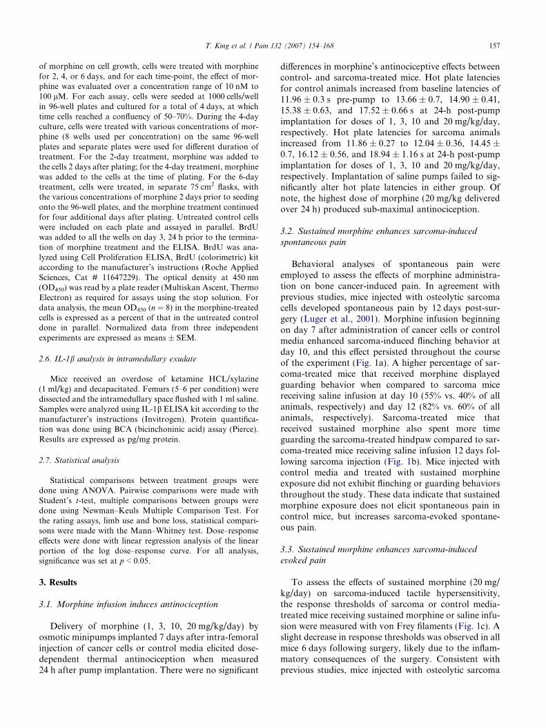
Behavioral analyses of spontaneous pain wereemployed to assess the effects of morphine administra-tion on bone cancer-induced pain. In agreement withprevious studies, mice injected with osteolytic sarcomacells developed spontaneous pain by 12 days post-sur-gery (Luger et al., 2001). Morphine infusion beginningon day 7 after administration of cancer cells or controlmedia enhanced sarcoma-induced flinching behavior atday 10, and this effect persisted throughout the courseof the experiment (Fig. 1a). A higher percentage of sar-coma-treated mice that received morphine displayedguarding behavior when compared to sarcoma micereceiving saline infusion at day 10 (55% vs. 40% of allanimals, respectively) and day 12 (82% vs. 60% of allanimals, respectively). Sarcoma-treated mice thatreceived sustained morphine also spent more timeguarding the sarcoma-treated hindpaw compared to sar-coma-treated mice receiving saline infusion 12 days fol-lowing sarcoma injection (Fig. 1b). Mice injected withcontrol media and treated with sustained morphineexposure did not exhibit flinching or guarding behaviorsthroughout the study. These data indicate that sustainedmorphine exposure does not elicit spontaneous pain incontrol mice, but increases sarcoma-evoked spontane-ous pain.
3.3. Sustained morphine enhances sarcoma-induced
evoked pain
To assess the effects of sustained morphine (20 mg/kg/day) on sarcoma-induced tactile hypersensitivity,the response thresholds of sarcoma or control media-treated mice receiving sustained morphine or saline infu-sion were measured with von Frey filaments (Fig. 1c). Aslight decrease in response thresholds was observed in allmice 6 days following surgery, likely due to the inflam-matory consequences of the surgery. Consistent withprevious studies, mice injected with osteolytic sarcoma
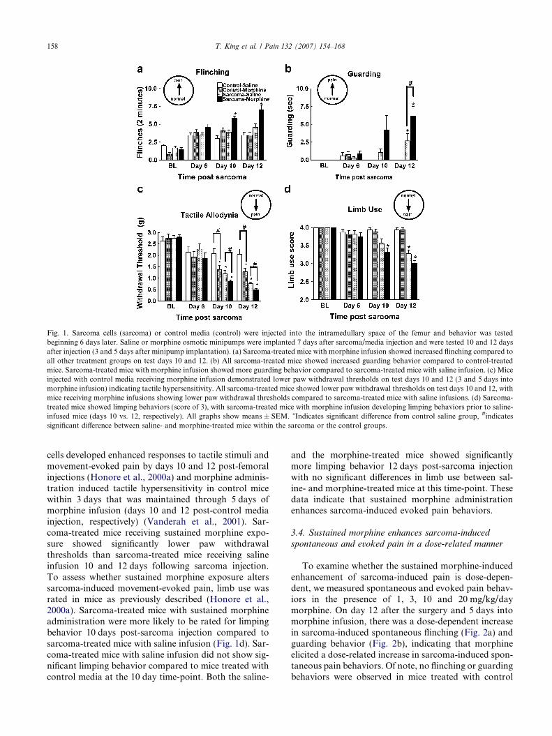
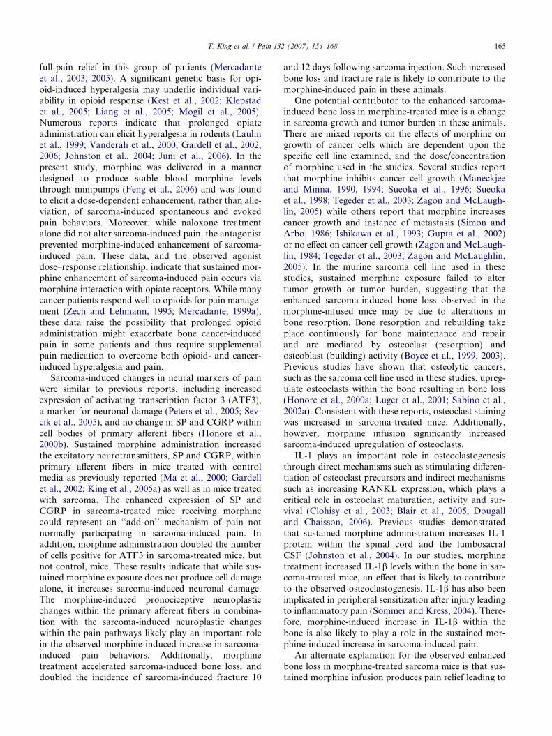
Fig. 1. Sarcoma cells (sarcoma) or control media (control) were injected into the intramedullary space of the femur and behavior was testedbeginning 6 days later. Saline or morphine osmotic minipumps were implanted 7 days after sarcoma/media injection and were tested 10 and 12 daysafter injection (3 and 5 days after minipump implantation). (a) Sarcoma-treated mice with morphine infusion showed increased flinching compared toall other treatment groups on test days 10 and 12. (b) All sarcoma-treated mice showed increased guarding behavior compared to control-treatedmice. Sarcoma-treated mice with morphine infusion showed more guarding behavior compared to sarcoma-treated mice with saline infusion. (c) Miceinjected with control media receiving morphine infusion demonstrated lower paw withdrawal thresholds on test days 10 and 12 (3 and 5 days intomorphine infusion) indicating tactile hypersensitivity. All sarcoma-treated mice showed lower paw withdrawal thresholds on test days 10 and 12, withmice receiving morphine infusions showing lower paw withdrawal thresholds compared to sarcoma-treated mice with saline infusions. (d) Sarcoma-treated mice showed limping behaviors (score of 3), with sarcoma-treated mice with morphine infusion developing limping behaviors prior to saline-infused mice (days 10 vs. 12, respectively). All graphs show means ± SEM. *Indicates significant difference from control saline group, #indicatessignificant difference between saline- and morphine-treated mice within the sarcoma or the control groups.
158 T. King et al. / Pain 132 (2007) 154–168
cells developed enhanced responses to tactile stimuli andmovement-evoked pain by days 10 and 12 post-femoralinjections (Honore et al., 2000a) and morphine adminis-tration induced tactile hypersensitivity in control micewithin 3 days that was maintained through 5 days ofmorphine infusion (days 10 and 12 post-control mediainjection, respectively) (Vanderah et al., 2001). Sar-coma-treated mice receiving sustained morphine expo-sure showed significantly lower paw withdrawalthresholds than sarcoma-treated mice receiving salineinfusion 10 and 12 days following sarcoma injection.To assess whether sustained morphine exposure alterssarcoma-induced movement-evoked pain, limb use wasrated in mice as previously described (Honore et al.,2000a). Sarcoma-treated mice with sustained morphineadministration were more likely to be rated for limpingbehavior 10 days post-sarcoma injection compared tosarcoma-treated mice with saline infusion (Fig. 1d). Sar-coma-treated mice with saline infusion did not show sig-nificant limping behavior compared to mice treated withcontrol media at the 10 day time-point. Both the saline-
and the morphine-treated mice showed significantlymore limping behavior 12 days post-sarcoma injectionwith no significant differences in limb use between sal-ine- and morphine-treated mice at this time-point. Thesedata indicate that sustained morphine administrationenhances sarcoma-induced evoked pain behaviors.
3.4. Sustained morphine enhances sarcoma-induced
spontaneous and evoked pain in a dose-related manner
To examine whether the sustained morphine-inducedenhancement of sarcoma-induced pain is dose-depen-dent, we measured spontaneous and evoked pain behav-iors in the presence of 1, 3, 10 and 20 mg/kg/daymorphine. On day 12 after the surgery and 5 days intomorphine infusion, there was a dose-dependent increasein sarcoma-induced spontaneous flinching (Fig. 2a) andguarding behavior (Fig. 2b), indicating that morphineelicited a dose-related increase in sarcoma-induced spon-taneous pain behaviors. Of note, no flinching or guardingbehaviors were observed in mice treated with control
T. King et al. / Pain 132 (2007) 154–168 159
media indicating that morphine fails to induce spontane-ous pain behaviors at any of the doses tested (data notshown). Sarcoma-induced evoked pain was measured atthe same time-point, and sustained morphine infusionenhanced sarcoma-induced tactile hypersensitivity(Fig. 2c) and decreased limb use (Fig. 2d) in a dose-depen-dent fashion, indicating that infusion of morphineincreases sarcoma-induced evoked pain behaviors in adose-dependent manner. Sustained morphine infusiondid not alter limb use of mice treated with control mediaat any of the doses tested (data not shown) but decreasedevoked paw withdrawal thresholds in the control mice(R2 = .99, p < 0.05). Collectively, these data indicate thatthe observed effects of sustained morphine administrationon spontaneous and evoked behaviors are dose related.
3.5. Sustained morphine-induced effects in DRG cells
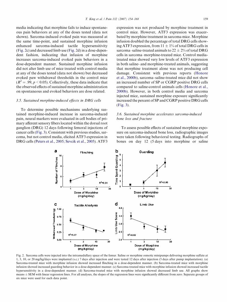
To determine possible mechanisms underlying sus-tained morphine-induced increase in sarcoma-inducedpain, neural markers were evaluated in cell bodies of pri-mary afferent sensory fibers located within the dorsal rootganglion (DRG) 12 days following femoral injections ofcancer cells (Fig. 3). Consistent with previous studies, sar-coma, but not control media, elicited ATF3 expression inDRG cells (Peters et al., 2005; Sevcik et al., 2005). ATF3
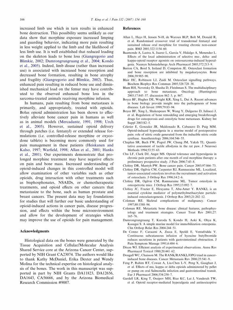
Fig. 2. Sarcoma cells were injected into the intramedullary space of the femur1, 3, 10, or 20 mg/kg/days were implanted (s.c.) 7 days after injection and wSarcoma-treated mice with morphine infusion showed increased flinching ininfusion showed increased guarding behavior in a dose-dependent manner. (c)hypersensitivity in a dose-dependent manner. (d) Sarcoma-treated mice wmeans ± SEM with linear regression lines. For all analyses, the slopes of the rsix mice were used for each data point.
expression was not produced by morphine treatment incontrol mice. However, ATF3 expression was exacer-bated by morphine treatment in sarcoma mice. Morphineinfusion doubled the percentage of total DRG cells show-ing ATF3 expression, from 11 ± 1% of total DRG cells insarcoma–saline-treated animals to 22 ± 2% of total DRGcells in sarcoma–morphine-treated mice. Control media-treated mice showed very low levels of ATF3 expressionin both saline- and morphine-treated animals, suggestingthat morphine treatment alone was not producing celldamage. Consistent with previous reports (Honoreet al., 2000b), sarcoma–saline-treated mice did not showan increased number of SP or CGRP positive DRG cellscompared to saline-control animals cells (Honore et al.,2000b). However, in both control media and sarcomainjected mice, sustained morphine exposure significantlyincreased the percent of SP and CGRP positive DRG cells(Fig. 3).
3.6. Sustained morphine accelerates sarcoma-induced
bone loss and fracture
To assess possible effects of sustained morphine expo-sure on sarcoma-induced bone loss, radiographic imageswere taken following behavioral testing. Radiographs ofbones on day 12 (5 days into morphine or saline
. Saline or morphine osmotic minipumps delivering morphine sulfate atere tested 12 days after injection (5 days after pump implantation). (a)
a dose-dependent manner. (b) Sarcoma-treated mice with morphineSarcoma-treated mice with morphine infusion showed increased tactile
ith morphine infusion showed decreased limb use. All graphs showegression lines were significantly different from zero. Separate groups of
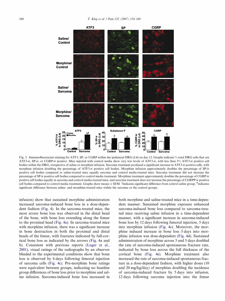
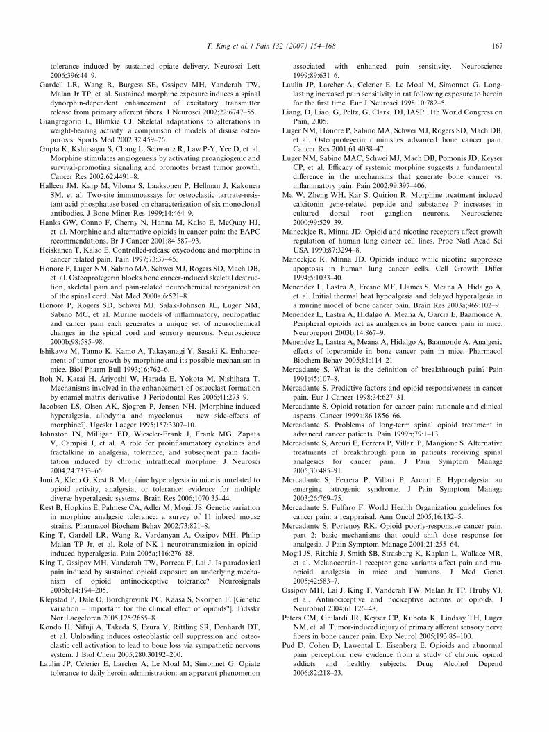
Fig. 3. Immunofluorescent staining for ATF3, SP, or CGRP within the ipsilateral DRG (L4) on day 12. Graphs indicate % total DRG cells that areATF3-ir, SP-ir, or CGRP-ir positive. Mice injected with control media show very low levels of ATF3-ir, with less than 5% ATF3-ir positive cellbodies within the DRG, irrespective of saline or morphine infusion. Sarcoma treatment produced a significant increase in ATF3-ir positive cells, withmorphine infusion doubling the percentage of ATF3-ir positive cell bodies. Morphine infusion approximately doubles the percentage of SP-irpositive cell bodies compared to saline-treated mice equally sarcoma and control media-treated mice. Sarcoma treatment did not increase thepercentage of SP-ir positive cell bodies compared to control media treatment. Morphine treatment approximately doubles the percentage of CGRP-irpositive cell bodies equally in sarcoma and control media-treated mice, and sarcoma treatment does not increase the percentage of CGRPP-ir positivecell bodies compared to control media treatment. Graphs show means ± SEM. *Indicates significant difference from control saline group, #indicatessignificant difference between saline- and morphine-treated mice within the sarcoma or the control groups.
160 T. King et al. / Pain 132 (2007) 154–168
infusion) show that sustained morphine administrationincreased sarcoma-induced bone loss in a dose-depen-dent fashion (Fig. 4). In the sarcoma-treated mice, themost severe bone loss was observed in the distal headof the bone, with bone loss extending along the femurto the proximal head (Fig. 4a). In sarcoma-treated micewith morphine infusion, there was a significant increasein bone destruction in both the proximal and distalheads of the femur, with fractures indicated by full-cor-tical bone loss as indicated by the arrows (Fig. 4a andb). Consistent with previous reports (Luger et al.,2001), visual ratings of the radiographs by an observerblinded to the experimental conditions show that boneloss is observed by 6 days following femoral injectionof sarcoma cells (Fig. 4c). Pre-morphine bone ratingswere equivalent between groups, indicating no baselinegroup differences of bone loss prior to morphine and sal-ine infusion. Sarcoma-induced bone loss increased in
both morphine and saline-treated mice in a time-depen-dent manner. Sustained morphine exposure enhancedsarcoma-induced bone loss compared to sarcoma-trea-ted mice receiving saline infusion in a time-dependentmanner, with a significant increase in sarcoma-inducedbone loss by 12 days following femoral injection, 5 daysinto morphine infusion (Fig. 4c). Moreover, the mor-phine induced increase in bone loss 5 days into mor-phine infusion was dose-dependent (Fig. 4d). Sustainedadministration of morphine across 3 and 5 days doubledthe rate of sarcoma-induced spontaneous fracture rate,indicated by bone loss across the full thickness of thecortical bone (Fig. 4e). Morphine treatment alsoincreased the rate of sarcoma-induced spontaneous frac-ture in a dose-dependent fashion, with higher doses (10and 20 mg/kg/day) of morphine doubling the incidenceof sarcoma-induced fracture by 5 days into infusion,12 days following sarcoma injection into the femur
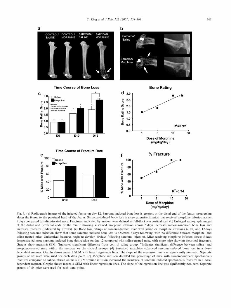
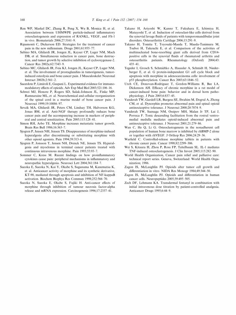
Fig. 4. (a) Radiograph images of the injected femur on day 12. Sarcoma-induced bone loss is greatest at the distal end of the femur, progressingalong the femur to the proximal head of the femur. Sarcoma-induced bone loss is more extensive in mice that received morphine infusion across5 days compared to saline-treated mice. Fractures, indicated by arrows, were defined as full-thickness cortical loss. (b) Enlarged radiograph imagesof the distal and proximal ends of the femur showing sustained morphine infusion across 5 days increases sarcoma-induced bone loss andincreases fractures (indicated by arrows). (c) Bone loss ratings of sarcoma-treated mice with saline or morphine infusions 6, 10, and 12 daysfollowing sarcoma injection show that some sarcoma-induced bone loss is observed 6 days following, with no difference between morphine- andsaline-treated mice. Unicortical fractures begin to develop 10 days following sarcoma injection. Mice receiving morphine infusion across 5 daysdemonstrated more sarcoma-induced bone destruction on day 12 compared with saline-treated mice, with more mice showing bicortical fractures.Graphs show means ± SEM. *Indicates significant difference from control saline group. #Indicates significant difference between saline- andmorphine-treated mice within the sarcoma or the control groups. (d) Sustained morphine enhanced sarcoma-induced bone loss in a dose-dependent manner. Graphs shows mean ± SEM with linear regression lines. The slope of the regression line was significantly non-zero. Separategroups of six mice were used for each data point. (e) Morphine infusion doubled the percentage of mice with sarcoma-induced spontaneousfractures compared to saline-infused animals. (f) Morphine infusion increased the incidence of sarcoma-induced spontaneous fractures in a dose-dependent manner. Graphs shows means ± SEM with linear regression lines. The slope of the regression line was significantly non-zero. Separategroups of six mice were used for each data point.
T. King et al. / Pain 132 (2007) 154–168 161
162 T. King et al. / Pain 132 (2007) 154–168
(Fig. 4e). Of note, in our experiments neither saline normorphine-infused control mice developed bone loss(Fig. 4a).
3.7. Naloxone antagonizes morphine-induced enhanced
pain and bone loss
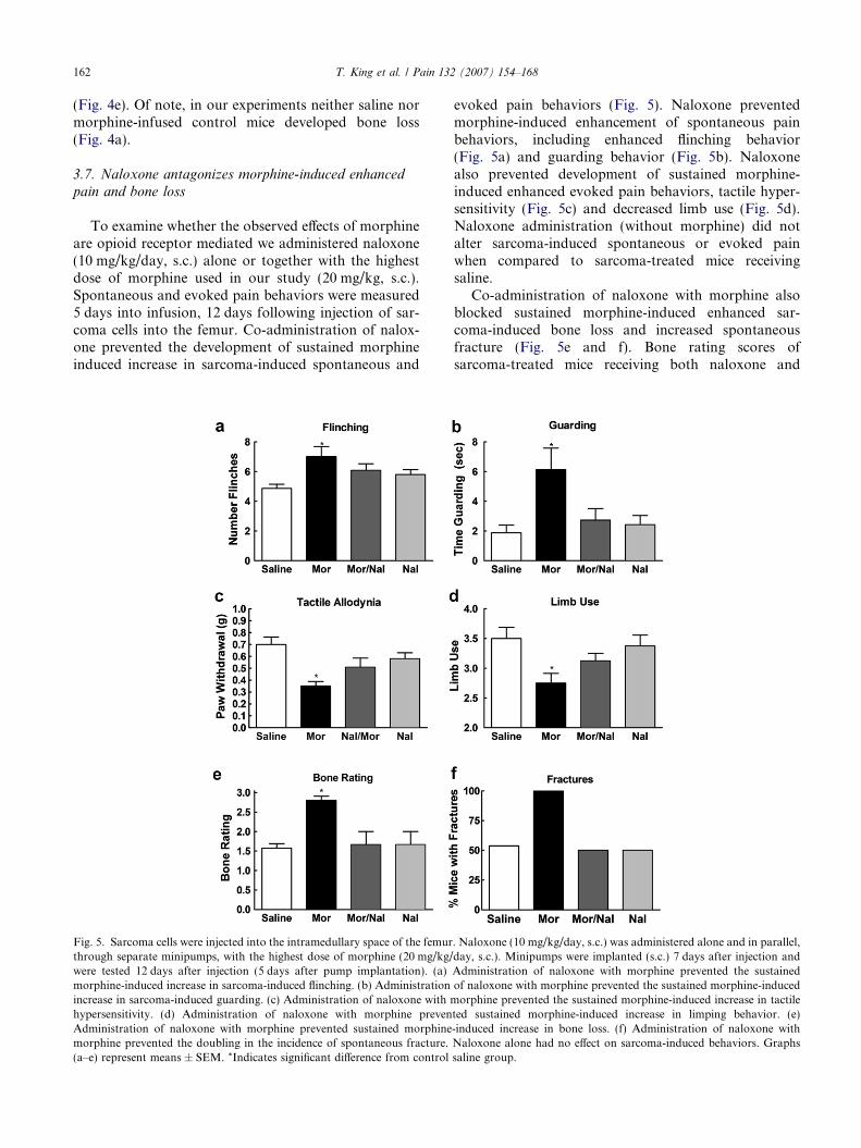
To examine whether the observed effects of morphineare opioid receptor mediated we administered naloxone(10 mg/kg/day, s.c.) alone or together with the highestdose of morphine used in our study (20 mg/kg, s.c.).Spontaneous and evoked pain behaviors were measured5 days into infusion, 12 days following injection of sar-coma cells into the femur. Co-administration of nalox-one prevented the development of sustained morphineinduced increase in sarcoma-induced spontaneous and
Fig. 5. Sarcoma cells were injected into the intramedullary space of the femurthrough separate minipumps, with the highest dose of morphine (20 mg/kg/were tested 12 days after injection (5 days after pump implantation). (a)morphine-induced increase in sarcoma-induced flinching. (b) Administrationincrease in sarcoma-induced guarding. (c) Administration of naloxone with mhypersensitivity. (d) Administration of naloxone with morphine prevenAdministration of naloxone with morphine prevented sustained morphinemorphine prevented the doubling in the incidence of spontaneous fracture.(a–e) represent means ± SEM. *Indicates significant difference from control
evoked pain behaviors (Fig. 5). Naloxone preventedmorphine-induced enhancement of spontaneous painbehaviors, including enhanced flinching behavior(Fig. 5a) and guarding behavior (Fig. 5b). Naloxonealso prevented development of sustained morphine-induced enhanced evoked pain behaviors, tactile hyper-sensitivity (Fig. 5c) and decreased limb use (Fig. 5d).Naloxone administration (without morphine) did notalter sarcoma-induced spontaneous or evoked painwhen compared to sarcoma-treated mice receivingsaline.
Co-administration of naloxone with morphine alsoblocked sustained morphine-induced enhanced sar-coma-induced bone loss and increased spontaneousfracture (Fig. 5e and f). Bone rating scores ofsarcoma-treated mice receiving both naloxone and
. Naloxone (10 mg/kg/day, s.c.) was administered alone and in parallel,day, s.c.). Minipumps were implanted (s.c.) 7 days after injection andAdministration of naloxone with morphine prevented the sustainedof naloxone with morphine prevented the sustained morphine-induced
orphine prevented the sustained morphine-induced increase in tactileted sustained morphine-induced increase in limping behavior. (e)-induced increase in bone loss. (f) Administration of naloxone withNaloxone alone had no effect on sarcoma-induced behaviors. Graphssaline group.
T. King et al. / Pain 132 (2007) 154–168 163
morphine infusion as well as naloxone alone did not dif-fer (p > 0.05) from sarcoma-treated mice receiving salineinfusion indicating full reversal of sustained morphine-induced increase in sarcoma-induced bone loss(Fig. 5e). Co-administration of naloxone with morphinealso completely blocked the increased rate of spontane-ous fracture, with 100% of the mice treated with mor-phine showing spontaneous sarcoma-induced fracture,and 50% of the mice treated with naloxone/morphineinfusion showing spontaneous sarcoma-induced frac-ture; this value was the same as that seen for sarcoma-treated mice treated with saline infusion or naloxoneinfusion (Fig. 5f). Administration of naloxone alonedid not alter sarcoma-induced effects on bone loss.
3.8. Sustained morphine fails to affect tumor burden but
enhances osteoclastogenesis
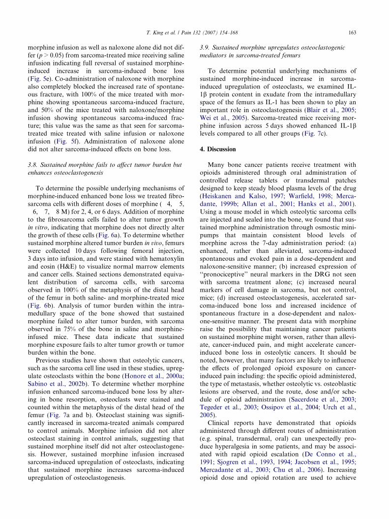
To determine the possible underlying mechanisms ofmorphine-induced enhanced bone loss we treated fibro-sarcoma cells with different doses of morphine (�4, �5,�6, �7, �8 M) for 2, 4, or 6 days. Addition of morphineto the fibrosarcoma cells failed to alter tumor growthin vitro, indicating that morphine does not directly alterthe growth of these cells (Fig. 6a). To determine whethersustained morphine altered tumor burden in vivo, femurswere collected 10 days following femoral injection,3 days into infusion, and were stained with hematoxylinand eosin (H&E) to visualize normal marrow elementsand cancer cells. Stained sections demonstrated equiva-lent distribution of sarcoma cells, with sarcomaobserved in 100% of the metaphysis of the distal headof the femur in both saline- and morphine-treated mice(Fig. 6b). Analysis of tumor burden within the intra-medullary space of the bone showed that sustainedmorphine failed to alter tumor burden, with sarcomaobserved in 75% of the bone in saline and morphine-infused mice. These data indicate that sustainedmorphine exposure fails to alter tumor growth or tumorburden within the bone.
Previous studies have shown that osteolytic cancers,such as the sarcoma cell line used in these studies, upreg-ulate osteoclasts within the bone (Honore et al., 2000a;Sabino et al., 2002b). To determine whether morphineinfusion enhanced sarcoma-induced bone loss by alter-ing in bone resorption, osteoclasts were stained andcounted within the metaphysis of the distal head of thefemur (Fig. 7a and b). Osteoclast staining was signifi-cantly increased in sarcoma-treated animals comparedto control animals. Morphine infusion did not alterosteoclast staining in control animals, suggesting thatsustained morphine itself did not alter osteoclastogene-sis. However, sustained morphine infusion increasedsarcoma-induced upregulation of osteoclasts, indicatingthat sustained morphine increases sarcoma-inducedupregulation of osteoclastogenesis.
3.9. Sustained morphine upregulates osteoclastogenic
mediators in sarcoma-treated femurs
To determine potential underlying mechanisms ofsustained morphine-induced increase in sarcoma-induced upregulation of osteoclasts, we examined IL-1b protein content in exudate from the intramedullaryspace of the femurs as IL-1 has been shown to play animportant role in osteoclastogenesis (Blair et al., 2005;Wei et al., 2005). Sarcoma-treated mice receiving mor-phine infusion across 5 days showed enhanced IL-1blevels compared to all other groups (Fig. 7c).
4. Discussion
Many bone cancer patients receive treatment withopioids administered through oral administration ofcontrolled release tablets or transdermal patchesdesigned to keep steady blood plasma levels of the drug(Heiskanen and Kalso, 1997; Warfield, 1998; Merca-dante, 1999b; Allan et al., 2001; Hanks et al., 2001).Using a mouse model in which osteolytic sarcoma cellsare injected and sealed into the bone, we found that sus-tained morphine administration through osmostic mini-pumps that maintain consistent blood levels ofmorphine across the 7-day administration period: (a)enhanced, rather than alleviated, sarcoma-inducedspontaneous and evoked pain in a dose-dependent andnaloxone-sensitive manner; (b) increased expression of‘‘pronociceptive’’ neural markers in the DRG not seenwith sarcoma treatment alone; (c) increased neuralmarkers of cell damage in sarcoma, but not control,mice; (d) increased osteoclastogenesis, accelerated sar-coma-induced bone loss and increased incidence ofspontaneous fracture in a dose-dependent and nalox-one-sensitive manner. The present data with morphineraise the possibility that maintaining cancer patientson sustained morphine might worsen, rather than allevi-ate, cancer-induced pain, and might accelerate cancer-induced bone loss in osteolytic cancers. It should benoted, however, that many factors are likely to influencethe effects of prolonged opioid exposure on cancer-induced pain including: the specific opioid administered,the type of metastasis, whether osteolytic vs. osteoblasticlesions are observed, and the route, dose and/or sche-dule of opioid administration (Sacerdote et al., 2003;Tegeder et al., 2003; Ossipov et al., 2004; Urch et al.,2005).
Clinical reports have demonstrated that opioidsadministered through different routes of administration(e.g. spinal, transdermal, oral) can unexpectedly pro-duce hyperalgesia in some patients, and may be associ-ated with rapid opioid escalation (De Conno et al.,1991; Sjogren et al., 1993, 1994; Jacobsen et al., 1995;Mercadante et al., 2003; Chu et al., 2006). Increasingopioid dose and opioid rotation are used to achieve
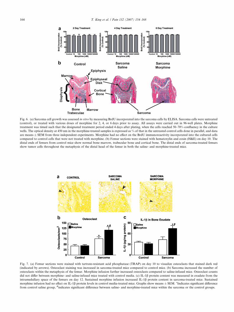
Fig. 6. (a) Sarcoma cell growth was assessed in vitro by measuring BrdU incorporated into the sarcoma cells by ELISA. Sarcoma cells were untreated(control), or treated with various doses of morphine for 2, 4, or 6 days prior to assay. All assays were carried out in 96-well plates. Morphinetreatment was timed such that the designated treatment period ended 4 days after plating, when the cells reached 50–70% confluency in the culturewells. The optical density at 450 nm in the morphine-treated samples is expressed as % of that in the untreated control cells done in parallel, and dataare means ± SEM from three independent experiments. Morphine had no effect on the BrdU immunoreactivity incorporated into the cultured cellscompared to control cells that were not treated with morphine. (b) Femur sections were stained with hematoxylin and eosin (H&E) on day 10. Thedistal ends of femurs from control mice show normal bone marrow, trabecular bone and cortical bone. The distal ends of sarcoma-treated femursshow tumor cells throughout the metaphysis of the distal head of the femur in both the saline- and morphine-treated mice.
Fig. 7. (a) Femur sections were stained with tartrate-resistant acid phosphatase (TRAP) on day 10 to visualize osteoclasts that stained dark red(indicated by arrows). Osteoclast staining was increased in sarcoma-treated mice compared to control mice. (b) Sarcoma increased the number ofosteoclasts within the metaphysis of the femur. Morphine infusion further increased osteoclasts compared to saline-infused mice. Osteoclast countsdid not differ between morphine- and saline-infused mice treated with control media. (c) IL-1b protein content was measured in exudate from theintramedullary space of the femurs on day 12. Sustained morphine infusion increased IL-1b protein content in sarcoma-treated mice. Sustainedmorphine infusion had no effect on IL-1b protein levels in control media-treated mice. Graphs show means ± SEM. *Indicates significant differencefrom control saline group, #indicates significant difference between saline- and morphine-treated mice within the sarcoma or the control groups.
164 T. King et al. / Pain 132 (2007) 154–168
T. King et al. / Pain 132 (2007) 154–168 165
full-pain relief in this group of patients (Mercadanteet al., 2003, 2005). A significant genetic basis for opi-oid-induced hyperalgesia may underlie individual vari-ability in opioid response (Kest et al., 2002; Klepstadet al., 2005; Liang et al., 2005; Mogil et al., 2005).Numerous reports indicate that prolonged opiateadministration can elicit hyperalgesia in rodents (Laulinet al., 1999; Vanderah et al., 2000; Gardell et al., 2002,2006; Johnston et al., 2004; Juni et al., 2006). In thepresent study, morphine was delivered in a mannerdesigned to produce stable blood morphine levelsthrough minipumps (Feng et al., 2006) and was foundto elicit a dose-dependent enhancement, rather than alle-viation, of sarcoma-induced spontaneous and evokedpain behaviors. Moreover, while naloxone treatmentalone did not alter sarcoma-induced pain, the antagonistprevented morphine-induced enhancement of sarcoma-induced pain. These data, and the observed agonistdose–response relationship, indicate that sustained mor-phine enhancement of sarcoma-induced pain occurs viamorphine interaction with opiate receptors. While manycancer patients respond well to opioids for pain manage-ment (Zech and Lehmann, 1995; Mercadante, 1999a),these data raise the possibility that prolonged opioidadministration might exacerbate bone cancer-inducedpain in some patients and thus require supplementalpain medication to overcome both opioid- and cancer-induced hyperalgesia and pain.
Sarcoma-induced changes in neural markers of painwere similar to previous reports, including increasedexpression of activating transcription factor 3 (ATF3),a marker for neuronal damage (Peters et al., 2005; Sev-cik et al., 2005), and no change in SP and CGRP withincell bodies of primary afferent fibers (Honore et al.,2000b). Sustained morphine administration increasedthe excitatory neurotransmitters, SP and CGRP, withinprimary afferent fibers in mice treated with controlmedia as previously reported (Ma et al., 2000; Gardellet al., 2002; King et al., 2005a) as well as in mice treatedwith sarcoma. The enhanced expression of SP andCGRP in sarcoma-treated mice receiving morphinecould represent an ‘‘add-on’’ mechanism of pain notnormally participating in sarcoma-induced pain. Inaddition, morphine administration doubled the numberof cells positive for ATF3 in sarcoma-treated mice, butnot control, mice. These results indicate that while sus-tained morphine exposure does not produce cell damagealone, it increases sarcoma-induced neuronal damage.The morphine-induced pronociceptive neuroplasticchanges within the primary afferent fibers in combina-tion with the sarcoma-induced neuroplastic changeswithin the pain pathways likely play an important rolein the observed morphine-induced increase in sarcoma-induced pain behaviors. Additionally, morphinetreatment accelerated sarcoma-induced bone loss, anddoubled the incidence of sarcoma-induced fracture 10
and 12 days following sarcoma injection. Such increasedbone loss and fracture rate is likely to contribute to themorphine-induced pain in these animals.
One potential contributor to the enhanced sarcoma-induced bone loss in morphine-treated mice is a changein sarcoma growth and tumor burden in these animals.There are mixed reports on the effects of morphine ongrowth of cancer cells which are dependent upon thespecific cell line examined, and the dose/concentrationof morphine used in the studies. Several studies reportthat morphine inhibits cancer cell growth (Maneckjeeand Minna, 1990, 1994; Sueoka et al., 1996; Sueokaet al., 1998; Tegeder et al., 2003; Zagon and McLaugh-lin, 2005) while others report that morphine increasescancer growth and instance of metastasis (Simon andArbo, 1986; Ishikawa et al., 1993; Gupta et al., 2002)or no effect on cancer cell growth (Zagon and McLaugh-lin, 1984; Tegeder et al., 2003; Zagon and McLaughlin,2005). In the murine sarcoma cell line used in thesestudies, sustained morphine exposure failed to altertumor growth or tumor burden, suggesting that theenhanced sarcoma-induced bone loss observed in themorphine-infused mice may be due to alterations inbone resorption. Bone resorption and rebuilding takeplace continuously for bone maintenance and repairand are mediated by osteoclast (resorption) andosteoblast (building) activity (Boyce et al., 1999, 2003).Previous studies have shown that osteolytic cancers,such as the sarcoma cell line used in these studies, upreg-ulate osteoclasts within the bone resulting in bone loss(Honore et al., 2000a; Luger et al., 2001; Sabino et al.,2002a). Consistent with these reports, osteoclast stainingwas increased in sarcoma-treated mice. Additionally,however, morphine infusion significantly increasedsarcoma-induced upregulation of osteoclasts.
IL-1 plays an important role in osteoclastogenesisthrough direct mechanisms such as stimulating differen-tiation of osteoclast precursors and indirect mechanismssuch as increasing RANKL expression, which plays acritical role in osteoclast maturation, activity and sur-vival (Clohisy et al., 2003; Blair et al., 2005; Dougalland Chaisson, 2006). Previous studies demonstratedthat sustained morphine administration increases IL-1protein within the spinal cord and the lumbosacralCSF (Johnston et al., 2004). In our studies, morphinetreatment increased IL-1b levels within the bone in sar-coma-treated mice, an effect that is likely to contributeto the observed osteoclastogenesis. IL-1b has also beenimplicated in peripheral sensitization after injury leadingto inflammatory pain (Sommer and Kress, 2004). There-fore, morphine-induced increase in IL-1b within thebone is also likely to play a role in the sustained mor-phine-induced increase in sarcoma-induced pain.
An alternate explanation for the observed enhancedbone loss in morphine-treated sarcoma mice is that sus-tained morphine infusion produces pain relief leading to
166 T. King et al. / Pain 132 (2007) 154–168
increased limb use which in turn results in enhancedbone destruction. This possibility seems unlikely as ourdata show that morphine exposure increased limpingand guarding behavior, indicating more pain resultingin less weight applied to the limb and the likelihood ofless limb use. It is well established that reduced loadingon the skeleton leads to bone loss (Giangregorio andBlimkie, 2002; Damrongrungruang et al., 2004; Kondoet al., 2005). Indeed, limb disuse (rather than increaseduse) is associated with increased bone resorption anddecreased bone formation, resulting in bone atrophyand fragility (Giangregorio and Blimkie, 2002). Thus,enhanced pain resulting in reduced bone use and dimin-ished mechanical load on the femur may have contrib-uted to the observed enhanced bone loss in thesarcoma-treated animals receiving morphine infusion.
In humans, pain resulting from bone metastases isprimarily, and appropriately, treated with opioids.Bolus opioid administration has been shown to effec-tively alleviate bone cancer pain in humans as wellas in animal models (Mercadante, 1991, 1998; Urchet al., 2005). However, sustained opioid deliverythrough patches (i.e. fentanyl) or extended release for-mulations (i.e. controlled-release morphine or oxyco-done tablets) is becoming more commonly used forpain management in these patients (Heiskanen andKalso, 1997; Warfield, 1998; Allan et al., 2001; Hankset al., 2001). Our experiments demonstrate that pro-longed morphine treatment may have negative effectson pain and bone mass. Increased understanding ofopioid-induced changes in this controlled model willallow examination of other variables such as otheropioids, drug interaction with other treatments suchas bisphosphonates, NSAIDs, or chemotherapeutictreatments, and opioid effects on other cancers thatmetastasize to the bone, such as human prostate andbreast cancers. The present data may lay foundationsfor studies that will further our basic understanding ofopioid-induced actions in cancer pain, disease progres-sion, and effects within the bone microenvironmentand allow for the development of strategies whichmay improve the use of opioids for pain management.
Acknowledgments
Histological data on the bones were generated by theTissue Acquisition and Cellular/Molecular AnalysisShared Service core at the Arizona Cancer Center, sup-ported by NIH Grant CA23074. The authors would liketo thank Kathy McDaniel, Erika Dexter and WendyMolina for the technical expertise on histological analy-sis of the bones. The work in this manuscript was sup-ported in part by NIH Grants DA11823, DA12656,DA1643, CA56666, and by the Arizona BiomedicalResearch Commission #9007.
References
Allan L, Hays H, Jensen N-H, de Waroux BLP, Bolt M, Donald R,et al. Randomised crossover trial of transdermal fentanyl andsustained release oral morphine for treating chronic non-cancerpain. BMJ 2001;322:1154–60.
Baamonde A, Lastra A, Juarez L, Garcia V, Hidalgo A, Menendez L.Effects of the local administration of selective mu-, delta- andkappa-opioid receptor agonists on osteosarcoma-induced hyperal-gesia. Naunyn Schmiedebergs Arch Pharmacol 2005;372:213–9.
Beeton CA, Bord S, Ireland D, Compston JE. Osteoclast formationand bone resorption are inhibited by megakaryocytes. Bone2006;39:985–90.
Blair HC, Robinson LJ, Zaidi M. Osteoclast signalling pathways.Biochem Biophys Res Commun 2005;328:728–38.
Blum RH, Novetsky D, Shasha D, Fleishman S. The multidisciplinaryapproach to bone metastases. Oncology (Huntington)2003;17:845–57. discussion 862–3, p. 867.
Boyce BF, Hughes DE, Wright KR, Xing L, Dai A. Recent advancesin bone biology provide insight into the pathogenesis of bonediseases. Lab Invest 1999;79:83–94.
Boyce BF, Xing L, Shakespeare W, Wang Y, Dalgarno D, Iuliucci J,et al. Regulation of bone remodeling and emerging breakthroughdrugs for osteoporosis and osteolytic bone metastases. Kidney IntSuppl 2003:S2–5.
Celerier E, Gonzalez JR, Maldonado R, Cabanero D, Puig MM.Opioid-induced hyperalgesia in a murine model of postoperativepain: role of nitric oxide generated from the inducible nitric oxidesynthase. Anesthesiology 2006;104:546–55.
Chaplan SR, Bach FW, Pogrel JW, Chung JM, Yaksh TL. Quanti-tative assessment of tactile allodynia in the rat paw. J NeurosciMethods 1994;53:55–63.
Chu LF, Clark DJ, Angst MS. Opioid tolerance and hyperalgesia inchronic pain patients after one month of oral morphine therapy: apreliminary prospective study. J Pain 2006;7:43–8.
Clohisy DR, Mantyh PW. Bone cancer pain. Cancer 2003;97:866–73.Clohisy DR, Ogilvie CM, Carpenter RJ, Ramnaraine ML. Localized,
tumor-associated osteolysis involves the recruitment and activationof osteoclasts. J Orthop Res 1996;14:2–6.
Clohisy DR, Ogilvie CM, Ramnaraine ML. Tumor osteolysis inosteopetrotic mice. J Orthop Res 1995;13:892–7.
Clohisy JC, Frazier E, Hirayama T, Abu-Amer Y. RANKL is anessential cytokine mediator of polymethylmethacrylate particle-induced osteoclastogenesis. J Orthop Res 2003;21:202–12.
Coleman RE. Skeletal complications of malignancy. Cancer1997;80:1588–94.
Coleman RE. Metastatic bone disease: clinical features, pathophys-iology and treatment strategies. Cancer Treat Rev 2001;27:165–76.
Damrongrungruang T, Kuroda S, Kondo H, Aoki K, Ohya K,Kasugai S. A simple murine model for immobilization osteopenia.Clin Orthop Relat Res 2004:244–51.
De Conno F, Caraceni A, Zecca E, Spoldi E, Ventafridda V.Continuous subcutaneous infusion of hyoscine butylbromidereduces secretions in patients with gastrointestinal obstruction. JPain Symptom Manage 1991;6:484–6.
Dixon WJ. Efficient analysis of experimental observations. Annu RevPharmacol Toxicol 1980;20:441–62.
Dougall WC, Chaisson M. The RANK/RANKL/OPG triad in cancer-induced bone diseases. Cancer Metastasis Rev 2006;25:541–9.
Feng P, Rahim RT, Cowan A, Liu-Chen L-Y, Peng X, Gaughan J,et al. Effects of mu, kappa or delta opioids administered by pelletor pump on oral Salmonella infection and gastrointestinal transit.Eur J Pharmacol 2006;534:250–7.
Gardell LR, King T, Ossipov MH, Rice KC, Lai J, Vanderah TW,et al. Opioid receptor-mediated hyperalgesia and antinociceptive
T. King et al. / Pain 132 (2007) 154–168 167
tolerance induced by sustained opiate delivery. Neurosci Lett2006;396:44–9.
Gardell LR, Wang R, Burgess SE, Ossipov MH, Vanderah TW,Malan Jr TP, et al. Sustained morphine exposure induces a spinaldynorphin-dependent enhancement of excitatory transmitterrelease from primary afferent fibers. J Neurosci 2002;22:6747–55.
Giangregorio L, Blimkie CJ. Skeletal adaptations to alterations inweight-bearing activity: a comparison of models of disuse osteo-porosis. Sports Med 2002;32:459–76.
Gupta K, Kshirsagar S, Chang L, Schwartz R, Law P-Y, Yee D, et al.Morphine stimulates angiogenesis by activating proangiogenic andsurvival-promoting signaling and promotes breast tumor growth.Cancer Res 2002;62:4491–8.
Halleen JM, Karp M, Viloma S, Laaksonen P, Hellman J, KakonenSM, et al. Two-site immunoassays for osteoclastic tartrate-resis-tant acid phosphatase based on characterization of six monoclonalantibodies. J Bone Miner Res 1999;14:464–9.
Hanks GW, Conno F, Cherny N, Hanna M, Kalso E, McQuay HJ,et al. Morphine and alternative opioids in cancer pain: the EAPCrecommendations. Br J Cancer 2001;84:587–93.
Heiskanen T, Kalso E. Controlled-release oxycodone and morphine incancer related pain. Pain 1997;73:37–45.
Honore P, Luger NM, Sabino MA, Schwei MJ, Rogers SD, Mach DB,et al. Osteoprotegerin blocks bone cancer-induced skeletal destruc-tion, skeletal pain and pain-related neurochemical reorganizationof the spinal cord. Nat Med 2000a;6:521–8.
Honore P, Rogers SD, Schwei MJ, Salak-Johnson JL, Luger NM,Sabino MC, et al. Murine models of inflammatory, neuropathicand cancer pain each generates a unique set of neurochemicalchanges in the spinal cord and sensory neurons. Neuroscience2000b;98:585–98.
Ishikawa M, Tanno K, Kamo A, Takayanagi Y, Sasaki K. Enhance-ment of tumor growth by morphine and its possible mechanism inmice. Biol Pharm Bull 1993;16:762–6.
Itoh N, Kasai H, Ariyoshi W, Harada E, Yokota M, Nishihara T.Mechanisms involved in the enhancement of osteoclast formationby enamel matrix derivative. J Periodontal Res 2006;41:273–9.
Jacobsen LS, Olsen AK, Sjogren P, Jensen NH. [Morphine-inducedhyperalgesia, allodynia and myoclonus – new side-effects ofmorphine?]. Ugeskr Laeger 1995;157:3307–10.
Johnston IN, Milligan ED, Wieseler-Frank J, Frank MG, ZapataV, Campisi J, et al. A role for proinflammatory cytokines andfractalkine in analgesia, tolerance, and subsequent pain facili-tation induced by chronic intrathecal morphine. J Neurosci2004;24:7353–65.
Juni A, Klein G, Kest B. Morphine hyperalgesia in mice is unrelated toopioid activity, analgesia, or tolerance: evidence for multiplediverse hyperalgesic systems. Brain Res 2006;1070:35–44.
Kest B, Hopkins E, Palmese CA, Adler M, Mogil JS. Genetic variationin morphine analgesic tolerance: a survey of 11 inbred mousestrains. Pharmacol Biochem Behav 2002;73:821–8.
King T, Gardell LR, Wang R, Vardanyan A, Ossipov MH, PhilipMalan TP Jr, et al. Role of NK-1 neurotransmission in opioid-induced hyperalgesia. Pain 2005a;116:276–88.
King T, Ossipov MH, Vanderah TW, Porreca F, Lai J. Is paradoxicalpain induced by sustained opioid exposure an underlying mecha-nism of opioid antinociceptive tolerance? Neurosignals2005b;14:194–205.
Klepstad P, Dale O, Borchgrevink PC, Kaasa S, Skorpen F. [Geneticvariation – important for the clinical effect of opioids?]. TidsskrNor Laegeforen 2005;125:2655–8.
Kondo H, Nifuji A, Takeda S, Ezura Y, Rittling SR, Denhardt DT,et al. Unloading induces osteoblastic cell suppression and osteo-clastic cell activation to lead to bone loss via sympathetic nervoussystem. J Biol Chem 2005;280:30192–200.
Laulin JP, Celerier E, Larcher A, Le Moal M, Simonnet G. Opiatetolerance to daily heroin administration: an apparent phenomenon
associated with enhanced pain sensitivity. Neuroscience1999;89:631–6.
Laulin JP, Larcher A, Celerier E, Le Moal M, Simonnet G. Long-lasting increased pain sensitivity in rat following exposure to heroinfor the first time. Eur J Neurosci 1998;10:782–5.
Liang, D, Liao, G, Peltz, G, Clark, DJ, IASP 11th World Congress onPain, 2005.
Luger NM, Honore P, Sabino MA, Schwei MJ, Rogers SD, Mach DB,et al. Osteoprotegerin diminishes advanced bone cancer pain.Cancer Res 2001;61:4038–47.
Luger NM, Sabino MAC, Schwei MJ, Mach DB, Pomonis JD, KeyserCP, et al. Efficacy of systemic morphine suggests a fundamentaldifference in the mechanisms that generate bone cancer vs.inflammatory pain. Pain 2002;99:397–406.
Ma W, Zheng WH, Kar S, Quirion R. Morphine treatment inducedcalcitonin gene-related peptide and substance P increases incultured dorsal root ganglion neurons. Neuroscience2000;99:529–39.
Maneckjee R, Minna JD. Opioid and nicotine receptors affect growthregulation of human lung cancer cell lines. Proc Natl Acad SciUSA 1990;87:3294–8.
Maneckjee R, Minna JD. Opioids induce while nicotine suppressesapoptosis in human lung cancer cells. Cell Growth Differ1994;5:1033–40.
Menendez L, Lastra A, Fresno MF, Llames S, Meana A, Hidalgo A,et al. Initial thermal heat hypoalgesia and delayed hyperalgesia ina murine model of bone cancer pain. Brain Res 2003a;969:102–9.
Menendez L, Lastra A, Hidalgo A, Meana A, Garcia E, Baamonde A.Peripheral opioids act as analgesics in bone cancer pain in mice.Neuroreport 2003b;14:867–9.
Menendez L, Lastra A, Meana A, Hidalgo A, Baamonde A. Analgesiceffects of loperamide in bone cancer pain in mice. PharmacolBiochem Behav 2005;81:114–21.
Mercadante S. What is the definition of breakthrough pain? Pain1991;45:107–8.
Mercadante S. Predictive factors and opioid responsiveness in cancerpain. Eur J Cancer 1998;34:627–31.
Mercadante S. Opioid rotation for cancer pain: rationale and clinicalaspects. Cancer 1999a;86:1856–66.
Mercadante S. Problems of long-term spinal opioid treatment inadvanced cancer patients. Pain 1999b;79:1–13.
Mercadante S, Arcuri E, Ferrera P, Villari P, Mangione S. Alternativetreatments of breakthrough pain in patients receiving spinalanalgesics for cancer pain. J Pain Symptom Manage2005;30:485–91.
Mercadante S, Ferrera P, Villari P, Arcuri E. Hyperalgesia: anemerging iatrogenic syndrome. J Pain Symptom Manage2003;26:769–75.
Mercadante S, Fulfaro F. World Health Organization guidelines forcancer pain: a reappraisal. Ann Oncol 2005;16:132–5.
Mercadante S, Portenoy RK. Opioid poorly-responsive cancer pain.part 2: basic mechanisms that could shift dose response foranalgesia. J Pain Symptom Manage 2001;21:255–64.
Mogil JS, Ritchie J, Smith SB, Strasburg K, Kaplan L, Wallace MR,et al. Melanocortin-1 receptor gene variants affect pain and mu-opioid analgesia in mice and humans. J Med Genet2005;42:583–7.
Ossipov MH, Lai J, King T, Vanderah TW, Malan Jr TP, Hruby VJ,et al. Antinociceptive and nociceptive actions of opioids. JNeurobiol 2004;61:126–48.
Peters CM, Ghilardi JR, Keyser CP, Kubota K, Lindsay TH, LugerNM, et al. Tumor-induced injury of primary afferent sensory nervefibers in bone cancer pain. Exp Neurol 2005;193:85–100.
Pud D, Cohen D, Lawental E, Eisenberg E. Opioids and abnormalpain perception: new evidence from a study of chronic opioidaddicts and healthy subjects. Drug Alcohol Depend2006;82:218–23.
168 T. King et al. / Pain 132 (2007) 154–168
Ren WP, Markel DC, Zhang R, Peng X, Wu B, Monica H, et al.Association between UHMWPE particle-induced inflammatoryosteoclastogenesis and expression of RANKL, VEGF, and Flt-1in vivo. Biomaterials 2006;27:5161–9.
Ripamonti C, Dickerson ED. Strategies for the treatment of cancerpain in the new millennium. Drugs 2001;61:955–77.
Sabino MA, Ghilardi JR, Jongen JL, Keyser CP, Luger NM, MachDB, et al. Simultaneous reduction in cancer pain, bone destruc-tion, and tumor growth by selective inhibition of cyclooxygenase-2.Cancer Res 2002a;62:7343–9.
Sabino MC, Ghilardi JR, Feia KJ, Jongen JL, Keyser CP, Luger NM,et al. The involvement of prostaglandins in tumorigenesis, tumor-induced osteolysis and bone cancer pain. J Musculoskelet NeuronalInteract 2002b;2:561–2.
Sacerdote P, Limiroli E, Gaspani L. Experimental evidence for immuno-modulatory effects of opioids. Adv Exp Med Biol 2003;521:106–16.
Schwei MJ, Honore P, Rogers SD, Salak-Johnson JL, Finke MP,Ramnaraine ML, et al. Neurochemical and cellular reorganizationof the spinal cord in a murine model of bone cancer pain. JNeurosci 1999;19:10886–97.
Sevcik MA, Ghilardi JR, Peters CM, Lindsay TH, Halvorson KG,Jonas BM, et al. Anti-NGF therapy profoundly reduces bonecancer pain and the accompanying increase in markers of periph-eral and central sensitization. Pain 2005;115:128–41.
Simon RH, Arbo TE. Morphine increases metastatic tumor growth.Brain Res Bull 1986;16:363–7.
Sjogren P, Jensen NH, Jensen TS. Disappearance of morphine-inducedhyperalgesia after discontinuing or substituting morphine withother opioid agonists. Pain 1994;59:313–6.
Sjogren P, Jonsson T, Jensen NH, Drenck NE, Jensen TS. Hyperal-gesia and myoclonus in terminal cancer patients treated withcontinuous intravenous morphine. Pain 1993;55:93–7.
Sommer C, Kress M. Recent findings on how proinflammatorycytokines cause pain: peripheral mechanisms in inflammatory andneuropathic hyperalgesia. Neurosci Lett 2004;361:184–7.
Sueoka E, Sueoka N, Kai Y, Okabe S, Suganuma M, Kanematsu K,et al. Anticancer activity of morphine and its synthetic derivative,KT-90, mediated through apoptosis and inhibition of NF-kappaBactivation. Biochem Biophys Res Commun 1998;252:566–70.
Sueoka N, Sueoka E, Okabe S, Fujiki H. Anti-cancer effects ofmorphine through inhibition of tumour necrosis factor-alpharelease and mRNA expression. Carcinogenesis 1996;17:2337–41.
Takano H, Ariyoshi W, Kanno T, Fukuhara E, Ichimiya H,Matayoshi T, et al. Induction of osteoclast-like cells derived fromthe synovial lavage fluids of patients with temporomandibular jointdisorders. Osteoarthritis Cartilage 2006;15:291–9.
Takano H, Tomita T, Toyosaki-Maeda T, Maeda-Tanimura M,Tsuboi H, Takeuchi E, et al. Comparison of the activities ofmultinucleated bone-resorbing giant cells derived from CD14-positive cells in the synovial fluids of rheumatoid arthritis andosteoarthritis patients. Rheumatology (Oxford) 2004;43:435–41.
Tegeder I, Grosch S, Schmidtko A, Haussler A, Schmidt H, Nieder-berger E, et al. G protein-independent G1 cell cycle block andapoptosis with morphine in adenocarcinoma cells: involvement ofp53 phosphorylation. Cancer Res 2003;63:1846–52.
Urch CE, Donovan-Rodriguez T, Gordon-Williams R, Bee LA,Dickenson AH. Efficacy of chronic morphine in a rat model ofcancer-induced bone pain: behavior and in dorsal horn patho-physiology. J Pain 2005;6:837–45.
Vanderah TW, Gardell LR, Burgess SE, Ibrahim M, Dogrul A, ZhongCM, et al. Dynorphin promotes abnormal pain and spinal opioidantinociceptive tolerance. J Neurosci 2000;20:7074–9.
Vanderah TW, Suenaga NM, Ossipov MH, Malan Jr TP, Lai J,Porreca F. Tonic descending facilitation from the rostral ventro-medial medulla mediates opioid-induced abnormal pain andantinociceptive tolerance. J Neurosci 2001;21:279–86.
Wan C, He Q, Li G. Osteoclastogenesis in the nonadherent cellpopulation of human bone marrow is inhibited by rhBMP-2 aloneor together with rhVEGF. J Orthop Res 2006;24:29–36.
Warfield C. Controlled-release morphine tablets in patients withchronic cancer pain. Cancer 1998;82:2299–306.
Wei S, Kitaura H, Zhou P, Ross FP, Teitelbaum SL. IL-1 mediatesTNF-induced osteoclastogenesis. J Clin Invest 2005;115:282–90.
World Health Organization, Cancer pain relief and palliative care:technical report series. Geneva, Switzerland: World Health Orga-nization; 1986.
Zagon IS, McLaughlin PJ. Opioids alter tumor cell growth anddifferentiation in vitro. NIDA Res Monogr 1984;49:344–50.
Zagon IS, McLaughlin PJ. Opioids and differentiation in humancancer cells. Neuropeptides 2005;39:495–505.
Zech DF, Lehmann KA. Transdermal fentanyl in combination withinitial intravenous dose titration by patient-controlled analgesia.Anticancer Drugs 1995;6:44–9.