Embed Size (px)
Citation preview
Morphine paradoxically prolongs neuropathic pain in ratsby amplifying spinal NLRP3 inflammasome activationPeter M. Gracea,b,c,1, Keith A. Stranda,b, Erika L. Galera,b, Daniel J. Urband, Xiaohui Wanga,b,e,f,g,h, Michael V. Barattaa,b,Timothy J. Fabisiaka,b, Nathan D. Andersona,b, Kejun Chengi, Lisa I. Greenea,b, Debra Berkelhammera,b,Yingning Zhanga,b, Amanda L. Ellisa,b, Hang Hubert Yinf,g,h,j, Serge Campeaua,b, Kenner C. Ricei, Bryan L. Rothd,Steven F. Maiera,b, and Linda R. Watkinsa,b
aDepartment of Psychology and Neuroscience, University of Colorado, Boulder, CO 80309; bThe Center for Neuroscience, University of Colorado, Boulder, CO 80309;cDiscipline of Pharmacology, School of Medicine, University of Adelaide, Adelaide, SA 5005, Australia; dDepartment of Pharmacology, University of North Carolina,Chapel Hill, NC 27599; eChemical Biology Laboratory, Changchun Institute of Applied Chemistry, Chinese Academy of Sciences, Changchun, Jilin 130022, China;fDepartment of Chemistry and Biochemistry, University of Colorado, Boulder, CO 80309; gBioFrontiers Institute, University of Colorado, Boulder, CO 80309; hTheCenter for Neuroscience, University of Colorado, Boulder, CO 80309; iChemical Biology Research Branch, National Institute on Drug Abuse and National Institute onAlcohol Abuse and Alcoholism, Bethesda, MD 20892; and jCenter of Basic Molecular Science, Department of Chemistry, Tsinghua University, Beijing 100082, China
Edited by David Julius, University of California, San Francisco, CA, and approved April 19, 2016 (received for review February 16, 2016)
Opioid use for painmanagement has dramatically increased, with littleassessment of potential pathophysiological consequences for theprimary pain condition. Here, a short course of morphine, starting10 d after injury in male rats, paradoxically and remarkably doubledthe duration of chronic constriction injury (CCI)-allodynia, months aftermorphine ceased. No such effect of opioids on neuropathic pain haspreviously been reported. Using pharmacologic and genetic ap-proaches, we discovered that the initiation and maintenance of thismultimonth prolongation of neuropathic pain was mediated by apreviously unidentified mechanism for spinal cord and pain—namely,morphine-induced spinal NOD-like receptor protein 3 (NLRP3) inflam-masomes and associated release of interleukin-1β (IL-1β). As spinaldorsal horn microglia expressed this signaling platform, these cellswere selectively inhibited in vivo after transfection with a novel De-signer Receptor Exclusively Activated by Designer Drugs (DREADD).Multiday treatment with the DREADD-specific ligand clozapine-N-oxide prevented and enduringly reversed morphine-induced persis-tent sensitization for weeks to months after cessation of clozapine-N-oxide. These data demonstrate both the critical importance ofmicroglia and that maintenance of chronic pain created by early ex-posure to opioids can be disrupted, resetting pain to normal. Thesedata also provide strong support for the recent “two-hit hypothesis”of microglial priming, leading to exaggerated reactivity after the sec-ond challenge, documented here in the context of nerve injury fol-lowed by morphine. This study predicts that prolonged pain is anunrealized and clinically concerning consequence of the abundantuse of opioids in chronic pain.
TLR4 | P2X7R | danger signals | DAMP | opioid-induced hyperalgesia
Recent reports are critical of the lack of controlled, long-termstudies to support the dramatic escalation of opioid treat-
ment for chronic pain over the past decade (1–5). Although onelong-term concern is that there may be no benefit, another isthat opioid treatment could have negative consequences forpain. For example, opioids are documented to paradoxicallyinduce nociceptive sensitization [opioid-induced hyperalgesia(OIH)], both in the presence and absence of a pain condition(6, 7). With only one exception (8), OIH has been observed inchronic pain populations and is amplified by the preexistingpain condition (9–16). However, the mechanistic interactionsbetween OIH and the pathophysiology of chronic pain areenigmatic, in part due to the absence of preclinical studies.Furthermore, the duration of OIH in either chronic pain pop-ulations or laboratory animals has never been assessed afterdiscontinuation of opioid treatment; rather, pain was only as-sessed concurrently with, or within a few hours after, opioidadministration. There would be major implications for howpain transitions to a chronic state if opioid treatment were toprolong the course of pain long after opioid cessation.
We predicted that opioid treatment would increase the magni-tude and/or duration of long-term neuropathic pain, based on threeinterrelated lines of evidence: (i) Spinal microglial reactivity istriggered after peripheral nerve injury, in part via spinal release ofdanger-associated molecular patterns (DAMPs) that initiate glialToll-like receptor 4 (TLR4) signaling (17). Chronic pain is gated byTLR4 in preclinical models, as the ensuing production of neuro-excitatory, immune mediators amplify nociceptive signaling in thespinal dorsal horn (17, 18); (ii) spinal microglial reactivity is alsotriggered by nonstereoselective opioid activation of TLR4 thatpromotes spinal release of neuroexcitatory immune mediators (7,19, 20); and (iii) an immunological phenomenon termed glial“priming” has been described (21, 22), wherein a primary immunechallenge (hit 1) confers a heightened neuroinflammatory responseto secondary challenge (hit 2). It therefore follows that neuropathicpain after peripheral nerve injury (hit 1) may be exacerbated andprolonged by opioid treatment (hit 2). However, it has not beenpreviously anticipated that opioids could contribute to chronic pain.In addition, the superimposition of peripheral nerve injury and
opioid treatment may activate a unique mechanism never pre-viously implicated in spinal cord, in opioid treatment, or for path-ological pain—namely, activation of the NOD-like receptor protein3 (NLRP3) inflammasome, a protein complex that activates
Significance
Pain after disease/damage of the nervous system is predominantlytreated with opioids, but without exploration of the long-termconsequences. We demonstrate that a short course of morphineafter nerve injury doubles the duration of neuropathic pain. Usinggenetic and pharmacological interventions, and innovative De-signer Receptor Exclusively Activated by Designer Drugs disrup-tion of microglia reactivity, we demonstrate that opioid-prolongedneuropathic pain arises from spinal microglia and NOD-like re-ceptor protein 3 inflammasome formation/activation. Inhibitingthese processes permanently resets amplified pain to basal levels,an effect not previously reported. These data support the “two-hithypothesis” of amplification of microglial activation—nerve injurybeing the first “hit,”morphine the second. The implications of suchpotent microglial “priming” has fundamental clinical implicationsfor pain and may extend to many chronic neurological disorders.
Author contributions: P.M.G., X.W., M.V.B., H.H.Y., S.F.M., and L.R.W. designed research;P.M.G., K.A.S., E.L.G., X.W., M.V.B., T.J.F., N.D.A., L.I.G., D.B., Y.Z., A.L.E., and S.C. performedresearch; D.J.U., K.C., S.C., K.C.R., and B.L.R. contributed new reagents/analytic tools; P.M.G.analyzed data; and P.M.G., X.W., S.C., S.F.M., and L.R.W. wrote the paper.
The authors declare no conflict of interest.
This article is a PNAS Direct Submission.1To whom correspondence should be addressed. Email: [email protected].
This article contains supporting information online at www.pnas.org/lookup/suppl/doi:10.1073/pnas.1602070113/-/DCSupplemental.
www.pnas.org/cgi/doi/10.1073/pnas.1602070113 PNAS Early Edition | 1 of 10
NEU
ROSC
IENCE
PNASPL
US
interleukin-1β (IL-1β), a “gatekeeper of inflammation” (summa-rized in Fig. S1) (23, 24). TLR4 signaling primes the inflam-masome by increasing the expression of NLRP3 and pro–IL-1β (25).A second signal, such as the purinergic receptor P2X7R—engaged bymorphine and after peripheral nerve injury (7, 17, 26)—leads to theassociation of NLRP3, the adaptor protein apoptosis-associatedspeck-like protein containing a CARD (ASC), and caspase-1,allowing proteolytic activation of IL-1β (25, 27). Therefore, the aimof the present study was to test whether morphine treatment afterperipheral nerve injury prolonged neuropathic pain in rats andwhether the prolonged pain was mediated by spinal NLRP3inflammasomes. Our data implicate the two superimposed challengesas both immunological in nature and as contributors to persistentneuropathic pain.
ResultsMorphine Induces Persistent Nociceptive Sensitization After PeripheralNerve Injury. To assess whether morphine could induce persistentsensitization under conditions of established neuropathic pain,morphine or saline was administered for 5 d (5 mg/kg, twice daily),beginning 10 d after sciatic chronic constriction injury (CCI) or shamsurgery.* Morphine treatment significantly prolonged CCI-allodyniain the Fischer 344 (F344) strain (Fig. 1A) and increased themagnitudeof CCI-allodynia in the Sprague–Dawley (SD) rat strain (Fig. 1B). The5-d morphine regimen induced only mild and transient mechanicalallodynia in sham-operated rats (Fig. 1 A and B), a recognized featureof opioid abstinence (30). The empirical observation that morphineincreased the vigor and speed of hindpaw withdrawal to the von Freyfilaments in SD rats was supported by increased startle (converted toforce; N) to a 0.2-mA shock (Fig. 1C). These data implicate morphinein the prolongation and amplification of neuropathic pain.
Morphine-Induced Persistent Nociceptive Sensitization Is Independent ofOpioid Receptors. To determine whether opioid receptors mediatedpersistent sensitization, the μ-, κ-, and δ-opioid receptor-inactivestereoisomer (+)-morphine (31) was administered in lieu of(−)-morphine. (+)-morphine recapitulated persistent sensitization(Fig. 2A), demonstrating that this effect can occur independently ofclassical opioid receptors. In support, knockdown of spinal Oprm1(encoding for the μ-opioid receptor) failed to prevent the devel-opment of morphine-induced persistent sensitization (Fig. 2B),despite knockdown of the target mRNA and protein sufficient toimpair (−)-morphine analgesia (Fig. S2 A and B). Because bothmorphine isomers are TLR4 agonists (7, 19, 20), the role of thisinnate immune receptor was assessed by substituting (−)-morphinewith the structurally distinct TLR4 agonist disulfide high mobilitygroup box-1 (ds-HMGB1) (32). Persistent sensitization was re-
capitulated with ds-HMGB1 (Fig. 2C). Therefore, mechanisms ofcentral immune signaling were investigated to explain morphine-induced persistent sensitization.
Central Immune Signaling Mediates Morphine-Induced PersistentNociceptive Sensitization. Morphine nonstereoselectively activatesinnate immunity, inducing production of the “gatekeeper of in-flammation” and neuroexcitatory cytokine IL-1β (7, 20, 23, 33, 34).Therefore, IL-1 receptor antagonist (IL-1ra) was intrathecally ad-ministered to test whether spinal IL-1 mediated morphine-inducedpersistent sensitization. Such a result would be congruent with theresults using (+)-morphine described above. Intrathecal IL-1ra in-fusion during morphine administration prevented persistent sensi-tization (Fig. 3A), whereas acute intrathecal IL-1ra during theperiod of persistent sensitization significantly attenuated mechanicalallodynia, in F344 rats (Fig. 3B) (for parallel data in SD rats, see Fig.S3). Inhibition of TNF and IL-6, cytokines that can be regulated byIL-1β (23), also attenuated morphine-induced persistent sensitiza-tion in F344 rats (Fig. 3H) (for parallel SD data, see Fig. S1). Thesedata indicate that the initiation and maintenance of morphine-induced persistent sensitization are dependent on proinflammatorycytokine signaling.There are several known mechanisms by which IL-1β may in-
crease the excitability of second-order nociceptive projection neu-rons, including phosphorylation of postsynaptic NR1 NMDAreceptor subunits (35), and down-regulation of both the astrocyteglutamate transporter GLT-1 (36) and neuronal G protein-coupledreceptor kinase 2 (GRK2; an enzymatic regulator of the homolo-gous desensitization of many G protein-coupled receptors thatprotects against overstimulation) (37). The respective levels of theseproteins were assessed in the ipsilateral lumbar dorsal horn duringthe period of persistent sensitization in F344 rats (5 wk after theconclusion of morphine or saline administration). Phospho-NR1was elevated, whereas GRK2 and GLT-1 were decreased by thesuperimposition of CCI and morphine (Fig. 3 D–F). These dataprovide biochemical validation of the prolonged allodynia pre-sented in Fig. 1A and additional supportive evidence that morphine-induced persistent sensitization was dependent on IL-1β signaling.
Morphine-Induced Persistent Sensitization Is Associated with SpinalCord Inflammasome Activation in Microglia. Inflammasomes regulateIL-1β activation in peripheral immune cells (Fig. S1), yet it is notknown whether parallel mechanisms exist in the spinal cord (24).Thus, expression of inflammasomes was quantified in the ipsilaterallumbar dorsal horn during the period of persistent sensitization inF344 rats (5 wk after the conclusion of morphine or saline ad-ministration). TLR4 mRNA and P2X7R protein levels, whichrepresent the respective first (priming) and second (activation)signals, were elevated by the combination of CCI and morphine,relative to sham and saline control (Fig. 4 A and B). Phosphorylatedp38 and the p65 subunit of NF-κB [which are responsible forNLRP3 and IL-1β transcription (25)], as well as NLRP3, were el-evated by the combination of CCI and morphine, relative to shamand saline control (Fig. 4 C–E). Expression of a negative regulatorof NLRP3, microRNA-223 (miR-223) (38), was decreased by thecombination of CCI and morphine, relative to sham and saline
Fig. 1. Repeated morphine increases the magnitudeand duration of CCI-allodynia. (A and B) Morphine/saline (5 d; shaded area) was administered 10 d afterCCI/sham surgery, and absolute thresholds for me-chanical allodynia were quantified in F344 (A) and SD(B) rats. (C) Startle force to 0.2-mA foot shocks atbaseline (BL), after CCI but before morphine (predose),and 5 wk after the conclusion of morphine dosing(5 wk). *P < 0.05; **P < 0.01; ***P < 0.001 (relative toCCI+saline); ###P < 0.001 (relative to sham+saline).Data are presented as mean ± SEM; n = 6 or 7 pergroup.
*The duration of mechanical allodynia after classic CCI (four sutures around the sciaticnerve) (28) is shorter in the F344 rat strain, relative to the SD rat strain (29). Therefore,the potential for morphine to increase the duration of CCI-allodynia was assessed byusing F344s. Conversely, both rat strains exhibit near maximal allodynia with classic CCI,so an increase in the magnitude of allodynia was not testable under this condition.Moving to a mild CCI (one suture around the sciatic nerve) (27) induced submaximalallodynia in SD rats, whereas F344s were still maximal on this measure. Therefore, theeffect of morphine on the magnitude of CCI-allodynia was assessed using SDs.
2 of 10 | www.pnas.org/cgi/doi/10.1073/pnas.1602070113 Grace et al.
control (Fig. 4F). The precursor enzyme procaspase-1, its activeform caspase-1, and the product IL-1β mRNA were elevated by thecombination of CCI and morphine, relative to sham and salinecontrol (Fig. 4G–I). These biochemical data support the behavioralattenuation of morphine-induced persistent sensitization by IL-1raand demonstrate that expression of the NLRP3 inflammasome bymicroglia is associated with such persistent sensitization.Lumbar dorsal spinal NLRP3 was colocalized with the microglia
marker Iba1 (Fig. 4J), but not GFAP (astrocytes) or NeuN (neurons)(Fig. S4A). Furthermore, the combination of CCI and morphineincreased the number of reactive lumbar dorsal spinal microglia(Iba1+ and phospho-p38+), relative to all other conditions, whenassessed 5 wk after the conclusion of morphine or saline adminis-tration (Fig. 4K). Therefore, the role of microglia in mediatingmorphine-induced persistent sensitization was functionally assessed.Current pharmacological methods to attenuate microglial reactivitylack selectivity, whereas the introduction of cellular debris to thelocal environment by depletion methods may present an immunestimulus in the central nervous system (CNS) (17). Therefore, wedeveloped an inhibitory (Gi) Designer Receptor Exclusively Acti-vated by a Designer Drug (DREADD) (39) under a CD68 promoterthat was intrathecally transfected via an AAV9 vector. Transfectionof the Gi or control constructs occurred before experimental ma-nipulation, to ensure that microglia would form the majority ofCD68+ cells in the spinal cord (40, 41). Gi-linked signaling waspredicted to attenuate microglial reactivity because activation of theM4 muscarinic receptor [the Gi DREADD progenitor (39)] inhibitsCa2+ influx in parasympathetic neurons (42), a process associatedwith decreased proinflammatory cytokine production in microglia(43, 44). DREADD expression was restricted to Iba1+ cells in thelumbar dorsal spinal cord (Fig. 4L), and not those expressing GFAPor NeuN (Fig. S4B). DREADDs were activated with the selective,biologically inert ligand clozapine-N-oxide (CNO). Intrathecal CNOinfusion during morphine administration prevented morphine-induced persistent sensitization in F344 rats expressing the GiDREADD (Fig. 4M). Intrathecal infusion of CNO at 5 wk after theconclusion of morphine administration [which is within the period ofpersistent sensitization induced by morphine, because mechanicalallodynia resolved in saline-treated CCI rats by this time (Fig. 1A)]reversed morphine-induced persistent sensitization in F344 ratsexpressing the Gi DREADD (Fig. 4N) (for parallel SD data, see Fig.S4C). Inhibition of proinflammatory signaling by Gi DREADDswas confirmed in vitro by using a Gi DREADD-transfected BV-2microglia cell line. HMGB1—a DAMP released spinally in chronicpain models (17, 45)—increased the expression of gene transcriptsencoding IκBα (a negative regulator induced by NF-κB), NLRP3,and IL-1β in a concentration-dependent manner (Fig. 4 O–Q). Suchincreases in gene expression were attenuated by coincubation with50 μMCNO (Fig. 4 O–Q). Similar results were found for expressionof gene transcripts encoding TNF and IL-6 (Fig. S4D). These datademonstrate that expression of the NLRP3 inflammasome bymicroglia is associated with morphine-induced persistent sensitization
and that the initiation and maintenance of such persistent sensiti-zation is dependent on microglial reactivity.
Spinal Cord Inflammasomes Mediate Initiation of Morphine-InducedPersistent Sensitization. The following experiments were designedto test whether spinal NLRP3 inflammasome activation was causalto the induction of morphine-induced persistent sensitization.Thus, the inflammasome platform was pharmacologically inhibitedat several levels during morphine administration and followed byassessment of the behavioral and biochemical consequences foropioid-induced persistent sensitization.The role of spinal TLR4—activated by both morphine (20) and
DAMPs (17)—was explored as the first signal for inflammasomeactivation. Intrathecal infusion of the TLR4 antagonist (+)-naloxone(46) during morphine administration prevented the development ofmorphine-induced persistent sensitization in F344 rats (Fig. 5A) (SDdata are in Fig. S5A). In support of the pharmacological data,knockdown of spinal Tlr4 (Fig. S5B), as well as TLR2/4 inhibition byoxidized 1-palmitoyl-2-arachidonyl-sn-3-glycero-phosphorylcholine(OxPAPC) (Fig. S5C), also prevented the development of mor-phine-induced persistent sensitization. Next, the role of spinalP2X7R—also activated by DAMPs (17)—was explored as the sec-ond signal for inflammasome activation. Intrathecal infusion ofA438079 (47), a selective P2X7R antagonist, during morphine ad-ministration prevented the development of morphine-induced per-sistent sensitization in F344 rats (Fig. 5B) (SD data are in Fig. S5D).In support of the A438079 results, P2X7R inhibition by BrilliantBlue G (48) likewise prevented the development of morphine-induced persistent sensitization in F344 rats and SD rats underidentical experimental designs (Fig. S5E). The role of spinal caspase-1 was then explored, because this is the enzyme responsible for theproteolytic activation of IL-1β (25). Intrathecal infusion ofN-Ac-Tyr-Val-Ala-Asp-chloromethyl ketone (ac-YVAD-cmk) (49) duringmorphine administration prevented the development of morphine-induced persistent sensitization in F344 rats (Fig. 5C) (SD data arein Fig. S5F). These data provide evidence that initiation of mor-phine-induced persistent sensitization is dependent on TLR4,P2X7R, and caspase-1 signaling during morphine administration.Markers of IL-1β–induced neuroexcitation were quantified in
the ipsilateral lumbar dorsal quadrant after coadministration of(+)-naloxone, A438079, or ac-YVAD-cmk with morphine (withinthe period of persistent sensitization in F344 rats; 5 wk after theconclusion of morphine administration). Each inhibitor decreasedexpression of phospho-NR1, and increased expression of GRK2and GLT-1, relative to vehicle controls (Fig. 5 D–F). These dataprovide biochemical support for the prevented allodynia presentedin Fig. 5 A–C and of attenuated IL-1β signaling.Expression of inflammasomes was quantified in the ipsilateral
lumbar dorsal quadrant within the period of persistent sensitizationin F344 rats (5 wk after the conclusion of morphine administration).(+)-naloxone, A438079 and ac-YVAD-cmk each decreased ex-pression of receptors mediating inflammasome priming (TLR4)and activation (P2X7R ) (Fig. 5 G–I). Furthermore, each inhibitor
Fig. 2. Opioid receptors do not mediate morphine-induced persistent sensitization. (A) The opioid-receptor inactive (+)-morphine or saline (5 d; shadedarea) was administered 10 d after CCI, and absolutethresholds for mechanical allodynia were quantifiedin F344 rats. (B) Oprm1 siRNA (7 d, beginning 8 dafter CCI; green hatched bar) and morphine (5 d,beginning 10 d after CCI; shaded area) were ad-ministered, and absolute thresholds for mechanicalallodynia were quantified in F344 rats. (C) The TLR4agonist ds-HMGB1 or saline (5 d; shaded area) wasadministered 10 d after CCI, and absolute thresholdsfor mechanical allodynia were quantified in F344rats. *P < 0.05; ***P < 0.001 (relative to CCI+saline).Data are presented as mean ± SEM; n = 6 per group.
Grace et al. PNAS Early Edition | 3 of 10
NEU
ROSC
IENCE
PNASPL
US
decreased expression of phospho-p38 and p65 NF-κB, and, con-sequently, NLRP3 (Fig. 5 J–L). Each inhibitor decreased expressionof procaspase-1, caspase-1, and IL-1βmRNA (with the exception ofprocaspase-1 expression, which was not altered by (+)-naloxone atthis timepoint) (Fig. 5 M–O). In support of a role for microglia inmorphine-induced persistent sensitization, the number of reactivelumbar dorsal spinal microglia (Iba1+ and phospho-p38+) was at-tenuated by (+)-naloxone, A438079, and ac-YVAD-cmk, relative tovehicle controls (Fig. 4 P-R). Together, these data demonstrate thatactivation of microglia and spinal cord inflammasomes is de-pendent on TLR4, P2X7R, and caspase-1 signaling during mor-phine administration and reveal underlying biochemical andmolecular changes likely responsible for the behavioral effects.Finally, the role of NLRP3 activation in the initiation of morphine-
induced persistent sensitization was confirmed by knockdown ofspinal Nlrp3, which prevented prolonged allodynia in F344 rats (Fig.5S). Knockdown of the target mRNA and protein was verified (Fig.S5G). By intrathecally inhibiting the first (TLR4) and second(P2X7R) signals, as well as NLRP3 and caspase-1, during morphineadministration, these affirmative data demonstrate a causal role forspinal NLRP3 inflammasomes in the initiation of morphine-inducedpersistent sensitization.
Spinal Cord Inflammasomes Mediate the Maintenance of PersistentSensitization. Because NLRP3 inflammasome expression remainedelevated within the period of morphine-induced persistent sensiti-zation (5 wk after the conclusion of morphine administration) (Fig.4), we tested whether such expression was causal to the mainte-nance of persistent sensitization. Thus, the inflammasome platformwas pharmacologically inhibited within the period of persistentsensitization (5 wk after the conclusion of morphine administrationfor F344 rats). Inhibition was accompanied by assessment of thebehavioral and biochemical consequences for opioid-inducedpersistent sensitization.The role of TLR4 was explored as the first signal for inflamma-
some activation. Intrathecal infusion of (+)-naloxone starting 5 wkafter morphine administration enduringly reversed established mor-phine-induced persistent sensitization in F344 rats (Fig. 6A) (SD dataare in Fig. S6A). The role of P2X7R was explored as the secondsignal for inflammasome activation. Intrathecal infusion of A438079starting 5 wk after morphine administration enduringly reversedestablished morphine-induced persistent sensitization in F344 rats(Fig. 6B) (SD data are in Fig. S6B). In support, Brilliant Blue G alsoreversed morphine-induced persistent sensitization in F344 rats and
SD rats under identical experimental designs (Fig. S6C). The role ofcaspase-1 was then explored, because it is the enzyme that is re-sponsible for the proteolytic activation of IL-1β. Intrathecal infusionof ac-YVAD-cmk beginning 5 wk after morphine administrationreversed morphine-induced persistent sensitization in F344 rats (Fig.6C) (SD data are in Fig. S6D). These data demonstrate that main-tenance of morphine-induced persistent sensitization is dependenton sustained TLR4, P2X7R, and caspase-1 signaling.Markers of IL-1–induced neuroexcitation were quantified in the
ipsilateral lumbar dorsal quadrant after reversal of morphine-induced persistent sensitization by (+)-naloxone, A438079, or ac-YVAD-cmk. Each inhibitor decreased expression of phospho-NR1,and increased expression of GRK2 and GLT-1, relative to vehiclecontrols (Fig. 6 D–F). These data provide biochemical support forthe reversed allodynia presented in Fig. 6 A–C and of attenuatedIL-1β signaling.Expression of inflammasomes was quantified in the ipsilateral
lumbar dorsal quadrant 1 d after the conclusion of inhibitor infusion(43 d after the conclusion of morphine administration) in F344 rats.(+)-naloxone, A438079, and ac-YVAD-cmk each decreased expres-sion of receptors mediating inflammasome priming and activationTLR4 and P2X7R (Fig. 6G–I). Furthermore, each inhibitor decreasedexpression of phospho-p38 and p65 NF-κB, and, consequently, NLRP3(Fig. 6 J–L). Each inhibitor decreased expression of procaspase-1,caspase-1, and IL-1β mRNA (Fig. 6 M–O). There were threeexceptions, where (+)-naloxone did not decrease expression ofP2X7R or procaspase-1, and ac-YVAD-cmk did not decrease ex-pression of P2X7R or procaspase-1 at this time point. These datademonstrate that the sustained activation of inflammasomes isdependent on TLR4, P2X7R, and caspase-1 signaling after mor-phine administration. Furthermore, this affirmative dataset dem-onstrates a causal role for spinal inflammasomes in the maintenanceof morphine-induced persistent sensitization.
DiscussionWe discovered that a brief course of morphine treatment, adminis-tered upon expression of neuropathic pain, drives persistent sensiti-zation for months after cessation of morphine. This persistentsensitization is (i) not dependent on opioid receptor signaling; (ii)correlated with increased expression of the ipsilateral spinal lumbardorsal inflammasome and localized to microglia; (iii) initiated bymorphine-induced spinal NLRP3 inflammasome activation, a proteinstructure that had not previously been identified in the spinal cord orlinked to pain; and (iv) maintained by spinal inflammasome activation.
Fig. 3. Morphine-induced persistent sensitization ismediated by central immune signaling. (A) IL-1ra(blue hatch; 5 d) was coadministered with morphine(5 d; shaded area), 10 d after CCI surgery, and abso-lute thresholds for mechanical allodynia were quan-tified in F344 rats. Morphine (5 d; shaded area) wasadministered 10 d after CCI surgery, (b) IL-1ra, (c)etanercept or TB-2–081 were intrathecally adminis-tered 5 wk after morphine conclusion, and absolutethresholds for mechanical allodynia quantified inF344 rats. Ipsilateral lumbar dorsal spinal cords werecollected from CCI/sham F344 rats, 5 wk after mor-phine/saline administration and phospho-NR1 (D),GRK2 (E), and GLT-1 (F) protein levels were quanti-fied. *P < 0.05; **P < 0.01; ***P < 0.001 [relative tovehicle (A–C) and relative to sham+saline (D–F)]; #P <0.05; ###P < 0.001 [TB-2-081 vs. vehicle (C) and relativeto sham+morphine (D–F); ^̂ P < 0.01; ^̂ ^P < 0.001[relative to CCI+saline[ (D–F)] . Data are presented asmean ± SEM; n = 5–7 per group.
4 of 10 | www.pnas.org/cgi/doi/10.1073/pnas.1602070113 Grace et al.
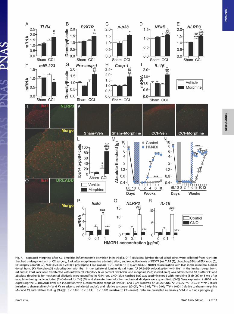
Fig. 4. Repeated morphine after CCI amplifies inflammasome activation in microglia. (A–I) Ipsilateral lumbar dorsal spinal cords were collected from F344 ratsthat had undergone sham or CCI surgery, 5 wk after morphine/saline administration, and respective levels of P2X7R (A), TLR4 (B), phospho-p38/total ERK ratio (C),NF-κB (p65 subunit) (D), NLRP3 (E), miR-223 (F), procaspase-1 (G), caspase-1 (H), and IL-1β (I) quantified. (J) NLRP3 colocalization with Iba1 in the ipsilateral lumbardorsal horn. (K) Phospho-p38 colocalization with Iba1 in the ipsilateral lumbar dorsal horn. (L) DREADD colocalization with Iba1 in the lumbar dorsal horn.(M and N) F344 rats were transfected with intrathecal inhibitory Gi or control DREADDs, and morphine (5 d; shaded area) was administered 10 d after CCI andabsolute thresholds for mechanical allodynia were quantified in F344 rats. CNO (blue hatched bar) was coadministered with morphine (5 d) (M) or 5 wk aftermorphine dosing had concluded (CNO dosed for 7 d) (N), and absolute thresholds for mechanical allodynia were quantified. (O–Q) Gene expression in BV-2 cellsexpressing the Gi DREADD after 4 h incubation with a concentration range of HMGB1, and 0 μM (control) or 50 μM CNO. *P < 0.05; **P < 0.01; ***P < 0.001[relative to sham+saline (A–I and K), relative to vehicle (M and N), and relative to control (O–Q)]; #P < 0.05; ##P < 0.01; ###P < 0.001 [relative to sham+morphine(A–I and K) and relative to 0 μg (O–Q)]; ^P < 0.05; ^̂ P < 0.01; ^̂ ^P < 0.001 (relative to CCI+saline). Data are presented as mean ± SEM; n = 6 or 7 per group.
Grace et al. PNAS Early Edition | 5 of 10
NEU
ROSC
IENCE
PNASPL
US
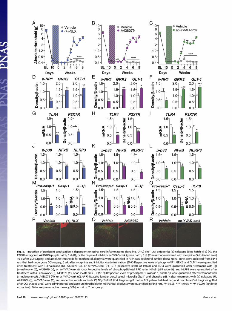
Fig. 5. Induction of persistent sensitization is dependent on spinal cord inflammasome signaling. (A–C) The TLR4 antagonist (+)-naloxone (blue hatch; 5 d) (A), theP2X7R antagonist A438079 (purple hatch; 5 d) (B), or the caspase-1 inhibitor ac-YVAD-cmk (green hatch; 5 d) (C) was coadministered with morphine (5 d; shaded area)10 d after CCI surgery, and absolute thresholds for mechanical allodynia were quantified in F344 rats. Ipsilateral lumbar dorsal spinal cords were collected from F344rats that had undergone CCI surgery, 5 wk after morphine and inhibitor coadministration. (D–F) Respective levels of phospho-NR1, GRK2, and GLT-1 were quantifiedafter treatment with (+)-naloxone (D), A438079 (E), or ac-YVAD-cmk (F). (G–I) Respective levels of P2X7R and TLR4 were quantified after treatment with (g)(+)-naloxone (G), A438079 (H), or ac-YVAD-cmk (I). (J–L) Respective levels of phospho-p38/total ERK ratio, NF-κB (p65 subunit), and NLRP3 were quantified aftertreatment with (+)-naloxone (J), A438079 (K), or ac-YVAD-cmk (L). (M–O) Respective levels of procaspase-1, caspase-1, and IL-1β were quantified after treatment with(+)-naloxone (M), A438079 (N), or ac-YVAD-cmk (O). (P–R) Reactive lumbar dorsal spinal microglia (Iba1+ and phospho-p38+) after treatment with (+)-naloxone (P),A438079 (Q), ac-YVAD-cmk (R), and respective vehicle controls. (S) Nlrp3 siRNA (7 d, beginning 8 d after CCI; yellow hatched bar) and morphine (5 d, beginning 10 dafter CCI; shaded area) were administered, and absolute thresholds for mechanical allodynia were quantified in F344 rats. *P < 0.05; **P < 0.01; ***P < 0.001 (inhibitorvs. control). Data are presented as mean ± SEM; n = 6 or 7 per group.
6 of 10 | www.pnas.org/cgi/doi/10.1073/pnas.1602070113 Grace et al.
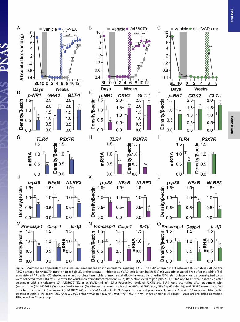
Fig. 6. Maintenance of persistent sensitization is dependent on inflammasome signaling. (A–C) The TLR4 antagonist (+)-naloxone (blue hatch; 5 d) (A), theP2X7R antagonist A438079 (purple hatch; 5 d) (B), or the caspase-1 inhibitor ac-YVAD-cmk (green hatch; 5 d) (C) was administered 5 wk after morphine (5 d,administered 10 d after CCI; shaded area), and absolute thresholds for mechanical allodynia were quantified in F344 rats. Ipsilateral lumbar dorsal spinal cordswere collected from F344 rats, 1 d after the conclusion of inhibitor treatment. (D–F) Respective levels of phospho-NR1, GRK2, and GLT-1 were quantified aftertreatment with (+)-naloxone (D), A438079 (E), or ac-YVAD-cmk (F). (G–I) Respective levels of P2X7R and TLR4 were quantified after treatment with(+)-naloxone (G), A438079 (H), or ac-YVAD-cmk (I). (J–L) Respective levels of phospho-p38/total ERK ratio, NF-κB (p65 subunit), and NLRP3 were quantifiedafter treatment with (+)-naloxone (J), A438079 (K), or ac-YVAD-cmk (L). (M–O) Respective levels of procaspase-1, caspase-1, and IL-1β were quantified aftertreatment with (+)-naloxone (M), A438079 (N), or (ac-YVAD-cmk (O). *P < 0.05; **P < 0.01; ***P < 0.001 (inhibitor vs. control). Data are presented as mean ±SEM; n = 6 or 7 per group.
Grace et al. PNAS Early Edition | 7 of 10
NEU
ROSC
IENCE
PNASPL
US
Mild OIH was induced in pain-free, previously opioid-naïvesubjects, but resolved within days, as reported in clinical and lab-oratory animal studies (6, 7). However, we discovered that mor-phine interacts with neuropathic pain pathophysiology to potentlyprolong this allodynia. We implicated the dorsal spinal NLRP3inflammasome in morphine-induced persistent sensitization, dis-covering that this signaling platform has a triumvirate of previouslyundocumented roles in: the spinal cord, a neuropathic pain model,and enhancement of its activity by morphine (24). Dorsal spinalNLRP3 inflammasomes mediate the initiation of morphine-induced persistent sensitization, because inhibition of TLR4, P2X7R,caspase-1, or IL-1 during morphine administration prevents pro-longed allodynia. Maintenance of morphine-induced persistentsensitization is also dependent on this pathway, because inhibitionof TLR4, P2X7R, caspase-1, or IL-1 reversed prolonged allodynia,an effect that was sustained after TLR4 or P2X7R antagonism. Itshould be noted that the role of TLR4 in OIH has been challenged(50, 51), although these data do not preclude a role for this receptorin morphine-induced persistent sensitization. Furthermore, TLR4 isposited to exclusively regulate male pain behaviors (26, 52). How-ever, ongoing studies indicate that morphine-induced persistentsensitization also occurs in female rodents.Expression of NLRP3 induced by persistent sensitization was lo-
calized to microglia, cells that also express TLR4 and P2X7R (17).The contribution of microglia to the induction and maintenance ofmorphine-induced persistent sensitization was confirmed by selec-tively inhibiting these cells with a Gi DREADD (Fig. 4). The novelapplication of DREADD technology represents an important tech-nical advance, because putative microglial inhibitors (e.g., minocycline,ibudilast, or propentofylline) have activity at other CNS cells,including neurons (17, 53). Expression of DREADDs before neu-ropathic pain induction prevented injury-induced recruitment ofmonocyte-derived cells from contributing to the observed effects.Although we predict that Gi-linked signaling inhibits Ca2+ influx inmicroglia to attenuate proinflammatory cytokine production (43,44), the precise mechanisms are the subject of ongoing investigation.Because microglial activity has not been selectively manipulated inany prior study, these data, to our knowledge, are the first to un-equivocally implicate microglia in a pathological pain state.The mechanism(s) by which inflammasomes remained activated
after cessation of morphine is an avenue for further investigation.Initial activation of inflammasomes may have induced several ad-aptations that create a positive feedback loop at TLR4 and P2X7R.One adaptation may be disrupted glutamate homeostasis, due toIL-1β–mediated down-regulation of GLT-1 (Fig. 3F). Elevatedglutamate may trigger ATP release from glia (54, 55), as well asexcitotoxicity and subsequent DAMP release (17). ATP and re-active oxygen species released after glial P2X7R activation (56,57), as well as additional DAMPs released as a consequence ofHMGB1-induced excitotoxicity (58), may also maintain inflam-masome signaling. However, whether spinal cord inflammasomesremain activated in the absence of morphine by reactive oxygenspecies and/or DAMP signaling at TLR4 and P2X7R, as part of apositive feedback loop, requires future examination.The implications of the present study are striking in light of the
“two-hit”model of glial priming and exaggerated neuroinflammation.Firstly, this model may provide a basis for understanding how opioids
exaggerate pain in preclinical models of peripheral inflammationand surgery (59, 60), as well as clinically after thoracotomy (61, 62).Secondly, opioids superimposed on CNS neuroinflammation mayhave far-ranging consequences beyond pain. For example, opioidsmay also serve as a second hit for glia primed by aging or in-flammation/trauma and may lead to cognitive decline in the elderly(63), postoperative cognitive decline (64), and impaired recoveryof motor function after spinal cord injury (65, 66). Whether themechanistic underpinnings revealed in the current series of studieswill prove to generalize to such opioid-related phenomena remainsto be defined. Finally, the implications of the present studies mayextend beyond opioids as the second hit. A broad range of repeatedneuroinflammatory challenges not only induce a transition fromacute to persistent pain (60, 67, 68), but also induce behaviors thatare comorbid with pain, including cognitive impairment (69), de-pression (70), and anxiety (71). Therefore, our data provide arationale to examine whether the ubiquitous management ofchronic pain with opioids contributes to the incidence of such pain,and potentially pain comorbidities—a hypothesis not previouslyconsidered or tested.In summary, the mechanisms underlying the transition from acute
to chronic pain are poorly understood (17, 72, 73). We discoveredthat a short course of morphine administered upon expression ofneuropathic pain remarkably doubled the duration of CCI-allodynia.This process was dependent upon dorsal spinal microglial re-activity and NLRP3 inflammasomes. These findings comport withprior demonstrations that repeated immune challenges induce atransition from acute to chronic pain (60, 67, 68), which may alsounderpin pain comorbidities (69–71). An evaluation of the long-term consequences of opioid treatment for chronic pain willidentify whether this phenomenon manifests clinically. Our datasuggest a unique strategy to prevent and reverse the deleteriouslong-term effects of opioid treatment without compromising mor-phine analgesia; μ-opioid receptor-mediated analgesia can be main-tained, while simultaneously eliminating inflammasome-mediatedpersistent sensitization.
Materials and MethodsSI Materials and Methods provides complete experimental methods. It includessubjects, drugs, RNA interference, surgery, catheter implantation, mechanicalallodynia, shock sensitivity, and thermal analgesia testing, in vitro Gi DREADDtransfection and stimulation, RT-PCR, Western blotting, and immunohisto-chemistry. Methods for statistical analysis are also included.
All animal procedures were approved by the Institutional Animal Care andUse Committee of the University of Colorado Boulder.
ACKNOWLEDGMENTS. This work was supported by the American PainSociety Future Leaders in Pain Research Grants Program (P.M.G.); NationalHealth and Medical Research Council CJ Martin Fellowship ID 1054091 (toP.M.G.); American Australian Association Sir Keith Murdoch Fellowship (P.M.G.);National Natural Science Foundation of China Grant 21543013 (to X.W.);Natural Science Foundation of Jilin Province Grant 20160101211JC (to X.W.);and NIH Grants DE021966, DA023132 (to L.R.W.), DA017204 (to D.J.U. andB.L.R.), U01MH105892 (to B.L.R.), and GM101279 (to H.H.Y.). The work ofthe Chemical Biology Research Branch was supported by the NIH IntramuralResearch Programs of the National Institute on Drug Abuse and the NationalInstitute of Alcohol Abuse and Alcoholism.
1. Manchikanti L, Fellows B, Ailinani H, Pampati V (2010) Therapeutic use, abuse, andnonmedical use of opioids: A ten-year perspective. Pain Physician 13(5):401–435.
2. Manchikanti L, et al. (2012) Opioid epidemic in the United States. Pain Physician 15(3,Suppl):ES9–ES38.
3. Daubresse M, et al. (2013) Ambulatory diagnosis and treatment of nonmalignant painin the United States, 2000-2010. Med Care 51(10):870–878.
4. Chou R, et al. (2014) The Effectiveness and Risks of Long-Term Opioid Treatment ofChronic Pain. (Agency for Healthcare Research and Quality, Rockville, MD). Available atwww.effectivehealthcare.ahrq.gov/ehc/products/557/1971/chronic-pain-opioid-treatment-report-141007.pdf).
5. Franklin GM; American Academy of Neurology (2014) Opioids for chronic noncancerpain: A position paper of the American Academy of Neurology. Neurology 83(14):1277–1284.
6. Chu LF, Angst MS, Clark D (2008) Opioid-induced hyperalgesia in humans: Molecularmechanisms and clinical considerations. Clin J Pain 24(6):479–496.
7. Grace PM, Maier SF, Watkins LR (2015) Opioid-induced central immune signaling:implications for opioid analgesia. Headache 55(4):475–489.
8. Chu LF, et al. (2012) Analgesic tolerancewithout demonstrable opioid-induced hyperalgesia:A double-blinded, randomized, placebo-controlled trial of sustained-release morphine fortreatment of chronic nonradicular low-back pain. Pain 153(8):1583–1592.
9. Hooten WM, Lamer TJ, Twyner C (2015) Opioid-induced hyperalgesia in community-dwelling adults with chronic pain. Pain 156(6):1145–1152.
10. Hina N, Fletcher D, Poindessous-Jazat F, Martinez V (2015) Hyperalgesia induced bylow-dose opioid treatment before orthopaedic surgery: An observational case-controlstudy. Eur J Anaesthesiol 32(4):255–261.
11. Basaria S, et al. (2015) Effects of testosterone replacement in men with opioid-induced androgen deficiency: A randomized controlled trial. Pain 156(2):280–288.
12. Suzan E, Eisenberg E, Treister R, Haddad M, Pud D (2013) A negative correlationbetween hyperalgesia and analgesia in patients with chronic radicular pain: Is hy-dromorphone therapy a double-edged sword? Pain Physician 16(1):65–76.
8 of 10 | www.pnas.org/cgi/doi/10.1073/pnas.1602070113 Grace et al.
13. Doehring A, Oertel BG, Sittl R, Lötsch J (2013) Chronic opioid use is associated withincreased DNA methylation correlating with increased clinical pain. Pain 154(1):15–23.
14. Chen L, et al. (2009) Altered quantitative sensory testing outcome in subjects withopioid therapy. Pain 143(1-2):65–70.
15. Ram KC, Eisenberg E, Haddad M, Pud D (2008) Oral opioid use alters DNIC but notcold pain perception in patients with chronic pain - new perspective of opioid-induced hyperalgesia. Pain 139(2):431–438.
16. Chu LF, Clark DJ, Angst MS (2006) Opioid tolerance and hyperalgesia in chronic painpatients after one month of oral morphine therapy: A preliminary prospective study.J Pain 7(1):43–48.
17. Grace PM, Hutchinson MR, Maier SF, Watkins LR (2014) Pathological pain and theneuroimmune interface. Nat Rev Immunol 14(4):217–231.
18. Nicotra L, Loram LC, Watkins LR, Hutchinson MR (2012) Toll-like receptors in chronicpain. Exp Neurol 234(2):316–329.
19. Hutchinson MR, et al. (2011) Exploring the neuroimmunopharmacology of opioids:An integrative review of mechanisms of central immune signaling and their impli-cations for opioid analgesia. Pharmacol Rev 63(3):772–810.
20. Wang X, et al. (2012) Morphine activates neuroinflammation in a manner parallel toendotoxin. Proc Natl Acad Sci USA 109(16):6325–6330.
21. Frank MG, Baratta MV, Sprunger DB, Watkins LR, Maier SF (2007) Microglia serve as aneuroimmune substrate for stress-induced potentiation of CNS pro-inflammatorycytokine responses. Brain Behav Immun 21(1):47–59.
22. Combrinck MI, Perry VH, Cunningham C (2002) Peripheral infection evokes exaggeratedsickness behaviour in pre-clinical murine prion disease. Neuroscience 112(1):7–11.
23. Dinarello CA (2011) A clinical perspective of IL-1β as the gatekeeper of inflammation.Eur J Immunol 41(5):1203–1217.
24. de Rivero Vaccari JP, Dietrich WD, Keane RW (2014) Activation and regulation ofcellular inflammasomes: Gaps in our knowledge for central nervous system injury.J Cereb Blood Flow Metab 34(3):369–375.
25. Latz E, Xiao TS, Stutz A (2013) Activation and regulation of the inflammasomes. NatRev Immunol 13(6):397–411.
26. Sorge RE, et al. (2012) Genetically determined P2X7 receptor pore formation regu-lates variability in chronic pain sensitivity. Nat Med 18(4):595–599.
27. Franceschini A, et al. (2015) The P2X7 receptor directly interacts with the NLRP3 in-flammasome scaffold protein. FASEB J 29(6):2450–2461.
28. Grace PM, Hutchinson MR, Manavis J, Somogyi AA, Rolan PE (2010) A novel animalmodel of graded neuropathic pain: Utility to investigate mechanisms of populationheterogeneity. J Neurosci Methods 193(1):47–53.
29. Bennett GJ, Xie YK (1988) A peripheral mononeuropathy in rat that produces disor-ders of pain sensation like those seen in man. Pain 33(1):87–107.
30. Li X, Angst MS, Clark JD (2001) A murine model of opioid-induced hyperalgesia. BrainRes Mol Brain Res 86(1-2):56–62.
31. Pert CB, Snyder SH (1973) Opiate receptor: Demonstration in nervous tissue. Science179(4077):1011–1014.
32. Yang H, et al. (2015) MD-2 is required for disulfide HMGB1-dependent TLR4 signaling.J Exp Med 212(1):5–14.
33. Hutchinson MR, et al. (2010) Possible involvement of toll-like receptor 4/myeloiddifferentiation factor-2 activity of opioid inactive isomers causes spinal proin-flammation and related behavioral consequences. Neuroscience 167(3):880–893.
34. Hutchinson MR, et al. (2007) Opioid-induced glial activation: Mechanisms of ac-tivation and implications for opioid analgesia, dependence, and reward.ScientificWorldJournal 7:98–111.
35. Zhang RX, et al. (2008) IL-1ra alleviates inflammatory hyperalgesia through pre-venting phosphorylation of NMDA receptor NR-1 subunit in rats. Pain 135(3):232–239.
36. Yan X, Yadav R, Gao M, Weng HR (2014) Interleukin-1 beta enhances endocytosis ofglial glutamate transporters in the spinal dorsal horn through activating protein ki-nase C. Glia 62(7):1093–1109.
37. Kleibeuker W, et al. (2008) IL-1 beta signaling is required for mechanical allodyniainduced by nerve injury and for the ensuing reduction in spinal cord neuronal GRK2.Brain Behav Immun 22(2):200–208.
38. Bauernfeind F, et al. (2012) NLRP3 inflammasome activity is negatively controlled bymiR-223. J Immunol 189(8):4175–4181.
39. Armbruster BN, Li X, Pausch MH, Herlitze S, Roth BL (2007) Evolving the lock to fit thekey to create a family of G protein-coupled receptors potently activated by an inertligand. Proc Natl Acad Sci USA 104(12):5163–5168.
40. Ginhoux F, Lim S, Hoeffel G, Low D, Huber T (2013) Origin and differentiation ofmicroglia. Front Cell Neurosci 7:45.
41. Zhang J, et al. (2007) Expression of CCR2 in both resident and bone marrow-derivedmicroglia plays a critical role in neuropathic pain. J Neurosci 27(45):12396–12406.
42. Cuevas J, Adams DJ (1997) M4 muscarinic receptor activation modulates calciumchannel currents in rat intracardiac neurons. J Neurophysiol 78(4):1903–1912.
43. Hayashi Y, et al. (2011) Microglial Ca(2+)-activated K(+) channels are possible mo-lecular targets for the analgesic effects of S-ketamine on neuropathic pain. J Neurosci31(48):17370–17382.
44. Hoffmann A, Kann O, Ohlemeyer C, Hanisch UK, Kettenmann H (2003) Elevation ofbasal intracellular calcium as a central element in the activation of brain macrophages(microglia): Suppression of receptor-evoked calcium signaling and control of releasefunction. J Neurosci 23(11):4410–4419.
45. Agalave NM, et al. (2014) Spinal HMGB1 induces TLR4-mediated long-lasting hyper-sensitivity and glial activation and regulates pain-like behavior in experimental ar-thritis. Pain 155(9):1802–1813.
46. Hutchinson MR, et al. (2008) Non-stereoselective reversal of neuropathic pain bynaloxone and naltrexone: Involvement of toll-like receptor 4 (TLR4). Eur J Neurosci28(1):20–29.
47. Nelson DW, et al. (2006) Structure-activity relationship studies on a series of novel,substituted 1-benzyl-5-phenyltetrazole P2X7 antagonists. J Med Chem 49(12):3659–3666.
48. Jiang LH, Mackenzie AB, North RA, Surprenant A (2000) Brilliant blue G selectivelyblocks ATP-gated rat P2X(7) receptors. Mol Pharmacol 58(1):82–88.
49. Rabuffetti M, et al. (2000) Inhibition of caspase-1-like activity by Ac-Tyr-Val-Ala-Asp-chloromethyl ketone induces long-lasting neuroprotection in cerebral ischemiathrough apoptosis reduction and decrease of proinflammatory cytokines. J Neurosci20(12):4398–4404.
50. Ferrini F, et al. (2013) Morphine hyperalgesia gated through microglia-mediateddisruption of neuronal Cl⁻ homeostasis. Nat Neurosci 16(2):183–192.
51. Mattioli TA, et al. (2014) Toll-like receptor 4 mutant and null mice retain morphine-induced tolerance, hyperalgesia, and physical dependence. PLoS One 9(5):e97361.
52. Stokes JA, Cheung J, Eddinger K, Corr M, Yaksh TL (2013) Toll-like receptor signalingadapter proteins govern spread of neuropathic pain and recovery following nerveinjury in male mice. J Neuroinflammation 10:148.
53. Grace PM, et al. (2014) Activation of adult rat CNS endothelial cells by opioid-inducedtoll-like receptor 4 (TLR4) signaling induces proinflammatory, biochemical, morpho-logical, and behavioral sequelae. Neuroscience 280:299–317.
54. Liu GJ, Kalous A, Werry EL, Bennett MR (2006) Purine release from spinal cord mi-croglia after elevation of calcium by glutamate. Mol Pharmacol 70(3):851–859.
55. Queiroz G, Meyer DK, Meyer A, Starke K, von Kügelgen I (1999) A study of themechanism of the release of ATP from rat cortical astroglial cells evoked by activationof glutamate receptors. Neuroscience 91(3):1171–1181.
56. Ficker C, et al. (2014) Astrocyte-neuron interaction in the substantia gelatinosa of thespinal cord dorsal horn via P2X7 receptor-mediated release of glutamate and reactiveoxygen species. Glia 62(10):1671–1686.
57. Suadicani SO, Brosnan CF, Scemes E (2006) P2X7 receptors mediate ATP releaseand amplification of astrocytic intercellular Ca2+ signaling. J Neurosci 26(5):1378–1385.
58. Balosso S, Liu J, Bianchi ME, Vezzani A (2014) Disulfide-containing high mobilitygroup box-1 promotes N-methyl-D-aspartate receptor function and excitotoxicity byactivating Toll-like receptor 4-dependent signaling in hippocampal neurons. AntioxidRedox Signal 21(12):1726–1740.
59. Célérier E, González JR, Maldonado R, Cabañero D, Puig MM (2006) Opioid-inducedhyperalgesia in a murine model of postoperative pain: Role of nitric oxide generatedfrom the inducible nitric oxide synthase. Anesthesiology 104(3):546–555.
60. Loram LC, et al. (2012) Prior exposure to repeated morphine potentiates mechanicalallodynia induced by peripheral inflammation and neuropathy. Brain Behav Immun26(8):1256–1264.
61. van Gulik L, et al. (2012) Remifentanil during cardiac surgery is associated withchronic thoracic pain 1 yr after sternotomy. Br J Anaesth 109(4):616–622.
62. Salengros JC, et al. (2010) Different anesthetic techniques associated with differentincidences of chronic post-thoracotomy pain: Low-dose remifentanil plus presurgicalepidural analgesia is preferable to high-dose remifentanil with postsurgical epiduralanalgesia. J Cardiothorac Vasc Anesth 24(4):608–616.
63. Puustinen J, et al. (2011) Use of CNS medications and cognitive decline in the aged: alongitudinal population-based study. BMC Geriatr 11:70.
64. Wang Y, Sands LP, Vaurio L, Mullen EA, Leung JM (2007) The effects of postoperativepain and its management on postoperative cognitive dysfunction. Am J GeriatrPsychiatry 15(1):50–59.
65. Hook MA, et al. (2007) The impact of morphine after a spinal cord injury. Behav BrainRes 179(2):281–293.
66. Hook MA, et al. (2009) Intrathecal morphine attenuates recovery of function after aspinal cord injury. J Neurotrauma 26(5):741–752.
67. Hains LE, et al. (2011) Prior laparotomy or corticosterone potentiates lipopolysac-charide-induced fever and sickness behaviors. J Neuroimmunol 239(1-2):53–60.
68. Hains LE, et al. (2010) Pain intensity and duration can be enhanced by prior chal-lenge: Initial evidence suggestive of a role of microglial priming. J Pain 11(10):1004–1014.
69. Barrientos RM, et al. (2006) Peripheral infection and aging interact to impair hippo-campal memory consolidation. Neurobiol Aging 27(5):723–732.
70. Fenn AM, et al. (2014) Immune activation promotes depression 1 month after diffusebrain injury: A role for primed microglia. Biol Psychiatry 76(7):575–584.
71. Spencer SJ, Heida JG, Pittman QJ (2005) Early life immune challenge—effects onbehavioural indices of adult rat fear and anxiety. Behav Brain Res 164(2):231–238.
72. Voscopoulos C, Lema M (2010) When does acute pain become chronic? Br J Anaesth105(Suppl 1):i69–i85.
73. Apkarian AV, Baliki MN, Farmer MA (2013) Predicting transition to chronic pain. CurrOpin Neurol 26(4):360–367.
74. Milligan ED, Hinde JL, Mehmert KK, Maier SF, Watkins LR (1999) A method for in-creasing the viability of the external portion of lumbar catheters placed in the spinalsubarachnoid space of rats. J Neurosci Methods 90(1):81–86.
75. Reagan-Shaw S, Nihal M, Ahmad N (2008) Dose translation from animal to humanstudies revisited. FASEB J 22(3):659–661.
76. APS-AAPM (2009) Clinical Guideline for the Use of Chronic Opioid Therapy in ChronicNoncancer Pain. Available at americanpainsociety.org/uploads/education/guidelines/chronic-opioid-therapy-cncp.pdf. Accessed April 4, 2016.
77. Chaplan SR, Bach FW, Pogrel JW, Chung JM, Yaksh TL (1994) Quantitative assessmentof tactile allodynia in the rat paw. J Neurosci Methods 53(1):55–63.
Grace et al. PNAS Early Edition | 9 of 10
NEU
ROSC
IENCE
PNASPL
US

78. Chacur M, et al. (2001) A new model of sciatic inflammatory neuritis (SIN): Inductionof unilateral and bilateral mechanical allodynia following acute unilateral peri-sciaticimmune activation in rats. Pain 94(3):231–244.
79. Milligan ED, et al. (2001) Intrathecal HIV-1 envelope glycoprotein gp120 inducesenhanced pain states mediated by spinal cord proinflammatory cytokines. J Neurosci21(8):2808–2819.
80. Harvey LO, Jr (1986) Efficient estimation of sensory thresholds. Behav Res MethodsInstrum Comput 18:623–632.
81. Treutwein B, Strasburger H (1999) Fitting the psychometric function. PerceptPsychophys 61(1):87–106.
82. Milligan ED, et al. (2000) Thermal hyperalgesia and mechanical allodynia produced byintrathecal administration of the human immunodeficiency virus-1 (HIV-1) envelopeglycoprotein, gp120. Brain Res 861(1):105–116.
83. Hargreaves K, Dubner R, Brown F, Flores C, Joris J (1988) A new and sensitive methodfor measuring thermal nociception in cutaneous hyperalgesia. Pain 32(1):77–88.
84. Chomczynski P, Sacchi N (1987) Single-step method of RNA isolation by acid guani-dinium thiocyanate-phenol-chloroform extraction. Anal Biochem 162(1):156–159.
85. Livak KJ, Schmittgen TD (2001) Analysis of relative gene expression data usingreal-time quantitative PCR and the 2(-Delta Delta C(T)) Method. Methods 25(4):402–408.
10 of 10 | www.pnas.org/cgi/doi/10.1073/pnas.1602070113 Grace et al.