Embed Size (px)
Citation preview
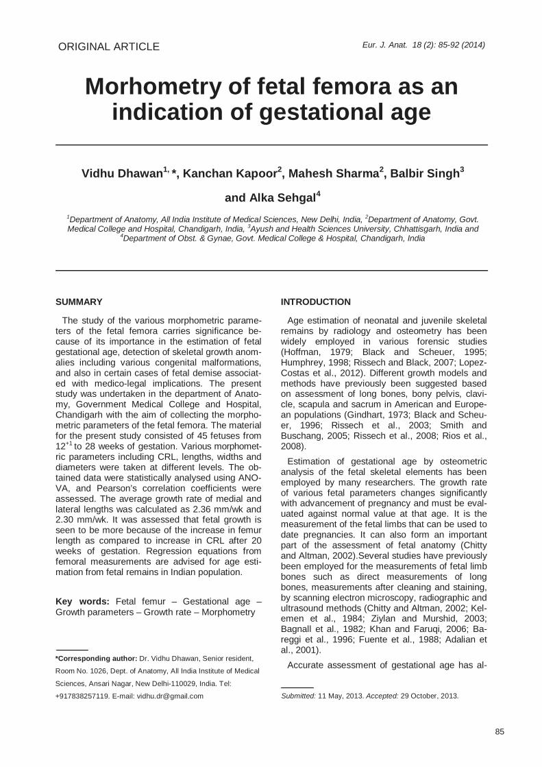
Morhometry of fetal femora as an indication of gestational age
ORIGINAL ARTICLE Eur. J. Anat. 18 (2): 85-92 (2014)
Vidhu Dhawan1, *, Kanchan Kapoor2, Mahesh Sharma2, Balbir Singh3
and Alka Sehgal4 1Department of Anatomy, All India Institute of Medical Sciences, New Delhi, India, 2Department of Anatomy, Govt. Medical College and Hospital, Chandigarh, India, 3Ayush and Health Sciences University, Chhattisgarh, India and
4Department of Obst. & Gynae, Govt. Medical College & Hospital, Chandigarh, India
SUMMARY
The study of the various morphometric parame-ters of the fetal femora carries significance be-cause of its importance in the estimation of fetal gestational age, detection of skeletal growth anom-alies including various congenital malformations, and also in certain cases of fetal demise associat-ed with medico-legal implications. The present study was undertaken in the department of Anato-my, Government Medical College and Hospital, Chandigarh with the aim of collecting the morpho-metric parameters of the fetal femora. The material for the present study consisted of 45 fetuses from 12+1 to 28 weeks of gestation. Various morphomet-ric parameters including CRL, lengths, widths and diameters were taken at different levels. The ob-tained data were statistically analysed using ANO-VA, and Pearson’s correlation coefficients were assessed. The average growth rate of medial and lateral lengths was calculated as 2.36 mm/wk and 2.30 mm/wk. It was assessed that fetal growth is seen to be more because of the increase in femur length as compared to increase in CRL after 20 weeks of gestation. Regression equations from femoral measurements are advised for age esti-mation from fetal remains in Indian population.
Key words: Fetal femur – Gestational age – Growth parameters – Growth rate – Morphometry
INTRODUCTION
Age estimation of neonatal and juvenile skeletal remains by radiology and osteometry has been widely employed in various forensic studies (Hoffman, 1979; Black and Scheuer, 1995; Humphrey, 1998; Rissech and Black, 2007; Lopez-Costas et al., 2012). Different growth models and methods have previously been suggested based on assessment of long bones, bony pelvis, clavi-cle, scapula and sacrum in American and Europe-an populations (Gindhart, 1973; Black and Scheu-er, 1996; Rissech et al., 2003; Smith and Buschang, 2005; Rissech et al., 2008; Rios et al., 2008).
Estimation of gestational age by osteometric analysis of the fetal skeletal elements has been employed by many researchers. The growth rate of various fetal parameters changes significantly with advancement of pregnancy and must be eval-uated against normal value at that age. It is the measurement of the fetal limbs that can be used to date pregnancies. It can also form an important part of the assessment of fetal anatomy (Chitty and Altman, 2002).Several studies have previously been employed for the measurements of fetal limb bones such as direct measurements of long bones, measurements after cleaning and staining, by scanning electron microscopy, radiographic and ultrasound methods (Chitty and Altman, 2002; Kel-emen et al., 1984; Ziylan and Murshid, 2003; Bagnall et al., 1982; Khan and Faruqi, 2006; Ba-reggi et al., 1996; Fuente et al., 1988; Adalian et al., 2001).
Accurate assessment of gestational age has al-
85
Submitted: 11 May, 2013. Accepted: 29 October, 2013.
*Corresponding author: Dr. Vidhu Dhawan, Senior resident,
Room No. 1026, Dept. of Anatomy, All India Institute of Medical
Sciences, Ansari Nagar, New Delhi-110029, India. Tel:
+917838257119. E-mail: [email protected]
Fetal femoral morphometry
86
ways been one of the most important functions of diagnostic ultrasound (Chitty and Altman, 2002; Chitty et al., 1994; Hadlock et al., 1982; Hadlock et al., 1984; Queenan et al., 1980; O’Brien et al., 1981). The antenatal ultrasonographic evaluation of the femur is based only on the measurements of the ossified length of the fetal femur, in contrast to the more valid direct measurements of the total lengths. The practical value of such measurements is debatable, as most of these studies have been concerned with the assessment of fetal maturity and have concentrated on fetus in vivo (in utero) near term. These measurements vary over a wide range due to differences in fetal position: difficul-ties occur in taking correct linear measurements because of variation of objective planes due to the movements of the fetuses (Chitty and Altman, 2002).
Several (Loughna et al., 2009; Chitty and Altman, 2002; Chitty et al., 1994) previously constructed growth charts of fetal size based on such ultraso-nographic measurements from European popula-tion are not easily reproducible on the fetal skeletal remains. The direct measurements thus provide an insight into the reliable method of estimation of gestational age in the abortuses. Long bone re-gressions produce the most accurate estimations, as long bones are highly correlated to the total stature. These studies are of importance to medico-legal authorities, particularly as it is sometimes necessary to determine if these skeletal remains are those of a full-term neonate or a pre-term fe-tus. This age estimation can also play an important role in the prosecution of forensic cases, specifi-cally in certain cases of criminal abortion or infanti-cide (Scheuer, 2001).
Some forensic studies have evaluated the diphyseal lengths of the fetal limb bones for esti-mation of gestational age (Fazekas and Kosa, 1978; Hoffman, 1979; Kelemen et al., 1984; Matsushitka et al., 1995; Watkins and German, 1996; Scheuer and Black, 2000, Ziylan and Murshid, 2003). The present study is an attempt to correlate the growth of various parameters like lengths, diameters, and other factors with the in-creasing gestational age. These data might be of immense clinical importance in the assessment of fetal age by direct or ultrasound method.
MATERIAL AND METHODS
The present study was carried out in the Depart-ment of Anatomy, Government Medical College and Hospital, Chandigarh, on 45 aborted human fetal specimens from 12th to 28th weeks of gesta-tional age. The specimens were the results of in-trauterine deaths or spontaneous abortions and were provided by the department of Obstetrics & Gynecology for routine fetal autopsy. Differentia-
tion based on sex of the fetus was not taken into account. The cases of congenital malformations of musculoskeletal system or visible skeletal defects were excluded. The mothers were registered ante-natally in the institute and gestational age was esti-mated by menstrual history, which was further con-firmed by ultrasonography. Consent for autopsy, measurements, and additional studies had been taken from the parents. For all fetuses, the crown rump and heel length (CRL and CHL) were meas-ured.
This study was conducted on the ethical guide-lines for biomedical research on human subjects as given in “Declaration of Helsinki” and by the Central Ethics Committee on Human Research (CECHR) of ICMR, New Delhi, and clearance was obtained from the Institutional Ethical Committee. After the routine autopsy, femur was separated after disarticulation of hip and knee joints taking care around the joints to preserve the cartilage at the ends of the bone. The bone was measured and then kept in 10% formalin for future reference.
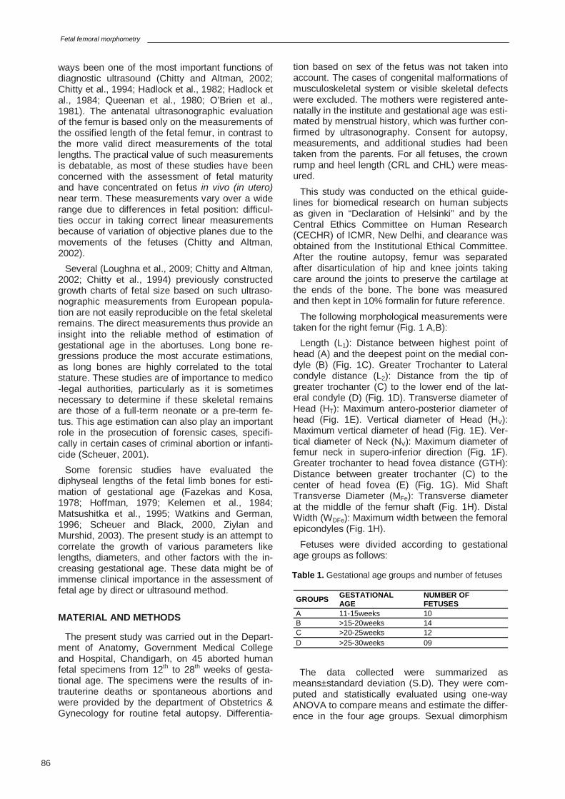
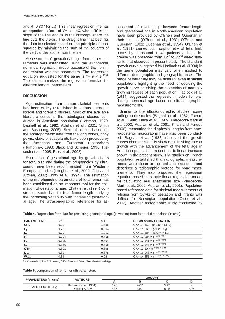
The following morphological measurements were taken for the right femur (Fig. 1 A,B):
Length (L1): Distance between highest point of head (A) and the deepest point on the medial con-dyle (B) (Fig. 1C). Greater Trochanter to Lateral condyle distance (L2): Distance from the tip of greater trochanter (C) to the lower end of the lat-eral condyle (D) (Fig. 1D). Transverse diameter of Head (HT): Maximum antero-posterior diameter of head (Fig. 1E). Vertical diameter of Head (HV): Maximum vertical diameter of head (Fig. 1E). Ver-tical diameter of Neck (NV): Maximum diameter of femur neck in supero-inferior direction (Fig. 1F). Greater trochanter to head fovea distance (GTH): Distance between greater trochanter (C) to the center of head fovea (E) (Fig. 1G). Mid Shaft Transverse Diameter (MFe): Transverse diameter at the middle of the femur shaft (Fig. 1H). Distal Width (WDFe): Maximum width between the femoral epicondyles (Fig. 1H).
Fetuses were divided according to gestational age groups as follows:
GROUPS GESTATIONAL AGE
NUMBER OF FETUSES
A 11-15weeks 10 B >15-20weeks 14 C >20-25weeks 12 D >25-30weeks 09
Table 1. Gestational age groups and number of fetuses
The data collected were summarized as means±standard deviation (S.D). They were com-puted and statistically evaluated using one-way ANOVA to compare means and estimate the differ-ence in the four age groups. Sexual dimorphism
V. Dhawan et al.
87
Fig. 1. Anatomical parameters of Femur. (A,B) Length (L1); (C,D) Length (L2); (E-H) HT- Transverse diameter Head, HV -Vertical diameter Head, NV-Vertical diameter Neck, GTH- Greater trochanter to head fovea distance, MFe -Mid shaft transverse diameter, WFe -Distal width.
A B C D
E F
H
G
was not assessed in the present study. In order to see the relationship between two variables, Pear-son correlation coefficients were analyzed. In or-der to determine the gestational age from various morphometric variables of femur, simple linear and exponential nonlinear regression models were applied with age as dependant variable in regression analysis. SPSS version 15 for Win-dows was used for the analysis. All statistical tests were two-sided and performed at a signifi-cance level of p=.05.
RESULTS
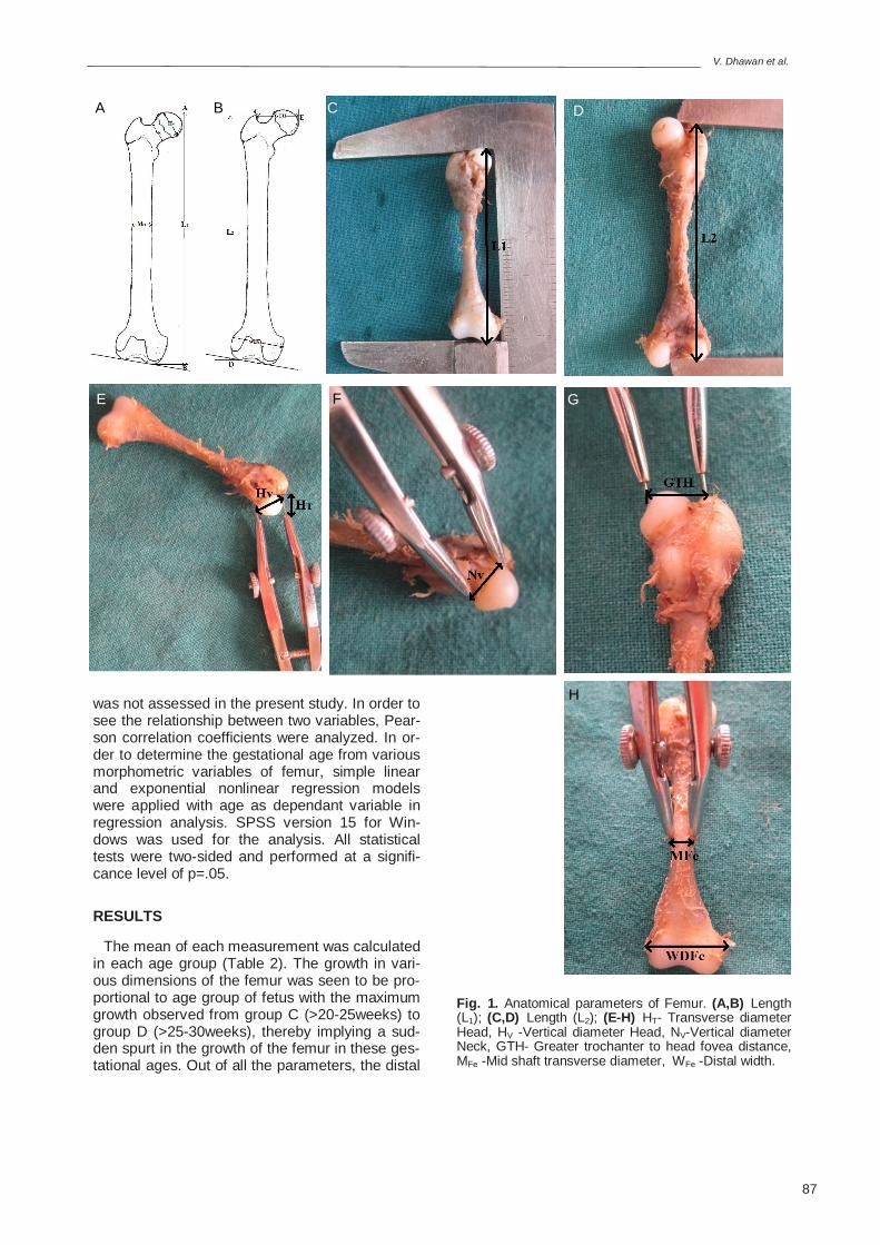
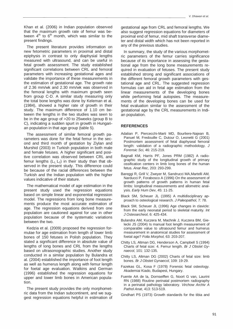
The mean of each measurement was calculated in each age group (Table 2). The growth in vari-ous dimensions of the femur was seen to be pro-portional to age group of fetus with the maximum growth observed from group C (>20-25weeks) to group D (>25-30weeks), thereby implying a sud-den spurt in the growth of the femur in these ges-tational ages. Out of all the parameters, the distal
Fetal femoral morphometry
88
Fig. 3. Approximate growth average per week assessed in every parameter.
Fig. 2. Trend of growth in various parameters of femur with increasing gestational ages.
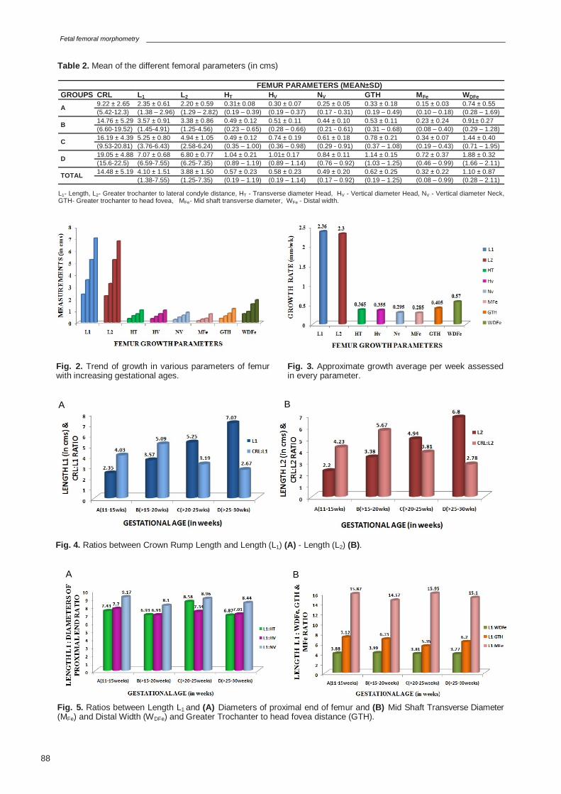
Fig. 5. Ratios between Length L1 and (A) Diameters of proximal end of femur and (B) Mid Shaft Transverse Diameter (MFe) and Distal Width (WDFe) and Greater Trochanter to head fovea distance (GTH).
Fig. 4. Ratios between Crown Rump Length and Length (L1) (A) - Length (L2) (B).
A B
A B
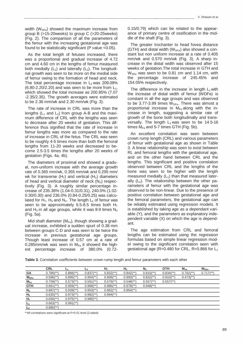
FEMUR PARAMETERS (MEAN±SD) GROUPS CRL L1 L2 HT HV NV GTH MFe WDFe
A 9.22 ± 2.65 2.35 ± 0.61 2.20 ± 0.59 0.31± 0.08 0.30 ± 0.07 0.25 ± 0.05 0.33 ± 0.18 0.15 ± 0.03 0.74 ± 0.55 (5.42-12.3) (1.38 – 2.96) (1.29 – 2.82) (0.19 – 0.39) (0.19 – 0.37) (0.17 - 0.31) (0.19 – 0.49) (0.10 – 0.18) (0.28 – 1.69)
B 14.76 ± 5.29 3.57 ± 0.91 3.38 ± 0.86 0.49 ± 0.12 0.51 ± 0.11 0.44 ± 0.10 0.53 ± 0.11 0.23 ± 0.24 0.91± 0.27 (6.60-19.52) (1.45-4.91) (1.25-4.56) (0.23 – 0.65) (0.28 – 0.66) (0.21 - 0.61) (0.31 – 0.68) (0.08 – 0.40) (0.29 – 1.28)
C 16.19 ± 4.39 5.25 ± 0.80 4.94 ± 1.05 0.49 ± 0.12 0.74 ± 0.19 0.61 ± 0.18 0.78 ± 0.21 0.34 ± 0.07 1.44 ± 0.40 (9.53-20.81) (3.76-6.43) (2.58-6.24) (0.35 – 1.00) (0.36 – 0.98) (0.29 - 0.91) (0.37 – 1.08) (0.19 – 0.43) (0.71 – 1.95)
D 19.05 ± 4.88 7.07 ± 0.68 6.80 ± 0.77 1.04 ± 0.21 1.01± 0.17 0.84 ± 0.11 1.14 ± 0.15 0.72 ± 0.37 1.88 ± 0.32 (15.6-22.5) (6.59-7.55) (6.25-7.35) (0.89 – 1.19) (0.89 – 1.14) (0.76 – 0.92) (1.03 – 1.25) (0.46 – 0.99) (1.66 – 2.11)
TOTAL 14.48 ± 5.19 4.10 ± 1.51 3.88 ± 1.50 0.57 ± 0.23 0.58 ± 0.23 0.49 ± 0.20 0.62 ± 0.25 0.32 ± 0.22 1.10 ± 0.87 (1.38-7.55) (1.25-7.35) (0.19 – 1.19) (0.19 – 1.14) (0.17 – 0.92) (0.19 – 1.25) (0.08 – 0.99) (0.28 – 2.11)
Table 2. Mean of the different femoral parameters (in cms)
L1- Length, L2- Greater trochanter to lateral condyle distance, HT - Transverse diameter Head, HV - Vertical diameter Head, NV - Vertical diameter Neck, GTH- Greater trochanter to head fovea, MFe- Mid shaft transverse diameter, WFe - Distal width.
V. Dhawan et al.
89
width (WDFe) showed the maximum increase from group B (>15-20weeks) to group C (>20-25weeks) (Fig. 2). The comparison of all the parameters of the femur with the increasing gestational age was found to be statistically significant (P value <0.05).
As the total length of fetuses increased, there was a proportional and gradual increase of 4.72 cm and 4.60 cm in the lengths of femur measured both medially (L1) and laterally (L2). The longitudi-nal growth was seen to be more on the medial side of femur owing to the formation of head and neck. The total percentage increase in L2 was 209.09% (6.80-2.20/2.20) and was seen to be more from L1, which showed the total increase as 200.85% (7.07-2.35/2.35). The growth averages were estimated to be 2.36 mm/wk and 2.30 mm/wk (Fig. 3).
The rate of increase in CRL was more than the lengths (L1 and L2) from group A-B and the maxi-mum difference of CRL with the lengths was seen to decrease after 20 weeks of gestation. This dif-ference thus signified that the rate of increase in femur lengths was more as compared to the rate of increase in CRL of the fetus. The CRL was seen to be roughly 4-5 times more than both the femoral lengths from 11-20 weeks and decreased to be-come 2.5-3.5 times the lengths after 20 weeks of gestation (Figs. 4a, 4b).
The diameters of proximal end showed a gradu-al, non-uniform increase with the average growth rate of 0.365 mm/wk, 0.355 mm/wk and 0.295 mm/wk for transverse (HT) and vertical (HV) diameters of head and vertical diameter of neck (NV) respec-tively (Fig. 3). A roughly similar percentage in-crease of 235.38% (1.04-0.31/0.31), 240.0% (1.02-0.30/0.30) and 236.0% (0.84-0.25/0.25) was calcu-lated for HT, HV and NV. The length L1 of femur was seen to be approximately 6.5-8.5 times both HT and HV in all age groups, while it was 8-9 times NV (Fig. 5a).
Mid shaft diameter (MFe), though showing a grad-ual increase, exhibited a sudden spurt of 0.38 mm between groups C-D and was seen to be twice the increase in previous gestational age groups. Though least increase of 0.57 cm at a rate of 0.285mm/wk was seen in MFe, it showed the high-est percentage increase of 380.0% (0.72-
0.15/0.79) which can be related to the appear-ance of primary centre of ossification in the mid-dle of the shaft (Fig. 3).
The greater trochanter to head fovea distance (GTH) and distal width (WDFe) also showed a con-stant but non uniform increase at a rate of 0.405 mm/wk and 0.570 mm/wk (Fig. 3). A sharp in-crease in the distal width was observed after 15 weeks of gestation.The total increase in GTH and WDFe was seen to be 0.81 cm and 1.14 cm, with the percentage increase of 245.45% and 154.05% respectively.
The difference in the increase in length L1 with the increase of distal width of femur (WDFe) is constant in all the age groups and was observed to be 3.77-3.99 times WDFe. There was almost a proportional increase in MFe along with the in-crease in length, suggesting a similar rate of growth of the bone both longitudinally and trans-versally. The length L1 was seen to be 14.5-16 times MFe and 5-7 times GTH (Fig. 5b).
An excellent correlation was seen between crown rump length (CRL) and various parameters of femur with gestational age as shown in Table 3. A linear relationship was seen to exist between RL and femoral lengths with the gestational age, and on the other hand between CRL and the lengths. This significant and positive correlation observed between CRL and the lengths of the bone was seen to be higher with the length measured medially (L1) than that measured later-ally (L2). The relationship between the other pa-rameters of femur with the gestational age was observed to be non-linear. Due to the presence of positive correlation between gestational age and the femoral parameters, the gestational age can be reliably estimated using regression models. It is established by taking age as a dependant vari-able (Y), and the parameters as explanatory inde-pendent variable (X) on which the age is depend-ant.
The age estimation from CRL and femoral lengths can be estimated using the regression formulas based on simple linear regression mod-el owing to the significant correlation seen with gestational age (R=0.480 for CRL, R=0.866 for L1
Table 3. Correlation coefficients between crown rump length and femur parameters with each other
CRL L1 L2 HT HV NV GTH MFe WDFe GA 0.780(**) 0.866(**) 0.837(**) 0.832(**) 0.842(**) 0.810(**) 0.834(**) 0.762(**) 0.717(**) WDFe 0.596(**) 0.895(**) 0.904(**) 0.906(**) 0.900(**) 0.832(**) 0.910(**) 0.473(**) MFe 0.709(**) 0.573(**) 0.591(**) 0.579(**) 0.548(**) 0.557(**) 0.557(**) GTH 0.661(**) 0.959(**) 0.968(**) 0.986(**) 0.978(**) 0.948(**) NV 0.687(**) 0.939(**) 0.953(**) 0.962(**) 0.954(**) HV 0.635(**) 0.973(**) 0.982(**) 0.994(**) HT 0.656(**) 0.975(**) 0.985(**) L1 0.663(**) 0.991(**) L2 0.680(**)
**All correlations were significant at P=0.01 level (2-tailed)
Fetal femoral morphometry
90
and R=0.837 for L2). This linear regression line has an equation in form of Y= a + bX, where ‘b’ is the slope of the line and ‘a’ is the intercept where the line cuts the y axis. The straight line that best fits the data is selected based on the principle of least squares by minimizing the sum of the squares of the vertical deviations from the line.
Assessment of gestational age from other pa-rameters was established using the exponential nonlinear regression model because of the nonlin-ear relation with the parameters. The regression equation suggested for the same is Y= a × e (bX). Table 4 summarizes the regression formulae for different femoral parameters.
DISCUSSION
Age estimation from human skeletal elements has been widely established in various anthropo-logical and forensic studies. Most of the available literature concerns the radiological studies con-ducted in American population (Hoffman, 1979; Bagnall et al., 1982; Adalian et al., 2001; Smith and Buschang, 2005). Several studies based on the anthropometric data from the long bones, bony pelvis, clavicle, scapula etc have been provided by the American and European researchers (Humphrey, 1998; Black and Scheuer, 1996; Ris-sech et al., 2008; Rios et al., 2008).
Estimation of gestational age by growth charts for fetal size and dating the pregnancies by ultra-sound have been recommended from Western-European studies (Loughna et al., 2009; Chitty and Altman, 2002; Chitty et al., 1994). The estimation of the morphometric parameters of fetal femur has been established as an important tool for the esti-mation of gestational age. Chitty et al. (1994) con-structed such chart for fetal femur length studying the increasing variability with increasing gestation-al age. The ultrasonographic references for as-
sessment of relationship between femur length and gestational age in North-American population have been provided by O’Brien and Queenan in their studies (O’Brien et al., 1981; O’Brien and Queenan, 1981; Queenan et al., 1994). O’Brien et al. (1981) carried out morphometry of fetal limb bones by ultrasound in 41 patients a linear in-crease was observed from 12th to 22nd week simi-lar to that observed in present study. The standard growth curve suggested by Hadlock et al. (1984) in the same population may vary when applied to different demographic and geographic areas. The range of variability may be different even in similar populations highlighting the need for deciding the growth curve satisfying the biometrics of normally growing fetuses of each population. Hadlock et al. (1984) suggested the regression models for pre-dicting menstrual age based on ultrasonographic measurements.
Similar to the ultrasonographic studies, some radiographic studies (Bagnall et al., 1982; Fuente et al., 1988; Kalifa et al., 1989; Piercecchi-Marti et al., 2002; Adalian et al., 2001; Khan and Faruqi, 2006), measuring the diaphysial lengths from ante-ro-posterior radiographs have also been conduct-ed. Bagnall et al. (1982) observed the growth curves characteristically show a diminishing rate of growth with the advancement of the fetal age in American population, in contrast to linear increase shown in the present study. The studies on French population established that radiographic measure-ments were closer to the real anatomic ones and described a radiographic protocol for bone meas-urements. They also proposed the regression equation based on simple linear regression model for calculating real anatomical size (Piercecchi-Marti et al., 2002; Adalian et al., 2001). Population based reference data for skeletal measurements of fetuses from 16wks of gestation and infants was defined for Norwegian population (Olsen et al., 2002). Another radiographic study conducted by
Table 5. comparison of femur length parameters
PARAMETERS (in cms) AUTHORS GROUPS A B C D
FEMUR LENGTH (L1) Kelemen et al (1984) 2.48 4.67 5.43 -
Present Study 2.35 3.57 5.25 7.07
Table 4. Regression formulae for predicting gestational age (in weeks) from femoral dimensions (in cms)
PARAMETERS R2 S.E REGRESSION EQUATION CRL 0.23 1.786 GA= 14.453+ (0.339 × CRL) L1 0.75 0.964 GA= 11.062 + (2.02 × L1) L2 0.70 1.013 GA= 11.684 + (1.974 × L2) HT 0.704 0.768 GA= 13.284 × e (0.63 × HT) HV 0.685 0.704 GA= 13.541 × e (0.63 × HV) NV 0.646 0.768 GA= 13.545 × e (0.71 × NV) GTH 0.691 0.698 GA= 13.50 × e (0.565 × GTH) MFe 0.52 0.678 GA= 16.046 × e (0.64 × MFe) WDFe 0.51 0.92 GA= 14.358 × e (0.262 ×WDFe)
R= Correlation, R2= R Squared, S.E= Standard Error, GA= Gestational Age
V. Dhawan et al.
91
Khan et al. (2006) in Indian population observed that the maximum growth rate of femur was be-tween 4th to 6th month, which was similar to the present findings.
The present literature provides information on new fetometric parameters in proximal and distal epiphysis in contrast to only diaphysial lengths measured with ultrasound, and can be useful in fetal growth assessment. The study established significant correlations between CRL and femoral parameters with increasing gestational ages and validate the importance of these measurements in the estimation of gestational age. The growth rate of 2.36 mm/wk and 2.30 mm/wk was observed in the femoral lengths with maximum growth seen from group C-D. A similar study measuring only the total bone lengths was done by Keleman et al. (1984), showed a higher rate of growth in their study. The maximum difference of 1.10 cm be-tween the lengths in the two studies was seen to be in the age group of >20 to 25weeks (group B to C), indicating a sudden spurt in growth in Hungari-an population in that age group (table 5).
The assessment of similar femoral growth pa-rameters was done for the fetal femur in the sec-ond and third month of gestation by Ziylan and Murshid (2003) in Turkish population in both male and female fetuses. A higher significant and posi-tive correlation was observed between CRL and femur lengths (L1, L2) in their study than that ob-served in the present study. This difference could be because of the racial differences between the Turkish and the Indian population with the higher values indicative of their stature.
The mathematical model of age estimation in the present study used the regression equations based on simple linear and exponential regression model. The regressions from long bone measure-ments produce the most accurate estimation of age. The regression equations derived from one population are cautioned against for use in other population because of the systematic variations between the two.
Kedzia et al. (2009) proposed the regression for-mulae for age estimation from length of lower limb bones of 150 fetuses in Polish population. They stated a significant difference in absolute value of lengths of long bones and CRL from the lengths based on ultrasonographic studies. Another study conducted in a similar population by Bulandra et al. (2004) established the importance of foot length as well as humerus length along with femur length for foetal age evaluation. Watkins and German (1996) established the regression equations for upper and lower limb bones in American popula-tion.
The present study provides the only morphomet-ric data from the Indian subcontinent, and we sug-gest regression equations helpful in estimation of
gestational age from CRL and femoral lengths. We also suggest regression equations for diameters of proximal end of femur, mid shaft transverse diame-ter and distal width which has not been reported in any of the previous studies.
In summary, the study of the various morphomet-ric parameters of the femur carries significance because of its importance in assessing the gesta-tional age from the long bone measurements re-quired in evaluation of fetuses. The present study established strong and significant associations of the different femoral growth parameters with ges-tational age and CRL. The suggested regression formulas can aid in fetal age estimation from the linear measurements of the developing bones while performing fetal autopsies. The measure-ments of the developing bones can be used for fetal evaluation similar to the assessment of the gestational age by the CRL measurements in Indi-an population.
REFERENCES
Adalian P, Piercecchi-Marti MD, Bourliere-Najean B, Panuel M, Fredouille C, Dutour O, Leonetti G (2001) Postmortem assessment of fetal diaphyseal femoral length: validation of a radiographic methodology. J Forensic Sci, 46: 215-219.
Bagnall KM, Harris PF, Jones PRM (1982) A radio-graphic study of the longitudinal growth of primary ossification centers in limb long bones of the human fetus. Anat Rec, 203: 293-299.
Bareggi R, Grill V, Zweyer M, Sandrucci MA,Martelli AM, Narducci P, Forabosco A (1996) On the assessment of growth patterns of growth patterns in human fetal limbs: longitudinal measurements and allometric anal-ysis. Early Hum Dev, 45: 11-25.
Black SM, Scheuer JL (1995) A multidisciplinary ap-proach to osteological research. J Paleopathol, 7: 78.
Black SM, Scheuer JL (1996) Age changes in clavicle: from the early neonatal period to skeletal maturity. Int J Osteoarcheol, 6: 425-434.
Bulandra AM, Kuczera M, Machnik J, Kuczera BM, Gie-lecki JS (2004) Is manual foot length measurement of comparable value to ultrasound femur and humerus measurement in anatomical studies for assessment of foetal age? Folia Morphol, 63: 203-207.
Chitty LS, Altman DG, Henderson A, Campbell S (1994) Charts of fetal size: 4. Femur length. Br J Obstet Gy-naecol, 101: 132-135.
Chitty LS, Altman DG (2002) Charts of fetal size: limb bones. Br J Obstet Gynaecol, 109: 19-29.
Fazekas GL, Kosa F (1978) Forensic fetal osteology. Akademiai Kiado, Budapest, Hungary.
Fuente AA de la, Dornseiffen G, Noort G van, Laurini RN (1988) Routine perinatal postmortem radiography in a perinatal pathology laboratory. Virchow Archiv A Pathol Anat, 413: 513-519.
Gindhart PS (1973) Growth standards for the tibia and
Fetal femoral morphometry
92
radius in children aged one month through eighteen years. Am J Phys Anthropol, 39: 41-48.
Hadlock FP, Harrist RB, Deter RL, Park SK (1982) Fetal femur length as a predictor of menstrual age: so-nographically measured. Am J Roentgenol, 138: 875-878.
Hadlock FP, Deter RL, Harrist RB, Park SK (1984) Esti-mating fetal age: computer-assisted analysis of multi-ple fetal growth parameters. Radiology, 152: 497-501.
Hoffman JM (1979) Age estimations from diaphyseal lengths: two months to twelve years. J Forensic Sci, 24: 461-469.
Humphrey LT (1998) Growth patterns in the modern human skeleton. Am J Phys Anthropol, 105: 57-72.
Kedzia A, Wozniak J, Dudek K (2009) Analysis of lower-extremity long bone growth during the fetal period. Adv Clin Exp Med, 18: 121-127.
Kelemen E, Janossa M, Calvo W, Fliedner TM (1984) Developmental age estimated by bone length meas-urement in human fetuses. Anat Rec, 209: 547-552.
Khan Z, Faruqi NA (2006) Determination of gestational age of human fetuses from diaphyseal lengths of long bones- A radiological study. J Anat Soc India, 55: 67-71.
Lopez-Costas O, Rissech C, Trancho G, Turbon (2012) Postnatal ontogenesis of the tibia. Implications for age and sex estimation. Forensic Sci Int, 3: 201.e1-11.
Loughna P, Chitty L, Evans T, Chudleigh T (2009) Fetal size and dating: charts recommended for clinical ob-stetric practice. Ultrasound, 17: 161-167.
Matsushitka K, Shinoda K, Akiyoshi T, Watanabe H (1995) Multivariate analysis of limb long bone growth during the human prenatal period. Tohoku L Exp Med, 176: 109-120.
O’Brien GD, Queenan JT (1981) Growth of the ultra-sound fetal femur length during normal pregnancy. Am J Obstet Gynecol, 141: 833-837.
O’Brien GD, Queenan JT, Campbell S (1981) Assess-
ment of gestational age in the second trimester by real-time ultrasound measurement of the femur length. Am J Obstet Gynaecol, 139: 540-545.
Olsen OE, Lie RT, Maartmann-Moe H, Pirhonen J, Lachman RS, Rosendahl K (2002) Skeletal measure-ments among infant who die during perinatal period: new population-based reference. Pediatr Radiol, 32: 667-673.
Piercecchi-Marti MD, Adalian P, Bourliere-Najean B, Gouvernet J, Maczel M, Dutour O, Leonetti G (2002) Validation of a radiographic method to establish new fetal growth standards: radio-anatomical correlation. J Forensic Sci, 47: 328-331.
Queenan JT, O’Brien GD, Campbell S (1980) Ultra-sound measurements of fetal limb bones. Am J Obstet Gynaecol, 138: 297-302.
Rios L, Weisensee K, Rissech C (2008) Sacral fusion as an aid in age estimation. Forensic Sci Int, 180: 111.e1-7.
Rissech C, Black SM (2007) Scapular development from neonatal period to skeletal maturity. A preliminary study. Int J Osteoarchaeol, 17: 451-464.
Rissech C, Schaefer M, Malgosa A (2008) Development of the femur-implications for age and sex determina-tion. Forensic Sci Int, 180: 1-9.
Scheuer JL (2001) Applicaion of osteology to forensic medicine. Clin Anat, 15: 297-312.
Scheuer JL, Musgrave JH, Evans SP (1980) The esti-mation of late fetal and perinatal age from limb bone length by linear and logarithmic regression. Ann Hum Biol, 7: 257-265.
Scheuer JL, Black SM (2000) Developmental juvenile osteology. Academic Press, London.
Watkins MA, German RZ (1996) Ontogenetic allometry of ossified fetal limb bones. Early Hum Dev, 45: 11-25.
Ziylan T, Murshid KA (2003) An assessment of femur growth parameters in human fetuses and their relation-ship to gestational age. Turk J Med Sci, 33: 27-32.