Embed Size (px)
Citation preview

0
GENERAL PATHOLOGY
-practical course-

1
I. DISORDERS OF BLOOD CIRCULATION
1. Acute pulmonary edema
Macroscopic – lung is distended; at compression, foamy liquid is eliminated (bubble of air + serum)
Microscopic – within alveolar septum – dilated capillaries and serum extravasation (septal edema) – intraalveolar – air (transparent spaces) + serum – pink (alveolar edema)
2. Chronic pulmonary congestion (brown induration of the lungs)
Macroscopic – congested and fleshy lungs, brown color Microscopic - within alveolar septum and intraalveolar – brown hemosiderin laden macrophages - alveolar septa are thickened (pulmonary fibrosis)
air
serum
h e m o s i d e r i n l a d e n m a c r o p h a g e sh e m o s i d e r i n l a d e n m a c r o p h a g e sh e m o s i d e r i n l a d e n m a c r o p h a g e sh e m o s i d e r i n l a d e n m a c r o p h a g e s

2
3. Hepatic congestion
Macroscopic – acute and chronic congestion – enlarged liver; on section: the alternance between yellow (hepatic parenchyma) and red colored areas (dilated central vein and sinusoid capillaries) - prolonged congestion – on palpation: induration (hardening) - on section: irregular gray areas of connective tissue (regeneration) Microscopic
- acute hepatic congestion - dilation of central veins (CLV) in hepatic lobules - chronic hepatic congestion (nutmeg liver) - dilation of central veins and sinusoid capillaries
(C) and their fusion with those of the neighbourn lobules - prolonged hepatic congestion (cardiac cirrhosis) - areas of fibrosis, respectively connective
tissue (F) followed by destruction of hepatocytes in those areas
CLVCLVCLVCLV
CLVCLVCLVCLV CLVCLVCLVCLV
CLVCLVCLVCLV
CCCC
CCCC
CCCC
CCCC
FFFF
FFFF FFFF
FFFF
FFFF
FFFF
ACUTE
CHRONIC
PROLONGED
3
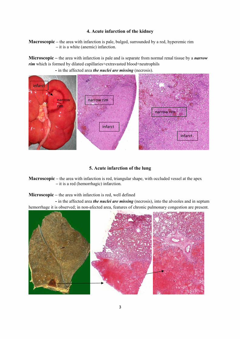
4. Acute infarction of the kidney
Macroscopic – the area with infarction is pale, bulged, surrounded by a red, hyperemic rim – it is a white (anemic) infarction.
Microscopic – the area with infarction is pale and is separate from normal renal tissue by a narrow rim which is formed by dilated capillaries+extravasted blood+neutrophils - in the affected area the nuclei are missing (necrosis).
5. Acute infarction of the lung
Macroscopic – the area with infarction is red, triangular shape, with occluded vessel at the apex – it is a red (hemorrhagic) infarction.
Microscopic – the area with infarction is red, well defined - in the affected area the nuclei are missing (necrosis), into the alveoles and in septum hemorrhage it is observed; in non-afected area, features of chronic pulmonary congestion are present.
narrow rim
narrow rim narrow rim
infarct
infarct
infarct

4
6. Mixed thrombus
Macroscopic – it is formed in living body - friable, adherent by the vessel wall Microscopic – it is located into the lumen of vessels - an alternance between pink (platelets) and red colored areas (erythrocytes)
5
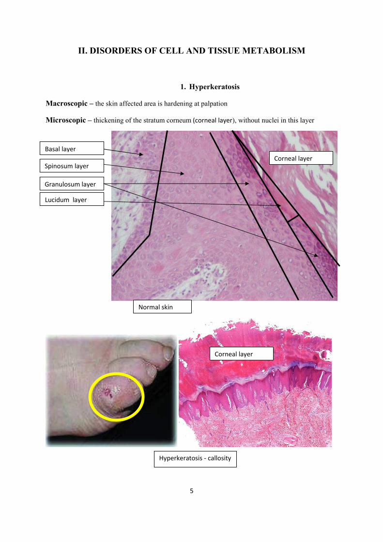
II. DISORDERS OF CELL AND TISSUE METABOLISM
1. Hyperkeratosis
Macroscopic – the skin affected area is hardening at palpation
Microscopic – thickening of the stratum corneum (corneal layer), without nuclei in this layer
Normal skin
Hyperkeratosis - callosity
Basal layer
Spinosum layer
Granulosum layer
Lucidum layer
Corneal layer
Corneal layer

6
2. Fatty liver (steatosis)
Macroscopic – the liver is enlarged, yellow, soft, greasy, friable
Microscopic – clear vacuoles (fat globules) within hepatocytes – the whole surface of hepatic lobules is affected (central and peripheral area)
3. Renal amyloidosis
Macroscopic – the kidney are enlarged, pale-shinny-glassy (on section)
Microscopic – accumulation of amyloid within the extracellular space of glomerules - in Hematoxylin-Eosine the amyloid is pink - with Congo Red dye the amyloid becomes orange
Hematoxylin-Eosine Congo red dye

7
III. INFLAMMATION
1. Fibrinous pericarditis
Macroscopic – villous membrane on the epicardial surface (“bread and butter” appearance) Microscopic – pink fibrin deposits on the epicardial surface
2. Hepatic abscess
Macroscopic – a localized cavity with pus and necrotic tissue within liver parenchyma Microscopic – into the cavity numerous neutrophils can be seen – around the cavity, the necrosis can be observed (acute abscess)
myocardium
epicardium
fibrin membrane
normal hepatocytes
portal space abscess

8
3. Foreign body granuloma
Macroscopic – a small palpable and well-defined nodule within breast Microscopic – the crystals of cholesterol are precipitated within the breast – elongated clear spaces surrounded by giant multinucleated cells (foreign body giant cells)
4. Tuberculous lymphadenitis
Macroscopic – the lymph node is increase in size (lymphadenopathy) – on section, yellow-white cheese-like necrosis (caseous necrosis) Microscopic – mixed tubercles within the lymph node – central caseous necrosis surrounded by palisaded epitheloid cells (elongated, uninucleated) and multinucleated Langhans’ cells with horeseshoe arrangement of peripheral nuclei; at the periphery - lymphocytes
granuloma
normal breast
cholesterol crystals foreign body giant cells
Caseous necrosis
epitheloid cells
Langhans’cell lymphocytes

9
IV. CONGENITAL MALFORMATIONS
Embryopathies = malformations which occur in first two months (8 weeks) Fetopathies = malformations which occur in first two months of the intrauterine life Inhibition of development Aplasia = lack of an organ development during embrionic life Agenesis = lack of an organ and its embriological structures Hypoplasia = insuficiency development of an organ during embriogenesis Atresia = lack of one lumen (ex: billiary atresia) Stenosis = narrowing of a lumen Excess of development Polydactyly = one or more extradigits Polymastia = accesory breasts Polythelia = extra nipples Macrocephaly = abnormally large head Macroglossia = enlargement of the tongue Development of organs in abnormal places Dystopia = abnormal position of an organ or a body part Heterotopia = the presence of a tissue in an abnormal location Situs inversus = right to left reversal of the thoracic and abdominal organs

10
Malformations of the cranium and brain Anencephaly = congenital absence of the brain Acrania = congenital absence of the cranium Exencephaly = the brain is located outside of the skull Cranioschisis = incomplete closure of the skull (schixo, gr. = split) Meningocele = protrusion of the meninges through a defect in the cranium or vertebral column Myelocele = protrusion of the spinal cord through a defect in the cranium or vertebral column Malformations of the face and mouth Cheiloschisis (cleft lip) = congenital cleft in the middle of the upper lip (cheilos, gr. = lip) Gnathoschisis (cleft jaw) = congenital cleft of the jaw (gnathos, gr. = jaw) Palatoschisis (cleft palate) = congenital cleft of the palate (roof of the mouth) Prosoposchisis = congenital cleft of the face (prosopon, gr. = face) Macrognathia = abnormally large jaws Agnathia = lack of the lower jaw Synotia = fusion of the lobe of the ears Cyclopia = presence of one eye Aprosopia = lack of the face Anophtalmia = lack of one or both eyes Malformations of the neck Branchial cyst = a fluid cavity that develops under the skin, in the cervical area Congenital hygroma = fluid accumulation in the soft tissue of the neck Malformations of the vertebral column Rachischisis = incomplete closure of the vertebral bone and the covered skin Spina bifida = the vertebral defect is covered by the skin Malformations of the limbs Syndactyly = fusion of fingers Oligodactyly = lack of one or more fingers Amelia = lack of all four limbs Abrachius = lack of both upper limbs Monobrachius = absence of one of the upper limbs Apus = lack of both lower limbs Phocomelia = congenital absence of the proximal portion of a limb; the hands are attached to abbreviated arms Sympus = fusion of the lower extremities Malformations of the twins Holoacardius = an unequal twin fetus which the heart is entirely absent Fetus papiraceus = mummification and compression of one of the twins Conjoined twins= Siamese twins = twins whose bodies are joined in utero Thoracopagus = conjoined twins in thorax region Pygopagus = conjoined twins in the sacral region (pygos, gr. = posterior) Dicephalic parapagus = one trunk and two heads

11
V. BENIGN TUMORS
1. Mucinous cystadenoma of the ovary
Macroscopic – uni- or multilocular cystic spaces, filled with gelatinous fluid rich in glycoproteins (mucin); the tumor is encapsulated, well defined, usually very large size Microscopic – the wall of the cysts are lined by tall columnar epithelium with apical mucin; the nuclei are basally located and are arranged on a single layer; no atypical cells are observed Immunohistochemistry – the tumor cells are marked by Cytokeratin
2. Fibroadenoma of the breast
Macroscopic – a well circumscribed, mobile and palpable mass, which especially occurs in young women; on section, grayish lobulated white nodule that bulge above the surrounding tissue and present slit like spaces Microscopic – proliferation of mammary ducts and connective tissue; depends on the growth manner, there are two histological types, without clinical significance a) pericanalicular fibroadenoma – proliferation of fibroblasts around ducts in a circumferential fashion b) intracanalicular fibroadenoma – the proliferating fibroblasts compress the ducts into clefts Immunohistochemistry – ductal cells are marked by Cytokeratin and fibroblast by Vimentin
pericanalicular fibroadenoma intracanalicular fibroadenoma

12
3. Leiomyoma of the uterine corpus
Macroscopic – well defined nodules, typically multiple, spherical and firm, without capsule, which bulge above the surrounding myometrium, from which they are easily shelled out; on section, white nodules with trabecular structure Microscopic – proliferation of smooth muscle cells, which are fusiform shape and form anastomosing fascicles; no atypical mitoses are observed Immunohistochemistry – the tumor cells are marked by Vimentin and Smooth Muscle Antigen (SMA)
4. Cavernous haemangioma of the liver
Macroscopic – ill defined red-blue, soft, spongy mass; on section, it contains venous blood Microscopic – proliferation of large (cavernous) vascular spaces, partly filled with blood; the tumor is sharply defined by the surrounding hepatocytes, but is not encapsulated Immunohistochemistry – the tumor cells are marked by CD31 and CD34, which are specifical antibodies (CD = Clusters of Differentiation) for blood cells
normal myometrium
leiomyoma
normal hepatocytes
haemangioma

13
VI. MALIGNANT TUMORS
1. Invasive squamos cell carcinoma of the skin, well differentiated
Macroscopic – a nodular lesion which can be ulcerated and can present hyperkeratosis; it is usually located on the skin of the face or the lip and also on the tongue Microscopic – in epidermis and dermis it is observed a proliferation of atypical cells which resemble the cells of the spinosum layer of the skin; the tumor cells are arranged in clusters and exhibit large zones of keratinization; they have a polygonal shape, large and atypical nuclei Immunohistochemistry – the tumor cells are marked by Cytokeratin
2. Invasive basal cell carcinoma of the skin
Macroscopic – a nodular lesion which is usually located on the skin of the face; it can be ulcerated or can be an erythematous plaque; it does not occur on mucosal surfaces Microscopic – in epidermis and dermis the tumor cells are arranged in small islands; the cells resemble those of the basal layer of the skin; they are small, atypical and round shape cells; the cells from the periphery of the islands tend to present a palisading arrangment (they are arranged radially with their long axes in parallel alignment) Immunohistochemistry – the tumor cells are marked by Cytokeratin
keratinization
palisading arrangement

14
3. Well-differentiated (grade 1) colorectal adenocarcinoma
Macroscopic – the malignant tumor can have the following aspects: polypoid - vegetant, ulcerated or infiltrative Microscopic – proliferation of atypical glands which infiltrate the mucosa, submucosa and muscularis layers of the colon wall Immunohistochemistry – the tumor cells are marked by Cytokeratin
4. Hodgkin’s lymphoma – nodular sclerosis
Macroscopic – enlarged lymph nodes, with a smooth surface; the cervical and axillary lymph nodes are first affected structures; on section: homogeneously white aspect, sometimes nodular aspect and fibrous septa Microscopic – the normal arhitecture of affected lymph nodes is replaced by cellular nodules which are separated by bands of collagen; within the cellular nodes the mixed cell infiltrate is composed by: Reed Sternberg cells - lacunar cell variant (uninucleated cells with large nuclei and prominent nucleoli and lipid-rich cytoplasm, with clear appearance in Hematoxilin-Eozine), Hodgkin’s cells (large uninucleated cells) and lymphocytes Immunohistochemistry – the Reed-Sternberg cells are marked by CD15 and CD30
ulcero-infiltrative tumor
collagen bands Reed-Sternberg cells

15
SYSTEMIC PATHOLOGY
-practical course-
I. PATHOLOGY OF THE HEART
1. Acute myocardial infarction
Macroscopic – the infarcted area is pale-yellow (1), surrounded by a hyperemic narrow rim (2)
Microscopic – 1. the myocardial fibers from the infarcted area – intense eosinophilic, without nuclei 2. the narrow rim – neutrophils + dilated capillaries 3. the normal myocardium – myocardial fibers with nuclei
1
2
3
3
2
1

16
2. Scarring myocardial infarction
Macroscopic – the infarcted area is hard and white, the normal myocardium is brown
Microscopic – the infarcted area – connective tissue (scar) – the normal myocardium – myocardial fibers with nuclei
3. Macroscopical lesions
1. Pericardial petechiae 2. Fibrinous pericarditis 3. Acute intramural myocardial infarction 4. Concentric hypertrophy of the left ventricles 5. Cardiac atrophy 6. Cardiac metastases from lung tumors 7. Atrial septal defect 8. Ventricular septal defect
scarring infarcted area
normal myocardium

17
II. PATHOLOGY OF THE HEART AND BLOOD VESSELS
1. Atherosclerosis - atheromas
Macroscopic - the intimal layer is covered by round, yellow and friable elevated plaques - on section, it can be seen a yellow friable substance Microscopic - the plaques are located in the arterial intima and are covered by endothelium - they are composed by cholesterol (elongated clear spaces) and cellular debris
2. Coronarosclerosis – hyaline plaques
Macroscopic - the lumen of coronary artery is narrowed by white hard and elevated plaques which can present rupture and can lead to formation of thrombi
Microscopic - the lumen of coronary artery is narrowed - the plaques are composed by a glassy-like substance (hyaline)
endothelium
atheroma intima
media
adventitia
myocardium coronary artery

18
3. Hemorrhoids
Macroscopic - the hemorrhoidal veins are dilated and elongated (varicose veins) Microscopic - the veins located within inferior rectum and anal canal are dilated - within venous lumen red thrombi can be observed
4. Macroscopical lesions
1. Infective endocarditis 2. Libman-Sacks endocarditis 3. Mitral stenosis and mitral incompetence 4. Aortic dissection 5. Aortic aneurysm 6. Atherosclerosis 7. Coronarosclerosis
rectum
anal canal
varicose veins
rectum
anal canal

19
III. PATHOLOGY OF THE RESPIRATORY SYSTEM
1. Bronchopneumonia
Macroscopic - enlarged grey-red lungs; the affected areas are congested and friable, the normal areas are pale, without friability („spotted lung”) . After formaline fixation the affected areas became yellow-grey.
Microscopic - neutrophils within alveolar lumen, dilated capillaries in alveolar septa - the pulmonary edema is associated.
2. Lung emphysema
Macroscopic – overdistended lungs, with spongy aspect on section
Microscopic – enlarged alveolar spaces, thinning alveolar septa.

20
3. Respiratory distress syndrome (Hyaline membrane disease)
Macroscopic - heavy dystelectasic lungs
Microscopic - the alveoli are linning by a thickening pink membranes (hyaline membranes)
4. Macroscopical lesions
1. Laryngeal obstruction by food 2. Laryngeal papillomatosis 3. Laryngeal carcinoma 4. Lung infarction 5. Bronchopneumonia 6. Lobar pneumonia 7. Hemorrhagic pneumonia 8. Lung emphysema 9. Bullos emphysema 10. Interstitial emphysema 11. Lung gangrene

21
5. Miliary tuberculosis of the lung
Macroscopic - small diffuse yellow-white nodes within lung parenchyma - on section, yellow-white cheese-like necrosis (caseous necrosis) Microscopic - caseous tubercles – expanded central caseous necrosis surrounded by epitheloid cells (elongated, uninucleated) and multinucleated Langhans’ cells with horeseshoe arrangement of peripheral nuclei; at the periphery - lymphocytes
6. Poorly-differentiated squamous cell carcinoma of the lung
Macroscopic – a nodular poorly-defined tumoral lesion which is usually located within the central part of the lung (hilum); sometimes, it can show cavitation Microscopic – a proliferation of atypical cells which resemble the cells of the spinosum layer of the skin; the tumor cells are arranged in clusters and do not exhibit keratinization; they have a polygonal shape, large and atypical nuclei
caseous necrosis
epitheloid cells
Langhans’ cells
medium-sized bronchi with cartilage
tumor

22
7. Small cell lung carcinoma (SCLC)
Macroscopic – perihilar soft, poorly-defined masses, with necrosis Microscopic – a proliferation of atypical small cells; the tumor cells present a diffuse arrangement (sheet-like growth); they are round-shape cells and measure less than the size of three small lymphocytes in diameter; the nuclear-to-cytoplasm ratio is high, the mitotic rate is increased
8. Macroscopical lesions
Lung tuberculosis: 1. Lung phtisis 2. Lung miliary tuberculosis
3. Lung apical cavities Lung tumors: 1. Perihilar (central) tumors 2. Peripheral tumors 3. Lung metastases (secondary tumor)
perihilar tumor

23
IV. PATHOLOGY OF THE GASTROINTESTINAL SYSTEM
1. Chronic hypertrophic and superficial gastritis
Macroscopic – sweling mucosa and thickening of the gastric folds Microscopic - lymphocytes and lymphoid structures with germinal centers within mucosa (M) - focal glandular hyperplasia in the glandular layer - no disorders of the foveolar layer, no inflammatory infiltrate in submucosa (SM)
2. Chronic peptic ulcer
Macroscopic – round sharply punched-out defect with straight walls; the margins are not ulcerated or elevated, the base is smooth and clean; the surrounding mucosal folds radiate from the ulcer in spokelike fashion (scarring) Microscopic – the chronic ulcer involves the mucosa, submucosa and muscularis; in the active ulcers four layers are observed: 1. inflammatory exudate (on the luminal surface); 2. fibrinoid necrosis; 3. granulation tissue; 4. fibrous scar (in the basis of the ulcer).
M SM
M
SM
1 2
3
4

24
3. Well-differentiated gastric adenocarcinoma
Macroscopic – an erosive crater is present within the gastric mucosa (ulcerated tumor); in contrast with the chronic ulcer, the margins are ulcerated and elevated, the base is ulcerated, with tissue debris and the surrounding mucosal folds do not radiate from the ulcer Microscopic – atypical well differentiated glands which infiltrate the mucosa, submucosa, muscularis and subserosa; one of the lymph nodes presents metastases
4. Macroscopical lesions
1. Necrotizing tonsillitis 2. Congenital pyloric stenosis 3. Chronic peptic ulcer 4. Gastric malignant tumors: polyp with malignant transformation
polypoid tumor ulcerated tumor infiltrative tumor.
lymph node with metastases
M
SM
muscularis
subserosa

25
V. PATHOLOGY OF THE INTESTINES, LIVER AND PANCREAS
1. Macroscopical lesions – intestines
1. Tuberculosis of the small intestine 2. Pedunculated polyps 3. Sessile polyps 4. Polyposis 5. Leiomyomas of the small intestine 6. Lymphomas of the small intestine 7. Adenocarcinoma of the large intestine
2. Liver cirrhosis
Macroscopic - hard liver with a diffuse nodular surface - on section, irregulary sized yellow nodules separated by scar areas
Microscopic - irregular nodules composed by hepatocytes, without centrolobular veins - the regenerative nodules are separated by large bands of connective tissue - within connective bands, on the periphery of nodules, small bile pseudo-ducts
regenerative nodules bile pseudo-ducts

26
3. Acute pancreatitis
Macroscopic – fat necrosis (yellow) in the pancreatic and peripancreatic fats + hemorrhages Microscopic – fat necrosis (soaps) are pink homogenous areas, without nuclei.
4. Macroscopical lesions – liver and pancreas
1. Amyloidosis of the spleen, liver and kidneys 2. Haemangioma of the liver 3. Liver cirrhosis 4. Esophageal and diaphragmatic varices 5. Colecystitis with stones (calculi) 6. Acute pancreatitis 7. Liver abscesses.
fat necrosis
fat necrosis
hemorrhages

27
VI. PATHOLOGY OF THE KIDNEY AND URINARY TRACT
1. Crescentic glomerulonephritis (Rapidly progressive glomerulonephritis)
Macroscopic –increasing size of the kidneys - minute hemorrhages on the kidney surface Microscopic – proliferation of the parietal epithelium of the Bowman’s capsule - thickening of the Bowman’s capsule (crescent-shaped masses) - compression of the capillaries - droplets of hyaline in the lumen of the tubules
Crescent - shaped masses
hyaline

28
2. Clear cell carcinoma of the kidney (Renal cell carcinoma, Grawitz tumor)
Macroscopic – the yellowish poorly-defined tumor is localized in the apex of the kidney Microscopic – clusters of atypical cells with clear cytoplasm
3. Macroscopical lesions
Kidney
1. Polycystic kidney 2. Renal infarction – acute and scarring infarction 3. Arteriosclerosis 4. Arteriolosclerosis 5. Shock kidney with and without DIC 6. Renal stones 7. Hydronephrosis 8. Glomerulonephritis 9. Grawitz tumor
Urinary bladder
1. Fibrinous cystitis 2. Ulcerated malignant tumor
Normal kidney
tumor

29
VII. PATHOLOGY OF THE FEMALE GENITAL SYSTEM AND THE BREAST
1. Invasive squamos cell carcinoma of the cervix, well differentiated
Macroscopic – an ulcerated lesion, an exophytic tumor which can protrude into vagina or an infiltrative tumor Microscopic – a proliferation of atypical cells which resemble the cells of the spinosum layer of the skin; the tumor cells are arranged in clusters and exhibit large zones of keratinization; they have a polygonal shape, large and atypical nuclei; they arise in the transformation zone and may infiltrate the endo- and exocervix.
2. Endometrial carcinoma
Macroscopic – an ulcerated, exophytic or infiltrative tumor Microscopic – a proliferation of atypical glands which infiltrate the myometrium
vagina
cervical tumor
uterine body
ovaries and fallopian tubes
exocervix (squamous epithelium)
endocervix (tumor)
exophytic tumor in uterine body
cervix

30
3. Fibrocystic change of the breast
Macroscopic – ill-defined thickening in the breast, smallest than 1 cm, painfully during menstruation Microscopic – cystic ducts dilatations, fibrosis, epithelial proliferation and apocrine metaplasia. It is not a tumor process.
4. Macroscopically lesions
Uterus
1. Leiomyomas 2. Cervical cancer 3. Endometrial cancer Ovaries 1. Polycystic ovaries 2. Cystadenoma – serous and mucinous 3. Teratoma (dermoid cyst)
Breast
1. Non-mucinos infiltrative tumor 2. Mucinous infiltrative tumor
epithelial proliferation
apocrine metaplasia
cystic ducts dilatations
fibrosis

31
VIII. PATHOLOGY OF THE MALE GENITAL SYSTEM
1. Nodular (benign) prostatic hyperplasia
Macroscopic – enlarged prostate, with nodular aspect on section; the urethra is compressed and the urinary bladder is hypertrophic Microscopic – a nodular proliferation of glands, surrounded by muscle fibers; the glands are dilated, without atypia.
urinary bladder

32
2. Seminoma
Macroscopic – enlarged testis with a homogenous, gray-white cut surface devoid of haemorrhage and necrosis Microscopic – sheets and nests of atypical spermatocyte-like cells separated by thin fibrous septa
3. Macroscopically lesions
1. Nodular prostatic hyperplasia 2. Hydrocele 3. Tuberculosis of the testis and epididymis 4. Seminoma 5. Immature teratoma of the testis 6. Carcinoma of the penis

33
IX. PATHOLOGY OF THE THYROID GLAND
1. Nodular goitre
Macroscopic – enlarged thyroid gland Microscopic – a nodular hyperplasia of the thyroidal colloid-filled follicles

34
2. Hashimoto’s thyroiditis
Macroscopic – the thyroid gland may be enlarged or atrophic Microscopic – the gland is infiltrated by lymphocytes with lymphoid follicle formation
3. Macroscopically lesions
Lesions of thyroid gland Goitre
Lesions of nervous system
Brain abscess Meningitis Meningeoma Glioblastoma
Lesions of bone
Osteosarcoma

35
Macroscopically lesions for the Practical Exam
1. Fibrinous pericarditis 2. Acute intramural myocardial infarction 3. Aortic dissection 4. Atherosclerosis 5. Lung infarction 6. Bronchopneumonia 7. Lobar pneumonia 8. Lung miliary tuberculosis 9. Lung apical cavities in tuberculosis 10. Perihilar (central) tumor of the lung 11. Lung metastases (secondary tumor) 12. Chronic peptic ulcer (stomach) 13. Infiltrative gastric malignant tumor 14. Sessile polyp of the intestine 15. Amyloidosis of the spleen, liver and kidneys 16. Liver cirrhosis 17. Acute pancreatitis 18. Haemangioma of the liver 19. Renal stones 20. Hydronephrosis 21. Grawitz tumor 22. Uterine leiomyomas 23. Cervical cancer 24. Endometrial cancer 25. Ovarian mucinous cystadenoma 26. Ovarian teratoma (dermoid cyst) 27. Infiltrative breast tumor 28. Nodular prostatic hyperplasia 29. Seminoma 30. Meningitis 31. Meningeoma 32. Glioblastoma 33. Osteosarcoma
36
Microscopically lesions for the Practical Exam
1. Acute pulmonary edema 2. Acute infarction of the kidney 3. Acute infarction of the lung 4. Fatty liver (steatosis) 5. Renal amyloidosis 6. Fibrinous pericarditis 7. Foreign body granuloma 8. Tuberculous lymphadenitis 9. Fibroadenoma of the breast 10. Leiomyoma of the uterine body 11. Cavernous haemangioma of the liver 12. Invasive squamos cell carcinoma of the skin, well differentiated 13. Invasive basal cell carcinoma of the skin 14. Well-differentiated (grade 1) colorectal adenocarcinoma 15. Acute myocardial infarction 16. Atherosclerosis – atheromas 17. Coronarosclerosis – hyaline plaques 18. Bronchopneumonia 19. Respiratory distress syndrome (Hyaline membrane disease) 20. Miliary tuberculosis of the lung 21. Poorly-differentiated squamous cell carcinoma of the lung 22. Chronic peptic ulcer 23. Well-differentiated gastric adenocarcinoma 24. Liver cirrhosis 25. Crescentic glomerulonephritis (Rapidly progressive glomerulonephritis) 26. Clear cell carcinoma of the kidney (Renal cell carcinoma, Grawitz tumor) 27. Invasive squamos cell carcinoma of the cervix, well differentiated 28. Endometrial carcinoma 29. Fibrocystic change of the breast 30. Nodular (benign) prostatic hyperplasia 31. Seminoma 32. Nodular goitre 33. Hashimoto’s thyroiditis