Embed Size (px)
Citation preview
More Evidence Supports Crevice Concept Soret shifts indicate that hemoglobins and other porphyrins have crevice, not plate structure
Shifts in the Soret peak in the absorption spectra of substituted hemoglobins and other porphyrin compounds give additional evidence that hemoglobin has a "crevice" rather than a "plate" structure, Dr. Alsoph H. Corwin of Johns Hopkins University, Baltimore, Md., told the ACS Southeastern Regional Meeting, at Charlotte, N.C. Although much work has been done in determining the amino acid sequence of the hemoglobin molecule (C&EN, Oct. 21, page 42) , the question of just how the porphyrin portion, heme, is attached hasn't been setttled conclusively.
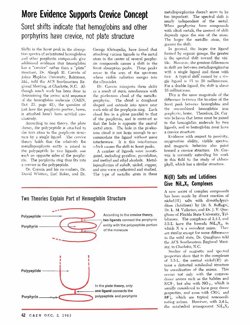
According to one theory, the plate theory, the polypeptide is attached to the iron atom in the porphyrin structure by a single ligand. The crevice theory holds that the relatively flat metalloporphyrin entity is joined to the polypeptide by two ligands, one each on opposite sides of the porphyrin. The porphyrin ring thus fits into a crevice in the polypeptide.
Dr. Corwin and his co-workers, Dr. David Whitten, Earl Baker, and Dr.
George Kleinspehn, have found that attaching various ligands to the metal atom in the center of several porphyrin compounds causes a shift in the Soret absorption peaks. These peaks occur in the area of the spectrum where visible radiation merges into the ultraviolet.
Dr. Corwin interprets these shifts as a result of steric interference with the pi-electron cloud of the metalloporphyrin. The cloud is doughnut shaped and extends into space near each side of the porphyrin ring. Each cloud lies in a plane parallel to that of the porphyrin, and is centered so that the hole is opposite the central metal atom. The hole in the pi-electron cloud is not large enough to accommodate the ligand without some interference. It is this interference which causes the shift in Soret peaks.
A number of ligands were investigated, including pyridine, pyrrolidine, and methyl and ethyl alcohols. Metal-loporphyrins of cobalt, nickel, copper, and zinc were synthesized and studied. The type of metallic atom in these
metalloporphyrins doesn't seem to be too important. The spectral shift is nearly independent of the metal. When porphyrins form compounds with alkali metals, the amount of shift depends upon the size of the atom. The larger the metallic atom, the greater the shift.
In general, the larger the ligand formed by organic groups, the greater is the spectral shift toward the visible. However, the greatest differences in shift are between metalloporphyrins with a single ligand and those with two. A typical shift caused by a single ligand is 15 to 20 millimicrons. For a double ligand, the shift is about 35 millimicrons.
This is the same magnitude of the difference between the location of the Soret peak between hemoglobin and the uncombined hemoglobin iron porphyrin, heme. Therefore, Dr. Corwin believes that heme must be joined to the hemoglobin molecule by two ligands, and so hemoglobin must have a crevice structure.
Evidence with respect to passivity, oxygenation ability, visible spectra, and magnetic behavior also point toward a crevice structure. Dr. Corwin is currently extending his work in this field to the study of chlorophyll, which has a similar structure.
Ni(ll) Salts and Lutidines Give N i L X Complexes A new series of complex compounds has been made by direct reaction of nickel (II) salts with dimethylpyri-dines (lutidines) by Dr. S. Buffagni, Dr. L. M. Vallarino, and Dr. J. V. Qua-gliano of Florida State University, Tallahassee. The complexes of 3,4-L and 3,5-L have the formula NiL4X2, in which X is a univalent anion. They are similar except for some differences in the solid state, Dr. Quagliano told the ACS Southeastern Regional Meeting, in Charlotte, N.C.
Studies of magnetic and spectral properties show that in the complexes of 3,5-L, the central nickel (II) attains a distorted octahedral structure by coordination of the anions. This occurs not only with the common donor anions such as the halides and NCS-, but also with N 0 3 j which is usually considered to have poor donor properties, and even with CIO 4 and BF4, which are typical noncoordi-nating anions. However, with 3,4-L, the octahedral arrangement NiL4X2
Two Theories Explain Part of Hemoglobin Structure
Polypeptide
Porphyrin
According to the crevice theory,
two ligands connect the porphyrin
entity with the polypeptide portion
of the molecule
Polypeptide
Porphyrin
In the plate theory, only
one ligand connects the
polypeptide and porphyrin
42 C & E N DEC. 2, 1963