Embed Size (px)
Citation preview

J.P. Glusker. 1994. X-ray crystallography of proteins Methods Biochem. Anal. 37: 1-72. (PubMed)
J.P. Wery and R.W. Schevitz. 1997. New trends in macromolecular x-ray crystallography Curr. Opin. Chem. Biol. 1: 365-369. (PubMed)
A.T. Brunger. 1997. X-ray crystallography and NMR reveal complementary views of structure and dynamics Nat. Struct. Biol. 4 (suppl.): 862-865. (PubMed)
K. Wüthrich. 1989. Protein structure determination in solution by nuclear magnetic resonance spectroscopy Science 243: 45-50. (PubMed)
G.M. Clore and A.M. Gronenborn. 1991. Structures of larger proteins in solution: Three- and four-dimensional heteronuclear NMR spectroscopy Science 252: 1390-1399. (PubMed)
Wüthrich, K., 1986. NMR of Proteins and Nucleic Acids. WileyInterscience.
Monoclonal antibodies and fluorescent molecules
G. Köhler and C. Milstein. 1975. Continuous cultures of fused cells secreting antibody of predefined specificity Nature 256: 495-497. (PubMed)
Goding, J. W., 1996. Monoclonal Antibodies: Principles and Practice. Academic Press.
Immunology Today, 2000. Volume 21, issue 8.
R.Y. Tsien. 1998. The green fluorescent protein Annu. Rev. Biochem. 67: 509-544. (PubMed)
J.M. Kendall and M.N. Badminton. 1998. Aequorea victoria bioluminescence moves into an exciting era Trends Biotechnol. 16: 216-234. (PubMed)
Chemical synthesis of proteins
K.H. Mayo. 2000. Recent advances in the design and construction of synthetic peptides: For the love of basics or just for the technology of it Trends Biotechnol. 18: 212-217. (PubMed)
J.A. Borgia and G.B. Fields. 2000. Chemical synthesis of proteins Trends Biotechnol. 18: 243-251. (PubMed)
I. The Molecular Design of Life
5. DNA, RNA, and the Flow of Genetic Information
DNA and RNA are long linear polymers, called nucleic acids, that carry information in a form that can be passed from one generation to the next. These macromolecules consist of a large number of linked nucleotides, each composed of a sugar, a phosphate, and a base. Sugars linked by phosphates form a common backbone, whereas the bases vary among four kinds. Genetic information is stored in the sequence of bases along a nucleic acid chain. The bases have an additional special property: they form specific pairs with one another that are stabilized by hydrogen bonds. The base pairing results in the formation of a double helix, a helical structure consisting of two strands. These base pairs provide a mechanism for copying the genetic information in an existing nucleic acid chain to form a new chain. Although RNA probably functioned as the genetic material very early in evolutionary history, the genes of all modern cells and many viruses are made of DNA. DNA is replicated by the action of DNA polymerase enzymes. These exquisitely specific enzymes copy sequences from nucleic acid templates with an error rate of less than 1 in 100 million nucleotides.
Genes specify the kinds of proteins that are made by cells, but DNA is not the direct template for protein synthesis. Rather, the templates for protein synthesis are RNA (ribonucleic acid) molecules. In particular, a class of RNA molecules called messenger RNA (mRNA) are the information-carrying intermediates in protein synthesis. Other RNA

molecules, such as transfer RNA (tRNA) and ribosomal RNA (rRNA), are part of the protein-synthesizing machinery. All forms of cellular RNA are synthesized by RNA polymerases that take instructions from DNA templates. This process of transcription is followed by translation, the synthesis of proteins according to instructions given by mRNA templates. Thus, the flow of genetic information, or gene expression, in normal cells is:
This flow of information is dependent on the genetic code, which defines the relation between the sequence of bases in DNA (or its mRNA transcript) and the sequence of amino acids in a protein. The code is nearly the same in all organisms: a sequence of three bases, called a codon, specifies an amino acid. Codons in mRNA are read sequentially by tRNA molecules, which serve as adaptors in protein synthesis. Protein synthesis takes place on ribosomes, which are complex assemblies of rRNAs and more than 50 kinds of proteins.
The last theme to be considered is the interrupted character of most eukaryotic genes, which are mosaics of nucleic acid sequences called introns and exons. Both are transcribed, but introns are cut out of newly synthesized RNA molecules, leaving mature RNA molecules with continuous exons. The existence of introns and exons has crucial implications for the evolution of proteins.
I. The Molecular Design of Life 5. DNA, RNA, and the Flow of Genetic Information

Having genes in common accounts for the resemblance of a mother and her daughters. Genes must be expressed to exert an effect, and proteins regulate such expression. One such regulatory protein, a zinc-finger protein (zinc ion is blue, protein is red), is shown bound to a control or promoter region of DNA (black). [Barnaby Hall/Photonica.] I. The Molecular Design of Life 5. DNA, RNA, and the Flow of Genetic Information
5.1. A Nucleic Acid Consists of Four Kinds of Bases Linked to a Sugar-Phosphate Backbone
The nucleic acids DNA and RNA are well suited to function as the carriers of genetic information by virtue of their covalent structures. These macromolecules are linear polymers built up from similar units connected end to end (Figure 5.1). Each monomer unit within the polymer consists of three components: a sugar, a phosphate, and a base. The sequence of bases uniquely characterizes a nucleic acid and represents a form of linear information.
5.1.1. RNA and DNA Differ in the Sugar Component and One of the Bases
The sugar in deoxyribonucleic acid (DNA) is deoxyribose. The deoxy prefix indicates that the 2 carbon atom of the
sugar lacks the oxygen atom that is linked to the 2 carbon atom of ribose (the sugar in ribonucleic acid, or RNA), as
shown in Figure 5.2. The sugars in nucleic acids are linked to one another by phosphodiester bridges. Specifically, the 3 -
hydroxyl (3 -OH) group of the sugar moiety of one nucleotide is esterified to a phosphate group, which is, in turn, joined
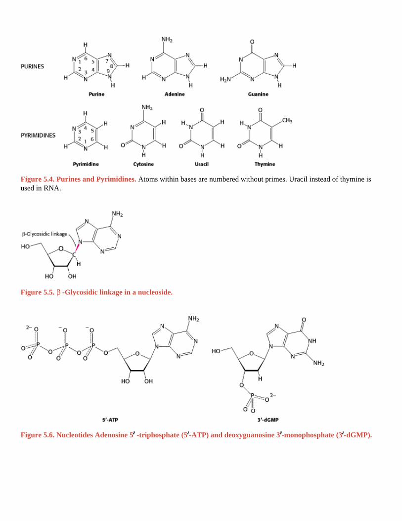
to the 5 -hydroxyl group of the adjacent sugar. The chain of sugars linked by phosphodiester bridges is referred to as the backbone of the nucleic acid (Figure 5.3). Whereas the backbone is constant in DNA and RNA, the bases vary from one monomer to the next. Two of the bases are derivatives of purine adenine (A) and guanine (G) and two of pyrimidine
cytosine (C) and thymine (T, DNA only) or uracil (U, RNA only), as shown in Figure 5.4.
RNA, like DNA, is a long unbranched polymer consisting of nucleotides joined by 3 5 phosphodiester bonds (see Figure 5.3). The covalent structure of RNA differs from that of DNA in two respects. As stated earlier and as indicated
by its name, the sugar units in RNA are riboses rather than deoxyriboses. Ribose contains a 2 -hydroxyl group not
present in deoxyribose. As a consequence, in addition to the standard 3 5 linkage, a 2 5 linkage is possible for RNA. This later linkage is important in the removal of introns and the joining of exons for the formation of mature RNA (Section 28.3.4). The other difference, as already mentioned, is that one of the four major bases in RNA is uracil (U) instead of thymine (T).
Note that each phosphodiester bridge has a negative charge. This negative charge repels nucleophilic species such as hydroxide ion; consequently, phosphodiester linkages are much less susceptible to hydrolytic attack than are other esters such as carboxylic acid esters. This resistance is crucial for maintaining the integrity of information stored in nucleic
acids. The absence of the 2 -hydroxyl group in DNA further increases its resistance to hydrolysis. The greater stability of DNA probably accounts for its use rather than RNA as the hereditary material in all modern cells and in many viruses.

5.1.2. Nucleotides Are the Monomeric Units of Nucleic Acids
Structural Insights, Nucleic Acids offers a three-dimensional perspective on nucleotide structure, base pairing, and other aspects of DNA and RNA structure.
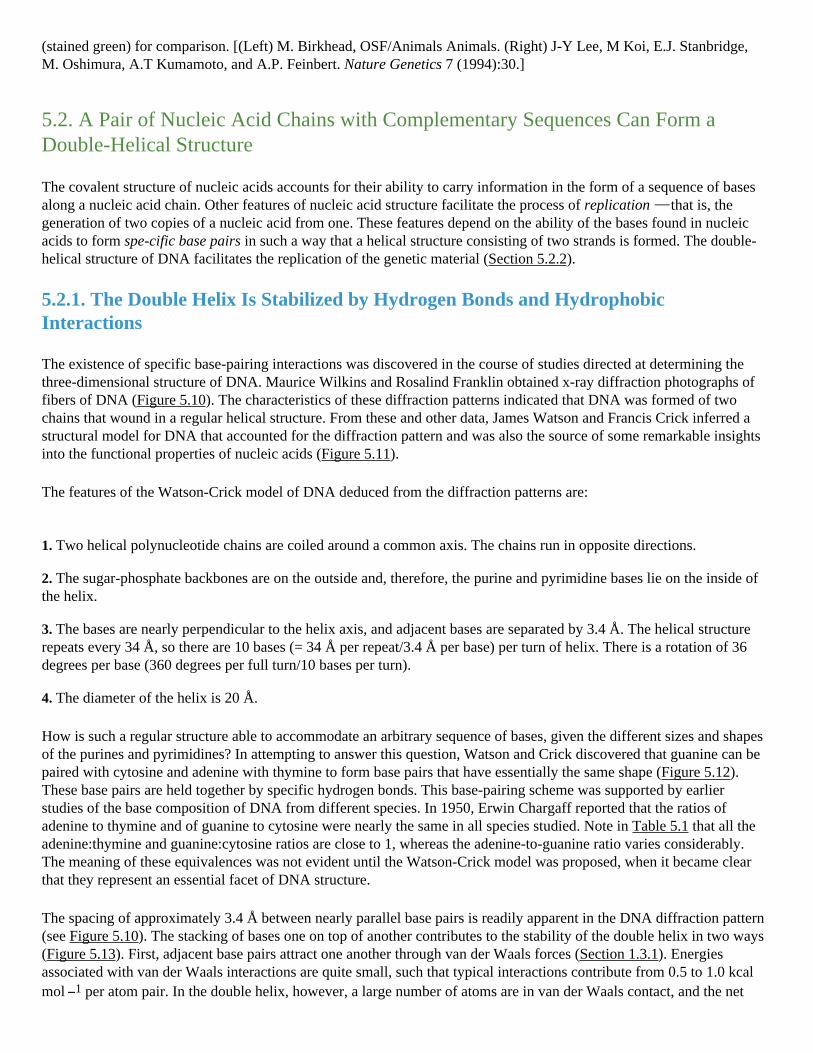
A unit consisting of a base bonded to a sugar is referred to as a nucleoside . The four nucleoside units in RNA are called adenosine, guanosine, cytidine, and uridine, whereas those in DNA are called deoxyadenosine, deoxyguanosine,
deoxycytidine, and thymidine. In each case, N-9 of a purine or N-1 of a pyrimidine is attached to C-1 of the sugar (Figure 5.5). The base lies above the plane of sugar when the structure is written in the standard orientation; that is, the configuration of the N-glycosidic linkage is β . A nucleotide is a nucleoside joined to one or more phosphate groups by an ester linkage. The most common site of esterification in naturally occurring nucleotides is the hydroxyl group
attached to C-5 of the sugar. A compound formed by the attachment of a phosphate group to the C-5 of a nucleoside
sugar is called a nucleoside 5 -phosphate or a 5 -nucleotide. For example, ATP is adenosine 5 -triphosphate. Another
nucleotide is deoxyguanosine 3 -monophosphate (3 -dGMP; Figure 5.6). This nucleotide differs from ATP in that it contains guanine rather than adenine, contains deoxyribose rather than ribose (indicated by the prefix "d"), contains one
rather than three phosphates, and has the phosphate esterified to the hydroxyl group in the 3 rather than the 5 position. Nucleotides are the monomers that are linked to form RNA and DNA. The four nucleotide units in DNA are called deoxyadenylate, deoxyguanylate, deoxycytidylate, and deoxythymidylate, and thymidylate. Note that thymidylate contains deoxyribose; by convention, the prefix deoxy is not added because thymine-containing nucleotides are only rarely found in RNA.
The abbreviated notations pApCpG or pACG denote a trinucleotide of DNA consisting of the building blocks deoxyadenylate monophosphate, deoxycytidylate monophosphate, and deoxyguanylate monophosphate linked by a
phosphodiester bridge, where "p" denotes a phosphate group (Figure 5.7). The 5 end will often have a phosphate
attached to the 5 -OH group. Note that, like a polypeptide (see Section 3.2), a DNA chain has polarity. One end of the
chain has a free 5 -OH group (or a 5 -OH group attached to a phosphate), whereas the other end has a 3 -OH group,
neither of which is linked to another nucleotide. By convention, the base sequence is written in the 5 -to-3 direction.
Thus, the symbol ACG indicates that the unlinked 5 -OH group is on deoxyadenylate, whereas the unlinked 3 -OH group is on deoxyguanylate. Because of this polarity, ACG and GCA correspond to different compounds.
A striking characteristic of naturally occurring DNA molecules is their length. A DNA molecule must comprise many nucleotides to carry the genetic information necessary for even the simplest organisms. For example, the DNA of a virus such as polyoma, which can cause cancer in certain organisms, is as long as 5100 nucleotides in length. We can quantify the information carrying capacity of nucleic acids in the following way. Each position can be one of four bases, corresponding to two bits of information (22 = 4). Thus, a chain of 5100 nucleotides corresponds to 2 × 5100 = 10,200 bits, or 1275 bytes (1 byte = 8 bits). The E. coli genome is a single DNA molecule consisting of two chains of 4.6 million nucleotides, corresponding to 9.2 million bits, or 1.15 megabytes, of information (Figure 5.8).
DNA molecules from higher organisms can be much larger. The human genome comprises approximately 3 billion nucleotides, divided among 24 distinct DNA molecules (22 autosomes, x and y sex chromosomes) of different sizes. One of the largest known DNA molecules is found in the Indian muntjak, an Asiatic deer; its genome is nearly as large as the human genome but is distributed on only 3 chromosomes (Figure 5.9). The largest of these chromosomes has chains of more than 1 billion nucleotides. If such a DNA molecule could be fully extended, it would stretch more than 1 foot in length. Some plants contain even larger DNA molecules.

I. The Molecular Design of Life 5. DNA, RNA, and the Flow of Genetic Information 5.1. A Nucleic Acid Consists of Four Kinds of Bases Linked to a Sugar-Phosphate Backbone
Figure 5.1. Polymeric Structure of Nucleic Acids. I. The Molecular Design of Life 5. DNA, RNA, and the Flow of Genetic Information 5.1. A Nucleic Acid Consists of Four Kinds of Bases Linked to a Sugar-Phosphate Backbone
Figure 5.2. Ribose and Deoxyribose. Atoms are numbered with primes to distinguish them from atoms in bases (see Figure 5.4). I. The Molecular Design of Life 5. DNA, RNA, and the Flow of Genetic Information 5.1. A Nucleic Acid Consists of Four Kinds of Bases Linked to a Sugar-Phosphate Backbone
Figure 5.3. Backbones of DNA and RNA. The backbones of these nucleic acids are formed by 3 -to-5 phosphodiester linkages. A sugar unit is highlighted in red and a phosphate group in blue.
I. The Molecular Design of Life 5. DNA, RNA, and the Flow of Genetic Information 5.1. A Nucleic Acid Consists of Four Kinds of Bases Linked to a Sugar-Phosphate Backbone
Figure 5.4. Purines and Pyrimidines. Atoms within bases are numbered without primes. Uracil instead of thymine is used in RNA. I. The Molecular Design of Life 5. DNA, RNA, and the Flow of Genetic Information 5.1. A Nucleic Acid Consists of Four Kinds of Bases Linked to a Sugar-Phosphate Backbone
Figure 5.5. β -Glycosidic linkage in a nucleoside. I. The Molecular Design of Life 5. DNA, RNA, and the Flow of Genetic Information 5.1. A Nucleic Acid Consists of Four Kinds of Bases Linked to a Sugar-Phosphate Backbone
Figure 5.6. Nucleotides Adenosine 5 -triphosphate (5 -ATP) and deoxyguanosine 3 -monophosphate (3 -dGMP).

I. The Molecular Design of Life 5. DNA, RNA, and the Flow of Genetic Information 5.1. A Nucleic Acid Consists of Four Kinds of Bases Linked to a Sugar-Phosphate Backbone
Figure 5.7. Structure of a DNA Chain. The chain has a 5 end, which is usually attached to a phosphate, and a 3 end, which is usually a free hydroxyl group. I. The Molecular Design of Life 5. DNA, RNA, and the Flow of Genetic Information 5.1. A Nucleic Acid Consists of Four Kinds of Bases Linked to a Sugar-Phosphate Backbone
Figure 5.8. Electron Micrograph of Part of the E. coli genome. [Dr. Gopal Murti/Science Photo Library/Photo Researchers.] I. The Molecular Design of Life 5. DNA, RNA, and the Flow of Genetic Information 5.1. A Nucleic Acid Consists of Four Kinds of Bases Linked to a Sugar-Phosphate Backbone
Figure 5.9. The Indian Muntjak and Its Chromosomes. Cells from a female Indian muntjak (right) contain three pairs of very large chromosomes (stained orange). The cell shown is a hybrid containing a pair of human chromosomes
(stained green) for comparison. [(Left) M. Birkhead, OSF/Animals Animals. (Right) J-Y Lee, M Koi, E.J. Stanbridge, M. Oshimura, A.T Kumamoto, and A.P. Feinbert. Nature Genetics 7 (1994):30.] I. The Molecular Design of Life 5. DNA, RNA, and the Flow of Genetic Information
5.2. A Pair of Nucleic Acid Chains with Complementary Sequences Can Form a Double-Helical Structure
The covalent structure of nucleic acids accounts for their ability to carry information in the form of a sequence of bases along a nucleic acid chain. Other features of nucleic acid structure facilitate the process of replication that is, the generation of two copies of a nucleic acid from one. These features depend on the ability of the bases found in nucleic acids to form spe-cific base pairs in such a way that a helical structure consisting of two strands is formed. The double-helical structure of DNA facilitates the replication of the genetic material (Section 5.2.2).
5.2.1. The Double Helix Is Stabilized by Hydrogen Bonds and Hydrophobic Interactions
The existence of specific base-pairing interactions was discovered in the course of studies directed at determining the three-dimensional structure of DNA. Maurice Wilkins and Rosalind Franklin obtained x-ray diffraction photographs of fibers of DNA (Figure 5.10). The characteristics of these diffraction patterns indicated that DNA was formed of two chains that wound in a regular helical structure. From these and other data, James Watson and Francis Crick inferred a structural model for DNA that accounted for the diffraction pattern and was also the source of some remarkable insights into the functional properties of nucleic acids (Figure 5.11).
The features of the Watson-Crick model of DNA deduced from the diffraction patterns are:
1. Two helical polynucleotide chains are coiled around a common axis. The chains run in opposite directions.
2. The sugar-phosphate backbones are on the outside and, therefore, the purine and pyrimidine bases lie on the inside of the helix.
3. The bases are nearly perpendicular to the helix axis, and adjacent bases are separated by 3.4 Å. The helical structure repeats every 34 Å, so there are 10 bases (= 34 Å per repeat/3.4 Å per base) per turn of helix. There is a rotation of 36 degrees per base (360 degrees per full turn/10 bases per turn).
4. The diameter of the helix is 20 Å.
How is such a regular structure able to accommodate an arbitrary sequence of bases, given the different sizes and shapes of the purines and pyrimidines? In attempting to answer this question, Watson and Crick discovered that guanine can be paired with cytosine and adenine with thymine to form base pairs that have essentially the same shape (Figure 5.12). These base pairs are held together by specific hydrogen bonds. This base-pairing scheme was supported by earlier studies of the base composition of DNA from different species. In 1950, Erwin Chargaff reported that the ratios of adenine to thymine and of guanine to cytosine were nearly the same in all species studied. Note in Table 5.1 that all the adenine:thymine and guanine:cytosine ratios are close to 1, whereas the adenine-to-guanine ratio varies considerably. The meaning of these equivalences was not evident until the Watson-Crick model was proposed, when it became clear that they represent an essential facet of DNA structure.
The spacing of approximately 3.4 Å between nearly parallel base pairs is readily apparent in the DNA diffraction pattern (see Figure 5.10). The stacking of bases one on top of another contributes to the stability of the double helix in two ways (Figure 5.13). First, adjacent base pairs attract one another through van der Waals forces (Section 1.3.1). Energies associated with van der Waals interactions are quite small, such that typical interactions contribute from 0.5 to 1.0 kcal mol 1 per atom pair. In the double helix, however, a large number of atoms are in van der Waals contact, and the net

effect, summed over these atom pairs, is substantial. In addition, the double helix is stabilized by the hydrophobic effect (Section 1.3.4): base stacking, or hydrophobic interactions between the bases, results in the exposure of the more polar surfaces to the surrounding water. This arrangement is reminiscent of protein folding, where hydrophobic amino acids are interior in the protein and hydrophilic are exterior (Section 3.4). Base stacking in DNA is also favored by the conformations of the relatively rigid five-membered rings of the backbone sugars. The sugar rigidity affects both the single-stranded and the double-helical forms.
5.2.2. The Double Helix Facilitates the Accurate Transmission of Hereditary Information
The double-helical model of DNA and the presence of specific base pairs immediately suggested how the genetic material might replicate. The sequence of bases of one strand of the double helix precisely determines the sequence of the other strand; a guanine base on one strand is always paired with a cytosine base on the other strand, and so on. Thus, separation of a double helix into its two component chains would yield two single-stranded templates onto which new double helices could be constructed, each of which would have the same sequence of bases as the parent double helix. Consequently, as DNA is replicated, one of the chains of each daughter DNA molecule would be newly synthesized, whereas the other would be passed unchanged from the parent DNA molecule. This distribution of parental atoms is achieved by semiconservative replication..
Matthew Meselson and Franklin Stahl carried out a critical test of this hypothesis in 1958. They labeled the parent DNA with 15N, a heavy isotope of nitrogen, to make it denser than ordinary DNA. The labeled DNA was generated by growing E. coli for many generations in a medium that contained 15NH4Cl as the sole nitrogen source. After the
incorporation of heavy nitrogen was complete, the bacteria were abruptly transferred to a medium that contained 14N, the ordinary isotope of nitrogen. The question asked was: What is the distribution of 14N and 15N in the DNA molecules after successive rounds of replication?
The distribution of 14N and 15N was revealed by the technique of density-gradient equilibrium sedimentation. A small amount of DNA was dissolved in a concentrated solution of cesium chloride having a density close to that of the DNA (1.7 g cm 3). This solution was centrifuged until it was nearly at equilibrium. The opposing processes of sedimentation and diffusion created a gradient in the concentration of cesium chloride across the centrifuge cell. The result was a stable density gradient, ranging from 1.66 to 1.76 g cm 3. The DNA molecules in this density gradient were driven by centrifugal force into the region where the solution's density was equal to their own. The genomic DNA yielded a narrow band that was detected by its absorption of ultraviolet light. A mixture of 14N DNA and 15N DNA molecules gave clearly separate bands because they differ in density by about 1% (Figure 5.14).
DNA was extracted from the bacteria at various times after they were transferred from a 15N to a 14N medium and centrifuged. Analysis of these samples showed that there was a single band of DNA after one generation. The density of this band was precisely halfway between the densities of the 14N DNA and 15N DNA bands (Figure 5.15). The absence of 15N DNA indicated that parental DNA was not preserved as an intact unit after replication. The absence of 14N DNA indicated that all the daughter DNA derived some of their atoms from the parent DNA. This proportion had to be half because the density of the hybrid DNA band was halfway between the densities of the 14N DNA and 15N DNA bands.
After two generations, there were equal amounts of two bands of DNA. One was hybrid DNA, and the other was 14N DNA. Meselson and Stahl concluded from these incisive experiments "that the nitrogen in a DNA molecule is divided equally between two physically continuous subunits; that following duplication, each daughter molecule receives one of these; and that the subunits are conserved through many duplications." Their results agreed perfectly with the Watson-Crick model for DNA replication (Figure 5.16).
5.2.3. The Double Helix Can Be Reversibly Melted
During DNA replication and other processes, the two strands of the double helix must be separated from one another, at

least in a local region. In the laboratory, the double helix can be disrupted by heating a solution of DNA. The heating disrupts the hydrogen bonds between base pairs and thereby causes the strands to separate. The dissociation of the double helix is often called melting because it occurs relatively abruptly at a certain temperature. The melting temperature (T m) is defined as the temperature at which half the helical structure is lost. Strands may also be separated
by adding acid or alkali to ionize the nucleotide bases and disrupt base pairing.
Stacked bases in nucleic acids absorb less ultraviolet light than do unstacked bases, an effect called hypochromism. Thus, the melting of nucleic acids is easily followed by monitoring their absorption of light, which peaks at a wavelength of 260 nm (Figure 5.17).
Separated complementary strands of nucleic acids spontaneously reassociate to form a double helix when the temperature is lowered below T m. This renaturation process is sometimes called annealing. The facility with which
double helices can be melted and then reassociated is crucial for the biological functions of nucleic acids. Of course, inside cells, the double helix is not melted by the addition of heat. Instead, proteins called helicases use chemical energy (from ATP) to disrupt the structure of double-stranded nucleic acid molecules.
The ability to reversibility melt and reanneal DNA in the laboratory provides a powerful tool for investigating sequence similarity as well as gene structure and expression. For instance, DNA molecules from two different organisms can be melted and allowed to reanneal or hybridize in the presence of each other. If the sequences are similar, hybrid DNA duplexes, with DNA from each organism contributing a strand of the double helix, can form. Indeed, the degree of hybridization is an indication of the relatedness of the genomes and hence the organisms. Similar hybridization experiments with RNA and DNA can locate genes in a cell's DNA that correspond to a particular RNA. We will return to this important technique in Chapter 6.
5.2.4. Some DNA Molecules Are Circular and Supercoiled
The DNA molecules in human chromosomes are linear. However, electron microscopic and other studies have shown that intact DNA molecules from some other organisms are circular (Figure 5.18A). The term circular refers to the continuity of the DNA chains, not to their geometrical form. DNA molecules inside cells necessarily have a very compact shape. Note that the E. coli chromosome, fully extended, would be about 1000 times as long as the greatest diameter of the bacterium.
A new property appears in the conversion of a linear DNA molecule into a closed circular molecule. The axis of the double helix can itself be twisted into a superhelix (Figure 5.18B). A circular DNA molecule without any superhelical turns is known as a relaxed molecule. Supercoiling is biologically important for two reasons. First, a supercoiled DNA molecule has a more compact shape than does its relaxed counterpart. Second, supercoiling may hinder or favor the capacity of the double helix to unwind and thereby affects the interactions between DNA and other molecules. These topological features of DNA will be considered further in Section 27.3.
5.2.5. Single-Stranded Nucleic Acids Can Adopt Elaborate Structures
Single-stranded nucleic acids often fold back on themselves to form well-defined structures. Early in evolutionary history, nucleic acids, particularly RNA, may have adopted complex and diverse structures both to store genetic information and to catalyze its transmission (Section 2.2.2). Such structures are also important in all modern organisms in entities such as the ribosome, a large complex of RNAs and proteins on which proteins are synthesized.
The simplest and most common structural motif formed is a stem-loop, created when two complementary sequences within a single strand come together to form double-helical structures (Figure 5.19). In many cases, these double helices are made up entirely of Watson-Crick base pairs. In other cases, however, the structures include mismatched or unmatched (bulged) bases. Such mismatches destabilize the local structure but introduce deviations from the standard double-helical structure that can be important for higher-order folding and for function (Figure 5.20).

Single-stranded nucleic acids can adopt structures more complex than simple stem-loops through the interaction of more widely separated bases. Often, three or more bases may interact to stabilize these structures. In such cases, hydrogen-bond donors and acceptors that ordinarily participate in Watson-Crick base pairs may participate in hydrogen bonds of nonstandard pairings. Metal ions such as magnesium ion (Mg2+) often assist in the stabilization of these more elaborate structures.
I. The Molecular Design of Life 5. DNA, RNA, and the Flow of Genetic Information 5.2. A Pair of Nucleic Acid Chains with Complementary Sequences Can Form a Double-Helical Structure
Figure 5.10. X-Ray Diffraction Photograph of a Hydrated DNA Fiber. The central cross is diagnostic of a helical structure. The strong arcs on the meridian arise from the stack of nucleotide bases, which are 3.4 Å apart. [Courtesy of Dr. Maurice Wilkins.] I. The Molecular Design of Life 5. DNA, RNA, and the Flow of Genetic Information 5.2. A Pair of Nucleic Acid Chains with Complementary Sequences Can Form a Double-Helical Structure

Figure 5.11. Watson-Crick Model of Double-Helical DNA. One polynucleotide chain is shown in blue and the other in red. The purine and pyrimidine bases are shown in lighter colors than the sugar-phosphate backbone. (A) Axial view. The structure repeats along the helical axis (vertical) at intervals of 34 Å, which corresponds to 10 nucleotides on each chain. (B) Radial view, looking down the helix axis. I. The Molecular Design of Life 5. DNA, RNA, and the Flow of Genetic Information 5.2. A Pair of Nucleic Acid Chains with Complementary Sequences Can Form a Double-Helical Structure
Figure 5.12. Structures of the Base Pairs Proposed by Watson and Crick. I. The Molecular Design of Life 5. DNA, RNA, and the Flow of Genetic Information 5.2. A Pair of Nucleic Acid Chains with Complementary Sequences Can Form a Double-Helical Structure
Table 5.1. Base compositions experimentally determined for a variety of organisms
Species A:T G:C A:G
Human being 1.00 1.00 1.56
Salmon 1.02 1.02 1.43
Wheat 1.00 0.97 1.22
Yeast 1.03 1.02 1.67
Escherichia coli 1.09 0.99 1.05
Serratia marcescens
0.95 0.86 0.70

I. The Molecular Design of Life 5. DNA, RNA, and the Flow of Genetic Information 5.2. A Pair of Nucleic Acid Chains with Complementary Sequences Can Form a Double-Helical Structure
Figure 5.13. Axial View of DNA. Base pairs are stacked nearly one on top of another in the double helix. I. The Molecular Design of Life 5. DNA, RNA, and the Flow of Genetic Information 5.2. A Pair of Nucleic Acid Chains with Complementary Sequences Can Form a Double-Helical Structure
Figure 5.14. Resolution of 14N DNA and 15 N DNA by density-gradient centrifugation. (A) Ultraviolet absorption photograph of a centrifuge cell showing the two distinct bands of DNA. (B) Densitometric tracing of the absorption photograph. [From M. Meselson and F. W. Stahl. Proc. Natl. Acad. Sci. U.S.A. 44(1958):671.] I. The Molecular Design of Life 5. DNA, RNA, and the Flow of Genetic Information 5.2. A Pair of Nucleic Acid Chains with Complementary Sequences Can Form a Double-Helical Structure


Figure 5.15. Detection of Semiconservative Replication of E. coli DNA by density-gradient centrifugation The position of a band of DNA depends on its content of 14N and 15N. After 1.0 generation, all of the DNA molecules were hybrids containing equal amounts of 14N and 15N. [From M. Meselson and F. W. Stahl. Proc. Natl. Acad. Sci. U.S.A. 44(1958):671.] I. The Molecular Design of Life 5. DNA, RNA, and the Flow of Genetic Information 5.2. A Pair of Nucleic Acid Chains with Complementary Sequences Can Form a Double-Helical Structure

Figure 5.16. Diagram of Semiconservative Replication. Parental DNA is shown in blue and newly synthesized DNA in red. [After M. Meselson and F. W. Stahl. Proc. Natl. Acad. Sci. U.S.A. 44(1958):671.] I. The Molecular Design of Life 5. DNA, RNA, and the Flow of Genetic Information 5.2. A Pair of Nucleic Acid Chains with Complementary Sequences Can Form a Double-Helical Structure

Figure 5.17. Hypochromism. (A) Single-stranded DNA absorbs light more effectively than does double-helical DNA. (B) The absorbance of a DNA solution at a wavelength of 260 nm increases when the double helix is melted into single strands. I. The Molecular Design of Life 5. DNA, RNA, and the Flow of Genetic Information 5.2. A Pair of Nucleic Acid Chains with Complementary Sequences Can Form a Double-Helical Structure
Figure 5.18. Electron Micrographs of Circular DNA from Mitochondria. (A) Relaxed form. (B) Supercoiled form. [Courtesy of Dr. David Clayton.]

I. The Molecular Design of Life 5. DNA, RNA, and the Flow of Genetic Information 5.2. A Pair of Nucleic Acid Chains with Complementary Sequences Can Form a Double-Helical Structure
Figure 5.19. Stem-Loop Structures. Stem-loop structures may be formed from single-stranded DNA and RNA molecules. I. The Molecular Design of Life 5. DNA, RNA, and the Flow of Genetic Information 5.2. A Pair of Nucleic Acid Chains with Complementary Sequences Can Form a Double-Helical Structure

Figure 5.20. Complex Structure of an RNA Molecule. A single-stranded RNA molecule may fold back on itself to form a complex structure. (A) The nucleotide sequence showing Watson-Crick base pairs and other nonstandard base pairings in stem-loop structures. (B) The three-dimensional structure and one important long-range interaction between three bases. Hydrogen bonds within the Watson-Crick base pair are shown as dashed black lines; additional hydrogen bonds are shown as dashed green lines I. The Molecular Design of Life 5. DNA, RNA, and the Flow of Genetic Information
5.3. DNA Is Replicated by Polymerases that Take Instructions from Templates
We now turn to the molecular mechanism of DNA replication. The full replication machinery in cells comprises more than 20 proteins engaged in intricate and coordinated interplay. In 1958, Arthur Kornberg and his colleagues isolated the first known of the enzymes, called DNA polymerases, that promote the formation of the bonds joining units of the DNA backbone.
5.3.1. DNA Polymerase Catalyzes Phosphodiester-Bond Formation
DNA polymerases catalyze the step-by-step addition of deoxyribonucleotide units to a DNA chain (Figure 5.21). Importantly, the new DNA chain is assembled directly on a preexisting DNA template. The reaction catalyzed, in its simplest form, is:
where dNTP stands for any deoxyribonucleotide and PPi is a pyrophosphate molecule. The template can be a single
strand of DNA or a double strand with one of the chains broken at one or more sites. If single stranded, the template DNA must be bound to a primer strand having a free 3 -hydroxyl group. The reaction also requires all four activated precursors that is, the deoxynucleoside 5 -triphosphates dATP, dGTP, dTTP, and dCTP as well as Mg2+ ion.
The chain-elongation reaction catalyzed by DNA polymerases is a nucleophilic attack by the 3 -hydroxyl group of the primer on the innermost phosphorus atom of the deoxynucleoside triphosphate (Figure 5.22). A phosphodiester bridge forms with the concomitant release of pyrophosphate. The subsequent hydrolysis of pyrophosphate by pyrophosphatase,
a ubiquitous enzyme, helps drive the polymerization forward. Elongation of the DNA chain proceeds in the 5 -to-3 direction.
DNA polymerases catalyze the formation of a phosphodiester bond efficiently only if the base on the incoming

nucleoside triphosphate is complementary to the base on the template strand. Thus, DNA polymerase is a template-directed enzyme that synthesizes a product with a base sequence complementary to that of the template. Many DNA polymerases also have a separate nuclease activity that allows them to correct mistakes in DNA by using a different reaction to remove mismatched nucleotides. These properties of DNA polymerases contribute to the remarkably high fidelity of DNA replication, which has an error rate of less than 10 8 per base pair.
5.3.2. The Genes of Some Viruses Are Made of RNA
Genes in all cellular organisms are made of DNA. The same is true for some viruses, but for others the genetic material is RNA. Viruses are genetic elements enclosed in protein coats that can move from one cell to another but are not capable of independent growth. One well-studied example of an RNA virus is the tobacco mosaic virus, which infects the leaves of tobacco plants. This virus consists of a single strand of RNA (6930 nucleotides) surrounded by a protein coat of 2130 identical subunits. An RNA-directed RNA polymerase catalyzes the replication of this viral RNA.
Another important class of RNA virus comprises the retroviruses, so called because the genetic information flows from RNA to DNA rather than from DNA to RNA. This class includes human immunodeficiency virus 1 (HIV-1), the cause of AIDS, as well as a number of RNA viruses that produce tumors in susceptible animals. Retrovirus particles contain two copies of a single-stranded RNA molecule. On entering the cell, the RNA is copied into DNA through the action of a viral enzyme called reverse transcriptase (Figure 5.23). The resulting double-helical DNA version of the viral genome can become incorporated into the chromosomal DNA of the host and is replicated along with the normal cellular DNA. At a later time, the integrated viral genome is expressed to form viral RNA and viral proteins, which assemble into new virus particles.
Note that RNA viruses are not vestiges of the RNA world. Instead, fragments of RNA in these viruses have evolved to encode their protein coats and other structures needed for transferring from cell to cell and replicating.
I. The Molecular Design of Life 5. DNA, RNA, and the Flow of Genetic Information 5.3. DNA Is Replicated by Polymerases that Take Instructions from Templates
Figure 5.21. Polymerization Reaction Catalyzed by DNA Polymerases. I. The Molecular Design of Life 5. DNA, RNA, and the Flow of Genetic Information 5.3. DNA Is Replicated by Polymerases that Take Instructions from Templates

Figure 5.22. DNA Replication. The formation of a phosphodiester bridge is catalyzed by DNA polymerases. I. The Molecular Design of Life 5. DNA, RNA, and the Flow of Genetic Information 5.3. DNA Is Replicated by Polymerases that Take Instructions from Templates
Figure 5.23. Flow of Information from RNA to DNA in Retroviruses. The RNA genome of a retrovirus is converted into DNA by reverse transcriptase, an enzyme brought into the cell by the infecting virus particle. Reverse transcriptase catalyzes the synthesis of a complementary DNA strand, the digestion of the RNA, and the subsequent synthesis of the DNA strand. I. The Molecular Design of Life 5. DNA, RNA, and the Flow of Genetic Information
5.4. Gene Expression Is the Transformation of DNA Information Into Functional Molecules
The information stored as DNA becomes useful when it is expressed in the production of RNA and proteins. This rich and complex topic is the subject of several chapters later in this book, but here we introduce the basics of gene expression. DNA can be thought of as archival information, stored and manipulated judiciously to minimize damage (mutations). It is expressed in two steps. First, an RNA copy is made. An RNA molecule that encodes proteins can be thought of as a photocopy of the original information it can be made in multiple copies, used, and then disposed of. Second, an RNA molecule can be further thought of as encoding directions for protein synthesis that must be translated to be of use. The information in messenger RNA is translated into a functional protein. Other types of RNA molecules exist to facilitate this translation. We now examine the transcription of DNA information into RNA, the translation of RNA information into protein, and the genetic code that links nucleotide sequence with amino acid sequence.

5.4.1. Several Kinds of RNA Play Key Roles in Gene Expression
Cells contain several kinds of RNA (Table 5.2).
1. Messenger RNA is the template for protein synthesis or translation. An mRNA molecule may be produced for each gene or group of genes that is to be expressed in E. coli, whereas a distinct mRNA is produced for each gene in eukaryotes. Consequently, mRNA is a heterogeneous class of molecules. In E. coli, the average length of an mRNA molecule is about 1.2 kilobases (kb).
Kilobase (kb)
A unit of length equal to 1000 base pairs of a double-stranded nucleic acid molecule (or 1000 bases of a single-stranded molecule).
One kilobase of double-stranded DNA has a contour length of 0.34 µ m and a mass of about 660 kd.
2. Transfer RNA carries amino acids in an activated form to the ribosome for peptide-bond formation, in a sequence dictated by the mRNA template. There is at least one kind of tRNA for each of the 20 amino acids. Transfer RNA consists of about 75 nucleotides (having a mass of about 25 kd), which makes it the smallest of the RNA molecules.
3. Ribosomal RNA (rRNA) ,the major component of ribosomes, plays both a catalytic and a structural role in protein synthesis (Section 29.3.1). In E. coli, there are three kinds of rRNA, called 23S, 16S, and 5S RNA because of their sedimentation behavior. One molecule of each of these species of rRNA is present in each ribosome.
Ribosomal RNA is the most abundant of the three types of RNA. Transfer RNA comes next, followed by messenger RNA, which constitutes only 5% of the total RNA. Eukaryotic cells contain additional small RNA molecules. Small nuclear RNA (snRNA) molecules, for example, participate in the splicing of RNA exons. A small RNA molecule in the cytosol plays a role in the targeting of newly synthesized proteins to intracellular compartments and extracellular destinations.
5.4.2. All Cellular RNA Is Synthesized by RNA Polymerases
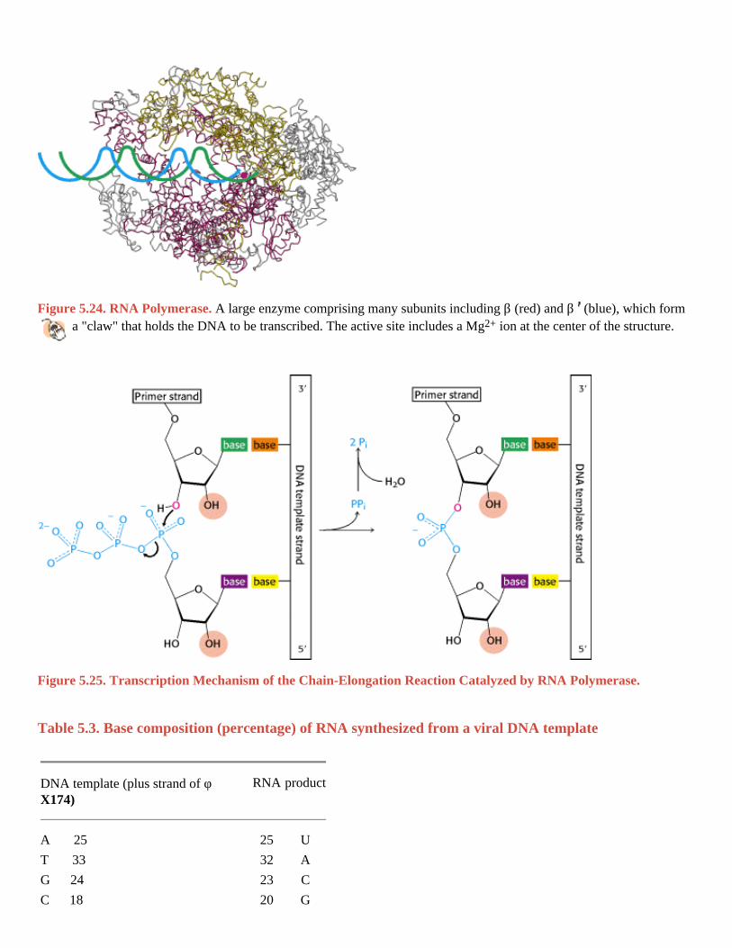
The synthesis of RNA from a DNA template is called transcription and is catalyzed by the enzyme RNA polymerase (Figure 5.24). RNA polymerase requires the following components:
1. A template. The preferred template is double-stranded DNA. Single-stranded DNA also can serve as a template. RNA, whether single or double stranded, is not an effective template; nor are RNA-DNA hybrids.
2. Activated precursors. All four ribonucleoside triphosphates ATP, GTP, UTP, and CTP are required.
3. A divalent metal ion. Mg2+ or Mn2+ are effective.
RNA polymerase catalyzes the initiation and elongation of RNA chains. The reaction catalyzed by this enzyme is:
The synthesis of RNA is like that of DNA in several respects (Figure 5.25). First, the direction of synthesis is 5 3 .

Second, the mechanism of elongation is similar: the 3 -OH group at the terminus of the growing chain makes a nucleophilic attack on the innermost phosphate of the incoming nucleoside triphosphate. Third, the synthesis is driven forward by the hydrolysis of pyrophosphate. In contrast with DNA polymerase, however, RNA polymerase does not require a primer. In addition, RNA polymerase lacks the nuclease capability used by DNA polymerase to excise mismatched nucleotides.
All three types of cellular RNA mRNA, tRNA, and rRNA are synthesized in E. coli by the same RNA polymerase according to instructions given by a DNA template. In mammalian cells, there is a division of labor among several different kinds of RNA polymerases. We shall return to these RNA polymerases in Chapter 28.
5.4.3. RNA Polymerases Take Instructions from DNA Templates
RNA polymerase, like the DNA polymerases described earlier, takes instructions from a DNA template. The earliest evidence was the finding that the base composition of newly synthesized RNA is the complement of that of the DNA template strand, as exemplified by the RNA synthesized from a template of single-stranded φ X174 DNA (Table 5.3). Hybridization experiments also revealed that RNA synthesized by RNA polymerase is complementary to its DNA template. In these experiments, DNA is melted and allowed to reassociate in the presence of mRNA. RNA-DNA hybrids will form if the RNA and DNA have complementary sequences. The strongest evidence for the fidelity of transcription came from base-sequence studies showing that the RNA sequence is the precise complement of the DNA template sequence (Figure 5.26).
5.4.4. Transcription Begins near Promoter Sites and Ends at Terminator Sites
RNA polymerase must detect and transcribe discrete genes from within large stretches of DNA. What marks the beginning of a transcriptional unit? DNA templates contain regions called promoter sites that specifically bind RNA
polymerase and determine where transcription begins. In bacteria, two sequences on the 5 (upstream) side of the first nucleotide to be transcribed function as promoter sites (Figure 5.27A). One of them, called the Pribnow box, has the
consensus sequence TATAAT and is centered at -10 (10 nucleotides on the 5 side of the first nucleotide transcribed, which is denoted by + 1). The other, called the -35 region, has the consensus sequence TTGACA. The first nucleotide transcribed is usually a purine.
Consensus sequence
The base sequences of promoter sites are not all identical. However, they do possess common features, which can be represented by an idealized consensus sequence. Each base in the consensus sequence TATAAT is found in a majority of prokaryotic promoters. Nearly all promoter sequences differ from this consensus sequence at only one or two bases.
Eukaryotic genes encoding proteins have promoter sites with a TATAAA consensus sequence, called a TATA box or a Hogness box, centered at about -25 (Figure 5.27B). Many eukaryotic promoters also have a CAAT box with a GGNCAATCT consensus sequence centered at about -75. Transcription of eukaryotic genes is further stimulated by
enhancer sequences, which can be quite distant (as many as several kilobases) from the start site, on either its 5 or its 3 side.
RNA polymerase proceeds along the DNA template, transcribing one of its strands until it reaches a terminator sequence. This sequence encodes a termination signal, which in E. coli is a base-paired hairpin on the newly synthesized RNA molecule (Figure 5.28). This hairpin is formed by base pairing of self-complementary sequences that are rich in G and C. Nascent RNA spontaneously dissociates from RNA polymerase when this hairpin is followed by a string of U residues.

Alternatively, RNA synthesis can be terminated by the action of rho, a protein. Less is known about the termination of transcription in eukaryotes. A more detailed discussion of the initiation and termination of transcription will be given in Chapter 28. The important point now is that discrete start and stop signals for transcription are encoded in the DNA template.
In eukaryotes, the mRNA is modified after transcription (Figure 5.29). A "cap" structure is attached to the 5 end, and a
sequence of adenylates the poly(A) tail is added to the 3 end. These modifications will be presented in detail in Section 28.3.1.
5.4.5. Transfer RNA Is the Adaptor Molecule in Protein Synthesis
We have seen that mRNA is the template for protein synthesis. How then does it direct amino acids to become joined in the correct sequence to form a protein? In 1958, Francis Crick wrote:
RNA presents mainly a sequence of sites where hydrogen bonding could occur. One would expect, therefore, that whatever went onto the template in a specific way did so by forming hydrogen bonds. It is therefore a natural hypothesis that the amino acid is carried to the template by an adaptor molecule, and that the adaptor is the part that actually fits onto the RNA. In its simplest form, one would require twenty adaptors, one for each amino acid.
This highly innovative hypothesis soon became established as fact. The adaptor in protein synthesis is transfer RNA. The structure and reactions of these remarkable molecules will be considered in detail in Chapter 29. For the moment, it suffices to note that tRNA contains an amino acidattachment site and a template-recognition site. A tRNA molecule carries a specific amino acid in an activated form to the site of protein synthesis. The carboxyl group of this amino acid
is esterified to the 3 - or 2 -hydroxyl group of the ribose unit at the 3 end of the tRNA chain (Figure 5.30). The joining of an amino acid to a tRNA molecule to form an aminoacyl-tRNA is catalyzed by a specific enzyme called an aminoacyl-tRNA synthetase (or acti-vating enzyme). This esterification reaction is driven by ATP. There is at least one specific synthetase for each of the 20 amino acids. The template-recognition site on tRNA is a sequence of three bases called an anticodon (Figure 5.31). The anticodon on tRNA recognizes a complementary sequence of three bases, called a codon, on mRNA.
I. The Molecular Design of Life 5. DNA, RNA, and the Flow of Genetic Information 5.4. Gene Expression Is the Transformation of DNA Information Into Functional Molecules
Table 5.2. RNA molecules in E. coli
Type Relative amount (%) Sedimentation coefficient (S) Mass (kd) Number of nucleotides
Ribosomal RNA (rRNA) 80 23 1.2 × 10³ 3700
16 0.55 × 10³ 1700
5 3.6 × 101 120
Transfer RNA (tRNA) 15 4 2.5 × 101 75
Messenger RNA (mRNA) 5 Heterogeneous
I. The Molecular Design of Life 5. DNA, RNA, and the Flow of Genetic Information 5.4. Gene Expression Is the Transformation of DNA Information Into Functional Molecules
Figure 5.24. RNA Polymerase. A large enzyme comprising many subunits including β (red) and β (blue), which form a "claw" that holds the DNA to be transcribed. The active site includes a Mg2+ ion at the center of the structure.
I. The Molecular Design of Life 5. DNA, RNA, and the Flow of Genetic Information 5.4. Gene Expression Is the Transformation of DNA Information Into Functional Molecules
Figure 5.25. Transcription Mechanism of the Chain-Elongation Reaction Catalyzed by RNA Polymerase. I. The Molecular Design of Life 5. DNA, RNA, and the Flow of Genetic Information 5.4. Gene Expression Is the Transformation of DNA Information Into Functional Molecules
Table 5.3. Base composition (percentage) of RNA synthesized from a viral DNA template
DNA template (plus strand of φ X174)
RNA product
A 25 25 U
T 33 32 A
G 24 23 C
C 18 20 G

I. The Molecular Design of Life 5. DNA, RNA, and the Flow of Genetic Information 5.4. Gene Expression Is the Transformation of DNA Information Into Functional Molecules
Figure 5.26. Complementarity between mRNA and DNA. The base sequence of mRNA (red) is the complement of that of the DNA template strand (blue). The sequence shown here is from the tryptophan operon, a segment of DNA containing the genes for five enzymes that catalyze the synthesis of tryptophan. The other strand of DNA (black) is called the coding strand because it has the same sequence as the RNA transcript except for thymine (T) in place of uracil (U). I. The Molecular Design of Life 5. DNA, RNA, and the Flow of Genetic Information 5.4. Gene Expression Is the Transformation of DNA Information Into Functional Molecules
Figure 5.27. Promoter Sites for Transcription. Promoter sites are required for the initiation of transcription in both (A) prokaryotes and (B) eukaryotes. Consensus sequences are shown. The first nucleotide to be transcribed is numbered +1. The adjacent nucleotide on the 5 side is numbered -1. The sequences shown are those of the coding strand of DNA.
I. The Molecular Design of Life 5. DNA, RNA, and the Flow of Genetic Information 5.4. Gene Expression Is the Transformation of DNA Information Into Functional Molecules
Figure 5.28. Base Sequence of the 3 end of an mRNA transcript in E. coli. A stable hairpin structure is followed by a sequence of uridine (U) residues.

I. The Molecular Design of Life 5. DNA, RNA, and the Flow of Genetic Information 5.4. Gene Expression Is the Transformation of DNA Information Into Functional Molecules
Figure 5.29. Modification of mRNA. Messenger RNA in eukaryotes is modified after transcription. A nucleotide "cap" structure is added to the 5 end, and a poly(A) tail is added at the 3 end.
I. The Molecular Design of Life 5. DNA, RNA, and the Flow of Genetic Information 5.4. Gene Expression Is the Transformation of DNA Information Into Functional Molecules
Figure 5.30. Attachment of an Amino Acid to a tRNA Molecule. The amino acid (shown in blue) is esterified to the 3 -hydroxyl group of the terminal adenosine of tRNA. I. The Molecular Design of Life 5. DNA, RNA, and the Flow of Genetic Information 5.4. Gene Expression Is the Transformation of DNA Information Into Functional Molecules
Figure 5.31. Symbolic Diagram of an Aminoacyl-tRNA. The amino acid is attached at the 3 end of the RNA. The anticodon is the template-recognition site.

I. The Molecular Design of Life 5. DNA, RNA, and the Flow of Genetic Information
5.5. Amino Acids Are Encoded by Groups of Three Bases Starting from a Fixed Point
The genetic code is the relation between the sequence of bases in DNA (or its RNA transcripts) and the sequence of amino acids in proteins. Experiments by Francis Crick, Sydney Brenner, and others established the following features of the genetic code by 1961:
1. Three nucleotides encode an amino acid. Proteins are built from a basic set of 20 amino acids, but there are only four bases. Simple calculations show that a minimum of three bases is required to encode at least 20 amino acids. Genetic experiments showed that an amino acid is in fact encoded by a group of three bases, or codon.
2. The code is nonoverlapping. Consider a base sequence ABCDEF. In an overlapping code, ABC specifies the first amino acid, BCD the next, CDE the next, and so on. In a nonoverlapping code, ABC designates the first amino acid, DEF the second, and so forth. Genetics experiments again established the code to be nonoverlapping.
3. The code has no punctuation. In principle, one base (denoted as Q) might serve as a "comma" between groups of three bases.
This is not the case. Rather, the sequence of bases is read sequentially from a fixed starting point, without punctuation.
4. The genetic code is degenerate. Some amino acids are encoded by more than one codon, inasmuch as there are 64 possible base triplets and only 20 amino acids. In fact, 61 of the 64 possible triplets specify particular amino acids and 3 triplets (called stop codons) designate the termination of translation. Thus, for most amino acids, there is more than one code word.
5.5.1. Major Features of the Genetic Code
All 64 codons have been deciphered (Table 5.4). Because the code is highly degenerate, only tryptophan and methionine are encoded by just one triplet each. The other 18 amino acids are each encoded by two or more. Indeed, leucine, arginine, and serine are specified by six codons each. The number of codons for a particular amino acid correlates with its frequency of occurrence in proteins.
Codons that specify the same amino acid are called synonyms. For example, CAU and CAC are synonyms for histidine.

Note that synonyms are not distributed haphazardly throughout the genetic code (depicted in Table 5.4). An amino acid specified by two or more synonyms occupies a single box (unless it is specified by more than four synonyms). The amino acids in a box are specified by codons that have the same first two bases but differ in the third base, as exemplified by GUU, GUC, GUA, and GUG. Thus, most synonyms differ only in the last base of the triplet. Inspection of the code shows that XYC and XYU always encode the same amino acid, whereas XYG and XYA usually encode the same amino acid. The structural basis for these equivalences of codons will become evident when we consider the nature of the anticodons of tRNA molecules (Section 29.3.9).
What is the biological significance of the extensive degeneracy of the genetic code? If the code were not degenerate, 20 codons would designate amino acids and 44 would lead to chain termination. The probability of mutating to chain termination would therefore be much higher with a nondegenerate code. Chain-termination mutations usually lead to inactive proteins, whereas substitutions of one amino acid for another are usually rather harmless. Thus, degeneracy minimizes the deleterious effects of mutations. Degeneracy of the code may also be significant in permitting DNA base composition to vary over a wide range without altering the amino acid sequence of the proteins encoded by the DNA. The G + C content of bacterial DNA ranges from less than 30% to more than 70%. DNA molecules with quite different G + C contents could encode the same proteins if different synonyms of the genetic code were consistently used.
5.5.2. Messenger RNA Contains Start and Stop Signals for Protein Synthesis
Messenger RNA is translated into proteins on ribosomes, large molecular complexes assembled from proteins and ribosomal RNA. How is mRNA interpreted by the translation apparatus? As already mentioned, UAA, UAG, and UGA designate chain termination. These codons are read not by tRNA molecules but rather by specific proteins called release factors (Section 29.4.4). Binding of the release factors to the ribosomes releases the newly synthesized protein. The start signal for protein synthesis is more complex. Polypeptide chains in bacteria start with a modified amino acid namely, formylmethionine (fMet). A specific tRNA, the initiator tRNA, carries fMet. This fMet-tRNA recognizes the codon AUG or, less frequently, GUG. However, AUG is also the codon for an internal methio-nine residue, and GUG is the codon for an internal valine residue. Hence, the signal for the first amino acid in a prokaryotic polypeptide chain must be more complex than that for all subsequent ones. AUG (or GUG) is only part of the initiation signal (Figure 5.32). In bacteria, the initiating AUG (or GUG) codon is preceded several nucleotides away by a purine-rich sequence that base-pairs with a complementary sequence in a ribosomal RNA molecule (Section 29.3.4). In eukaryotes, the AUG closest to
the 5 end of an mRNA molecule is usually the start signal for protein synthesis. This particular AUG is read by an initiator tRNA conjugated to methionine. Once the initiator AUG is located, the reading frame is established groups of three nonoverlapping nucleotides are defined, beginning with the initiator AUG codon.
5.5.3. The Genetic Code Is Nearly Universal
Is the genetic code the same in all organisms? The base sequences of many wild-type and mutant genes are known, as are the amino acid sequences of their encoded proteins. In each case, the nucleotide change in the gene
and the amino acid change in the protein are as predicted by the genetic code. Furthermore, mRNAs can be correctly translated by the proteinsynthesizing machinery of very different species. For example, human hemoglobin mRNA is correctly translated by a wheat germ extract, and bacteria efficiently express recombinant DNA molecules encoding human proteins such as insulin. These experimental findings strongly suggested that the genetic code is universal.

A surprise was encountered when the sequence of human mitochondrial DNA became known. Human mitochondria read UGA as a codon for tryptophan rather than as a stop signal (Table 5.5). Furthermore, AGA and AGG are read as stop signals rather than as codons for arginine, and AUA is read as a codon for methionine instead of isoleucine. Mitochondria of other species, such as those of yeast, also have genetic codes that differ slightly from the standard one. The genetic code of mitochondria can differ from that of the rest of the cell because mitochondrial DNA encodes a distinct set of tRNAs. Do any cellular protein-synthesizing systems deviate from the standard genetic code? Ciliated protozoa differ from most organisms in reading UAA and UAG as codons for amino acids rather than as stop signals; UGA is their sole termination signal. Thus, the genetic code is nearly but not absolutely universal. Variations clearly exist in mitochondria and in species, such as ciliates, that branched off very early in eukaryotic evolution. It is interesting to note that two of the codon reassignments in human mitochondria diminish the information content of the third base of the triplet (e.g., both AUA and AUG specify methionine). Most variations from the standard genetic code are in the direction of a simpler code.
Why has the code remained nearly invariant through billions of years of evolution, from bacteria to human beings? A mutation that altered the reading of mRNA would change the amino acid sequence of most, if not all, proteins synthesized by that particular organism. Many of these changes would undoubtedly be deleterious, and so there would be strong selection against a mutation with such pervasive consequences.
I. The Molecular Design of Life 5. DNA, RNA, and the Flow of Genetic Information 5.5. Amino Acids Are Encoded by Groups of Three Bases Starting from a Fixed Point
Table 5.4. The genetic code
First position (5 end) Second position Third position (3 end)
U C A G
Phe Ser Tyr Cys U
U Phe Ser Tyr Cys C
Leu Ser Stop Stop A
Leu Ser Stop Trp G
Leu Pro His Arg U
C Leu Pro His Arg C
Leu Pro Gln Arg A
Leu Pro Gln Arg G
Ile Thr Asn Ser U
A Ile Thr Asn Ser C
Ile Thr Lys Arg A
Met Thr Lys Arg G
Val Ala Asp Gly U
G Val Ala Asp Gly C
Val Ala Glu Gly A
Val Ala Glu Gly G