Embed Size (px)
Citation preview
INFECTION AND IMMUNITY,0019-9567/00/$04.0010
Dec. 2000, p. 7028–7038 Vol. 68, No. 12
Copyright © 2000, American Society for Microbiology. All Rights Reserved.
Molecular Variation among Type IV Pilin (bfpA) Genes fromDiverse Enteropathogenic Escherichia coli Strains
T. ERIC BLANK,1 HAILANG ZHONG,1 ALISON L. BELL,2 THOMAS S. WHITTAM,2
AND MICHAEL S. DONNENBERG1*
Division of Infectious Diseases, Department of Medicine, University of Maryland School of Medicine,Baltimore, Maryland 21201,1 and Institute of Molecular Evolutionary Genetics and Department
of Biology, Pennsylvania State University, University Park, Pennsylvania 168022
Received 12 June 2000/Returned for modification 7 September 2000/Accepted 13 September 2000
Typical enteropathogenic Escherichia coli (EPEC) strains produce bundle-forming pili (BFP), type IVBfimbriae that have been implicated in EPEC virulence, antigenicity, autoaggregation, and localized adherenceto epithelial cells (LA). BFP are polymers of bundlin, a pilin protein that is encoded by the bfpA gene foundon a large EPEC plasmid. Striking sequence variation has previously been observed among type IV pilin genesof other gram-negative bacterial pathogens (e.g., Pseudomonas and Neisseria spp.). In contrast, the establishedsequences of bfpA genes from two distantly related prototype EPEC strains vary by only a single base pair. Todetermine whether bundlin sequences vary more extensively, we used PCR to amplify the bfpA genes from 19EPEC strains chosen for their various serotypes and sites and years of isolation. Eight different bfpA alleleswere identified by sequencing of the PCR products. These alleles can be classified into two major groups. Thea group contains three alleles derived from strains carrying O55, O86, O111, O119, O127, or O128 somaticantigens. The b group contains five alleles derived from strains carrying O55, O110, O128ab, O142, ornontypeable antigens. Sequence comparisons show that bundlin has highly conserved and variable regions,with most of the variation occurring in the C-terminal two-thirds of the protein. The results of multilocusenzyme electrophoresis support the hypothesis that bfpA sequences have spread horizontally across distantlyrelated clonal lineages. Strains with divergent bundlin sequences express bundlin protein, produce BFP, andcarry out autoaggregation and LA. However, four strains lack most or all of these phenotypes despite havingan intact bfpA gene. These results have important implications for our understanding of bundlin structure,transmission of the bfp gene cluster among EPEC strains, and the role of bundlin variation in the evasion ofhost immune system responses.
Enteropathogenic Escherichia coli (EPEC) is one of severalpathovars of E. coli capable of causing diarrhea (45). Whilehuman EPEC infections, which are manifest primarily in in-fants, were once commonplace in industrialized nations (56),they are now identified primarily in developing countries (10).EPEC strains possess distinct virulence factors not found inmost E. coli strains (45). Typical EPEC strains produce bundle-forming pili (BFP), long, flexible, rope-like structures com-posed of intertwining fibers (19). Based on protein sequenceanalysis and morphology, BFP belong to the type IV group offimbriae or pili found on a variety of gram-negative bacteria,many of which are human, animal, or plant pathogens (67, 70,77). BFP have recently been shown to elicit an antibody re-sponse in natural infections (37, 38, 53) and a modest responsein experimentally infected adults (13). BFP are required fortwo phenotypes of EPEC that can be studied in vitro andprobably play a role in colonization in vivo. The first is auto-aggregation, which is the ability of EPEC to reversibly formmulticellular clusters in liquid culture (5, 76). The second islocalized adherence (LA), which is the capacity of EPEC toform defined multicellular clusters (microcolonies) upon epi-thelial cells (7, 59) and upon human intestinal tissue (26). Bothof these phenomena seem to be manifestations of the ability ofBFP to intertwine, creating a fibrous network connecting indi-
vidual bacteria. BFP also appear to adhere directly to epithe-lial cells (19, 31), although no BFP receptor on epithelial cellshas been identified. During the course of tissue culture infec-tion, BFP bundles undergo a transformation to a longer andthicker form that is correlated with dispersal of bacteria frommicrocolonies (31). In a recent clinical study, volunteers fedmutant EPEC strains lacking BFP developed diarrhea at amuch lower frequency and of less severity than did volunteersreceiving equivalent doses of an isogenic strain producing BFP(5). Thus, BFP are dynamic fimbriae that contribute to EPECpathogenesis.
BFP biogenesis is specified by a cluster of 14 bfp genes (65,66) located on large (;50- to 70-MDa) EPEC adherence fac-tor (EAF) plasmids (3, 47, 48). The first gene of this cluster isbfpA, which encodes bundlin, a pilin protein that is the onlyknown structural component of BFP. Like other type IV pilins,bundlin is synthesized with a leader peptide removed by aprepilin peptidase, has a hydrophobic region at its mature Nterminus, and has two cysteine residues forming a disulfidebond near the C terminus. Bundlin is membrane associated (2,76). It seems likely that the ultimate function of most of the bfpgene products is to remove bundlin subunits from the bacterialmembrane and assemble them into pili (2).
Little is known about the structure of bundlin monomers orhow they assemble into BFP. However, some information onthese topics can be inferred from studies of other type IV pili.The crystal structures of type IV pilin monomers from Neisse-ria gonorrhoeae and Pseudomonas aeruginosa have been solved(15, 25, 52). These pilins are ladle shaped with a long N-terminal a-helical handle and a globular head. Models of the
* Corresponding author. Mailing address: Division of InfectiousDiseases, Department of Medicine, University of Maryland School ofMedicine, 10 South Pine St., Medical School Teaching Facility 9-00,Baltimore, MD 21201. Phone: (410) 706-7560. Fax: (410) 706-8700.E-mail: [email protected].
7028
on July 5, 2018 by guesthttp://iai.asm
.org/D
ownloaded from
way in which pilin monomers pack together into the pilus fiberhave been proposed for the gonococcal and Pseudomonas pili(16, 25, 52), as well as the toxin-coregulated pili (TCP) ofVibrio cholerae (R. Chattopadhyaya and A. C. Ghose, unpub-lished data; Protein Data Bank accession number 1QQZ).
Extensive sequence variability has been observed in the typeIV pilins of Dichelobacter nodosus, Eikenella corrodens, Morax-ella bovis, P. aeruginosa (reviewed in reference 70), N. gonor-rhoeae (52), N. meningitidis (1), and Vibrio cholerae (27, 43, 50,55). In some of these species, pilin variability is clearly linkedto changes in antigenicity. The host immune response mayhave played a major role in selecting for pilin variants withaltered sequences. In the N. gonorrhoeae genome, extra copiesof partial pilin genes are also present. These normally silentcopies can be exchanged with the active copy via recombina-tion, leading to antigenic variation that can aid the gonococcusin evading the host immune response (reviewed in references41 and 62). A comparison of many gonococcal pilin gene prod-ucts shows that extensive sequence variation occurs in aminoacid residues of the “head” region but hardly any occurs inresidues of the “handle” region (52). A hypervariable region ofgonococcal pilin occurs between the disulfide-bonded cysteineresidues. This region, as well as a disaccharide moiety and theextreme C terminus of the pilin, is predicted to be exposed onthe outside of the pilus fiber (16, 52). In P. aeruginosa pilins,the disulfide region is also variable in sequence and serves as abinding domain for cell surface receptors (reviewed in refer-ence 23).
In contrast to the considerable variation noted in other typeIV pilins, the bundlins encoded by the two known bfpA allelescontain only a single amino acid difference (11, 64). Bundlinproteins from diverse EPEC strains might be expected to ex-hibit more significant sequence variation than has previouslybeen noted. The current study was undertaken to assess thelevel of sequence diversity in bundlin proteins among EPECstrains and to gain insight into structural and functional con-straints on different regions of the molecule.
MATERIALS AND METHODS
EPEC strains. The origins of the EPEC strains examined in this study arelisted in Table 1. To analyze the motility of each strain, a loopful of culture wasstabbed into a motility agar plate (1% Bacto-Tryptone, 0.5% NaCl, 0.35% agar)and examined after overnight incubation at 37°C. Based on this analysis, strainslacking H serotype information were designated H1 (motile) or NM (nonmo-tile) (Table 1).
Isolation and sequencing of bfpA genes. Partial and complete bfpA genes wereamplified by PCR using DeepVent DNA polymerase in accordance with themanufacturer’s (New England Biolabs) instructions. EPEC colonies were pickedfrom Luria agar plates and boiled for 10 min in ThermoPol buffer (New EnglandBiolabs) containing 2 to 6 mM MgSO4. DeepVent DNA polymerase (2 U),oligonucleotide primers (0.35 to 1.2 mM), and deoxynucleoside triphosphates(400 mM each) were added to bring the total reaction volume to 100 ml beforebeginning the PCR. Primer pairs used for PCR were Donne-28 and -29, -362 and-363, -423 and -363, -6 and -382, and -423 and -382 (Table 2). The precisereaction conditions varied with the strain and primers used. The PCR productswere examined by agarose gel electrophoresis. If the PCR mixture contained asingle DNA species of the expected size, it was purified directly using the WizardPCR Preps DNA Purification System (Promega). If additional DNA specieswere present, the desired PCR product was isolated from a preparative agarosegel. Automated DNA sequencing of PCR products was carried out by the staff ofThe Biopolymer Laboratory (University Of Maryland School of Medicine) using
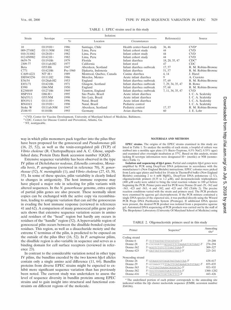
TABLE 1. EPEC strains used in this study
Strain SerotypeIsolation:
Reference(s) SourceYr Location Circumstances
10 O119:H1 1986 Santiago, Chile Health center-based study 36, 46 CVDa
009-271082 O111:NM 1982 Lima, Peru Infant cohort study 44 CVD010-311082 O128:H1 1982 Lima, Peru Infant cohort study 44 CVD012-050982 O142:H1 1982 Lima, Peru Infant cohort study 44 CVD0659-79 O119:H6 1979 Florida Infant diarrhea 18, 20, 35, 47 CDCb
2309-77 O111ab:H2 1977 California Infant stool 47 CDCBeta O55:H6 1947 Aberdeen, Scotland Infant diarrhea outbreak 17, 57 R. M. Robins-BrowneC771 O142:H6 Pre-1960 Djakarta, Indonesia Infant diarrhea 51, 57 R. M. Robins-BrowneCA89-4221 NTc:H1 1989 Montreal, Quebec, Canada Canine diarrhea 4, 14 J. HarelDIF043256 O111:H2 1986 Morelos, Mexico Acute infant diarrhea 9 A. CraviotoE56/54 O128ab:H2 1953 England Infant diarrhea outbreak 57, 69 R. M. Robins-BrowneE851/71 O142:H6 1971 Glasgow, Scotland Infant diarrhea outbreak 7, 29, 34, 35, 47 B. RoweE990 O86:NM 1950 England Infant diarrhea outbreak 57, 68 R. M. Robins-BrowneE2348/69 O127:H6 1969 Taunton, England Infant diarrhea outbreak 7, 11, 34, 35, 47 CVDHSP19/4 O86:H1 1995 Sao Paulo, Brazil Acute infant diarrhea I. C. A. ScaletskyMA551/1 O55:NM 1998 Sao Luiz, Brazil Acute infant diarrhea I. C. A. ScaletskyRN191/1 O111:H1 1996 Natal, Brazil Acute infant diarrhea I. C. A. ScaletskyRN410/1 O119:H1 1998 Natal, Brazil Pediatric control I. C. A. ScaletskyStoke W O111ab:NM 1947 Aberdeen, Scotland Pediatric control 17, 57 R. M. Robins-BrowneZ188-93 0110:H6 1993 Germany Avian diarrhea 60 J. E. Lohr
a CVD, Center for Vaccine Development, University of Maryland School of Medicine, Baltimore.b CDC, Centers for Disease Control and Prevention, Atlanta, Ga.c NT, nontypeable.
TABLE 2. Oligonucleotide primers used in this study
Primer Sequencea Annealingsitea
Coding strandDonne-6 59-TCTTGGTGCTTGCGTGTC-39 19–208Donne-28 59-CGCGGATCCATGGTTTCTAAAATCATGAAT-39 274–294Donne-362 59-AGGTCTGTCTTTGATTGAA-39 309–327Donne-423 59-GATTATTCCGTGACCTATT-39 223–241
Noncoding strandDonne-26 59-GTAAGCGTCAGATAGTAACCAA-39 638–617Donne-29 59-GCGAAGCTTTTACTTCATAAAATATGTAAC-39 855–835Donne-363 59-CCTGAGTAAAACAGGATAC-39 953–935Donne-382 59-TCCTTCGGGTGATTGTGTA-39 1300–1282Donne-416 59-GTTGCGCTCATTACTTCT-39 443–426
a The underlined portion of each primer corresponds to the annealing siteindicated within the bfp cluster nucleotide sequence (EMBL accession numberZ68186).
VOL. 68, 2000 TYPE IV PILIN SEQUENCE VARIATION IN EPEC 7029
on July 5, 2018 by guesthttp://iai.asm
.org/D
ownloaded from
two or more of the primers listed in Table 2. For some strains, the entiredouble-stranded sequence of the bfpA gene could be obtained using only primersDonne-423 and Donne-363.
Sequence analysis. DNASIS (Hitachi Software) was used to align bfpA nucle-otide sequences from each strain and determine a consensus sequence. Multiplesequence alignment was performed on the bfpA gene sequences and the deducedbundlin amino acid sequences using DNASIS and CLUSTAL W (71). Genetrees were constructed with the computer program MEGA (Molecular Evolu-tionary Genetics Analysis, version 1.0; S. Kumar, K. Tamura, and M. Nei,Institute of Molecular Evolutionary Genetics, The Pennsylvania State University,1993 [http://www.bio.psu.edu/People/Faculty/Nei/Lab/megaform.txt]). The pro-portions of polymorphic synonymous (pS) and nonsynonymous (pN) sites werecalculated by the method of Nei and Gojobori (49). To examine variation in thefunctional constraints of different parts of the molecule, these statistics weretabulated in a sliding-window analysis of 30 codons along the gene by theprogram PSWIN. The theoretical three-dimensional structure of bundlin wasanalyzed and colored using RASMOL (58) (http://www.umass.edu/microbio/rasmol/).
MLEE. Every strain was characterized by its profile of electromorphs for 20enzymes by multilocus enzyme electrophoresis (MLEE) (63, 78, 79). Geneticrelationships between strains were determined based on a matrix of geneticdistances between all pairs constructed by comparison of the allelic arrays. Adendrogram was constructed with MEGA using the neighbor-joining algorithm.
Immunoblotting. Overnight cultures of EPEC in LB were diluted 1:100 into 20ml of Dulbecco’s modified Eagle medium–nutrient mixture F-12 (DMEM–F-12)containing 15 mM HEPES buffer and lacking phenol red (Gibco-BRL LifeTechnologies catalog no. 11039-021). After 6 h of growth at 37°C with shaking,the bacteria were pelleted by centrifugation at 2,500 3 g for 5 min and thenresuspended in 1.2 ml of cell lysis buffer (20 mM Tris-HCl [pH 8.0], 500 mMNaCl, 0.1 mM EDTA, 0.1% Triton X-100). The optical density at 600 nm of aportion of these samples was measured on a spectrophotometer. Aliquots (75 mlor less) of these samples were mixed with 25 ml of 43 sodium dodecyl sulfate-polyacrylamide gel electrophoresis loading buffer (2) and then adjusted to a totalvolume of 100 ml with additional cell lysis buffer such that the resulting gelsamples were derived from roughly equivalent densities of bacteria. The gelsamples were boiled for 10 min, and then 5 ml of each was loaded per lane of asodium dodecyl sulfate–15% polyacrylamide gel. After electrophoretic separa-tion, the protein in the gel was electrotransferred to a polyvinylidene difluoridemembrane. The membrane was blocked with phosphate-buffered saline contain-ing 0.1% Tween 20 plus 5% nonfat dry milk and then incubated sequentially witha polyclonal antibody (1:5,000 dilution) directed against six-His-tagged prebund-lin (81), an anti-rabbit horseradish peroxidase conjugate (1:30,000 dilution), andECL Western blotting detection reagents (Amersham Pharmacia Biotech).
Transmission electron microscopy. To prepare samples for electron micros-copy, 1-ml aliquots were removed from EPEC cultures grown in DMEM–F-12for 5 h and the bacteria were pelleted by brief centrifugation. Most of themedium was decanted, and the bacterial pellet was gently resuspended in theremainder. Aliquots (10 ml) were spotted onto electron microscopy grids, whichwere dried for 10 min and then washed, stained, and examined for BFP asdescribed previously (66).
Autoaggregation assay. EPEC strains were cultured overnight at 37°C in LB.The resulting stationary-phase cultures were diluted 1:250 (making appropriateadjustments for optical density at 600 nm) into 20 ml of DMEM–F-12. Thesecultures were shaken at 250 rpm for 5 h in 50-ml conical polypropylene tubes at37°C. Autoaggregation was initially gauged by visually inspecting the cultures forbacterial aggregates and sedimentation. For some strains, it was necessary to uselight microscopy in order to detect aggregates. Culture aliquots (5 ml) wereexamined in hanging-drop slides at 340 magnification.
LA assay. LA to HeLa epithelial cells was assayed as described previously (12)using the eight-well chamber slide modification.
Nucleotide sequence accession numbers. The nucleotide sequences of thebfpA genes isolated from all of the strains listed in Table 1 (except prototypestrain E2348/69) have been deposited in GenBank under accession numbersAF304468 through AF304486.
RESULTS
Identification of novel bfpA alleles. The goal of this studywas to assess the amount of sequence variation among bfpAgenes found in EPEC strains. To this end, we initially analyzed13 strains from our laboratory collection (a subset of thoselisted in Table 1) representing a variety of serotypes and well-documented outbreaks (or sporadic cases) of human diarrhea.These strains were originally isolated from diverse geographi-cal locations between 1947 and 1986. They belong to classicalEPEC O serogroups O55, O86, O111, O119, O127, O128, andO142. Three of them (C771, E990, and Stoke W) are typestrains for the preparation of O antisera. Canine EPEC strainCA89-4221 and avian EPEC strain Z188-93, which are both
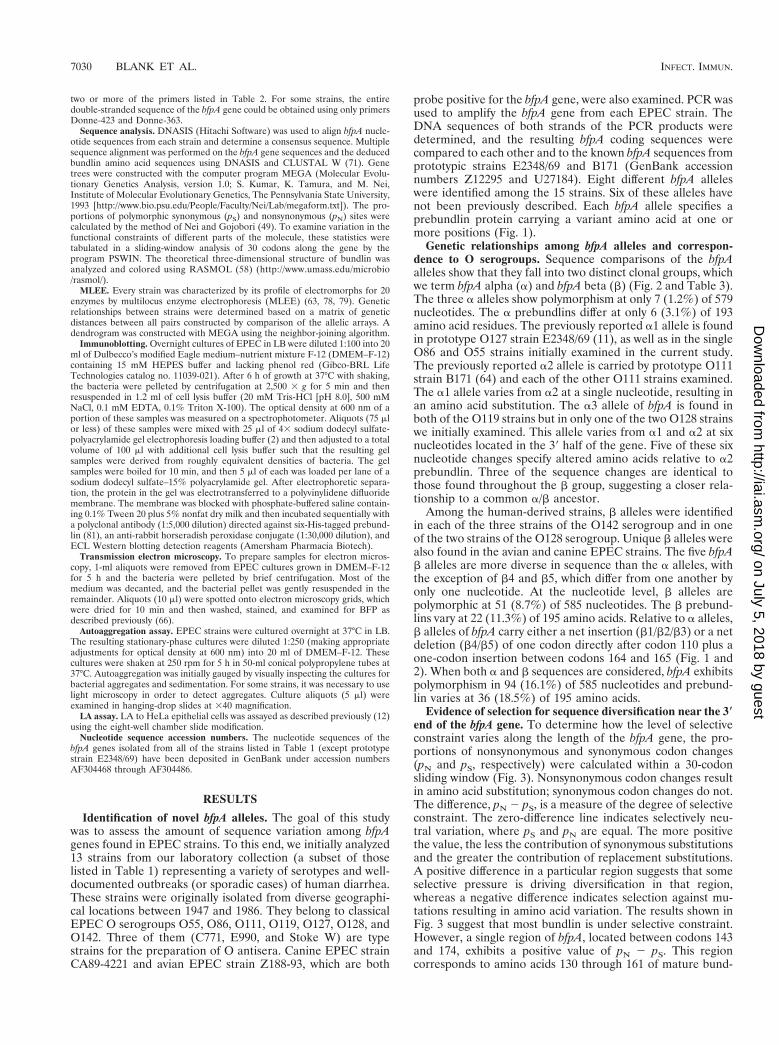
probe positive for the bfpA gene, were also examined. PCR wasused to amplify the bfpA gene from each EPEC strain. TheDNA sequences of both strands of the PCR products weredetermined, and the resulting bfpA coding sequences werecompared to each other and to the known bfpA sequences fromprototypic strains E2348/69 and B171 (GenBank accessionnumbers Z12295 and U27184). Eight different bfpA alleleswere identified among the 15 strains. Six of these alleles havenot been previously described. Each bfpA allele specifies aprebundlin protein carrying a variant amino acid at one ormore positions (Fig. 1).
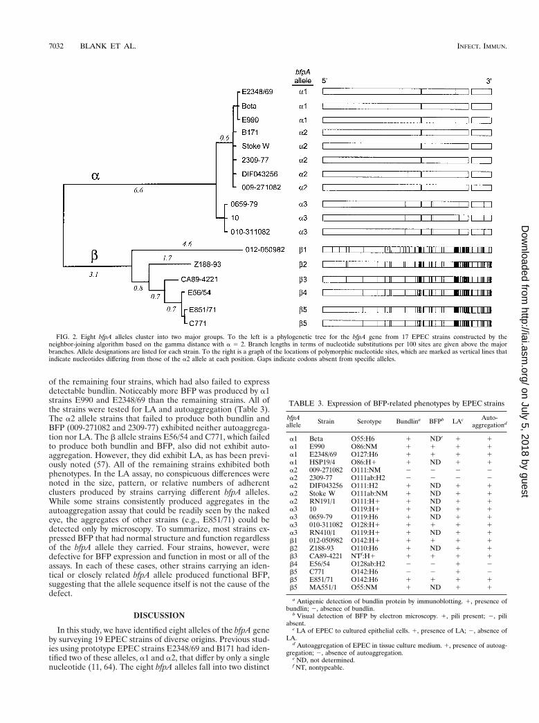
Genetic relationships among bfpA alleles and correspon-dence to O serogroups. Sequence comparisons of the bfpAalleles show that they fall into two distinct clonal groups, whichwe term bfpA alpha (a) and bfpA beta (b) (Fig. 2 and Table 3).The three a alleles show polymorphism at only 7 (1.2%) of 579nucleotides. The a prebundlins differ at only 6 (3.1%) of 193amino acid residues. The previously reported a1 allele is foundin prototype O127 strain E2348/69 (11), as well as in the singleO86 and O55 strains initially examined in the current study.The previously reported a2 allele is carried by prototype O111strain B171 (64) and each of the other O111 strains examined.The a1 allele varies from a2 at a single nucleotide, resulting inan amino acid substitution. The a3 allele of bfpA is found inboth of the O119 strains but in only one of the two O128 strainswe initially examined. This allele varies from a1 and a2 at sixnucleotides located in the 39 half of the gene. Five of these sixnucleotide changes specify altered amino acids relative to a2prebundlin. Three of the sequence changes are identical tothose found throughout the b group, suggesting a closer rela-tionship to a common a/b ancestor.
Among the human-derived strains, b alleles were identifiedin each of the three strains of the O142 serogroup and in oneof the two strains of the O128 serogroup. Unique b alleles werealso found in the avian and canine EPEC strains. The five bfpAb alleles are more diverse in sequence than the a alleles, withthe exception of b4 and b5, which differ from one another byonly one nucleotide. At the nucleotide level, b alleles arepolymorphic at 51 (8.7%) of 585 nucleotides. The b prebund-lins vary at 22 (11.3%) of 195 amino acids. Relative to a alleles,b alleles of bfpA carry either a net insertion (b1/b2/b3) or a netdeletion (b4/b5) of one codon directly after codon 110 plus aone-codon insertion between codons 164 and 165 (Fig. 1 and2). When both a and b sequences are considered, bfpA exhibitspolymorphism in 94 (16.1%) of 585 nucleotides and prebund-lin varies at 36 (18.5%) of 195 amino acids.
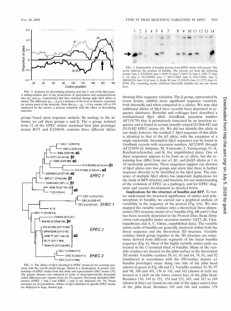
Evidence of selection for sequence diversification near the 3*end of the bfpA gene. To determine how the level of selectiveconstraint varies along the length of the bfpA gene, the pro-portions of nonsynonymous and synonymous codon changes(pN and pS, respectively) were calculated within a 30-codonsliding window (Fig. 3). Nonsynonymous codon changes resultin amino acid substitution; synonymous codon changes do not.The difference, pN 2 pS, is a measure of the degree of selectiveconstraint. The zero-difference line indicates selectively neu-tral variation, where pS and pN are equal. The more positivethe value, the less the contribution of synonymous substitutionsand the greater the contribution of replacement substitutions.A positive difference in a particular region suggests that someselective pressure is driving diversification in that region,whereas a negative difference indicates selection against mu-tations resulting in amino acid variation. The results shown inFig. 3 suggest that most bundlin is under selective constraint.However, a single region of bfpA, located between codons 143and 174, exhibits a positive value of pN 2 pS. This regioncorresponds to amino acids 130 through 161 of mature bund-
7030 BLANK ET AL. INFECT. IMMUN.
on July 5, 2018 by guesthttp://iai.asm
.org/D
ownloaded from
lin, which are located between the disulfide-bonded cysteines.An attractive possibility is that this region of bundlin is locatedon the surface of the BFP filament and that the host immunesystem has selected for antigenic diversity at this site.
Genetic relationships between EPEC strains. MLEE wascarried out to define the overall phylogenetic relationshipsbetween selected EPEC strains. A dendrogram resulting fromthis analysis (Fig. 4) shows that all of the a2 alleles are foundin a closely related cluster of O111 strains. Among the remain-ing EPEC strains, however, there is a curious lack of corre-spondence between the deduced clonal pattern of the strainsand the pattern of bfpA alleles they possess. Alleles of both thea and b types are found among strains in either of the twomajor clonal EPEC groups, EPEC 1 and EPEC 2 (54, 78). Inthe EPEC 1 group, the a1, a3, b1, b3, and b5 alleles areinterspersed among each other in a way that obviously does notcorrelate with the overall strain lineage. These results strong-ly suggest that the bfpA gene has been recently transferredbetween EPEC strains through multiple horizontal transferevents.
Examination of bfpA alleles in recent EPEC isolates. Todetermine whether bfpA a alleles have remained stable overrecent decades, we examined a set of four EAF1 EPEC strainsof very recent Brazilian origin (Table 1). The bfpA allelesderived from these strains were isolated by PCR and se-quenced. As expected from their serogroups, the O86, O111,and O119 strains (HSP19/4, RN191/1, and RN410/1) possessedbfpA genes of the a1, a2, and a3 varieties, respectively. Theseresults indicate that bfpA a sequences have been retainedunchanged in current EPEC strains. The O55 strain (MA551/
1), however, did not carry the a1 allele found in the previouslyexamined O55 EPEC (strain b). Rather, it carried a b5 alleleidentical to that carried by O142 strains C771 and E851/71.This unexpected finding provides a further suggestion of thehorizontal spread of a particular bfpA allele.
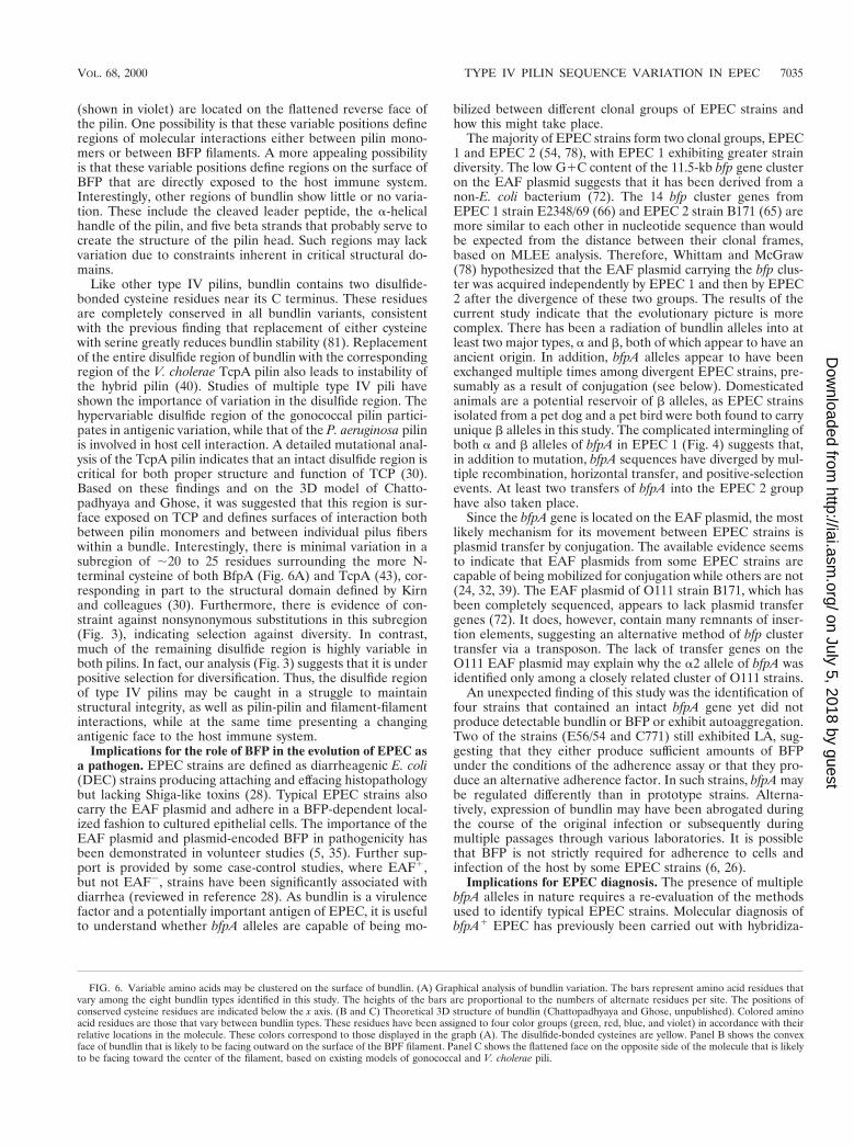
Expression and function of BFP variants. Selected EPECstrains were examined to determine whether they produceddetectable bundlin and BFP and whether they exhibited theBFP-dependent phenotypes of autoaggregation and LA. Someof these characteristics have been previously reported for asubset of these strains (see references in Table 1). PrototypeEPEC strain E2348/69 served as a positive control for all of ourstudies. To identify bundlin, a polyclonal antiserum was used toprobe whole-cell extracts from each of the EPEC strains (Fig.5 and Table 3). Bundlin protein was readily detected in extractsfrom 10 out of 12 strains tested carrying a alleles of bfpA and5 of 7 strains carrying b alleles. Bundlin was not identified instrain 009-271082 or 2309-77 carrying the a2 allele or in strainE56/54 or C771 carrying the b4 or b5 allele. Particular bbundlins migrated more slowly (b1 and b2) or more quickly(b5) than did a bundlins (Fig. 5 and data not shown). Therelative migration correlates well with the lengths (180 aminoacids for a1 and b5 and 182 amino acids for b1 and b2) andmolecular masses (19,986 Da for b5, 20,269 Da for a1, 20,306Da for b2, and 20,328 Da for b1) predicted for these bundlintypes. Such a migration anomaly has been noted for bundlinfrom other strains in previous studies (18, 20, 38).
Ten of the strains were examined by electron microscopy forthe presence of BFP fimbriae. Fimbrial bundles were readilydetected in six strains (Table 3). No BFP was seen in samples
FIG. 1. Variable amino acids are clustered in prebundlin. An alignment of the prebundlin amino acid sequences encoded by the eight bfpA alleles described in thisstudy is shown. Note that amino acids 1 to 13 comprise the cleaved leader peptide. The a1 (ALPHA1.AMI) prebundlin prototype sequence is listed on the top line.In the remaining seven sequences (ALPHA2.AMI through ALPHA3.AMI and BETA1.AMI through BETA5.AMI), invariant amino acids are indicated by dots.Variant amino acids are boxed and indicated by single-letter abbreviations.
VOL. 68, 2000 TYPE IV PILIN SEQUENCE VARIATION IN EPEC 7031
on July 5, 2018 by guesthttp://iai.asm
.org/D
ownloaded from
of the remaining four strains, which had also failed to expressdetectable bundlin. Noticeably more BFP was produced by a1strains E990 and E2348/69 than the remaining strains. All ofthe strains were tested for LA and autoaggregation (Table 3).The a2 allele strains that failed to produce both bundlin andBFP (009-271082 and 2309-77) exhibited neither autoaggrega-tion nor LA. The b allele strains E56/54 and C771, which failedto produce both bundlin and BFP, also did not exhibit auto-aggregation. However, they did exhibit LA, as has been previ-ously noted (57). All of the remaining strains exhibited bothphenotypes. In the LA assay, no conspicuous differences werenoted in the size, pattern, or relative numbers of adherentclusters produced by strains carrying different bfpA alleles.While some strains consistently produced aggregates in theautoaggregation assay that could be readily seen by the nakedeye, the aggregates of other strains (e.g., E851/71) could bedetected only by microscopy. To summarize, most strains ex-pressed BFP that had normal structure and function regardlessof the bfpA allele they carried. Four strains, however, weredefective for BFP expression and function in most or all of theassays. In each of these cases, other strains carrying an iden-tical or closely related bfpA allele produced functional BFP,suggesting that the allele sequence itself is not the cause of thedefect.
DISCUSSION
In this study, we have identified eight alleles of the bfpA geneby surveying 19 EPEC strains of diverse origins. Previous stud-ies using prototype EPEC strains E2348/69 and B171 had iden-tified two of these alleles, a1 and a2, that differ by only a singlenucleotide (11, 64). The eight bfpA alleles fall into two distinct
FIG. 2. Eight bfpA alleles cluster into two major groups. To the left is a phylogenetic tree for the bfpA gene from 17 EPEC strains constructed by theneighbor-joining algorithm based on the gamma distance with a 5 2. Branch lengths in terms of nucleotide substitutions per 100 sites are given above the majorbranches. Allele designations are listed for each strain. To the right is a graph of the locations of polymorphic nucleotide sites, which are marked as vertical lines thatindicate nucleotides differing from those of the a2 allele at each position. Gaps indicate codons absent from specific alleles.
TABLE 3. Expression of BFP-related phenotypes by EPEC strains
bfpAallele Strain Serotype Bundlina BFPb LAc Auto-
aggregationd
a1 Beta O55:H6 1 NDe 1 1a1 E990 O86:NM 1 1 1 1a1 E2348/69 O127:H6 1 1 1 1a1 HSP19/4 O86:H1 1 ND 1 1a2 009-271082 O111:NM 2 2 2 2a2 2309-77 O111ab:H2 2 2 2 2a2 DIF043256 O111:H2 1 ND 1 1a2 Stoke W O111ab:NM 1 ND 1 1a2 RN191/1 O111:H1 1 ND 1 1a3 10 O119:H1 1 ND 1 1a3 0659-79 O119:H6 1 ND 1 1a3 010-311082 O128:H1 1 1 1 1a3 RN410/1 O119:H1 1 ND 1 1b1 012-050982 O142:H1 1 1 1 1b2 Z188-93 O110:H6 1 ND 1 1b3 CA89-4221 NTf:H1 1 1 1 1b4 E56/54 O128ab:H2 2 2 1 2b5 C771 O142:H6 2 2 1 2b5 E851/71 O142:H6 1 1 1 1b5 MA551/1 O55:NM 1 ND 1 1
a Antigenic detection of bundlin protein by immunoblotting. 1, presence ofbundlin; 2, absence of bundlin.
b Visual detection of BFP by electron microscopy. 1, pili present; 2, piliabsent.
c LA of EPEC to cultured epithelial cells. 1, presence of LA; 2, absence ofLA.
d Autoaggregation of EPEC in tissue culture medium. 1, presence of autoag-gregation; 2, absence of autoaggregation.
e ND, not determined.f NT, nontypeable.
7032 BLANK ET AL. INFECT. IMMUN.
on July 5, 2018 by guesthttp://iai.asm
.org/D
ownloaded from
groups based upon sequence analysis. By analogy to the in-timins, we call these groups a and b. The a group, isolatedfrom 12 of the EPEC strains examined here plus prototypestrains B171 and E2348/69, contains three different alleles
showing little sequence variation. The b group, represented byseven strains, exhibits more significant sequence variation,both internally and when compared to a alleles. We note thatadditional alleles of bfpA have recently been deposited in se-quence databases. Bortolini and colleages have described anonfunctional bfpA allele (GenBank accession numberAF119170) that is prematurely truncated by an insertion se-quence and is found in certain clonally related O128ab:H2 andO119:H2 EPEC strains (6). We did not identify this allele inour study; however, the residual 59 bfpA sequence of this alleleis identical to that of the b3 allele, with the exception of asingle nucleotide. Incomplete bfpA sequences can be found inGenBank records with accession numbers AF233895 throughAF233899 (S. Subpasu, M. Yamasaki, J. Yatsuyanagi, O.-A.Ratchatrachenchai, and K. Ito, unpublished data). One ofthese sequences appears to be from an a1 allele, but the re-maining four differ from our a3, b1, and b4/b5 alleles at 1 to10 nucleotide positions. These sequences support our divisionof bfpA alleles into two groups and attest that there is furthersequence diversity to be identified in the bfpA gene. The exis-tence of multiple bfpA alleles has important implications forthe study of BFP structure and function, for our understandingof the evolution of EPEC as a pathogen, and for EPEC diag-nosis and vaccine development as detailed below.
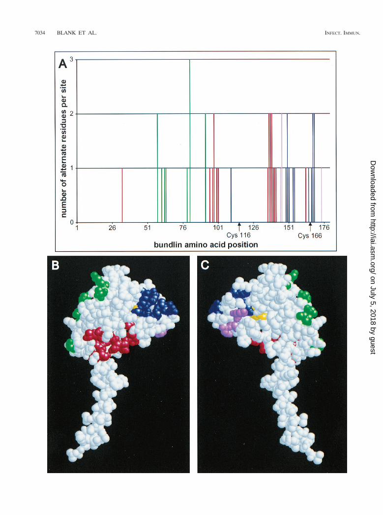
Implications for the structure of bundlin and BFP. To bet-ter understand the structural significance of amino acid poly-morphism in bundlin, we carried out a graphical analysis ofvariability in the sequence of the protein (Fig. 6A). We alsomapped the variable residues onto a theoretical three-dimen-sional (3D) structure model of a1 bundlin (Fig. 6B and C) thathas been recently deposited in the Protein Data Bank (http://www.rcsb.org/pdb) under accession number 1QT2 (R. Chat-topadhyaya and A. C. Ghose, unpublished data). The variableamino acids of bundlin are generally clustered, within both thelinear sequence and the theoretical 3D structure. Variableresidues which group together in the 3D structure are some-times derived from different segments of the linear bundlinsequence (Fig. 6). Most of the highly variable amino acids arelocated in the C-terminal third of bundlin. Many of the vari-able residues are located on the pilin surface in the theoretical3D model. Variable residues 58, 61, 63 and 64, 79, 81, and 92(numbered in accordance with the 180-residue mature a1bundlin prototype) occur along one side of the pilin head(shown in green in Fig. 6B and C). Variable residues 33, 95, 97and 98, 100 and 101, 136 to 142, and 163 (shown in red) arelocated in a cleft on the lower convex face of the pilin head.Residues 110, 149 to 151, 154 and 155, 165, and 167 to 169(shown in blue) are found on one side of the upper convex faceof the pilin head. Residues 145 and 146 and residue 174
FIG. 3. Evidence for diversifying selection near the 39 end of the bfpA gene.A sliding-window plot of the proportions of synonymous and nonsynonymoussites (pS and pN, respectively) that have mutated among eight bfpA alleles isshown. The difference (pN 2 pS) is a measure of the level of selective constrainton various parts of the molecule. Note that pN 2 pS . 0 for codons 143 to 174(indicated by the arrow), a pattern consistent with the effect of diversifyingselection.
FIG. 4. The alleles of bfpA identified in EPEC strains do not correlate pre-cisely with the overall clonal lineage. Shown is a dendrogram of genetic rela-tionships of EPEC strains from this study and representative DEC strains (79).The genetic distance was estimated in terms of electrophoretically detectablecodon differences per enzyme locus for 20 enzymes. Previously identified DECclusters (EPEC 1 and 2 and EHEC 1 and 2) are indicated (54, 78). Strainserotypes are in parentheses. Alleles of bfpA identified in specific EPEC strainsare displayed in large, shaded type.
FIG. 5. Immunoblot of bundlin protein from EPEC whole-cell extracts. Thearrow indicates the position of bundlin. The extracts are from the followingstrains: lane 1, E2348/69; lane 2, 0659-79; lane 3, E851/71; lane 4, 2309-77; lane5, 10; lane 6, 012-050982; lane 7, 009-271082; lane 8, 010-311082; lane 9,DIF043256; lane 10, b; lane 11, Stoke W; lane 12, E56/54; lane 13, C771; lane 14,E990. The remaining strains exhibited detectable bundlin but are not shownhere.
VOL. 68, 2000 TYPE IV PILIN SEQUENCE VARIATION IN EPEC 7033
on July 5, 2018 by guesthttp://iai.asm
.org/D
ownloaded from
7034 BLANK ET AL. INFECT. IMMUN.
on July 5, 2018 by guesthttp://iai.asm
.org/D
ownloaded from
(shown in violet) are located on the flattened reverse face ofthe pilin. One possibility is that these variable positions defineregions of molecular interactions either between pilin mono-mers or between BFP filaments. A more appealing possibilityis that these variable positions define regions on the surface ofBFP that are directly exposed to the host immune system.Interestingly, other regions of bundlin show little or no varia-tion. These include the cleaved leader peptide, the a-helicalhandle of the pilin, and five beta strands that probably serve tocreate the structure of the pilin head. Such regions may lackvariation due to constraints inherent in critical structural do-mains.
Like other type IV pilins, bundlin contains two disulfide-bonded cysteine residues near its C terminus. These residuesare completely conserved in all bundlin variants, consistentwith the previous finding that replacement of either cysteinewith serine greatly reduces bundlin stability (81). Replacementof the entire disulfide region of bundlin with the correspondingregion of the V. cholerae TcpA pilin also leads to instability ofthe hybrid pilin (40). Studies of multiple type IV pili haveshown the importance of variation in the disulfide region. Thehypervariable disulfide region of the gonococcal pilin partici-pates in antigenic variation, while that of the P. aeruginosa pilinis involved in host cell interaction. A detailed mutational anal-ysis of the TcpA pilin indicates that an intact disulfide region iscritical for both proper structure and function of TCP (30).Based on these findings and on the 3D model of Chatto-padhyaya and Ghose, it was suggested that this region is sur-face exposed on TCP and defines surfaces of interaction bothbetween pilin monomers and between individual pilus fiberswithin a bundle. Interestingly, there is minimal variation in asubregion of ;20 to 25 residues surrounding the more N-terminal cysteine of both BfpA (Fig. 6A) and TcpA (43), cor-responding in part to the structural domain defined by Kirnand colleagues (30). Furthermore, there is evidence of con-straint against nonsynonymous substitutions in this subregion(Fig. 3), indicating selection against diversity. In contrast,much of the remaining disulfide region is highly variable inboth pilins. In fact, our analysis (Fig. 3) suggests that it is underpositive selection for diversification. Thus, the disulfide regionof type IV pilins may be caught in a struggle to maintainstructural integrity, as well as pilin-pilin and filament-filamentinteractions, while at the same time presenting a changingantigenic face to the host immune system.
Implications for the role of BFP in the evolution of EPEC asa pathogen. EPEC strains are defined as diarrheagenic E. coli(DEC) strains producing attaching and effacing histopathologybut lacking Shiga-like toxins (28). Typical EPEC strains alsocarry the EAF plasmid and adhere in a BFP-dependent local-ized fashion to cultured epithelial cells. The importance of theEAF plasmid and plasmid-encoded BFP in pathogenicity hasbeen demonstrated in volunteer studies (5, 35). Further sup-port is provided by some case-control studies, where EAF1,but not EAF2, strains have been significantly associated withdiarrhea (reviewed in reference 28). As bundlin is a virulencefactor and a potentially important antigen of EPEC, it is usefulto understand whether bfpA alleles are capable of being mo-
bilized between different clonal groups of EPEC strains andhow this might take place.
The majority of EPEC strains form two clonal groups, EPEC1 and EPEC 2 (54, 78), with EPEC 1 exhibiting greater straindiversity. The low G1C content of the 11.5-kb bfp gene clusteron the EAF plasmid suggests that it has been derived from anon-E. coli bacterium (72). The 14 bfp cluster genes fromEPEC 1 strain E2348/69 (66) and EPEC 2 strain B171 (65) aremore similar to each other in nucleotide sequence than wouldbe expected from the distance between their clonal frames,based on MLEE analysis. Therefore, Whittam and McGraw(78) hypothesized that the EAF plasmid carrying the bfp clus-ter was acquired independently by EPEC 1 and then by EPEC2 after the divergence of these two groups. The results of thecurrent study indicate that the evolutionary picture is morecomplex. There has been a radiation of bundlin alleles into atleast two major types, a and b, both of which appear to have anancient origin. In addition, bfpA alleles appear to have beenexchanged multiple times among divergent EPEC strains, pre-sumably as a result of conjugation (see below). Domesticatedanimals are a potential reservoir of b alleles, as EPEC strainsisolated from a pet dog and a pet bird were both found to carryunique b alleles in this study. The complicated intermingling ofboth a and b alleles of bfpA in EPEC 1 (Fig. 4) suggests that,in addition to mutation, bfpA sequences have diverged by mul-tiple recombination, horizontal transfer, and positive-selectionevents. At least two transfers of bfpA into the EPEC 2 grouphave also taken place.
Since the bfpA gene is located on the EAF plasmid, the mostlikely mechanism for its movement between EPEC strains isplasmid transfer by conjugation. The available evidence seemsto indicate that EAF plasmids from some EPEC strains arecapable of being mobilized for conjugation while others are not(24, 32, 39). The EAF plasmid of O111 strain B171, which hasbeen completely sequenced, appears to lack plasmid transfergenes (72). It does, however, contain many remnants of inser-tion elements, suggesting an alternative method of bfp clustertransfer via a transposon. The lack of transfer genes on theO111 EAF plasmid may explain why the a2 allele of bfpA wasidentified only among a closely related cluster of O111 strains.
An unexpected finding of this study was the identification offour strains that contained an intact bfpA gene yet did notproduce detectable bundlin or BFP or exhibit autoaggregation.Two of the strains (E56/54 and C771) still exhibited LA, sug-gesting that they either produce sufficient amounts of BFPunder the conditions of the adherence assay or that they pro-duce an alternative adherence factor. In such strains, bfpA maybe regulated differently than in prototype strains. Alterna-tively, expression of bundlin may have been abrogated duringthe course of the original infection or subsequently duringmultiple passages through various laboratories. It is possiblethat BFP is not strictly required for adherence to cells andinfection of the host by some EPEC strains (6, 26).
Implications for EPEC diagnosis. The presence of multiplebfpA alleles in nature requires a re-evaluation of the methodsused to identify typical EPEC strains. Molecular diagnosis ofbfpA1 EPEC has previously been carried out with hybridiza-
FIG. 6. Variable amino acids may be clustered on the surface of bundlin. (A) Graphical analysis of bundlin variation. The bars represent amino acid residues thatvary among the eight bundlin types identified in this study. The heights of the bars are proportional to the numbers of alternate residues per site. The positions ofconserved cysteine residues are indicated below the x axis. (B and C) Theoretical 3D structure of bundlin (Chattopadhyaya and Ghose, unpublished). Colored aminoacid residues are those that vary between bundlin types. These residues have been assigned to four color groups (green, red, blue, and violet) in accordance with theirrelative locations in the molecule. These colors correspond to those displayed in the graph (A). The disulfide-bonded cysteines are yellow. Panel B shows the convexface of bundlin that is likely to be facing outward on the surface of the BPF filament. Panel C shows the flattened face on the opposite side of the molecule that is likelyto be facing toward the center of the filament, based on existing models of gonococcal and V. cholerae pili.
VOL. 68, 2000 TYPE IV PILIN SEQUENCE VARIATION IN EPEC 7035
on July 5, 2018 by guesthttp://iai.asm
.org/D
ownloaded from
tion probes consisting of restriction digest fragments, PCRproducts, or labeled oligonucleotides (4, 18, 42, 45, 64). PCRamplification has also been described for the identification ofbfpA from E. coli (22, 74, 75, 80). The current results indicatethat oligonucleotides for bfpA identification should be de-signed such that the annealing regions represent sequencesconserved in all known bfpA alleles. Use of a poorly conservedoligonucleotide sequence might result in the incorrect desig-nation of bonafide bfpA1 strains as BFP negative. Results fromthe functional studies we conducted, along with the findings ofothers (38), show the limitations of using probes or PCR fordiagnosis—some strains carrying an intact bfpA gene may notactually produce readily detectable levels of bundlin or BFP.Therefore, accurate designation of an E. coli strain as BFPpositive ultimately requires one or more tests of BFP expres-sion or function. Our results have shown for the first time thatdiverse EPEC strains exhibit autoaggregation, a phenotypethat is BFP dependent. While the autoaggregation assay is notyet in common use, it appears to be an acceptable and facilesubstitute for LA in those laboratories lacking tissue culturefacilities but having a simple microscope.
Antigenic variation of bundlin and implications for vaccinedevelopment. BFP has been proposed as a potential compo-nent of an EPEC vaccine (33, 61). This concept is supported bythe detection of antibundlin antibodies in children with naturalEPEC infections (38) and in healthy children and mothersliving in areas where EPEC is endemic (37, 53). Further sup-port is provided by the finding that antisera raised against BFPblocks LA (19). Studies of EPEC infections in adult volunteershave demonstrated a correlation between the level of anti-bundlin antibodies and the propensity for loss of the plasmidencoding BFP (13). Since the plasmid-cured strain is lesspathogenic than the plasmid-containing strain (35), this asso-ciation establishes a link between antibundlin antibodies andvirulence. The results of the current study have a direct bearingon considerations for vaccine formulations. Strains of sero-groups O55, O111, and O119 have the highest prevalence inareas where EPEC is endemic (8, 21, 36, 73). Our results,which demonstrate that O55 strains, at least, may possess ei-ther a or b bfpA alleles, suggest that successful vaccines mayneed to include both classes of antigen. Further studies areneeded to test the hypotheses that immune responses discrim-inate between these classes of bundlin proteins and that theimmune response is a significant factor driving the evolution ofbfpA allelic variants of EPEC.
In summary, our analysis of 19 EPEC strains indicates thatthere is heretofore unappreciated allelic diversity in the bfpAgene. Although some strains with intact bfpA alleles failed toproduce BFP, most of the strains produced BFP having readilydetectable expression and function. The bfpA gene containsboth variable and invariable regions, whose locations are likelyto correlate with their role in bundlin and BFP filament struc-ture. In particular, the most highly variable stretch of bundlinis located in a region of the molecule that is predicted to besurface exposed and under diversifying selection from a hostimmune response. This study additionally establishes evidencefor the horizontal transfer of bfpA across diverse EPEC lin-eages, which likely contributed to the evolution of this impor-tant pathogen.
ACKNOWLEDGMENTS
We are grateful to each of the researchers who previously provid-ed EPEC strains that were used in this study, including AlejandroCravioto, Josee Harel, J. E. Lohr, Roy Robins-Browne, Bernard Rowe,Isabel Scaletsky, and Nancy Strockbine. We thank Jorge Giron forcritical review of the manuscript.
This investigation was supported by a Public Health Service grant(AI37606) to M.S.D. and a National Research Service Award postdoc-toral training fellowship (AI10191) to T.E.B.
REFERENCES1. Aho, E. L., J. W. Botten, R. J. Hall, M. K. Larson, and J. K. Ness. 1997.
Characterization of a class II pilin expression locus from Neisseria meningi-tidis: evidence for increased diversity among pilin genes in pathogenic Neis-seria species. Infect. Immun. 65:2613–2620.
2. Anantha, R. P., K. D. Stone, and M. S. Donnenberg. 2000. Effects of bfpmutations on biogenesis of functional enteropathogenic Escherichia coli typeIV pili. J. Bacteriol. 182:2498–2506.
3. Baldini, M. M., J. B. Kaper, M. M. Levine, D. C. Candy, and H. W. Moon.1983. Plasmid-mediated adhesion in enteropathogenic Escherichia coli. J. Pe-diatr. Gastroenterol. Nutr. 2:534–538.
4. Beaudry, M., C. Zhu, J. M. Fairbrother, and J. Harel. 1996. Genotypic andphenotypic characterization of Escherichia coli isolates from dogs manifest-ing attaching and effacing lesions. J. Clin. Microbiol. 34:144–148.
5. Bieber, D., S. W. Ramer, C. Y. Wu, W. J. Murray, T. Tobe, R. Fernandez, andG. K. Schoolnik. 1998. Type IV pili, transient bacterial aggregates, andvirulence of enteropathogenic Escherichia coli. Science 280:2114–2118.
6. Bortolini, M. R., L. R. Trabulsi, R. Keller, G. Frankel, and V. Sperandio.1999. Lack of expression of bundle-forming pili in some clinical isolates ofenteropathogenic Escherichia coli (EPEC) is due to a conserved large dele-tion in the bfp operon. FEMS Microbiol. Lett. 179:169–174.
7. Cravioto, A., R. J. Gross, S. M. Scotland, and B. Rowe. 1979. An adhesivefactor found in strains of Escherichia coli belonging to the traditional infan-tile enteropathogenic serotypes. Curr. Microbiol. 3:95–99.
8. Cravioto, A., R. E. Reyes, F. Trujillo, F. Uribe, A. Navarro, J. M. De La Roca,J. M. Hernandez, G. Perez, and V. Vazquez. 1990. Risk of diarrhea duringthe first year of life associated with initial and subsequent colonization byspecific enteropathogens. Am. J. Epidemiol. 131:886–904.
9. Cravioto, A., A. Tello, A. Navarro, J. Ruiz, H. Villafan, F. Uribe, and C.Eslava. 1991. Association of Escherichia coli HEp-2 adherence patterns withtype and duration of diarrhoea. Lancet 337:262–264.
10. Donnenberg, M. S. 1995. Enteropathogenic Escherichia coli, p. 709–726. InM. J. Blaser, P. D. Smith, J. I. Ravdin, H. B. Greenberg, and R. L. Guerrant(ed.), Infections of the gastrointestinal tract. Raven Press, Ltd., New York,N.Y.
11. Donnenberg, M. S., J. A. Giron, J. P. Nataro, and J. B. Kaper. 1992. Aplasmid-encoded type IV fimbrial gene of enteropathogenic Escherichia coliassociated with localized adherence. Mol. Microbiol. 6:3427–3437.
12. Donnenberg, M. S., and J. P. Nataro. 1995. Methods for studying adhesionof diarrheagenic Escherichia coli. Methods Enzymol. 253:324–336.
13. Donnenberg, M. S., C. O. Tacket, G. Losonsky, G. Frankel, J. P. Nataro, G.Dougan, and M. M. Levine. 1998. Effect of prior experimental human en-teropathogenic Escherichia coli infection on illness following homologousand heterologous rechallenge. Infect. Immun. 66:52–58.
14. Drolet, R., J. M. Fairbrother, J. Harel, and P. Helie. 1994. Attaching andeffacing and enterotoxigenic Escherichia coli associated with enteric coliba-cillosis in the dog. Can. J. Vet. Res. 58:87–92.
15. Forest, K. T., S. A. Dunham, M. Koomey, and J. A. Tainer. 1999. Crystal-lographic structure reveals phosphorylated pilin from Neisseria: phospho-serine sites modify type IV pilus surface chemistry and fibre morphology.Mol. Microbiol. 31:743–752.
16. Forest, K. T., and J. A. Tainer. 1997. Type-4 pilus-structure: outside to insideand top to bottom—a minireview. Gene 192:165–169.
17. Giles, C., G. Sangster, and J. Smith. 1949. Epidemic gastroenteritis ofinfants in Aberdeen during 1947. Arch. Dis. Child. 24:45–53.
18. Giron, J. A., M. S. Donnenberg, W. C. Martin, K. G. Jarvis, and J. B. Kaper.1993. Distribution of the bundle-forming pilus structural gene (bfpA) amongenteropathogenic Escherichia coli. J. Infect. Dis. 168:1037–1041.
19. Giron, J. A., A. S. Y. Ho, and G. K. Schoolnik. 1991. An inducible bundle-forming pilus of enteropathogenic Escherichia coli. Science 254:710–713.
20. Giron, J. A., F. Qadri, T. Azim, K. J. Jarvis, J. B. Kaper, and M. J. Albert.1995. Monoclonal antibodies specific for the bundle-forming pilus of enter-opathogenic Escherichia coli. Infect. Immun. 63:4949–4952.
21. Gomes, T. A. T., M. A. M. Vieira, I. K. Wachsmuth, P. A. Blake, and L. R.Trabulsi. 1989. Serotype-specific prevalence of Escherichia coli strains withEPEC adherence factor genes in infants with and without diarrhea in SaoPaulo, Brazil. J. Infect. Dis. 160:131–135.
22. Gunzburg, S. T., N. G. Tornieporth, and L. W. Riley. 1995. Identification ofenteropathogenic Escherichia coli by PCR-based detection of the bundle-forming pilus gene. J. Clin. Microbiol. 33:1375–1377.
23. Hahn, H. P. 1997. The type-4 pilus is the major virulence-associated adhesinof Pseudomonas aeruginosa—a review. Gene 192:99–108.
24. Hales, B. A., C. A. Hart, R. M. Batt, and J. R. Saunders. 1992. The largeplasmids found in enterohemorrhagic and enteropathogenic Escherichia coliconstitute a related series of transfer-defective Inc F-IIA replicons. Plasmid28:183–193.
25. Hazes, B., P. A. Sastry, K. Hayakawa, R. J. Read, and R. T. Irvin. 2000.Crystal structure of Pseudomonas aeruginosa PAK pilin suggests a main-
7036 BLANK ET AL. INFECT. IMMUN.
on July 5, 2018 by guesthttp://iai.asm
.org/D
ownloaded from
chain-dominated mode of receptor binding. J. Mol. Biol. 299:1005–1017.26. Hicks, S., G. Frankel, J. B. Kaper, G. Dougan, and A. D. Phillips. 1998. Role
of intimin and bundle-forming pili in enteropathogenic Escherichia coli ad-hesion to pediatric intestinal tissue in vitro. Infect. Immun. 66:1570–1578.
27. Iredell, J. R., and P. A. Manning. 1994. Biotype-specific tcpA genes in Vibriocholerae. FEMS Microbiol. Lett. 121:47–54.
28. Kaper, J. B. 1996. Defining EPEC. Rev. Microbiol. Sao Paulo 27(Suppl.1):130–133.
29. Kennedy, D. H., G. H. Walker, R. J. Fallon, J. F. Boyd, R. J. Gross, and B.Rowe. 1973. An outbreak of infantile gastroenteritis due to E. coli 0142.J. Clin. Pathol. 26:731–737.
30. Kirn, T. J., M. J. Lafferty, C. M. Sandoe, and R. K. Taylor. 2000. Delineationof pilin domains required for bacterial association into microcolonies andintestinal colonization by Vibrio cholerae. Mol. Microbiol. 35:896–910.
31. Knutton, S., R. K. Shaw, R. P. Anantha, M. S. Donnenberg, and A. A.Zorgani. 1999. The type IV bundle-forming pilus of enteropathogenicEscherichia coli undergoes dramatic alterations in structure associatedwith bacterial adherence, aggregation and dispersal. Mol. Microbiol. 33:499–509.
32. Laporta, M. Z., M. L. M. Silva, I. C. A. Scaletsky, and L. R. Trabulsi. 1986.Plasmids coding for drug resistance and localized adherence to HeLa cells inenteropathogenic Escherichia coli O55:H2 and O55:H6. Infect. Immun.51:715–717.
33. Levine, M. M. 1996. Vaccines against enteropathogenic Escherichia coli.Rev. Microbiol. Sao Paulo 27(Suppl. 1):126–129.
34. Levine, M. M., E. J. Bergquist, D. R. Nalin, D. H. Waterman, R. B. Hornick,C. R. Young, S. Sotman, and B. Rowe. 1978. Escherichia coli strains thatcause diarrhoea but do not produce heat-labile or heat-stable enterotoxinsand are non-invasive. Lancet i:1119–1122.
35. Levine, M. M., J. P. Nataro, H. Karch, M. M. Baldini, J. B. Kaper, R. E.Black, M. L. Clements, and A. D. O’Brien. 1985. The diarrheal response ofhumans to some classic serotypes of enteropathogenic Escherichia coli isdependent on a plasmid encoding an enteroadhesiveness factor. J. Infect.Dis. 152:550–559.
36. Levine, M. M., V. Prado, R. Robins-Browne, H. Lior, J. B. Kaper, S. L.Moseley, K. Gicquelais, J. P. Nataro, P. Vial, and B. Tall. 1988. Use of DNAprobes and HEp-2 cell adherence assay to detect diarrheagenic Escherichiacoli. J. Infect. Dis. 158:224–228.
37. Loureiro, I., G. Frankel, J. Adu-Bobie, G. Dougan, L. R. Trabulsi, andM. M. Carneiro-Sampaio. 1998. Human colostrum contains IgA antibod-ies reactive to enteropathogenic Escherichia coli virulence-associated pro-teins: intimin, BfpA, EspA, and EspB. J. Pediatr. Gastroenterol. Nutr.27:166–171.
38. Martinez, M. B., C. R. Taddei, A. Ruiz-Tagle, L. R. Trabulsi, and J. A. Giron.1999. Antibody response of children with enteropathogenic Escherichia coliinfection to the bundle-forming pilus and locus of enterocyte effacement-encoded virulence determinants. J. Infect. Dis. 179:269–274.
39. McConnell, M. M., H. Chart, S. M. Scotland, H. R. Smith, G. A. Willshaw,and B. Rowe. 1989. Properties of adherence factor plasmids of enteropatho-genic Escherichia coli and the effect of host strain on expression of adherenceto HEp-2 cells. J. Gen. Microbiol. 135:1123–1134.
40. McNamara, B. P., and M. S. Donnenberg. 2000. Evidence for specificity intype 4 pilus biogenesis by enteropathogenic Escherichia coli. Microbiology146:719–729.
41. Meyer, T. F., C. P. Gibbs, and R. Haas. 1990. Variation and control ofprotein expression in Neisseria. Annu. Rev. Microbiol. 44:451–477.
42. Nagayama, K., Z. Bi, T. Oguchi, Y. Takarada, S. Shibata, and T. Honda.1996. Use of an alkaline phosphatase-conjugated oligonucleotide probe forthe gene encoding the bundle-forming pilus of enteropathogenic Escherichiacoli. J. Clin. Microbiol. 34:2819–2821.
43. Nandi, B., R. K. Nandy, A. C. Vicente, and A. C. Ghose. 2000. Molecularcharacterization of a new variant of toxin-coregulated pilus protein (TcpA)in a toxigenic non-O1/non-O139 strain of Vibrio cholerae. Infect. Immun.68:948–952.
44. Nataro, J. P., M. M. Baldini, J. B. Kaper, R. E. Black, N. Bravo, and M. M.Levine. 1985. Detection of an adherence factor of enteropathogenic Esche-richia coli with a DNA probe. J. Infect. Dis. 152:560–565.
45. Nataro, J. P., and J. B. Kaper. 1998. Diarrheagenic Escherichia coli. Clin.Microbiol. Rev. 11:142–201.
46. Nataro, J. P., J. B. Kaper, R. Robins-Browne, V. Prado, P. Vial, and M. M.Levine. 1987. Patterns of adherence of diarrheagenic Escherichia coli toHEp-2 cells. Pediatr. Infect. Dis. J. 6:829–831.
47. Nataro, J. P., K. O. Maher, P. Mackie, and J. B. Kaper. 1987. Character-ization of plasmids encoding the adherence factor of enteropathogenic Esch-erichia coli. Infect. Immun. 55:2370–2377.
48. Nataro, J. P., I. C. A. Scaletsky, J. B. Kaper, M. M. Levine, and L. R.Trabulsi. 1985. Plasmid-mediated factors conferring diffuse and localizedadherence of enteropathogenic Escherichia coli. Infect. Immun. 48:378–383.
49. Nei, M., and T. Gojobori. 1986. Simple methods for estimating the numbersof synonymous and nonsynonymous nucleotide substitutions. Mol. Biol.Evol. 3:418–426.
50. Novais, R. C., A. Coelho, C. A. Salles, and A. C. Vicente. 1999. Toxin-co-regulated pilus cluster in non-O1, non-toxigenic Vibrio cholerae: evidence ofa third allele of pilin gene. FEMS Microbiol. Lett. 171:49–55.
51. Ørskov, F., I. Ørskov, T. A. Rees, and K. Sahab. 1960. Two new E. coliO-antigens: O141 and O142 and two new coli K-antigens: K85 and K86. ActaPathol. Microbiol. Scand. 48:48–50.
52. Parge, H. E., K. T. Forest, M. J. Hickey, D. A. Christensen, E. D. Getzoff, andJ. A. Tainer. 1995. Structure of the fibre-forming protein pilin at 2.6 Åresolution. Nature 378:32–38.
53. Parissi-Crivelli, A., J. M. Parissi-Crivelli, and J. A. Giron. 2000. Recognitionof enteropathogenic Escherichia coli virulence determinants by human co-lostrum and serum antibodies. J. Clin. Microbiol. 38:2696–2700.
54. Reid, S. D., C. J. Herbelin, A. C. Bumbaugh, R. K. Selander, and T. S.Whittam. 2000. Parallel evolution of virulence in pathogenic Escherichia coli.Nature 406:64–67.
55. Rhine, J. A., and R. K. Taylor. 1994. TcpA pilin sequences and colonizationrequirements for O1 and O139 Vibrio cholerae. Mol. Microbiol. 13:1013–1020.
56. Robins-Browne, R. M. 1987. Traditional enteropathogenic Escherichia coli ofinfantile diarrhea. Rev. Infect. Dis. 9:28–53.
57. Robins-Browne, R. M., W. C. Yam, L. E. O’Gorman, and K. A. Bettelheim.1993. Examination of archetypal strains of enteropathogenic Escherichia colifor properties associated with bacterial virulence. J. Med. Microbiol. 38:222–226.
58. Sayle, R. A., and E. J. Milner-White. 1995. RASMOL: biomolecular graphicsfor all. Trends Biochem. Sci. 20:374.
59. Scaletsky, I. C. A., M. L. M. Silva, and L. R. Trabulsi. 1984. Distinctivepatterns of adherence of enteropathogenic Escherichia coli to HeLa cells.Infect. Immun. 45:534–536.
60. Schremmer, C., J. E. Lohr, U. Wastlhuber, J. Kosters, K. Ravelshofer, H.Steinruck, and L. H. Wieler. 1999. Enteropathogenic Escherichia coli inPsittaciformes. Avian Pathol. 28:349–354.
61. Schriefer, A., J. R. Maltez, N. Silva, M. Y. Stoeckle, M. Barral-Netto, andL. W. Riley. 1999. Expression of a pilin subunit BfpA of the bundle-formingpilus of enteropathogenic Escherichia coli in an aroA live Salmonella vaccinestrain. Vaccine 17:770–778.
62. Seifert, H. S. 1996. Questions about gonococcal pilus phase and antigenicvariation. Mol. Microbiol. 21:433–440.
63. Selander, R. K., D. A. Caugant, H. Ochman, J. M. Musser, M. N. Gilmour,and T. S. Whittam. 1986. Methods of multilocus enzyme electrophoresis forbacterial population genetics and systematics. Appl. Environ. Microbiol.51:873–884.
64. Sohel, I., J. L. Puente, W. J. Murray, J. Vuopio-Varkila, and G. K. Schoolnik.1993. Cloning and characterization of the bundle-forming pilin gene ofenteropathogenic Escherichia coli and its distribution in Salmonella sero-types. Mol. Microbiol. 7:563–575.
65. Sohel, I., J. L. Puente, S. W. Ramer, D. Bieber, C.-Y. Wu, and G. K.Schoolnik. 1996. Enteropathogenic Escherichia coli: identification of a genecluster coding for bundle-forming pilus morphogenesis. J. Bacteriol. 178:2613–2628.
66. Stone, K. D., H.-Z. Zhang, L. K. Carlson, and M. S. Donnenberg. 1996. Acluster of fourteen genes from enteropathogenic Escherichia coli is sufficientfor biogenesis of a type IV pilus. Mol. Microbiol. 20:325–337.
67. Strom, M. S., and S. Lory. 1993. Structure-function and biogenesis of thetype IV pili. Annu. Rev. Microbiol. 47:565–596.
68. Taylor, J., and R. E. Charter. 1952. The isolation of serological types of Bact.coli in two residential nurseries and their relation to infantile gastroenteritis.J. Pathol. Bacteriol. 64:715–728.
69. Taylor, J., and R. E. Charter. 1955. Escherichia coli O.128 causing gastro-enteritis of infants. J. Clin. Pathol. 8:276–281.
70. Tennent, J. M., and J. S. Mattick. 1994. Type 4 fimbriae, p. 127–146. In P.Klemm (ed.), Fimbriae: adhesion, genetics, biogenesis, and vaccines. CRCPress, Inc., Boca Raton, Fla.
71. Thompson, J. D., D. G. Higgins, and T. J. Gibson. 1994. CLUSTAL W:improving the sensitivity of progressive multiple sequence alignment throughsequence weighting, position-specific gap penalties and weight matrix choice.Nucleic Acids Res. 22:4673–4680.
72. Tobe, T., T. Hayashi, C. G. Han, G. K. Schoolnik, E. Ohtsubo, and C.Sasakawa. 1999. Complete DNA sequence and structural analysis of theenteropathogenic Escherichia coli adherence factor plasmid. Infect. Immun.67:5455–5462.
73. Toledo, M. R., M. Alvariza, J. Murahovschi, S. R. Ramos, and L. R. Tra-bulsi. 1983. Enteropathogenic Escherichia coli serotypes and endemic diar-rhea in infants. Infect. Immun. 39:586–589.
74. Tornieporth, N. G., J. John, K. Salgado, P. de Jesus, E. Latham, M. C. Melo,S. T. Gunzburg, and L. W. Riley. 1995. Differentiation of pathogenic Esch-erichia coli strains in Brazilian children by PCR. J. Clin. Microbiol. 33:1371–1374.
75. Tsukamoto, T. 1996. PCR methods for detection of enteropathogenic Esch-erichia coli (localized adherence) and enteroaggregative Escherichia coli.Kansenshogaku Zasshi 70:569–573.
76. Vuopio-Varkila, J., and G. K. Schoolnik. 1991. Localized adherence by
VOL. 68, 2000 TYPE IV PILIN SEQUENCE VARIATION IN EPEC 7037
on July 5, 2018 by guesthttp://iai.asm
.org/D
ownloaded from
enteropathogenic Escherichia coli is an inducible phenotype associated withthe expression of new outer membrane proteins. J. Exp. Med. 174:1167–1177.
77. Wall, D., and D. Kaiser. 1999. Type IV pili and cell motility. Mol. Microbiol.32:1–10.
78. Whittam, T. S., and E. A. McGraw. 1996. Clonal analysis of EPEC sero-groups. Rev. Microbiol. Sao Paulo 27(Suppl. 1):7–16.
79. Whittam, T. S., M. L. Wolfe, I. K. Wachsmuth, F. Ørskov, I. Ørskov, andR. A. Wilson. 1993. Clonal relationships among Escherichia coli strains that
cause hemorrhagic colitis and infantile diarrhea. Infect. Immun. 61:1619–1629.
80. Wieler, L. H., E. Vieler, C. Erpenstein, T. Schlapp, H. Steinruck, R. Bauer-feind, A. Byomi, and G. Baljer. 1996. Shiga toxin-producing Escherichia colistrains from bovines: association of adhesion with carriage of eae and othergenes. J. Clin. Microbiol. 34:2980–2984.
81. Zhang, H.-Z., and M. S. Donnenberg. 1996. DsbA is required for stability ofthe type IV pilin of enteropathogenic Escherichia coli. Mol. Microbiol. 21:787–797.
Editor: A. D. O’Brien
7038 BLANK ET AL. INFECT. IMMUN.
on July 5, 2018 by guesthttp://iai.asm
.org/D
ownloaded from