Embed Size (px)
Citation preview

RESEARCH ARTICLE Open Access
Molecular typing of multi-drug resistantAcinetobacter baumannii isolates fromclinical and environmental specimens inthree Iranian hospitals by pulsed field gelelectrophoresisAli Mohammadi Bardbari1, Parviz Mohajeri2, Mohammad Reza Arabestani1, Manoochehr Karami3, Fariba Keramat4,5,Saba Asadollahi2, Amir Khodavirdipour6 and Mohammad Yousef Alikhani1,5*
Abstract
Background: Multi-drug resistant (MDR) Acinetobacter baumannii is one of the most important causes ofnosocomial infections. The purpose of this study was to identify antibiotic resistance patterns, biofilm formation andthe clonal relationship of clinical and environmental isolates of A. baumannii by Pulsed Field Gel Electrophoresismethod. Forty-three clinical and 26 environmental isolates of the MDR A. baumannii were collected and recognizedvia API 20NE. Antibiotic resistance of the isolates was assessed by the disk diffusion method, and the biofilmformation test was done by the microtiter plate method. Pulsed Field Gel Electrophoresis (PFGE) was used to assessthe genomic features of the bacterial isolates.
Results: The resistance rate of clinical and environmental isolates against antibiotics were from 95 to 100%. Thedifference in antibiotic resistance rates between clinical and environmental isolates was not statistically significant(p > 0.05). Biofilm production capabilities revealed that 31 (44.9%), and 30 (43.5%) isolates had strong and moderatebiofilm producer activity, respectively. PFGE typing exhibited eight different clusters (A, B, C, D, E, F, G, and H) withtwo significant clusters included A and G with 21 (30.4%) and 16 (23.2%) members respectively, which comprisesup to 53.6% of all isolates. There was no relationship between biofilm formation and antibiotic resistance patternswith PFGE pulsotypes.
Conclusions: The results show that there is a close relationship between environmental and clinical isolates of A.baumannii. Cross-contamination is also very important that occurs through daily clinical activities betweenenvironmental and clinical isolates. Therefore, in order to reduce the clonal contamination of MDR A. baumanniienvironmental and clinical isolates, it is necessary to use strict infection control strategies.
Keywords: A.baumannii, MDR, Biofilm formation, Antibiotic resistance, PFGE
© The Author(s). 2020 Open Access This article is licensed under a Creative Commons Attribution 4.0 International License,which permits use, sharing, adaptation, distribution and reproduction in any medium or format, as long as you giveappropriate credit to the original author(s) and the source, provide a link to the Creative Commons licence, and indicate ifchanges were made. The images or other third party material in this article are included in the article's Creative Commonslicence, unless indicated otherwise in a credit line to the material. If material is not included in the article's Creative Commonslicence and your intended use is not permitted by statutory regulation or exceeds the permitted use, you will need to obtainpermission directly from the copyright holder. To view a copy of this licence, visit http://creativecommons.org/licenses/by/4.0/.The Creative Commons Public Domain Dedication waiver (http://creativecommons.org/publicdomain/zero/1.0/) applies to thedata made available in this article, unless otherwise stated in a credit line to the data.
* Correspondence: [email protected]; [email protected] of Microbiology, Faculty of Medicine, Hamadan University ofMedical Sciences, Hamadan, Iran5Brucellosis Research Center, Hamadan University of Medical Sciences,Hamadan, IranFull list of author information is available at the end of the article
Mohammadi Bardbari et al. BMC Microbiology (2020) 20:101 https://doi.org/10.1186/s12866-020-01792-w
BackgroundAcinetobacter baumannii is a gram-negative bacteriumand one of the important pathogens of nosocomial infec-tions, including pneumonia, meningitis, bacteremia, urinarytract infections, surgical wounds and soft tissue infections[1]. It has a key role in worldwide nosocomial infections, es-pecially in the adult intensive care units (ICUs) [2, 3]. Dueto numerous factors, including prolonged hospital admis-sion, loss of the skin barrier, and complex treatment proto-cols, patients admitted to ICU wards are significantlysusceptible to nosocomial infections [4].Recently, due to the use of broad-spectrum antibiotics,
antimicrobial resistance between A. baumannii isolateshas increased significantly. Therefore, the emergence ofmulti-drug resistant (MDR) and extensively drug-resistant (XDR) A. baumannii isolates as an importantcause of nosocomial infections is one of the major healthproblems in different countries of the world [2, 5, 6].The impervious outer membrane and environmental ex-posure to a large pool of resistance genes are consideredas selective pressures that cause XDR isolates in thesebacteria [7]. This pathogen possesses a remarkable abil-ity to survive and widely spreading in hospital environ-ments and mucosal surfaces [8]. Long-term survival islikely to be a major cause of hospital transmission of thisorganism, especially in ICU wards and through health-care staff [1]. For this reason, particular attention hasbeen paid to the capability of A. baumannii to causeoutbreaks of nosocomial infections and to obtain resist-ance to antibiotics [4]. The ability of A. baumannii toform biofilms on living and non-living surfaces is an im-portant factor in the persistence of bacteria because itprotects them against environmental stress conditions,such as desiccation and exposure to antibiotics and dis-infectants, which makes biofilm infections persistent andchallenging to treat [9]. For epidemiological studies, sev-eral typing methods have been used to investigate out-breaks caused by A. baumannii. The usually appliedmethods focus on differences in the phenotypic proper-ties that have insufficient reproducibility and discrimin-atory power. Molecular approaches such as PFGE thatcompare the DNA differences of bacteria have been ac-cepted because of establishing the clonal association inmany bacteria including A. baumannii isolates [10]. Sofar, few investigations have been done on the relation-ship between environmental and clinical isolates of A.baumannii in patients admitted to intensive care units.Concurrent typing of clinical and environmental iso-lates of A. baumannii is an important tool for findingsources and ways of transmission of such epidemic iso-lates. This research aimed to identify antibiotic resist-ance patterns, biofilm formation and clonal associationof clinical and environmental isolates of A. baumanniiby PFGE technique.
ResultsSusceptibility to antibioticsThe results of the antimicrobial susceptibility test shownin Fig. 1. All clinical and environmental isolates of A.baumannii (100%) were susceptible to colistin and tige-cycline and all isolates (100%) were resistant to cipro-floxacin and cefepime. The resistance rate againstampicillin-sulbactam, meropenem, imipenem, and ami-kacin in the clinical isolates were 43(100%), 42(97.7%),42(97.7%), 43(100%) and in environmental isolates were24(92.3%), 26(100%), 25(96.2%), and 23(88.5%), respect-ively. Most clinical (95.3%) and environmental (84.6%)isolates of A. baumannii were resistant to all tested an-tibiotics and designated as extensively drug-resistance(XDR). The difference in antibiotic resistance rates be-tween clinical and environmental isolates was not sta-tistically significant (p > 0.05).
Biofilm formationOne of the major virulence-related features of A. bau-mannii is the ability of biofilm formation. Therefore, wedecided to measure potential biofilm formation in XDRof clinical and environmental isolates. In our study, wefound that 68 (98.6%) of the isolates were capable offorming biofilm. The mean OD 595 values for clinicaland environmental isolates were 0.680 ± 0.289 and0.540 ± 0.265, respectively. Biofilm production capabil-ities revealed that 31 (44.9%), 30 (43.5%), 7 (10.2%), and1 (1.4%) isolates had strong, moderate, weak, and no bio-film producer activity in the microplate assay, respect-ively. No statistically significant difference in biofilmformation was seen among the clinical and the environ-mental isolates (p > 0.05).
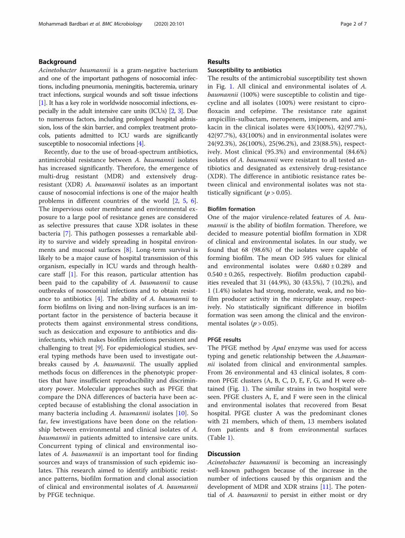
PFGE resultsThe PFGE method by ApaI enzyme was used for accesstyping and genetic relationship between the A.bauman-nii isolated from clinical and environmental samples.From 26 environmental and 43 clinical isolates, 8 com-mon PFGE clusters (A, B, C, D, E, F, G, and H were ob-tained (Fig. 1). The similar strains in two hospital wereseen. PFGE clusters A, E, and F were seen in the clinicaland environmental isolates that recovered from Besathospital. PFGE cluster A was the predominant cloneswith 21 members, which of them, 13 members isolatedfrom patients and 8 from environmental surfaces(Table 1).
DiscussionAcinetobacter baumannii is becoming an increasinglywell-known pathogen because of the increase in thenumber of infections caused by this organism and thedevelopment of MDR and XDR strains [11]. The poten-tial of A. baumannii to persist in either moist or dry
Mohammadi Bardbari et al. BMC Microbiology (2020) 20:101 Page 2 of 7

Fig. 1 Dendrogram cluster analysis of PFGE data for clinical and environmental A.baumannii isolates. Clin; clinical, Env; environmental, H1; Besathospital, H2; Behashti hospital, H3; Sina hospital, R; resistance, SAM; ampicillin-sulbactam, AK; amikacin, CIP; ciprofloxacin, MEM; meropenem, IPM;imipenem, CPM; cefepime, N; non-biofilm producer, W; weak, I; intermediate, and S; strong biofilm producer
Mohammadi Bardbari et al. BMC Microbiology (2020) 20:101 Page 3 of 7

conditions in the hospital environment is a consequenceof the presence of multiple antibacterial resistance genesand biofilm formation makes this bacterium a successfulpathogen among nosocomial bacteria [12]. The uniqueability of this bacterium to survive in the environmentfor a long time demonstrates its role in the outbreaks ofnosocomial infections [13]. Contaminated environmentalsurfaces can contribute directly to the transmission ofpathogens to patients or from the hands of health careworkers to patients [14].The results of this study show high environmental pol-
lution in the three intensive care units in our area. Theincidence of XDR A. baumannii isolated from environ-mental surfaces 22(84.6%) which, were resistant to alltested antibiotics was greater than that detected in previ-ous studies in Germany (7.3%), United States (9.8%), and13.1% in China [1, 13, 15]. These results can probably beattributed to inappropriate strategies of disinfection andhand washing by health workers in hospitals. Further-more, 41(95.3%) clinical isolates were resistant to alltested antibiotics and were XDR, which agrees withother investigations conducted in Iran [16, 17]. Of the 8antimicrobials tested, the most potent ones were colistinand tigecycline (100%) for all clinical and environmentalisolates. In agreement with previously research who re-quested, the most effective drug in controlling A. bau-mannii is polymyxin B [18].Of the 41 XDR A. baumannii strains isolated from pa-
tients’ respiratory tracts, the bacteria isolated from tra-cheal aspirate specimens were the most commonrespiratory isolates, which is consistent with previousstudies [19, 20]. Consistent with the earlier study, the re-sistance rate of clinical and environmental isolates of A.baumannii to antibiotics was 95–100% and there was nosignificant difference between antibiotic resistance inclinical and environmental isolates (p > 0.05) [21]. Oneof the important features related to the virulence of A.baumannii is its ability to form biofilms. In our study,we determined that 68 (98.6%) isolates of XDR A.
baumannii formed biofilm, which is in agreement withprevious studies [2, 17, 22]. According to our results,44.9% of isolates showed strong ability to biofilm forma-tion. Our results are consistent with previous reportswhich showed that more than 75% of A. baumannii iso-lates form biofilms [23, 24]. Previous studies have re-ported a positive relationship between biofilm formationand antibiotic resistance in A. baumannii isolates [17,25, 26]. In our study, all strong biofilm forming A. bau-mannii isolates were XDR.To track and evaluate the outbreaks, the genetic associ-
ation of the isolates, and to attribute one strain to the rele-vant clonal lineage, several molecular typing techniqueshave been developed [27, 28]. Among these methods,PFGE is considered the gold standard due to its discrimin-atory power, reproducibility, and sensitivity, and to deter-mine the single-colonal pattern of hospital outbreaks, theprevalence of pathogens within and between hospitals andtheir stability in the environment are used [28].In the current study typing of XDR A. baumannii iso-
lates was done for tracks of outbreak and analyses of apopulation survey of bacteria based on their genotypes,predominant genotypes, distribution and probabilitytransmission of isolates between patient and environ-mental surfaces. By the PFGE technique, 43 clinical and26 environmental A. baumannii isolates were typed.PFGE typing showed 8 different PFGE cluster (A, B, C,D, E, F, G, and H) with two major cluster A and G with21 (30.4%) and 6 (23.2%) members, respectively, whichcontains up to 53.6% of all isolates. In our study, a closegenetic relationship between clinical and environmentalisolates of A. baumannii was observed that is consistentwith other studies [4, 21]. These results indicate that thehospital environment is frequently colonized by differentA. baumannii clones, which may be responsible for thetransmission of A. baumannii isolates between patientsand their surroundings.In our study, two clinical isolates (No 5 and 31) which
were found in two distinct hospitals were clustered into
Table 1 The frequency distribution of A. baumannii in clinical and environmental isolates based on the location of specimencollection and PFGE type
Pattern Clinical isolates No (%) Environmental isolates No (%) Total No (%) Status of pulsotypes
A 13 (30.2) 8 (30.8) 21(30.4) Major pulsotype
B 7 (16.3) 3 (11.5) 10 (14.5) Intermediate pulsotype
C 2 (4.6) 2 (7.7) 4 (5.8) Minor pulsotype
D 5 (11.6) 4 (15.4) 9 (13.0) Intermediate pulsotype
E 2 (4.6) 1 (3.8) 3 (4.3) Minor pulsotype
F 2 (4.6) 1 (3.8) 3 (4.3) Minor pulsotype
G 10 (23.3) 6 (23.1) 16 (23.2) Major pulsotype
H 2 (4.6) 1 (3.8) 3 (4.3) Minor pulsotype
Total 43(100) 26(100) 69(100) –
Mohammadi Bardbari et al. BMC Microbiology (2020) 20:101 Page 4 of 7

pulsotype A. This issue may indicate the possible trans-fer of related isolates from one ICU to another in thesame hospital or different hospitals from patients admit-ted to the ICUs or the hospital health team in the samecity. This type of transmission has been reported in sev-eral countries [2, 29]. Comparing the frequency of bio-film formation ability in clinical and environmentalisolates with pulsotypes, no significant correlation wasfound, which is consistent with the study of Wroblewskaet al. [30].In our study, the correlation analysis of PFGE typing
and antibiotic resistance profiles showed that most iso-lates were XDR and no difference in antibiotic resistancewas found in the PFGE clusters. Therefore, there is nosignificant relationship between different PFGE clustersand antimicrobial resistance patterns. This indicates thatantimicrobial resistance patterns have low discriminatorypower for bacterial typing and highlights the necessity ofgenotyping techniques such as PFGE to categorize iso-lates with similar phenotypes and distinct genetic re-latedness during the evaluation of outbreak episodes orhorizontal transmission of isolates in the hospital envi-ronments [31].
ConclusionsOur investigation shown the high frequency of biofilmforming XDR A. baumannii with a high prevalence ofbiofilm formation. Tracing the sources of environmentalisolates indicates that there is a close genetic link be-tween environmental and clinical isolates of A. bauman-nii. Besides, it suggests that the occurrence of cross-contamination events is likely to occur between environ-mental and clinical isolates during routine clinical activ-ities. Therefore, the use of strict infection controlstrategies to reduce cross-contamination of endemicclones of A. baumannii isolates is essential.
MethodsBacterial isolatesIn this cross-sectional study, 43 MDR A. baumanniiwere collected from respiratory tracts of patients admit-ted to ICU wards of Besat, Sina, and Beheshti educa-tional hospitals of Hamadan University of MedicalSciences in Hamadan, west of Iran, during a period be-tween November 2015 and August 2016.The Besat hospital is a major tertiary referral hospital
where patients are referred from neighboring provincesand Sina and Beheshti hospitals have infectious and in-ternal medicine departments respectively, which acceptpatients in Hamadan province. Simultaneously, 26 MDRA. baumannii strains isolated from different environ-ments and equipment surfaces such as ventilators, sink,and ground, hands of Staff, trolleys, bedside table, pillowand linens. For sampling from the environment and
equipment of ICU wards, an area of about 10 cm2 wasselected and sampled using a sterile humidified swabwith physiological serum.
Culture and identificationAfter taking the samples, the swabs were inoculated inBrian heart infusion broth (BHI) media and incubatedovernight at 35 °C and further subcultured on MacCon-key’s agar plates at 37 °C for 24 h. The Acinetobacterspp. were identified by colony morphology, growth at44 °C, oxidase, OF (Oxidation and fermentation), Simoncitrate, and API 20NE system (BioMérieux Co, France).The A. baumannii isolates identification was confirmedby PCR of the blaOXA-51 gene. A. baumannii ATTC19606 was used as a reference strain [32].
Antibacterial susceptibility testAntimicrobial susceptibility test was accomplished by theKirby-Bauer disk diffusion method using the ampicillin/sulbactam (10 μg /10 μg), imipenem (10 μg), meropenem(10 μg), amikacin (30 μg), cefepime (30 μg), colistin(10 μg), tigecycline (15 μg), and ciprofloxacin (5 μg), anti-biotic disks (Mast Group Co, UK). The results interpretedaccording to Clinical and Laboratory Standard Instituteguidelines (CLSI) [33]. Pseudomonas aeruginosa ATCC27853 was used as a control strain. MDR A. baumanniiisolates were defined as resistant to three or more classesof antibiotics as previously described [34].
Biofilm assayThe ability of A. baumannii isolates to produce biofilmwas assessed by the microtiter plate method as previ-ously described [35]. Briefly, biofilm formation was per-formed in triplicate from overnight cultures diluted inTryptic soy broth (TSB) medium supplied with 1% glu-cose to an optical density (OD) of 0.01 at 600 nm anddeposited in 96-well plates. TSB medium without inocu-lum was used as a negative control. The plate was incu-bated at 37 °C for 24 h with gentle shaking. The wellswere washed three times with Phosphate Buffer Saline(PBS) solution. Absolute methanol was added per well tobiofilm fixation. Biofilm was stained with 1% crystal vio-let (w/v) and quantified at 595 nm after solubilizationwith absolute ethanol for 15 min at room temperature.Biofilm production was interpreted according to the cri-teria of Stepannovic et al. [36]. The optical density cut-off value (ODc) was established as three standard devia-tions (SD) above the mean of the optical density (OD) ofthe negative control as showed in the following formula:ODc = average OD of negative control+(3 SD of negativecontrol).The results were divided into the four following cat-
egories according to their optical densities as strong bio-film producer (4 ODc < OD); medium biofilm producer
Mohammadi Bardbari et al. BMC Microbiology (2020) 20:101 Page 5 of 7

(2 ODc < OD ≤ 4 ODc); weak biofilm producer (ODc <OD ≤ 2 ODc); and non-biofilm (OD ≤ODc) [36].
Pulsed-field gel electrophoresis typingGenetic similarities among clinical and environmentalisolates of A. baumannii were investigated by PFGE aspreviously described [37]. Briefly, an overnight culture ofbacteria was suspended in 100 μl of cell suspension bufferand was mixed with an identical volume of 2% low melt-ing agarose and distributed in a plug mold. GenomicDNA in agarose plugs was lysed in the cell lysis solutions Iand II, washed and digested with ApaI restriction enzyme(Thermo Scientific, USA). The Lambda PFG Ladder (NewEngland, Biolabs) was used as a DNA size marker. Electro-phoresis of digested DNA was performed in a pulsed-fieldelectrophoresis system (Chef Mapper; Bio-Rad Laborator-ies, USA) by programming two states with the followingconditions: temperature 14 °C; voltage 6 V/cm; switchangle, 120°; switch ramp 2.2–35 s for 19 h.
Cluster analysisGel images were studied by BioNumerics software ver-sion 7.5 (Applied Maths, StMartens-.Latem, Belgium). Dendrograms were obtained for all
of the isolates. A comparison of the banding patternswas done by the unweighted pair group method withmathematical averaging (UPGMA), and DNA similaritywas considered by using the band-based Dice coefficientwith a tolerance setting of 1.5% band tolerance and 1.5%optimization setting were applied during comparison ofthe DNA patterns. The PFGE results were compared ac-cording to the criteria by Tenover et al.; a PFGE clusterwas based on a similarity cutoff of 80% [38].
Statistical analysisStatistical analysis was performed using SPSS 23.0 (SPSS,Chicago, IL, USA). The frequency of susceptibility andbiofilm formation category were determined in clinicaland environmental isolates. The relationship among bio-film formation and the antibiotic resistance with PFGEtype were made using chi-square tests. A P-value of lessthan 0.05 was considered as statistically significant.
AbbreviationsMDR: Multidrug-resistant; PFGE: Pulsed Field Gel Electrophoresis;ICUs: Intensive care units; XDR: Extensively drug resistant; CLSI: Clinical andLaboratory Standard Institute guidelines; TSB: Tryptic soy broth; OD: Opticaldensity; PBS: Phosphate Buffer Saline
AcknowledgmentsThe authors would like to acknowledge Vice-chancellor of Research andTechnology, Hamadan University of Medical Sciences, Hamadan, Iran, andmicrobiology laboratory staff.
Authors’ contributionsMYA designed and organized this study and wrote the manuscript, PMperformed the experiments, MRA revised the manuscript, AMB and SAcollaborate in collecting samples and doing experiments. FK collaboration in
introducing patients and clinical examination MK analyzing the statistical resultsof the study, AK improvement of the discussion and English editing of therevised manuscript. All authors have read and approved the manuscript.
FundingThis research was supported by a grant from Vice-chancellor of Research andTechnology, Hamadan University of Medical Sciences, Hamadan, Iran (GrantNumber: 9412187238).The funder have no role in conducting this research.
Availability of data and materialsThe datasets used and/or analyzed during the current study available fromthe corresponding author on reasonable request.
Ethics approval and consent to participateThe ethics committee of the Hamadan University of Medical Sciencesapproved the study protocol (Ethical approval code: IR.UMSHA.REC.1394.531).The Written consent was obtained from all participants.
Consent for publicationNot applicable.
Competing interestsThe authors declare that they have no competing interests.
Author details1Department of Microbiology, Faculty of Medicine, Hamadan University ofMedical Sciences, Hamadan, Iran. 2Department of Microbiology, Faculty ofMedicine, Kermanshah University of Medical Sciences, kermanshah, Iran.3Department of Epidemiology, School of Public Health, Hamadan Universityof Medical Sciences, Hamadan, Iran. 4Department of Infectious Diseases,Faculty of Medicine, Hamadan University of Medical Sciences, Hamadan, Iran.5Brucellosis Research Center, Hamadan University of Medical Sciences,Hamadan, Iran. 6Division of Human Genetics, Department of Anatomy, St.John’s Hospital, Bangalore, India.
Received: 14 October 2019 Accepted: 19 April 2020
References1. Ying C, Li Y, Wang Y, Zheng B, Yang C. Investigation of the molecular
epidemiology of Acinetobacter baumannii isolated from patients andenvironmental contamination. J Antibiot. 2015;68(9):562–7.
2. Castilho SRA, de Miranda Godoy CS, Guilarde AO, Cardoso JL, André MCP,Junqueira-Kipnis AP, et al. Acinetobacter baumannii strains isolated frompatients in intensive care units in Goiânia, Brazil: molecular and drugsusceptibility profiles. PLoS One. 2017;12(5):e0176790.
3. Safari M, Nejad ASM, Bahador A, Jafari R, Alikhani MY. Prevalence of ESBLand MBL encoding genes in Acinetobacter baumannii strains isolated frompatients of intensive care units (ICU). Saudi J Biological Sci. 2015;22(4):424–9.
4. Gong Y, Shen X, Huang G, Zhang C, Luo X, Yin S, et al. Epidemiology andresistance features of Acinetobacter baumannii isolates from the wardenvironment and patients in the burn ICU of a Chinese hospital. J Microbiol.2016;54(8):551–8.
5. Li P, Li H, Lei H, Liu W, Zhao X, Guo L, et al. Rapid detection ofAcinetobacter baumannii and molecular epidemiology of carbapenem-resistant A. baumannii in two comprehensive hospitals of Beijing, China.Front Microbiol. 2015;6:997.
6. Safari M, Saidijam M, Bahador A, Jafari R, Alikhani MY. High prevalence ofmultidrug resistance and metallo-beta-lactamase (MβL) producingAcinetobacter baumannii isolated from patients in ICU wards, Hamadan,Iran. J Res Health Sci. 2013;13(2):162–7.
7. Eliopoulos GM, Maragakis LL, Perl TM. Acinetobacter baumannii:epidemiology, antimicrobial resistance, and treatment options. Clin InfectDis. 2008;46(8):1254–63.
8. Chen Z, Liu W, Zhang Y, Li Y, Jian Z, Deng H, et al. Molecular epidemiologyof carbapenem-resistant Acinetobacter spp. from XiangYa hospital, inHunan Province, China. J Basic Microbiol. 2013;53(2):121–7.
9. Kaliterna V, Kaliterna M, Hrenović J, Barišić Z, Tonkić M, Goic-Barisic I.Acinetobacter baumannii in southern Croatia: clonal lineages, biofilmformation, and resistance patterns. Infect Dis Ther. 2015;47(12):902–7.
Mohammadi Bardbari et al. BMC Microbiology (2020) 20:101 Page 6 of 7

10. Dehbalaei MA, Najar-Peerayeh S, Taherikalani M, Behmanesh M. ClinicalIsolates of Acinetobacter baumannii From Tehran Hospitals: Pulsed-field GelElectrophoresis Characterization, Clonal Lineages, Antibiotic Susceptibility,and Biofilm-forming Ability. Jundishapur J Microbiol. 2017;10(7).
11. McConnell MJ, Actis L, Pachón J. Acinetobacter baumannii: humaninfections, factors contributing to pathogenesis and animal models. FEMSMicrobiol Rev. 2013;37(2):130–55.
12. Doughari HJ, Ndakidemi PA, Human IS, Benade S. The ecology, biology andpathogenesis of Acinetobacter spp.: an overview. Microbes Environ. 2011;26(2):101–12.
13. Thom KA, Johnson JK, Lee MS, Harris AD. Environmental contaminationbecause of multidrug-resistant Acinetobacter baumannii surroundingcolonized or infected patients. Am J Infect Control. 2011;39(9):711–5.
14. Daef EA, Mohamad IS, Ahmad AS, El-Gendy SG, Ahmed EH, Sayed IM.Relationship between clinical and environmental isolates of Acinetobacterbaumannii in Assiut University hospitals. J Am Sci. 2013;9(11s):67–73.
15. Lemmen S, Häfner H, Zolldann D, Stanzel S, Lütticken R. Distribution ofmulti-resistant gram-negative versus gram-positive bacteria in the hospitalinanimate environment. J Hosp Infect. 2004;56(3):191–7.
16. Saffari F, Monsen T, Karmostaji A, Bahadori Azimabad F, Widerström M.Significant spread of extensively drug-resistant Acinetobacter baumanniigenotypes of clonal complex 92 among intensive care unit patients in auniversity hospital in southern Iran. J Med Microbiol. 2017;66(11):1656–62.
17. Zeighami H, Valadkhani F, Shapouri R, Samadi E, Haghi F. Virulence characteristicsof multidrug resistant biofilm forming Acinetobacter baumannii isolated fromintensive care unit patients. BMC Infect Dis. 2019;19:629.
18. Hassan PA, Khider AK. Correlation of biofilm formation and antibioticresistance among clinical and soil isolates of Acinetobacter baumannii inIraq. Acta Microbiol Immunol Hung. 2019:1–10.
19. Behnia M, Logan SC, Fallen L, Catalano P. Nosocomial and ventilator-associated pneumonia in a community hospital intensive care unit: aretrospective review and analysis. BMC Res Notes. 2014;7(1):1.
20. Ebrahimi M, Khansari-nejad B, Ghaznavi-Rad E. High frequency of ventilatorassociated pneumonia nosocomial co-infection caused by methicillinresistant Staphylococcus aureus and carbapenem resistant A. baumannii inintensive care unit. J Iranian Clin Res. 2015;1(2):67–71.
21. Uwingabiye J, Lemnouer A, Roca I, Alouane T, Frikh M, Belefquih B, et al.Clonal diversity and detection of carbapenem resistance encoding genesamong multidrug-resistant Acinetobacter baumannii isolates recoveredfrom patients and environment in two intensive care units in a Moroccanhospital. Antimicrob Resist Infect Control. 2017;6(1):99.
22. Gurung J, Khyriem AB, Banik A, Lyngdoh WV, Choudhury B, Bhattacharyya P.Association of biofilm production with multidrug resistance among clinicalisolates of Acinetobacter baumannii and Pseudomonas aeruginosa fromintensive care unit. Indian J Critical Care Med. 2013;17(4):214.
23. Thummeepak R, Kongthai P, Leungtongkam U, Sitthisak S. Distribution ofvirulence genes involved in biofilm formation in multi-drug resistantAcinetobacter baumannii clinical isolates. Int Microbiol. 2016;19(2):121–9.
24. Youn SJ. Molecular characterization and antimicrobial susceptibility ofbiofilm-forming Acinetobacter baumannii clinical isolates from Daejeon,Korea. Korean J Clin Lab Sci. 2018;50(2):100–9.
25. Qi L, Li H, Zhang C, Liang B, Li J, Wang L, Du X, Liu X, Qiu S, Song H.Relationship between antibiotic resistance, biofilm formation, and biofilmspecific resistance in Acinetobacter baumannii. Front Microbiol. 2016;7:483.
26. Babapour E, Haddadi A, Mirnejad R, Angaji S-A, Amirmozafari N. Biofilmformation in clinical isolates of nosocomial Acinetobacter baumannii and itsrelationship with multidrug resistance. Asian Pac J Trop Biomed. 2016;6(6):528–33.
27. Chang K-M, Huang W-C, Chiou C-S, Shen G-H, Huang C-C, Wen F-S. Suitablerestriction enzyme for standardization of pulsed-field gel electrophoresisprotocol and interlaboratory comparison of Acinetobacter baumannii. JMicrobiol Immunol Infect. 2013;46(3):195–201.
28. Rafei R, Dabboussi F, Hamze M, Eveillard M, Lemarié C, Gaultier M-P, et al.Molecular analysis of Acinetobacter baumannii strains isolated in Lebanonusing four different typing methods. PLoS One. 2014;9(12):e115969.
29. Fournier PE, Richet H. The epidemiology and control of Acinetobacterbaumannii in health care facilities. Clin Infect Dis. 2006;42(5):692–9.
30. Wroblewska MM, Sawicka-Grzelak A, Marchel H, Luczak M, Sivan A. Biofilmproduction by clinical strains of Acinetobacter baumannii isolatedfrompatients hospitalized in two tertiary care hospitals. FEMS Immunol MedMicrobiol. 2008;53(1):140–4.
31. Ying J, Lu J, Zong L, Li A, Pan R, Cheng C, et al. Molecular epidemiologyand characterization of genotypes of Acinetobacter baumannii isolates fromregions of South China. Japanese J Infect Dis. 2016;69(3):180–5 27.
32. Oh MH, Lee JC, Kim J, Choi CH, Han K. Simple method for markerless genedeletion in multidrug-resistant Acinetobacter baumannii. Appl EnvironMicrobiol. 2015;81(10):3357–68.
33. Performance standards for antimicrobial susceptibility testing. Wayne, PA:Clinical and Laboratory Standards Institute; 2018.
34. Azizi O, Shakibaie MR, Modarresi F, Shahcheraghi F. Molecular detection ofclass-D OXA carbapenemase genes in biofilm and non-biofilm forming clinicalisolates of Acinetobacter baumannii. Jundishapur J Microbiol. 2015;8(1).
35. Bardbari AM, Arabestani MR, Karami M, Keramat F, Aghazadeh H, Alikhani MY,et al. Highly synergistic activity of melittin with imipenem and colistin inbiofilm inhibition against multidrug-resistant strong biofilm producer strains ofAcinetobacter baumannii. Eur J Clin Microbiol Infect Dis. 2018:1–12.
36. Stepanović S, Vuković D, Hola V, Bonaventura GD, Djukić S, Ćirković I, et al.Quantification of biofilm in microtiter plates: overview of testing conditionsand practical recommendations for assessment of biofilm production bystaphylococci. Apmis. 2007;115(8):891–9.
37. Mohajeri P, Farahani A, Feizabadi M, Norozi B. Clonal evolution multi-drugresistant Acinetobacter baumannii by pulsed-field gel electrophoresis.Indian J Med Microbiol. 2015;33(1):87.
38. Tenover FC, Arbeit RD, Goering RV, et al. Interpreting chromosomal DNArestriction patterns produced by pulsed-field gel electrophoresis: criteria forbacterial strain typing. J Clin Microbiol. 1995;33:2233–9.
Publisher’s NoteSpringer Nature remains neutral with regard to jurisdictional claims inpublished maps and institutional affiliations.
Mohammadi Bardbari et al. BMC Microbiology (2020) 20:101 Page 7 of 7