Embed Size (px)
Citation preview
1996 88: 386-401
E Yang and SJ Korsmeyer Molecular thanatopsis: a discourse on the BCL2 family and cell death
http://www.bloodjournal.org/content/88/2/386.citation.full.htmlUpdated information and services can be found at:
Articles on similar topics can be found in the following Blood collections
http://www.bloodjournal.org/site/misc/rights.xhtml#repub_requestsInformation about reproducing this article in parts or in its entirety may be found online at:
http://www.bloodjournal.org/site/misc/rights.xhtml#reprintsInformation about ordering reprints may be found online at:
http://www.bloodjournal.org/site/subscriptions/index.xhtmlInformation about subscriptions and ASH membership may be found online at:
Copyright 2011 by The American Society of Hematology; all rights reserved.Society of Hematology, 2021 L St, NW, Suite 900, Washington DC 20036.Blood (print ISSN 0006-4971, online ISSN 1528-0020), is published weekly by the American
For personal use only.on November 3, 2014. by guest www.bloodjournal.orgFrom For personal use only.on November 3, 2014. by guest www.bloodjournal.orgFrom
REVIEW ARTICLE
Molecular Thanatopsis: A Discourse on the BCLZ Family and Cell Death
By Elizabeth Yang and Stanley J. Korsmeyer
BCL2 PREVENTS MULTIPLE FORMS OF CELL DEATH AND DEFINES A NEW CLASS OF ONCOGENES
IGHTY-FIVE PERCENT of follicular lymphomas and E 20% of diffuse B-cell lymphomas have a characteristic t( 14; 18) translocation.’.’ In this translocation, the proto-on- cogene BCL2 at chromosome segment 18q21 is juxtaposed with the Ig heavy chain locus at 14q32, resulting in deregu- lated expression of BCL2.3-6 The discovery that BCL2, unlike oncogenes studied previously, functions in preventing pro- grammed cell death (PCD) instead of promoting proliferation established a new class of oncogene^.^.^
The initial observation of BCL2’s ability to enhance cell survival was that overex- pression of BCL2 increased the viability of certain cytokine- dependent cells upon cytokine withdrawal. In interleukin-3 (IL-3)-dependent pro-B -cell lines and promyeloid cell lines, BCL2 overexpression prolonged cell survival upon IL- 3 withdrawal and maintained the cells in Go7,’ The observa- tion was extended to IL-4- and granulocyte-macrophage colony-stimulating factor (GM-CSF)-dependent cells’ and in certain IL-2-dependent” and IL-6-dependent” cells. BCL2 was also capable of protecting T cells against a variety of apoptotic signals, including glucocorticoids, y -irradiation, phorbol esters, ionomycin, and cross-linking of cell surface molecules by anti-CD3 antibody. The protective effects were observed in T-cell hybridomas transfected with BCL2 and in thymocytes and peripheral T cells from transgenic mice with expression of BCL2 under the control of the proximal promoter of lck (ZC~J’?’~ or the Ig heavy chain enhancer
Overexpression of BCL2 alters lymphoid development and leads to neoplasia. The in vivo effects of BCL2 were ini- tially investigated using transgenic mice with BCL2 overex- pression targeted to B cells or to T cells. Transgenic mice bearing a BCL2-Zg minigene harbor expanded B-cell com- partments. Mice in which the BCLZ transgene expression is targeted to T cells by the lck proximal promoter exhibit increased CD3med and increased CD4-CD8+ single-positive thymocytes compared with littermate control^.'^ When the BCL2 transgene is expressed in B lymphocytes, the mice develop follicular hyperplasia, some of which progress to high-grade monoclonal (Fig 1). When ex- pression is directed to T cells, fully one third of the mice
BCLZ prolongs cell survival.
(E,).’4
From the Howard Hughes Medical Institute, Division of Molecu- lar Oncology, Departments of Medicine, Pathology, and Pediatrics, Washington University School of Medicine, St Louis, MO.
Submitted August 9, 1995; accepted February 16, 1996. E.Y. is supported by a Pjzer fellowship. Address reprint requests to Stanley J . Korsmeyer, MD, Howard
Hughes Medical Institute, Division of Molecular Oncology, Depart- ments of Medicine, Pathology, Washington University School of Medicine, 660 S Euclid Ave, Box 8022, St Louis, MO 63110. 0 1996 by The American Society of Hematology. 0006-4971/96/8802-0036$3.00/0
develop peripheral T-cell lymphomas” (Fig 1). A long la- tency and progression from polyclonal hyperplasia to mono- clonal malignancy are consistent with the hypothesis that oncogenic events in addition to BCL2 overexpression are necessary for tumor formation. In lymphomas arising in BCL2-Zg transgenic mice, a common second hit is transloca- tion of the Myc oncogene.I6 (The interaction between Myc and BCLZ will be specifically discussed in a later section.) These transgenic mice experiments illustrated that cell death is normally a well-regulated process in lymphoid develop- ment and that lack of cell death is tumorigenic. Deleterious mutations that would have resulted in cell death can be re- tained when apoptosis is inhibited. The progression to lymphoma in these BCL2 transgenic mice constitutes in vivo evidence that the t( 14;18) and BCL2 overexpression play a primary role in oncogenesis.
Prompted by these studies, BCL2 has been found to protect against death in a variety of cell types. Notably, BCL2 protects against neuronal cell death induced by various apoptotic stimuli. BCL2 inhibited apoptosis in PC 12 pheochromocy- toma cells after nerve growth factor (NGF) withdrawal.”.” Microinjection of a BCLZ construct driven by the neuron- specific enolase promoter into cultured rat sympathetic neu- rons also resulted in the prevention of programmed death after NGF deprivation.22 Other experiments suggested that not all neuronal cell deaths are inhibitable by BCL2. For example, BCL2 rescued embryonic chick sensory neurons dependent on nerve growth factor, brain-derived neuro- trophic factor, and neurotrophin-3, but not ciliary neurons dependent on ciliary neurotrophic factor.z3 In addition to growth factor dependency, BCL2 has been shown to counter death in a neuronal cell line after serum and glucose with- drawal, membrane peroxidation, and treatment with calcium ionophore and menadione, an inducer of free radical forma- tion.” In animal models, overexpression of BCL2 under the neuron-specific enolase or phosphoglycerate kinase pro- moter led to neuronal hypertrophy by decreasing naturally occurring cell death. The brains from transgenic animals were larger than wild-type littermates by 12%.25 The number of motoneurons in the facial nucleus and the ganglion cell layer of the retina was increased by 40% to 50%.25 Overex- pression of BCL2 in these animals also protected against experimental cell death. This is evidenced by a 50% reduc- tion in the volume of brain infarction in transgenic mice after occlusion of the middle cerebral arteryz5 and by continued survival of facial motoneurons after axotomy in transgenic animals, whereas those in wild-type mice degeneratesz6 The role of BCLZ in normal neuronal physiology has been ex- plored in Bcl2-deficient mice. Superior cervial ganglion cells from Bc12-’- mice died more rapidly after NGF deprivation than those from wild-type mice,27 suggesting that BCLZ is an important regulator of sympathetic neuron survival during the period of naturally occurring programmed neuronal death.
BCL2 protects against neuronal cell deaths.
386 Blood, VOI 88, NO 2 (Jul~lB), 1996: pp 386-401
For personal use only.on November 3, 2014. by guest www.bloodjournal.orgFrom

MOLECULAR THANATOPSIS: BCL2 387
-Q- contld(n=56) - bd-2-lg (-133) - Idc-bd-Z(rid8) - Q - I I Y C ( ~ Z ~ ) - b d - 2 - l ~ + Q - ~ c ( ~ l Z )
0 10 20 age (months)
Fig 1. BCU overexpression leads to neoplasia, which is syner- gized by EMYCoverexpression. Cumulative tumor incidence in BCLZ- ig, Ickp'-BCLZ, and E,-MYC transgenic mice and BCLZ-lg + E,-MYC double transgenic mice compared with littermate c o n t r o l ~ . ' ~ ' ~ ~ ' ' ~
Virus-induced cell death can be blocked by BCL2. Upon infection of host cells, adenovirus expresses several virally encoded genes. Expression of the adenovirus E1A oncogene alone stimulates host cell proliferation accompanied by apoptosis, which can be p53 dependent.28*29 Simultaneous expression of the E1B 19-kD protein suppresses E1A-in- duced apoptosis,2* allowing foci formation after adenovirus infection. BCL2 shares limited homology with E1B 19-kD protein and can substitute for its ability to inhibit E1A-in- duced cell death.30,3' This is an example of BCL2's ability to repress cell death due to abnormal proliferation. BCL2 can also block apoptosis in serum-deprived cells expressing the Tax protein of human T-cell leukemia virus human T- cell lymphotropic virus type 1 (HTLV-1).32 In another sys- tem, BCL2 is able to inhibit programmed cell death induced by lytic infection of the alphavirus, Sindbis, allowing the establishment of persistent viral infection.33
Numer- ous examples now exist in which apoptosis due to external toxic stimuli can be rescued by BCL2 (Table 1). An interest- ing system is the "apoptosis" of nuclei in cell-free Xenopus oocyte extracts, which can be inhibited by BCL2.34 This in vitro system offers the potential for dissecting individual components of apoptosis. Other examples in which BCL2 plays a role include transforming growth factor 0 (TGFP)- induced growth arrest and cell death in M1 and chemotherapeutic drug-induced apoptosis in cancer
Despite numerous positive examples, BCL2 does not prevent every cell death. BCL2 does not have a substantial effect on negative selection
BCL2 functions in multiple cell death systems.
BCL2 does not repress all cell deaths.
of thymocyte^.'^ Also, it does not easily prevent apoptosis in targets of cytotoxic T-cell killing."5 However, observations that BCL2 can occasionally affect outcomes by these stimuli suggest that results can be dose-related. Because BCL2 is able to inhibit apoptosis resulting from so many different signals and intracellular pathways, it must act after the con- vergence of many signals in the apoptotic pathway. Because overexpression of BCL2 does not protect every example of cell death, it is theorectically possible that more than one distal pathway of cell death exists. Alternatively, individual BCL2 family members may prove more effective in certain contexts than others.
BIOCHEMICAL AND CELL BIOLOGICAL STUDIES OF BCL2 ACTIVITY
BCL2's ful l activity requires an integral membrane posi- tion. The carboxy terminus of BCL2 contains a hydropho- bic 19-amino acid stretch reminiscent of a membrane spanning domain. Subcellular fractionation, immuno- fluorescence, and confocal microscopy studies using anti-
Table 1. Cell Deaths Repressed by BCL-2
Lymphoid Factor withdrawal-IL-2, IL-3, IL-4, IL-6, GM-CSF Glucocorticoid y Irradiation Phorbol esters Calcium Cross-linking by anti-CD3
Factor withdrawal-NGF, BDNF, Neurotrophin-3 Serum withdrawal Calcium Infarction Axotomy Naturally occurring cell death
Fibroblasts Serum deprivation and MYC induction
Oncogene-related MYC-induced E l A-induced p53-mediated
Viral infections Adenovirus Sindbis virus
Neuronal
HTLV-1 Chemotherapeutic drugs
DNA synthesis inhibitors Alkylating agents Topoisomerase inhibitors Microtubule inhibitors Antimetabolites
Oxidant stress H A Menadione Membrane peroxidation
Others TGF-8 Staurosporine Loss of extracellular matrix
For personal use only.on November 3, 2014. by guest www.bloodjournal.orgFrom
388 YANG AND KORSMEYER
BCL2 antibodies indicated that BCL2 is an intracellular membrane protein whose distribution varies somewhat de- pending on cell type. BCL2 has been most convincingly localized to mitochondria, its predominant site in hematopoi- etic cells, as well as smooth endoplasmic reticulum and peri- nuclear m e m b ~ a n e . ' ' * ~ ~ ~ Targeting studies using purified mi- tochondria and in vitro-translated BCL2 protein showed that the carboxy terminus functions as a signal anchor sequence responsible for targeting and insertion into the mitochondrial outer membrane. This exposes most of the polypeptide to the cytosol, in which it remains sensitive to protease dige~tion.~' BCL2 devoid of the signal anchor sequence is only partially functional in protection against apoptosis. However, a por- tion of the truncated BCL2 is still bound to its membrane- associated heterodimerizing partner, BAX.51 Substitution of the BCL2 mitochondrial anchor sequence with the yeast outer membrane protein Mas70p signal anchor sequence re- targets the protein into the mitochondrial outer membrane and fully restores BCL2's activity, as measured by the ability to inhibit E l A-induced cytotoxi~ity.~~ A fusion protein of BCL2pm-2 receptor transmembrane domain produced sim- ilar results.53 These studies argue that BCL2's full function depends on its subcellular membrane localization. Most of the amino portion of BCL2 is exposed, in which it may interact with proteins in the cytosol or other BCL2-like mole- cules similarly anchored in the mitochondria. BCL2 function is not dependent on an intact electron transportloxidative phosphorylation chain, as is shown by BCL2's ability to block apoptosis in cells lacking mitochondrial DNA and unable to carry out electron
The mito- chondrial outer membrane, the endoplasmic reticulum, and the nuclear envelope are all sites implicated in the production of reactive oxygen species (ROS). The localization of BCL2 to these sites prompted investigation into the role of ROS in programmed cell death. BCL2 can protect cells against H202 and t-butyl hydroperoxide or menadione, which gener- ate 02-.s's5s At low concentrations, these oxidant stresses kill cells by an apoptotic process. Agents that decrease reactive oxygen species, such as N-acetylcysteine, glutathione perox- idase, and desfemoxamine, can partially protect against apopt~sis.~' Furthermore, BCL2 can protect against death induced by agents that decrease intracellular glutathione (GSH), such as buthionine sulfoximine and ethacrynic This suggests that reactive oxygen species may be involved in apoptotic pathways rescuable by BCL2. The endogenous production of intracellular peroxides, as measured by the conversion of the oxidation-sensitive fluorescent dye DCFH to DCF, is not significantly changed in the presence of BCL2. BCL2 also does not have a significant effect on the genera- tion of superoxide, 02-. BCL2 does inhibit lipid peroxida- tion, a downstream event in oxidative damage and a frequent accompaniment of apopt~sis.~' However, subsequent reports of BCL2's ability to rescue cells from programmed cell death occurring under hypoxic conditions in which the generation of ROS is greatly reduced suggest that ROS are not essential for PCD.56,57 Thus, BCL2's death repressor function does not solely depend on the protection of cellular constituents from oxidative damage. Although BCL2 can block oxidant-
BCL2 can inhibit oxidant induced apoptosis.
induced apoptosis, in the absence of a proven biochemical activity, it remains an open question whether BCL2 has a direct or indirect role on the oxidant pathway.
Another area of investigation into BCL2 function that relates to BCL2's lo- calization to the endoplasmic reticulum is intracellular cal- cium homeostasis. Caz+ has been implicated in apoptosis because of the Ca" dependence of certain internucleosomal DNA fragmentations and the ability of Ca ionophores A23187 and ionomycin to induce lymphocyte apoptosis. Al- though the total cellular Ca2+ content has not been consis- tently shown to change with the induction of cell death, a redistribution of intracellular Caz+ can result.s8 Studies using thapsigargin, an inhibitor of the ER-associated Ca2+ pump, indicated that apoptosis is associated with an efflux of Ca2+ from the ER into the cytosol and that BCL2 can block this flux of CaZ+ across the ER Although intrigu- ing, mobilization of intracellular Ca" stores is but one step in the complex cell death pathway. Whether BCL2's effect on calcium homeostasis is direct or indirect is still uncertain.
BCL2 and intracellular calcium fluxes.
BCLZ FOUNDS A FAMILY OF CELL DEATH REGULATORS
BAX, a heterodimerizing partner of BCL2, is a death pro- moter. Identification of a number of BCL2 homologs, some of which bind to BCL2, suggests that BCL2 functions, at least in part, through protein-protein interactions. The first of these homologs, BAX, was identified by coimmunopreci- pitation with BCL2 protein. BAX is a 21-kD protein that shares homology with BCL2 principally clustered in two conserved regions, BH1 and BH2 (Fig 2). In addition, an exon juncture in BH2 is conserved. BAX heterodimerizes with BCL2 and homodimerizes with itself.6' Site-directed mutagenesis of BH1 and BH2 in BCLZ showed that these two domains were important for binding to BAX. When binding was disrupted, BCL2's protective function was also eliminated, suggesting that BCL2 must bind BAX to exert its effect. Most noteworthy are the substitutions of a single amino acid Gly145 in BH1 with either alanine or glutamic acid and Trp188 with alanine in BH2, which completely disrupted binding to BAX and abrogated BCL2's death-re- pressor effect.62 When BAX was overexpressed in cells, apo- ptotic death in response to a death signal was accelerated, earning its designation as a death promoter. When BCL2 was overexpressed, it heterodimerized with BAX and death was repressed.6' Thus, the ratio of BCL2 to BAX determines the amount of BCL2BAX heterodimers versus BAX/BAX homodimers and is important in determining susceptibility to apoptosis (Fig 3). BAX protein contains a hydrophobic carboxy terminus like BCL2 and has been colocalized to mitochondria with BCL2 (unpublished observations). BAX is widely expressed in tissues, including a number of sites in which cells die during normal matura t i~n .~ ' .~~ Moreover, the BCL2 to BAX ratio varies during the developmental history of a given lineage, such as T lymphocytes. For exam- ple, BCL2 is present in the immature, double-negative thy- mocytes and in the mature, single-positive T cells. However, it is absent at the double-positive stage when selection oc-
For personal use only.on November 3, 2014. by guest www.bloodjournal.orgFrom
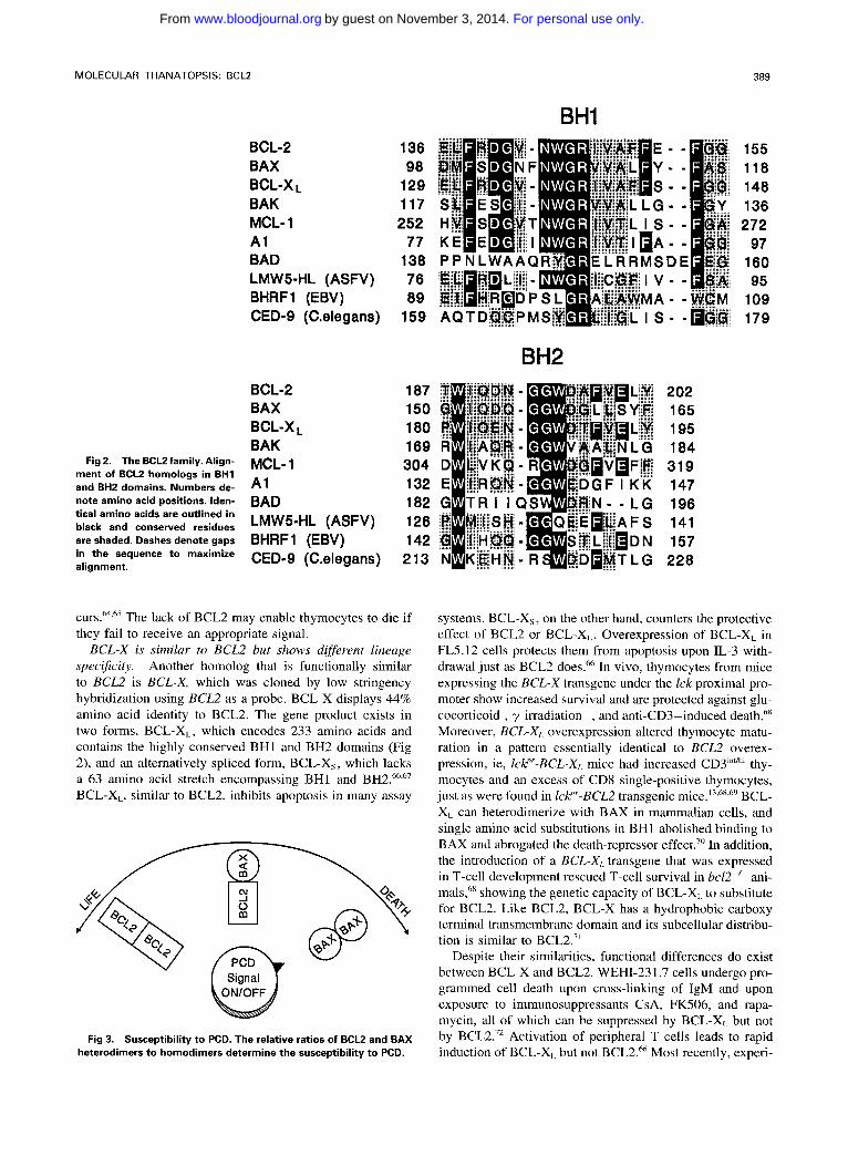
MOLECULAR THANATOPSIS: BCLP 389
BHI
Fig 2. The BCL2 family. Align- ment of BCLZ homologs in BH1 and BH2 domains. Numbers de- note amino acid positions. Iden- tical amino acids are outlined in black and conserved residues are shaded. Dashes denote gaps in the sequence to maximize alignment.
BCL-2 BAX
BAK MCL- 1 A1 BAD LMW5-HL (ASFV) BHRF 1 (EBV) CED-9 (C-elegans)
BCL-X L
BCL-2 BAX
BAK
A 1 BAD LMW5-HL (ASFV) BHRF 1 (EBV) CED-9 (C.elegans)
BCL-X L
MCL- 1
136 98
129 117 252
77 138 76 89
159
187 150 180 169 304 132 182 126 142
213
C U ~ S . ~ ~ . ' ~ The lack of BCL2 may enable thymocytes to die if they fail to receive an appropriate signal.
BCL-X is similar to BCLZ but shows different lineage spec$city. Another homolog that is functionally similar to BCL2 is BCL-X, which was cloned by low stringency hybridization using BCL2 as a probe. BCL-X displays 44% amino acid identity to BCL2. The gene product exists in two forms, BCL-XL, which encodes 233 amino acids and contains the highly conserved BHI and BH2 domains (Fig 2), and an alternatively spliced form, BCL-X,, which lacks a 63 amino acid stretch encompassing BH1 and BH2.663'7 BCL-XL, similar to BCL2, inhibits apoptosis in many assay
Fig 3. Susceptibility to PCD. The relative ratios of BCLZ and BAX heterodimers to homodimers determine the susceptibility to PCD.
BH2 2 02 165 195 184 319 147 196 141 157 228
systems. BCL-Xs, on the other hand, counters the protective effect of BCL2 or BCL-XL. Overexpression of BCL-XL in FL5.12 cells protects them from apoptosis upon IL-3 with- drawal just as BCL2 does.66 In vivo, thymocytes from mice expressing the BCL-X transgene under the Zck proximal pro- moter show increased survival and are protected against glu- cocorticoid-, y-irradiation-, and anti-CD3-induced death." Moreover, BCL-XL overexpression altered thymocyte matu- ration in a pattern essentially identical to BCL2 overex- pression, ie, ZcV"BCL-XL mice had increased CD3i"'h' thy- mocytes and an excess of CD8 single-positive thymocytes, just as were found in lcP-BCL2 transgenic mice.'',"8,6' BCL- X,. can heterodimerize with BAX in mammalian cells, and single amino acid substitutions in BH1 abolished binding to BAX and abrogated the death-repressor effect.70 In addition, the introduction of a BCL-X,, transgene that was expressed in T-cell development rescued T-cell survival in bcZ2"- ani- mals," showing the genetic capacity of BCL-XL, to substitute for BCL2. Like BCL2, BCL-X has a hydrophobic carboxy terminal transmembrane domain and its subcellular distribu- tion is similar to BCL2.7'
Despite their similarities, functional differences do exist between BCL-X and BCL2. WEHI-23 1.7 cells undergo pro- grammed cell death upon cross-linking of IgM and upon exposure to immunosuppressants CsA, FK506, and rapa- mycin, all of which can be suppressed by BCL-XL but not by BCL2." Activation of peripheral T cells leads to rapid induction of BCL-X,- but not BCL2." Most recently, experi-
For personal use only.on November 3, 2014. by guest www.bloodjournal.orgFrom
390 YANG AND KORSMEYER
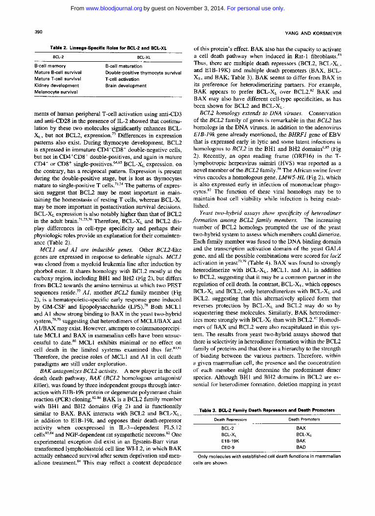
Table 2. Lineage-Specific Roles for BCL-2 and BCL-XL
BCL-2 BCL-XL
B-cell memory B-cell maturation Mature B-cell survival Mature T-cell survival T-cell activation Kidney development Brain development Melanocyte survival
Double-positive thymocyte survival
ments of human peripheral T-cell activation using anti-CD3 and anti-CD28 in the presence of IL-2 showed that costimu- lation by these two molecules significantly enhances BCL- XL, but not BCL2, expres~ion .~~ Differences in expression patterns also exist. During thymocyte development, BCL2 is expressed in immature CD4-CD8- double-negative cells, but not in CD4+CD8+ double-positives, and again in mature CD4' or CD8+ s ingle-po~i t ives .~~~~ BCL-XL expression, on the contrary, has a reciprocal pattern. Expression is present during the double-positive stage, but is lost as thymocytes mature to single-positive T ~ e l l s . ~ ' , ~ ~ The patterns of expres- sion suggest that BCL2 may be most important in main- taining the homeostasis of resting T cells, whereas BCL-XL may be more important in postactivation survival decisions. BCL-XL expression is also notably higher than that of BCL2 in the adult brain.7',7',76 Therefore, BCL-XL and BCL2 dis- play differences in cell-type specificity and perhaps their physiologic roles provide an explanation for their comainten- ance (Table 2).
Other BCL2-like genes are expressed in response to definable signals. MCLl was cloned from a myeloid leukemia line after induction by phorbol ester. It shares homology with BCL2 mostly at the carboxy region, including BH1 and BH2 (Fig 2), but differs from BCL2 towards the amino terminus at which two PEST sequences reside.77 A l , another BCL2 family member (Fig 2), is a hematopoietic-specific early response gene induced by GM-CSF and lipopolysaccharide (LPS).78 Both MCLl and A1 show strong binding to BAX in the yeast two-hybrid ~ y s t e m , ~ ' . ~ ~ suggesting that heterodimers of MCLUBAX and A 1BAX may exist. However, attempts to coimmunoprecipi- tate MCLl and BAX in mammalian cells have been unsuc- cessful to date.80 MCLl exhibits minimal or no effect on cell death in the limited systems examined thus far."~s' Therefore, the precise roles of MCLl and A1 in cell death paradigms are still under exploration.
BAK antagonizes BCL2 activity. A new player in the cell death death pathway, BAK (BCL2 homologous antagonist/ killer), was found by three independent groups through inter- action with E1B-19k protein or degenerate polymerase chain reaction (PCR) c l ~ n i n g . ~ ~ - * ~ BAK is a BCL2 family member with BH1 and BH2 domains (Fig 2) and is functionally similar to BAX. BAK interacts with BCL2 and BCL-XL, in addition to ElB-lgk, and opposes their death-repressor activity when coexpressed in IL-3-dependent FL5.12 ~ e l l s ~ ~ . ~ ~ and NGF-dependent rat sympathetic neurons.82 One experimental exception did exist in an Epstein-Barr virus- transformed lymphoblastoid cell line WI-L2, in which BAK actually enhanced survival after serum deprivation and men- adione treatment.x4 This may reflect a context dependence
MCLl and A1 are inducible genes.
of this protein's effect. BAK also has the capacity to activate a cell death pathway when induced in Rat-1 fibroblast^.'^ Thus, there are multiple death repressors (BCL2, BCL-XL, and E1B-19K) and multiple death promoters (BAX, BCL- Xs, and BAK; Table 3). BAK seems to differ from BAX in its preference for heterodimerizing partners. For example, BAK appears to prefer BCL-XL over BCL2." BAK and BAX may also have different cell-type specificities, as has been shown for BCL2 and BCL-XL.
Conservation of the BCLZ family of genes is remarkable in that BCL2 has homologs in the DNA viruses. In addition to the adenovirus ElB-19k gene already mentioned, the BHRFl gene of EBV that is expressed early in lytic and some latent infections is homologous to BCL2 in the BH1 and BH2 domains6," (Fig 2). Recently, an open reading frame (ORF16) in the T- lymphotropic herpesvirus saimiri (HVS) was reported as a novel member of the BCL2 family.86 The African swine fever virus encodes a homologous gene, LMWS-HL (Fig 2), which is also expressed early in infection of mononuclear phago- c y t e ~ . ~ ~ The function of these viral homologs may be to maintain host cell viability while infection is being estab- lished.
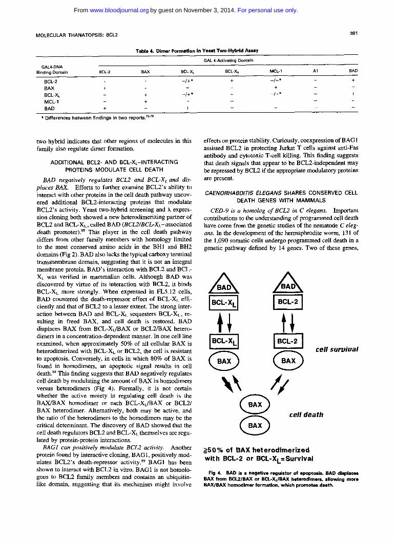
Yeast two-hybrid assays show speci$city of heterodimer formation among BCL2 family members. The increasing number of BCL2 homologs prompted the use of the yeast two-hybrid system to assess which members could dimerize. Each family member was fused to the DNA binding domain and the transcription activation domain of the yeast GAL4 gene, and all the possible combinations were scored for lacZ activation in (Table 4). BAX was found to strongly heterodimerize with BCL-XL, MCLl, and AI, in addition to BCL2, suggesting that it may be a common partner in the regulation of cell death. In contrast, BCL-Xs, which opposes BCL-XL and BCL2, only heterodimerizes with BCL-X,< and BCL2, suggesting that this alternatively spliced form that reverses protection by BCL-XL and BCL2 may do so by sequestering these molecules. Similarly, BAK heterodimer- izes more strongly with BCL-XL than with BCL2." Homodi- mers of BAX and BCL2 were also recapitulated in this sys- tem. The results from yeast two-hybrid assays showed that there is selectivity in heterodimer formation within the BCL2 family of proteins and that there is a hierarchy to the strength of binding between the various partners. Therefore, within a given mammalian cell, the presence and the concentration of each member might determine the predominant dimer species. Although BHl and BH2 domains in BCLZ are es- sential for heterodimer formation, deletion mapping in yeast
BCL2 homology extends to DNA viruses.
Table 3. BCL-2 Family Death Repressors and Death Promoters
Death Repressors Death Promoters
BCL-2 BCL-XL EIB-19K CED-9
BAX
BAK BAD
BCL-Xs
Only molecules with established cell death functions in mammalian cells are shown.
For personal use only.on November 3, 2014. by guest www.bloodjournal.orgFrom
39 1 MOLECULAR THANATOPSIS: BCLZ
Table 4. Dimer Formation in Yeast Two-Hybrid h a y
GAL 4-Activating Domain GAL4-DNA
Binding Domain BCL-2 BAX BCL-XL BCL-X. MCL-1 A1 BAD
BCL-2 + + -I+* + -I+* + + BAX + + + + +
+ + + -I+* -I+* - BCL-XL MCL-1 -
BAD + -
* Differences between findings in two r e p o ~ t s ? ~ ~ ~ ~
- -
- - + - - - -
+ - - - -
two hybrid indicates that other regions of molecules in this family also regulate dimer formation.
ADDITIONAL BCLZ- AND BCL-XL-INTERACTING PROTEINS MODULATE CELL DEATH
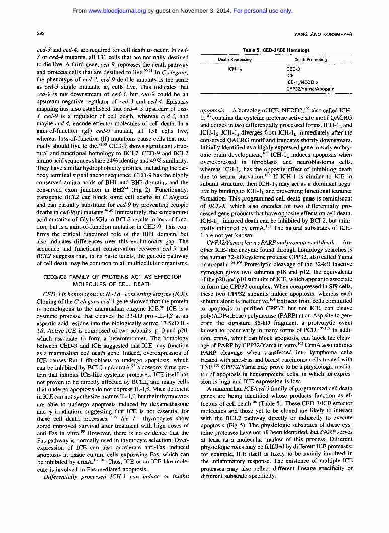
BAD negatively regulates BCLZ and BCL-X, and dis- places BAX. Efforts to further examine BCL2’s ability to interact with other proteins in the cell death pathway uncov- ered additional BCL2-interacting proteins that modulate BCL2’s activity. Yeast two-hybrid screening and A expres- sion cloning both showed a new heterodimerizing partner of BCL2 and BCL-XL, called BAD (BCL2/BCL-XL-associated death promoter).88 This player in the cell death pathway differs from other family members with homology limited to the most conserved amino acids in the BH1 and BH2 domains (Fig 2). BAD also lacks the typical carboxy terminal transmembrane domain, suggesting that it is not an integral membrane protein. BAD’S interaction with BCL2 and BCL- XL was verified in mammalian cells. Although BAD was discovered by virtue of its interaction with BCL2, it binds BCL-X, more strongly. When expressed in FL5.12 cells, BAD countered the death-repressor effect of BCL-XL effi- ciently and that of BCLZ to a lesser extent. The strong inter- action between BAD and BCL-XL sequesters BCL-X,, re- sulting in freed BAX, and cell death is restored. BAD displaces BAX from BCL-XJBAX or BCL2/BAX hetero- dimers in a concentration-dependent manner. In one cell line examined, when approximately 50% of all cellular BAX is heterodimerized with BCL-XL or BCL2, the cell is resistant to apoptosis. Conversely, in cells in which 80% of BAX i s found in homodimers, an apoptotic signal results in cell death.88 This finding suggests that BAD negatively regulates cell death by modulating the amount of BAX in homodimers versus heterodimers (Fig 4). Formally, it is not certain whether the active moiety in regulating cell death is the BAX/BAX homodimer or each BCL-XJBAX or BCL2/ BAX heterodimer. Alternatively, both may be active, and the ratio of the heterodimers to the homodimers may be the critical determinant. The discovery of BAD showed that the cell death regulators BCL2 and BCL-X, themselves are regu- lated by protein-protein interactions.
Another protein found by interactive cloning, BAG1, positively mod- ulates BCL2’s death-repressor activity.89 BAGl has been shown to interact with BCL2 in vitro. BAGl is not homolo- gous to BCL2 family members and contains an ubiquitin- like domain, suggesting that its mechanism might involve
BAGl can positively modulate BCL2 activity.
effects on protein stability. Curiously, coexpression of BAGl assisted BCL2 in protecting Jurkat T cells against anti-Fas antibody and cytotoxic T-cell killing. This finding suggests that death signals that appear to be BCL2-independent may be repressed by BCL2 if the appropriate modulatory proteins are present.
CAENORHABDITIS ELEGANS SHARES CONSERVED CELL DEATH GENES WITH MAMMALS
CED-9 is a homolog of BCL2 in C elegans. Important contributions to the understanding of programmed cell death have come from the genetic studies of the nematode C eleg- ans. In the development of the hermaphrodite worm, 131 of the 1,090 somatic cells undergo programmed cell death in a genetic pathway defined by 14 genes. Two of these genes,
cell suruiual
\\ r / @
cell death n w 250% of BAX heterodimerized with BCL-2 or BCL-XL=Survival
Fig 4. BAD is a negative regulator of apoptosis. BAD displaces BAX from BCU/BAX or BCL-Xr/BAX heterodimen, allowing more BAX/BAX homodimer formation, which promotes death.
For personal use only.on November 3, 2014. by guest www.bloodjournal.orgFrom
392 YANG AND KORShAEYER
ced-3 and ced-4, are required for cell death to occur. In ced- 3 or ced-4 mutants, all 131 cells that are normally destined to die live. A third gene, ced-9, represses the death pathway and protects cells that are destined to In C elegans, the phenotype of ced-3, ced-9 double mutants is the same as ced-3 single mutants, ie, cells live. This indicates that ced-9 is not downstream of ced-3, but ced-9 could be an upstream negative regulator of ced-3 and ced-4. Epistasis mapping has also established that ced-4 is upstream of ced- 3. ced-9 is a regulator of cell death, whereas ced-3, and maybe ced-4, encode effector molecules of cell death. In a gain-of-function (gf) ced-9 mutant, all 131 cells live, whereas loss-of-function (If) mutations cause cells that nor- mally should live to die.92,93 CED-9 shows significant struc- tural and functional homology to BCL2. CED-9 and BCL2 amino acid sequences share 24% identity and 49% similarity. They have similar hydrophobicity profiles, including the car- boxy terminal signal anchor sequence. CED-9 has the highly conserved amino acids of BHl and BH2 domains and the conserved exon junction in BH2" (Fig 2). Functionally, transgenic BCL2 can block some cell deaths in C elegans and can partially substitute for ced-9 by preventing ectopic deaths in ced-9(lf) Interestingly, the same amino acid mutation of Gly145Glu in BCL2 results in loss of func- tion, but is a gain-of-function mutation in CED-9. This con- firms the critical functional role of the BHl domain, but also indicates differences over this evolutionary gap. The sequence and functional conservation between ced-9 and BCL2 suggests that, in its basic tenets, the genetic pathway of cell death may be common to all multicellular organisms.
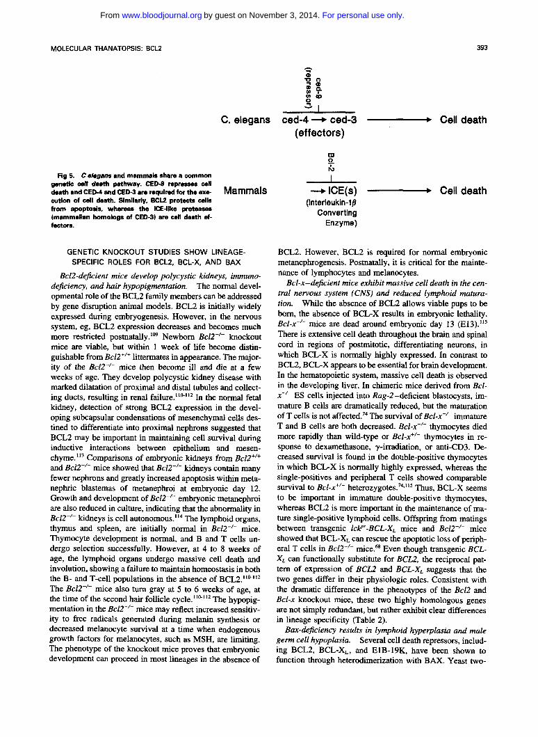
CED3KE FAMILY OF PROTEINS ACT AS EFFECTOR MOLECULES OF CELL DEATH
CED-3 is homologous to IL-l&converting enzyme (ICE). Cloning of the C elegans ced-3 gene showed that the protein is homologous to the mammalian enzyme ICE?' ICE is a cysteine protease that cleaves the 33-kD pro-IL-Ip at an aspartic acid residue into the biologically active 17.5kD IL- lp . Active ICE is composed of two subunits, p10 and p20, which associate to form a heterotetramer. The homology between CED-3 and ICE suggested that ICE may function as a mammalian cell death gene. Indeed, overexpression of ICE causes Rat-1 fibroblasts to undergo apoptosis, which can be inhibited by BCL2 and ~ r m A , 9 ~ a cowpox virus pro- tein that inhibits ICE-like cysteine proteases. ICE itself has not proven to be directly affected by BCL2, and many cells that undergo apoptosis do not express IL-1p. Mice deficient in ICE can not synthesize mature IL-Ip, but their thymocytes are able to undergo apoptosis induced by dexamethasone and y-irradiation, suggesting that ICE is not essential for these cell death proces~es.~' .~~ Ice-l- thymocytes show some improved survival after treatment with high doses of anti-Fas in vitro.w However, there is no evidence that the Fas pathway is normally used in thymocyte selection. Over- expression of ICE can also accelerate anti-Fas-induced apoptosis in tissue culture cells expressing Fas, which can be inhibited by ~ r m A . ' ~ . ' ~ ' Thus, ICE or an ICE-like mole- cule is involved in Fas-mediated apoptosis.
Diflerentially processed ICH- I can induce or inhibit
Table 5. CED3IICE Homologs
Death-Repressing Death-Promoting
ICH-ls CED-3 ICE ICE-1JNEDD 2 CPPBZ/Yama/Apopain
apoptosis. A homolog of ICE, NEDD2,"' also called ICH- 1,'03 contains the cysteine protease active site motif QACRG and comes in two differentially processed forms, ICH- 1 and ICH-1s. ICH-1s diverges from ICH-IL immediately after the conserved QACRG motif and truncates shortly downstream. Initially identified as a highly expressed gene in early embry- onic brain development," ICH- lL induces apoptosis when overexpressed in fibroblasts and neuroblastoma cells, whereas ICH-1, has the opposite effect of inhibiting death due to serum ~tarvation. '~~ If ICH-1 is similar to ICE in subunit structure, then ICH-ls may act as a dominant nega- tive by binding to ICH- lL and preventing functional tetramer formation. This programmed cell death gene is reminiscent of BCL-X, which also encodes for two differentially pro- cessed gene products that have opposite effects on cell death. ICH-1,-induced death can be inhibited by BCL2, but mini- mally inhibited by crmA.'03 The natural substrates of ICH- 1 are not yet known.
CPP32Nama cleaves PARPandpromotes celldeath. An- other ICE-like enzyme found through homology searches is the human 32-kD cysteine protease CPP32, also called Yama or apopain.'M-'06 Proteolytic cleavage of the 32-kD inactive zymogen gives two subunits p18 and p12, the equivalents of the p20 and p10 subunits of ICE, which appear to associate to form the CPP32 complex. When coexpressed in Sf9 cells, these two CPP32 subunits induce apoptosis, whereas each subunit alone is ineffective." Extracts from cells committed to apoptosis or purified CPP32, but not ICE, can cleave poly(ADP-ribose) polymerase (PARP) at an Asp site to gen- erate the signature 85-kD fragment, a proteolytic event known to occur early in many forms of PCD.106.'07 In addi- tion, crmA, which can block apoptosis, can block the cleav- age of P A W by CPP32Nama in vitro.'05 CrmA also inhibits PARP cleavage when transfected into lymphoma cells treated with anti-Fas and breast carcinoma cells treated with TNF.'" CPP32Nama may prove to be a physiologic media- tor of apoptosis in hematopoietic cells, in which its expres- sion is high and ICE expression is low.
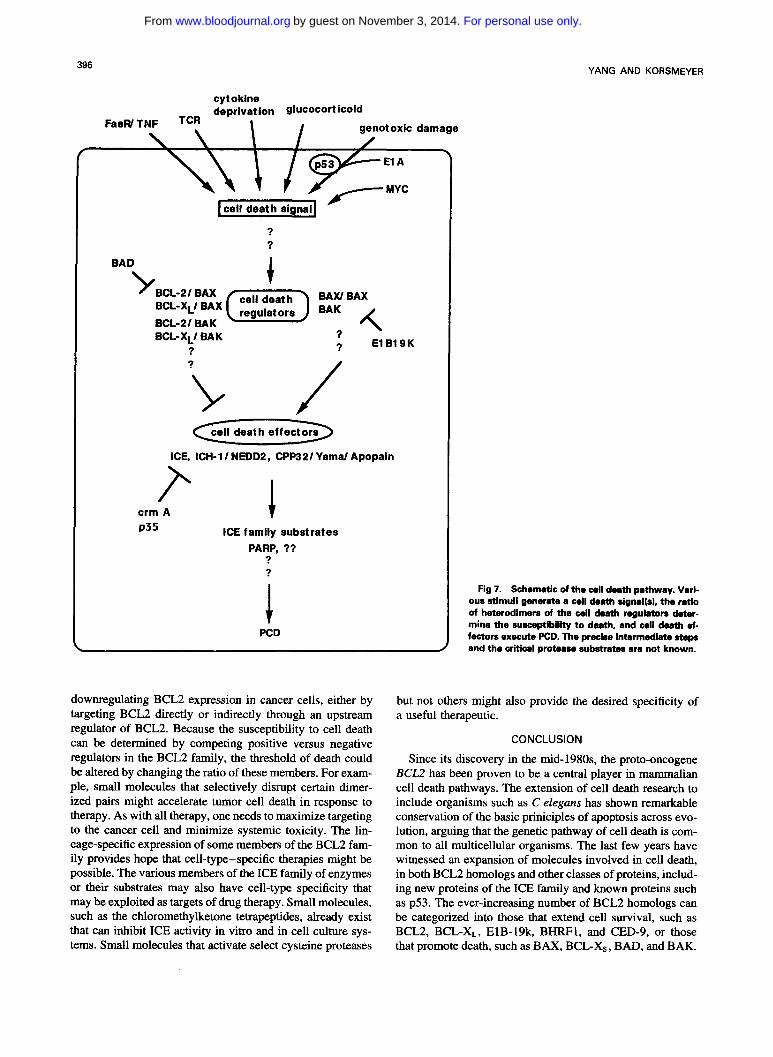
A mammalian ZCWced-3 family of programmed cell death genes are being identified whose products function as ef- fectors of cell death"' (Table 5). These CED-3LCE effector molecules and those yet to be cloned are likely to interact with the BCL2 pathway directly or indirectly to execute apoptosis (Fig 5). The physiologic substrates of these cys- teine proteases have not all been identified, but PARP serves at least as a molecular marker of this process. Different physiologic roles may be fulfilled by different ICE proteases; for example, ICE itself is likely to be mainly involved in the inflammatory response. The existence of multiple ICE proteases may also reflect different lineage specificity or different substrate specificity.
For personal use only.on November 3, 2014. by guest www.bloodjournal.orgFrom
MOLECULAR THANATOPSIS: BCLZ 393
C. elegans
Fig 5. C elegans and mammals share a common genetic cell death pathway. CED-9 represses cell death and CED-4 and CED-3 are required for the ex8- cution of cell death. Similarly, BCK protects cells from apoptosis, whereas the ICE-like proteases (mammalian homologs of CED-31 are cell death ef- fectors.
Mammals
GENETIC KNOCKOUT STUDIES SHOW LINEAGE- SPECIFIC ROLES FOR BCLS, BCL-X, AND BAX
Bcl2-dejcient mice develop polycystic kidneys, immuno- dejciency, and hair hypopigmentation. The normal devel- opmental role of the BCL2 family members can be addressed by gene disruption animal models. BCL2 is initially widely expressed during embryogenesis. However, in the nervous system, eg, BCL2 expression decreases and becomes much more restricted postnatally.'Og Newbom Bc12-'- knockout mice are viable, but within 1 week of life become distin- guishable from Bc12+/+ littermates in appearance. The major- ity of the BcZ2-/- mice then become ill and die at a few weeks of age. They develop polycystic kidney disease with marked dilatation of proximal and distal tubules and collect- ing ducts, resulting in renal failure."@"2 In the normal fetal kidney, detection of strong BCL2 expression in the devel- oping subcapsular condensations of mesenchymal cells des- tined to differentiate into proximal nephrons suggested that BCL2 may be important in maintaining cell survival during inductive interactions between epithelium and mesen- chyme.113 Comparisons of embryonic kidneys from Bc12+/+ and Bc12-/- mice showed that Bc12-'- kidneys contain many fewer nephrons and greatly increased apoptosis within meta- nephric blastemas of metanephroi at embryonic day 12. Growth and development of Bc12-'- embryonic metanephroi are also reduced in culture, indicating that the abnormality in Bc12-'- kidneys is cell autonomou~."~ The lymphoid organs, thymus and spleen, are initially normal in Bc12-/- mice. Thymocyte development is normal, and B and T cells un- dergo selection successfully. However, at 4 to 8 weeks of age, the lymphoid organs undergo massive cell death and involution, showing a failure to maintain homeostasis in both the B- and T-cell populations in the absence of BCL2.l'@'l2 The Bc12-/- mice also turn gray at 5 to 6 weeks of age, at the time of the second hair follicle The hypopig- mentation in the Bc12-" mice may reflect increased sensitiv- ity to free radicals generated during melanin synthesis or decreased melanocyte survival at a time when endogenous growth factors for melanocytes, such as MSH, are limiting. The phenotype of the knockout mice proves that embryonic development can proceed in most lineages in the absence of
ced-4 + ced-3 - Cell death (effectors)
m P I; I +ICE(s) - Cell death
(Interleukin-1p Converting
Enzyme)
BCL2. However, BCL2 is required for normal embryonic metanephrogenesis. Postnatally, it is critical for the mainte- nance of lymphocytes and melanocytes.
Bcl-x-dejcient mice exhibit mamive cell death in the cen- tral nervous system (CNS) and reduced lymphoid matura- tion. While the absence of BCLZ allows viable pups to be born, the absence of BCL-X results in embryonic lethality. Bcl-x-/- mice are dead around embryonic day 13 (E13).'15 There is extensive cell death throughout the brain and spinal cord in regions of postmitotic, differentiating neurons, in which BCL-X is normally highly expressed. In contrast to BCL2, BCL-X appears to be essential for brain development. In the hematopoietic system, massive cell death is observed in the developing liver. In chimeric mice derived from Bcl- x-'- ES cells injected into Rag-2-deficient blastocysts, im- mature B cells are dramatically reduced, but the maturation of T cells is not affected." The survival of Bcl-x-/- immature T and B cells are both decreased. Bcl-x-/- thymocytes died more rapidly than wild-type or Bcl-x+/- thymocytes in re- sponse to dexamethasone, y-irradiation, or anti-CD3. De- creased survival is found in the double-positive thymocytes in which BCL-X is normally highly expressed, whereas the single-positives and peripheral T cells showed comparable survival to Bcl-x+/- heterozygote^."*"^ Thus, BCL-X seems to be important in immature double-positive thymocytes, whereas BCL2 is more important in the maintenance of ma- ture single-positive lymphoid cells. Offspring from matings between transgenic IcW-BCL-XL mice and Bc12-/- mice showed that BCL-X, can rescue the apoptotic loss of periph- eral T cells in Bc12-'- mice?* Even though transgenic BCL- X, can functionally substitute for BCL2, the reciprocal pat- tem of expression of BCU and BCL-XL suggests that the two genes differ in their physiologic roles. Consistent with the dramatic difference in the phenotypes of the Bc12 and BcZ-x knockout mice, these two highly homologous genes are not simply redundant, but rather exhibit clear differences in lineage specificity (Table 2).
Bax-de$ciency results in lymphoid hyperplasia and male germ cell hypoplasia. Several cell death repressors, includ- ing BCL2, BCL-XL, and ElB-l9K, have been shown to function through heterodimerization with BAX. Yeast two-
For personal use only.on November 3, 2014. by guest www.bloodjournal.orgFrom
394 YANG AND KORSMEYER
hybrid assays also showed that BAX interacts widely with other family members. These findings suggested that BAX may have a central role in the regulation of apoptosis. One prediction is that BAX may be necessary for cell death; alternatively, heterodimers of BAX may be required for death repression. Interestingly, whether BAX deficiency re- sults in hyperplasia or hypoplasia appears to be tissue spe- cific. Bar-'- mice appear to be healthy, indicating that BAX is not essential for development of the organism. Thymocyte numbers of Bax-/- mice are increased 1.6-fold over wild- type controls, and the splenic B cells are similarly increased l.&fold. On the other hand, male Bax-/- mice are infertile, and BUY- testes exhibited a marked increase in cell death clustered in the germ cells. The seminiferous tubules were abnormal, and multinucleated giant cells and pyknotic cells were present. The complete cessation of mature sperm cell production was accompanied by an expansion of the premei- otic 2N cell population, suggesting a role for BAX in meio- sis. However, Bax-/- ovaries display an accumulation of atrophic granulosa cells that presumably failed to undergo apoptosis. Thus, the phenotypic abnormalities of Bar-'- can be either hyperplasia or hypoplasia, depending on the cell type. Because the affected tissues of Bax-'- mice are not identical to the affected tissues of Bc12-'- mice, BCLZ may not always act through interaction with BAX. The Bar? mice dramatically illustrated that not only is there lineage specificity in the BCL2 family members, but that, depending on cell type, the same molecule can have a positive or a negative effect on cell death.Iz7
BCLZ FAMILY COOPERATES WITH OTHER CANCER GENES
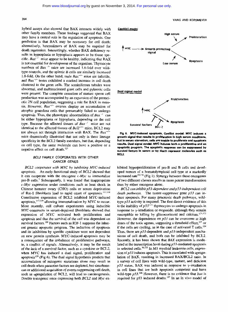
BCL2 cooperates with MYC by inhibiting MYC-induced apoptosis. An early functional study of BCL2 showed that it can cooperate with the oncogene c-Myc to immortalize pre-B cells.' Subsequently, it was found that inappropriate c-Myc expression under conditions such as heat shock in Chinese hamster ovary (CHO) cells or serum deprivation of Rat-1 fibroblasts lead to rapid onset of apopto~is."'~"~ Constitutive expression of BCL2 inhibited MYC-induced apoptosis,' I 7 . ' l 9 allowing immortalization by MYC to occur. Most recently, cell culture experiments using inducible MYC-constructs in serum-deprived fibroblasts showed that expression of MYC activated both proliferation and apoptosis and that the survival of the cell was dependent on survival factors.lZ0 Factors such as IGF-1 suppress the inher- ent genetic apoptotic program. The induction of apoptosis and its inhibition by specific cytokines were not dependent on new protein synthesis. MYC-induced apoptosis may be a consequence of the imbalance of proliferative pathways, ie, a conflict of signals. Alternatively, it may be the result of the lack of a survival factor, such as a cytokine or BCLZ, when MYC has induced a dual signal, proliferation and apoptosis'" (Fig 6). The dual signal hypothesis predicts that accumulation of mitogenic mutations alone may result in cell death when paracrine factors are depleted, but simultane- ous or additional acquisition of events suppressing cell death, such as upregulation of BCL2, will lead to carcinogenesis. Double transgenic mice expressing both BCL2 and Myc ex-
I conflict mode
High serum Proliferation Y + MYC .-> Growth promoting
\ signal
Low serum y L Apoptosis
Dual signal model
Proliferation
/ /
MYC
Fig 6. MYCinduced apuptoris. Conflict mod&. MYC induces a growth signal that results in proliration in high serum conditions, but in serum starvation, cells are unable to proliierate and apoptods results. Dual signal model: MYC induces both a profierative and an apoptotic program. The apoptotie response can be suppressed by survival factors in serum or by death repressor molecules such as BCK.
hibited hyperproliferation of pre-B and B cells and devel- oped tumors of a hematolymphoid cell type at a markedly increased (Fig 1). Synergy between these oncogenes of two different classes results in more potent transformation than by either oncogene alone.
BCL2 can inhibit p53-dependent andp53-independent cell death pathways. The tumor-suppressor gene p53 can in- duce apoptosis. For many genotoxic death pathways, wild- type p53 activity is required. The first direct evidence of this is the inability of p53-/- thymocytes to undergo apoptosis in response to y-irradiation or etoposide, although they remain susceptible to killing by glucocorticoid and calcium.1213122 However, the dependence on p53 can be overcome at high doses of the toxic agents, suggesting a threshold effect,Iz3 or if the cells are cycling, as in the case of activated T cells.lz4 Thus, there are p53-dependent and p53-independent mecha- nisms of cell death, and both can be inhibited by BCL2. Recently, it has been shown that BAX expression is modu- lated at the transcription level during p53-mediated apoptosis in selected cell^.^^,'^ In MI myeloid leukemia cells, expres- sion of p53 induces apoptosis. This is correlated with upregu- lation of BAX, resulting in increased BAXIBCL2 ratio. In a survey of cell lines with wild-type, mutant, and deficient p53 status, BAX was induced in response to y-irradiation in cell lines that are both apoptosis competent and have wild-type ~ 5 3 . ' ~ However, there is no evidence that bax is required for p53 induced deaths.'" In an in vivo model of
For personal use only.on November 3, 2014. by guest www.bloodjournal.orgFrom

MOLECULAR THANATOPSIS: BCLZ 395
choroid plexus tumor progression comparing p53+'- with p53-'- mice, it was found that aggressive tumor progression occurred in the absence of p53 function attributable to de- creased apoptosis.12' The function o f p 5 3 as a tumor-suppres- sor gene may be largely explained by its role in promoting cell death.
CLINICAL ASPECTS OF BCLZ
Studies of t(14;18) in lymphomas support the multi-hit oncogenesis model. BCL2 was first described as the dereg- ulated oncogene in t(14; 18) lymphomas. One initial study of 20 patients with follicular lymphoma possessing a large- cell component correlated the presence of the t(14; 18) with a poor response to therapy.'29 Subsequent larger studies com- posed of both large-cell and small-cleaved cell follicular lymphomas have not shown a prognostic significance of hav- ing a t( 14; 18).130-'32 However, many B-cell lymphomas that lack the t(14; 18) also have high levels of BCL2 p r ~ t e i n . ' ~ ~ . ' ~ ~ Clones harboring the t( 14; 18) translocation are commonly found in normal individuals. A large percentage of normal tonsils were found to contain cells positive by PCR for t( 14; 18),'35,136 and many healthy individuals harbored t( 14; 18)-containing B cells in their peripheral bl00d.l~' An- other study found peripheral blood lymphocytes from 55% of normal individuals and 35% of autopsied spleens con- tained cells with PCR-detectable t( 14; 18).13' These findings confirm the conclusion from transgenic mice experiments that translocation involving BCL2 alone is not sufficient to cause cancer, ie, additional events are necessary for malig- nant transformation to occur. Moreover, the frequency of translocations increased significantly with age, being 40 times greater in the spleen and 13 times greater in the periph- eral blood in the oldest age group (>60 years) compared with the youngest age group (<20 years)."' The increase in the frequency oft( 14; 18) cells with age parallels the increase in lymphoma incidence with age. It is likely that both t(14; 18)-bearing cells and secondary hits increase over time. These epidemiologic correlates illustrate the importance of extended cell survival as a primary event in a multihit onco- genesis model.
The presence of t(14;18) provides a convenient way to observe patients after therapy. It has been shown that cells positive for t( 14; 18) persist in patients in prolonged com- plete r e m i s ~ i o n , ' ~ ~ . ' ~ but whether this predicts imminent re- lapse remains uncertain. More recently, investigators have looked for the disappearance of translocation-bearing cells from bone marrows after myeloablative therapy or in vitro p ~ r g i n g . ' ~ ' , ' ~ ~ Detection of the translocation by PCR provides a means to assess the success of the elimination of the lymphoma clone upon myeloablation or purging. The litera- ture suggests that detection of cells with translocation may correlate with shorter remission.
BCL2 expression is found in tumors of some hormonally responsive epithelium. BCL2 expression has been investi- gated in nonlymphoid tumors. It is well established that some breast carcinomas, prostate cancers, and non-small-cell lung cancers express BCL2. In breast carcinoma, BCL2 expres- sion is positively correlated with estrogen receptor and pro- gesterone receptor positivity. Conversely, loss of BCL2 ex-
pression is associated with known poor prognostic indicators, such as estrogen receptor negativity, epidermal growth factor receptor positivity, p53 mutation, and high histologic grade. 143-145 The normal epithelium from which breast carcinoma arises expresses BCL2, suggesting that BCL2 expression allows cells to live longer and accumulate genetic alterations. Loss of BCL2 is likely to be a late event accompanied by additional genetic changes. In multivariate analysis, it appears that the prognostic role of BCL2 is re- lated to p53 status, which itself has independent prognostic significance.
BCL2 expression is also found in cancers of another hor- monally responsive tissue, the prostate. High levels of BCL2 are observed in androgen-independent tumors, 146 in particu- lar those tumors that persist after androgen ablation ther- apy,14' leading to the speculation that BCL2 function allows the neoplastic prostate cells to survive in a hormonally de- prived environment.
High BCL2 expression is correlated with poor response to chemotherapy. Programmed cell death is not only an important normal physiological process, but it is also how cancer cells die when treated with a variety of chemothera- peutic drugs, including inhibitors of DNA synthesis, alkylat- ing agents, topoisomerase inhibitors, microtubule inhibitors, and antimetabolites. The ability of BCL2 to inhibit cell death induced by many of these agents with different mechanisms of action is consistent with BCL2 being a downstream mole- cule in the apoptotic pathway."^“" Cell lines transfected with BCL2 show increased resistance to nitrogen mustard, camp- tothecin, VP- 16, platinum compounds, methotrexate, Ara-C, adriamycin, and cyclophosphamide.4043." These observa- tions are borne out in the clinical arena. High BCLZ expres- sion is associated with low remission rate in acute myeloid 1e~kemia.I~' In an analysis of acute lymphocytic leukemia (ALL) and acute myeloid leukemia (AML) patients at diag- nosis and relapse, it was found that both the percentage of BCL2 expressing cells and the intensity of BCL2 staining were higher at relapse than at presentation. In de novo AML and ALL, the intensity of BCL2 staining and the number of positive cells were lower in cases that responded to chemo- therapy than in nonresponders; therefore, high BCL2 expres- sion is an indicator of poor response in acute 1e~kemia . I~~
In a cell line model, cells selected for acquired resistance to cytotoxic drugs associated with overexpression of the MDRl gene showed induction of BCL-XL. These cells were also resistant to y-irradiation induced apop to~ i s . ' ~~ Thus, in- duction of BCL-XL may play a role in the etiology of chemo- therapy and radiation-resistant tumors and may prove to have prognostic significance as well.
Given that inappropri- ate survival can be a primary event in tumorigenesis and that cells undergo apoptosis in response to chemotherapy, the outcome of cancer may be affected by changing the setpoint at which cells undergo apoptosis in response to a signal (Fig 7). In cancers that overexpress BCL2, decreasing BCL2 expression may allow a cell that contains otherwise intolerable genetic alterations to die. Altering the threshold for cell death, one may render the cancer cell more sensitive to chemotherapeutic agents. This might be approached by
Apoptosis as a therapeutic target.
For personal use only.on November 3, 2014. by guest www.bloodjournal.orgFrom
396
cell death signal
YANG AND KORSMEYER
BAD
\
? ?
BCL-XLI BAK ?
? ? E l B l 9 K
ICE, ICH-1 I NEODP, CPP321 Yamal Apopain
crm A P35 ICE family substrates
PARP, ?? ? ?
PCD
downregulating BCL2 expression in cancer cells, either by targeting BCL2 directly or indirectly through an upstream regulator of BCL2. Because the susceptibility to cell death can be determined by competing positive versus negative regulators in the BCL2 family, the threshold of death could be altered by changing the ratio of these members. For exam- ple, small molecules that selectively disrupt certain dimer- ized pairs might accelerate tumor cell death in response to therapy. As with all therapy, one needs to maximize targeting to the cancer cell and minimize systemic toxicity. The lin- eage-specific expression of some members of the BCL2 fam- ily provides hope that cell-type-specific therapies might be possible. The various members of the ICE family of enzymes or their substrates may also have cell-type specificity that may be exploited as targets of drug therapy. Small molecules, such as the chloromethylketone tetrapeptides, already exist that can inhibit ICE activity in vitro and in cell culture sys- tems. Small molecules that activate select cysteine proteases
Fig 7. Schematic of the cell death pathway. Vari- ous stimuli generate a cell death signells1, the ratio of heterodimers of the cell doath regulators dater- mine the wwtibility to death, and cell death ef- fectors execute PCD. The precise intermediate steps and the critical protease substrates are not known.
but not others might also provide the desired specificity of a useful therapeutic.
CONCLUSION
Since its discovery in the mid-l980s, the proto-oncogene BCL2 has been proven to be a central player in mammalian cell death pathways. The extension of cell death research to include organisms such as C elegans has shown remarkable conservation of the basic priniciples of apoptosis across evo- lution, arguing that the genetic pathway of cell death is com- mon to all multicellular organisms. The last few years have witnessed an expansion of molecules involved in cell death, in both BCL2 homologs and other classes of proteins, includ- ing new proteins of the ICE family and known proteins such as p53. The ever-increasing number of BCL2 homologs can be categorized into those that extend cell survival, such as BCL2, BCL-XL, ElB-l9k, BHRFl, and CED-9, or those that promote death, such as BAX, BCL-Xs, BAD, and BAK.
For personal use only.on November 3, 2014. by guest www.bloodjournal.orgFrom
MOLECULAR THANATOPSIS: BCL2 397
Knockout mice argue that each member of the BCL2 family will serve a pivotal role in select tissues. In addition, the identification of new proteins, such as BAD, which modulate BCL2 and BCL-XL activity, suggests that the multiple cell death proteins must be tightly regulated. Although the pre- cise biochemical activity of BCL2 remains uncertain, genetic studies of the BCL2 family members have established the importance of these genes in the normal development and maintenance of the organism. Inappropriate cell survival re- sulting from the deregulation of cell death genes can be a first step in oncogenesis. Once a tumor is established, its response to therapy can also be affected by its propensity to undergo programmed cell death. A remaining challenge is to define the intermediate steps that connect the sets of signals, regulators, and effectors that comprise apoptosis (Fig 7). Perhaps this knowledge will provide the basis for designing effective cancer therapies that target the cell death pathway.
REFERENCES 1. Fukuhara S, Rowley JD, Variakojis D, Golomb HM: Chromo-
some abnormablities in poorly differentiated lymphocytic lymphoma. Cancer Res 39:3119, 1979
2. Yunis JJ, Frizzera G, Oken MM, McKenna J, Theologides A, Arnesen M: Multiple recurrent genomic defects in follicular lymphoma. N Engl J Med 316:79, 1987
3. Tsujimoto Y, Gorham J, Cossman J, Jaffe E, Croce CM: The t( 14; 18) chromosome translocations involved in B-cell neoplasms result from mistakes in VDJ joining. Science 229:1390, 1985
4. Bakhshi A, Jensen JP, Goldman P, Wright JJ, McBride OW, Epstein AL, Korsmeyer SJ: Cloning the chromosomal breakpoint of t( 14; 18) human lymphomas: Clustering around JH on chromosome 14 and near a transcriptional unit on 18. Cell 41:889, 1985
5. Cleary ML, Sklar I: Nucleotide sequence of a t(14; 18) chromo- somal breakpoint in follicular lymphoma and demonstration of a breakpoint cluster region near a transcriptionally active locus on chromosome 18. Proc Natl Acad Sci USA 82:7439, 1985
6. Cleary ML, Smith SD, Sklar J: Cloning and structural analysis of cDNAs for bc2-2 and a hybrid bcl-Zfimmunoglobulin transcript resulting from the t(14; 18) translocation. Cell 47:19, 1986
7. Vaux DL, Cory S, Adams JM: Bcl-2 oncogene promotes haemopoietic cell survival and cooperates with c-myc to immortalize pre-B cells. Nature 335:440, 1988
8. Nunez G, London L, Hockenbery D, Alexander M, McKeam J, Korsmeyer SJ: Deregulated Bcl-2 gene expression selectively pro- longs survival of growth factor-deprived hemopoietic cell lines. J Immunol 144:3602, 1990
9. Korsmeyer SJ: Bcl-2 initiates a new category of oncogenes: Regulators of cell death. Blood 80:879, 1992
10. Hockenbery D, Nunez G, Milliman C, Schreiber RD, Kors- meyer SJ: Bcl-2 is an inner mitochondrial membrane protein that blocks programmed cell death. Nature 348:334, 1990
11. Deng G, Podack ER: Suppression of apoptosis in a cytotoxic T-cell line by interleukin 2-mediated gene transcription and deregu- lated expression of the protooncogene bcl-2. Proc Natl Acad Sci USA 90:2189, 1993
12. Schwarze MMK, Hawley RG: Prevention of myeloma cell apoptosis by ectopic bcl-2 expression or interleukin 6-mediated up- regulation of bcl-x,Cancer Res 55:2262, 1995
13. Sentman DL, Shutter JR, Hockenbery D. Kanagawa 0, Kors- meyer SJ: Bcl-2 inhibits multiple forms of apoptosis but not negative selection in thymocytes. Cell 671879, 1991
14. Strasser A, Harris AW, Cory S: Bcl-2 transgene inhibits T cell death and perturbs thymic self-censorship. Cell 67:889, 1991
15. McDonnell TJ, Deanne N, Platt FM, Nunez G, Jaeger U, McKearn JP, Korsmeyer SJ: Bcf-2-immunoglobulin transgenic mice demonstrate extended B cell survival and follicular lymphoprolifera- tion. Cell 57:79, 1989
16. McDonnell TJ, Korsmeyer SJ: Progression from lymphoid hyperplasia to high-grade malignant lymphoma in mice transgenic for the t(14; 18). Nature 349:6306, 1991
17. Strasser A, Harris AW, Cory S: E, -bcl-2 transgene facilitates spontaneous transformation of early pre-B and immunoglobulin-se- creting cells but not T cells. Oncogene 8:1, 1993
18. Marin MC, Hsu B, Stephens LC, Brisbay S, McDonnell TJ: The functional basis of c-myc and bcl-2 complementation during multistep lymphomagenesis in vivo. Exp Cell Res 217:240, 1995
19. Linette GP, Hess JL, Sentman CL, Korsmeyer SJ: Diffuse malignant T cell lymphoma in IckPr-bcl-2 transgenic mice. Blood 86:1255, 1995
20. Batistatou A, Merry DE, Korsmeyer SJ, Green LA: Expres- sion of Bcl-2 proto-oncogene rescues PC12 cells from death caused by withdrawal of trophic support. J Neurosci 13:4422, 1993
21. Mah SP, Zhong LT, Liu Y, Roghani A, Edwards RH, Brede- sen DE: The protooncogene bcl-2 inhibits apoptosis in PC12 cells. J Neurochem 60: 1183, 1993
22. Garcia I, Martinou I, Tsujimoto Y, Martinou J-C: Prevention of programmed cell death of sympathetic neurons by the bcl-2 proto- oncogene. Science 258:302, 1992
23. Allsopp TE, Wyatt S, Paterson HF, Davies AM: The proto- oncogene bcl-2 can selectively rescue neurotrophic factor-dependent neurons from apoptosis. Cell 73:295, 1993
24. Zhong LT, Sarafian T, Kane DJ, Charles AC, Mah SP, Ed- wards RH, Bredesen DE: bcl-2 inhibits death of central neural cells induced by multiple agents. Proc Natl Acad Sci USA 90:4533, 1993
25. Martinou J-C, Dubois-Dauphin M, Staple JK, Rodriguez I, Frankowski H, Missotten M, Albertini P, Talabot D, Catsicas S, Pietra C, Huarte J: Overexpression of BCL2 in transgenic mice protects neurons from naturally occurring cell death and experimen- tal ischemia. Neuron 13:1017, 1994
26. Dubois-Dauphin M, Frankowski H, Tsujimoto Y, Huarte J, Martinou J-C: Neonatal motoneurons overexpressing the bcl-2 pm- tooncogene in transgenic mice are protected from axotomy-induced cell death. Proc Natl Acad Sci USA 91:3309, 1994
27. Greenlund LJS, Korsmeyer SJ, Johnson EM: Role of bcl-2 in the survival and function of developing and mature sympathetic neurons. Neuron 15647, 1995
28. Rao L, Debbas M, Sabbatini P, Hockenbery D, Korsmeyer S, White E: The adenovirus ElA proteins induce apoptosis, which is inhibited by the EIB I9kDa and Bcl-2 proteins. Proc Natl Acad Sci USA 89:7742, 1992
29. Debbas M, White E: Wild-type p53 mediates apoptosis by ElA, which is inhibited by E1B. Genes Dev 7:546, 1993
30. Chiou S-K, Tseng C-C, Rao L, White E: Functional comple- mentation of the adenovirus EIB 19-kilodalton protein with Bcl-2 in the inhibition of apoptosis in infected cells. J Virol68:6553, 1994
31. Chiou S-K, Rao L, White E: Bcl-2 blocks p53-dependent apoptosis. Mol Cell Biol 14:2556, 1994
32. Yamada T, Yamaoka S, Goto T, Nakai M, Tsujimoto Y, Hatanaka M: The human T-cell leukemia virus type I Tax protein induces apoptosis which is blocked by the Bcl-2 protein. J Virol 68:3374, 1994
33. Levine B, Huang Q, Isaacs JT, Reed JC, Griffin DE, Hardwick M: Conversion of lytic to persistent alphavirus infection by the bcl- 2 cellular oncogene. Nature 361:739, 1993
34. Newmeyer DD, Farschon DM, Reed JC: Cell-free apoptosis in Xenopus egg extracts: Inhibition by Bcl-2 and requirement for an organelle fraction enriched in mitochondria. Cell 79:353, 1994
35. Selvakumaran M, Lin HK, Sjin RT, Reed JC, Liebemann
For personal use only.on November 3, 2014. by guest www.bloodjournal.orgFrom

398 YANG AND KORSMEYER
DA, Hoffman B: The novel primary response gene MyD118 and the proto-oncogenes myb, myc, and bcl-2 modulate transforming growth factor 01-induced apoptosis of myeloid leukemia cells. Mol Cell Biol 14:2352, 1994
36. Selvakumaran M, Lin HK, Miyashita T, Wang HG, Krajewski S, Reed JC, Hoffman B, Liebermann D: Immediate early up-regula- tion of bax expression by p53 but not TGFpl: A paradigm for distinct apoptotic pathways. Oncogene 9:1791, 1994
37. Miyashita T, Reed JC: Bcl-2 gene transfer increases relative resistance of S49.1 and “7.2 lymphoid cells to cell death and DNA fragmentation induced by glucocorticoids and multiple chemo- therapeutic drugs. Cancer Res 52:5407, 1992
38. Desoize B: Anticancer drug resistance and inhibition of apoptosis. Anticancer Res 14:229 1, 1994
39. Lowe SW, Ruley HE, Jacks T, Housman DE: p53-dependent apoptosis modulates the cytotoxicity of anticancer agents. Cell 74:957, 1993
40. Walton MI, Whysong D, O’Connor PM, Hockenbery D, Korsmeyer SJ, Kohn KW. Constitutive expression of human E d 2 modulates nitrogen mustard and camptothecin induced apoptosis. Cancer Res 53:1853, 1993
41. Fisher TC, Milner AE, Gregory CD, Jackman AL, Aheme W, Hartley JA, Dive C, Hickman JA: bcl-2 modulation of apoptosis induced by anticancer drugs: Resistance to thymidylate stress is independent of classical resistance pathways. Cancer Res 53:3321, 1993
42. Reed JC, Kitada S, Takayama S, Miyashita T: Regulation of chemoresistance by the bcl-2 oncoprotein in non-Hodgkin’s lymphoma and lymphocytic leukemia cell lines. Ann Oncol 5:S61, 1994
43. Miyashita T, Reed JC: Bcl-2 oncoprotein blocks chemother- apy-induced apoptosis in a human leukemia cell line. Blood 8 1 : 15 1, I993
44. Reed JC, Kitada S, Takayama S, Miyashita T: Regulation of chemoresistance by the bcl-2 oncoprotein in non-Hodgkin’s lymphoma and lymphocytic leukemia cell lines. Ann Oncol 5:S61, I994
45. Vaux DL, Aguila HL, Weissman IL: Bcl-2 prevents death of factor-deprived cells but fails to prevent apoptosis in targets of cell mediated killing. Int Immunol 42321, 1992
46. Chen-Levy Z, Nourse J, Cleary ML: The bcl-2 candidate proto-oncogene product is a 24-hlodalton integral-membrane pro- tein highly expressed in lymphoid cell lines and lymphomas carrying the t(14;18) translocation. Mol Cell Biol 9:701, 1989
47. Monaghan P, Robertson D, Amos T, Dyer M, Mason D, Greaves M: Ultrastructural localization of Bcl-2 protein. J Histochem Cytochem 40:1819, 1992
48. Krajewski S, Tanaka S, Takayama S, Schibler MJ, Fenton W, Reed JC: Investigation of the subcellular distribution of the bcl- 2 oncoprotein: Residence in the nuclear envelope, endoplasmic retic- ulum, and outer mitochondrial membranes. Cancer Res 53:4701, 1993
49. de Jong D, Prins FA, Mason DY, Reed JC, van O m e n GN, Kluin PM: Subcellular localization of the bcl-2 protein in malignant and normal lymphoid cells. Cancer Res 54:256, 1994
50. Nguyen M, Millar DG, Yong VW, Korsmeyer SJ, Shore GC: Targeting of Bcl-2 to the mitochondrial outer membrane by a COOH-terminal signal anchor sequence. J Biol Chem 268:25265, 1993
51. Hockenbery DM, Oltvai ZN, Yin X-M, Milliman CL, Kors- meyer SJ: Bcl-2 functions in an antioxidant pathway to prevent apoptosis. Cell 75:241, 1993
52. Nguyen M, Branton PE, Walton PA, Oltvai ZN, Korsmeyer SJ, Shore GC: Role of membrane anchor domain of Bcl-2 in suppres-
sion of apoptosis caused by E1B-defective adenovirus. J Biol Chem 269:16521, 1994
53. Tanaka S, Saito K, Reed JC: Structure-function analysis of the Bcl-2 oncoprotein. Addition of a heterologous transmembrane domain to portions of the Bcl-2P protein restores function as a regulator of cell survival. J Biol Chem 268:10921, 1993
54. Jacobson MD, Bume JF, King MP, Miyashita T, Reed JC, Raff MC: Bcl-2 blocks apoptosis in cells lacking mitochondrial DNA. Nature 361:365, 1993
55. Kane DJ, Sarafian TA, Anton R, Hahn H, Gralla EB, Valen- tine JS, Ord T, Bredesen DE: Bcl-2 inhibition of neural death: De- creased generation of reactive oxygen species. Science 262: 1274, 1993
56. Shimizu S, Eguchi Y, Kosaka H, Kamiike W, Matsude H, Tsujimoto Y: Prevention of hypoxia-induced cell death by Bci-2 and Bcl-x,. Nature 37231 1, 1995
57. Jacobson MD, Raff MC: Programmed cell death and Bcl-2 protection in very low oxygen. Nature 374:814, 1995
58. Lam M, Dubyak G, Distelhorst CW: Effect of glucocortico- steroid treatment on intracellular calcium homeostasis in mouse lymphoma cells. Mol Endocrinol 5:686, 1993
59. Lam M, Kubyak G, Chen L, Nunez G, Miesfeld RL, Distel- horst CW: Evidence that BCL2 represses apoptosis by regulating endoplasmic reticulum-associated Ca” fluxes. Proc Natl Acad Sci USA 91:6569, 1994
60. Baffy G , Miyashita T, Williamson JR, Reed JC: Apoptosis induced by withdrawal of interleukin-3 (IL-3) from an IL-3-depen- dent hematopoietic cell line is associated with repartitioning of intra- cellular calcium and is blocked by enforced Bcl-2 oncoprotein pro- duction. J Biol Chem 268:6511, 1993
61. Oltvai ZN, Milliman CL, Korsmeyer: Bcl-2 heterodimerizes in vivo with a conserved homolog, Bax, that accelerates programmed cell death. Cell 74:609, 1993
62. Yin X-M, Oltvai ZN, Korsmeyer SJ: BH1 and BH2 domains of Bcl-2 are required for inhibition of apoptosis and heterodimeriza- tion with Bax. Nature 369:321, 1994
63. Krajewski S, Krajewski M, Shabaik A, Miyashita T, Wang HG, Reed JC: Immunohistochemical determination of in vivo distri- bution of Bax, a dominant inhibitor of Bcl-2. Am J Pathol 145: 1323, 1994
64. Veis DJ, Sentman CL, Back EA, Korsmeyer SJ: Expression of the Bcl-2 protein in murine and human thymocytes and in periph- eral T lymphocytes. J Immunol 151:2546, 1993
65. Gratiot-Deans J, Ding L, Turka LA, Nunez G: Bcl-2 proto- oncogene expression during human T cell development. J Immunol 151233, 1993
66. Boise LH, Gonzalez-Garcia M. Postema CE, Ding L, Lindsten T, Turka LA, Mao X, Nunez G, Thompson CB: bcl-x, a bcl-2-related gene that functions as a dominant regulator of apoptotic cell death. Cell 74:597, 1993
67. Fang W, Rivard JJ, Mueller DL, Behrens TW: Cloning and molecular characterization of mouse bcl-x in B and T lymphocytes. J Immunol 153:4388, 1994
68. Chao DT, Linette GP, Boise LH, White LS, Thompson CB, Korsmeyer SJ: Bcl-x,. and Bcl-2 repress a common pathway of cell death. J Exp Med 182:821, 1995
69. Linette GP, Brusby MJ, Hedrick SM, Hansen TH, Glimcher LH, Korsmeyer SJ: Bcl-2 is upregulated at the CD4+CD8+ stage during positive selection and promotes thymocyte differentiation at several control points. Immunity 1: 197, 1994
70. Sedlak TW, Oltvai ZN, Yang E, Wang K, Boise LH, Thomp- son CB, Korsmeyer SJ: Multiple Bcl-2 family members demonstrate selective dimerizations with Bax. h o c Natl Acad Sci USA 92:7834, 1995
71. Gonzalez-Garcia M, Perez-Ballestero R, Ding L, Duan L,
For personal use only.on November 3, 2014. by guest www.bloodjournal.orgFrom
MOLECULAR THANATOPSIS: BCLL 399
Boise LH, Thompson CB, Nunez G: bcl-x, is the major bcl-x mRNA form expressed during murine development and its product localizes to mitochondria. Development 120:3033, 1994
72. Gottschalk AR, Boise LH, Thompson CB, Quintans J: Identi- fication of immunosuppressant-induced apoptosis in a murine B-cell line and its prevention by bcl-x but not bcl-2. Proc Natl Acad Sci USA 91:7350, 1994
73. Boise LH, Minn AJ, Noel PJ, June CH, Accavitti MA, Linds- ten T, Thompson CB: CD28 costimulation can promote T cell sur- vival by enhancing the expression of Bcl-xL. Immunity 3:87, 1995
74. Ma A, Pena JC, Chang B, Margosian E, Davidson L, Alt RW, Thompson CB: Bcl-x regulates the survival of double positive thymocytes. Proc Natl Acad Sci USA 92:4763, 1995
75. Krajewski S, Krajewska M, Shabaik A, Wang H-G, Irie S, Fong L, Reed JC: Immunohistochemical analysis of in vivo patterns of Bcl-x expression. Cancer Res 54:5501, 1994
76. Motoyama N, Wang F, Roth KA, Sawa H, Nakayama K-i, Nakayama K, Negishi I, Senju S, Zhang Q, Fujii S, Loh DY: Massive cell death of immature hematopoietic cells and neurons in Bcl-x- deficient mice. Science 267:1506, 1995
77. Kozopas KM, Yang T, Buchan HL, Zhou P, Craig RW: MCLI, a gene expressed in programmed myeloid cell differentiation, has sequence similarity to BCL2. Proc Natl Acad Sci USA 90:3516, 1993
78. Lin EY, Orlofsky A, Berger MS, Prystowsky MB: Character- ization of Al , a novel hemopoietic-specific early-response gene with sequence similarity to bcl-2. J Immunol 151:1979, 1993
79. Sat0 T, Hanada M, Bodrug S, hie S, Iwama N, Boise LH, Thompson CB, Golemis E, Fong L, Wang H-G, Reed JC: Interac- tions among members of the Bcl-2 protein family analyzed with a yeast two-hybrid system. Proc Natl Acad Sci USA 91:9238, 1994
80. Bodrug SE, Aime-Sempe C, Sat0 T, Krajewski S, Hanada M, Reed JC: Biochemical and functional comparisons of Mcl-1 and Bcl-2 proteins: Evidence for a novel mechanism of regulating Bcl- 2 family protein function. Cell Death Differ 2:173, 1995
81. Reynolds JE, Yang T, Qian L, Jenkinson JD, Zhou P, Eastman A, Craig RW: Mcl-1, a member of the Bcl-2 family, delays apoptosis induced by c-Myc overexpression in Chinese hamster ovary cells. Cancer Res 64:6348, 1994
82. Farrow SN, White JHM, Martinou I, Raven T, Pun K-T, Grinham CJ, Martinou J-C, Brown R: Cloning of a bcl-2 homologue by interaction with adenovirus E1B 19K. Nature 374:731, 1995
83. Chittenden T, Harrington EA, O'Connor R, Flemington C, Lutz RJ, Evan GI, Guild BC: Induction of apoptosis by the Bcl-2 homologue Bak. Nature 374:733, 1995
84. Kiefer MC, Brauer MJ, Powers VC, Wu JJ, Umansky SR, Tomei LD, Barr PJ: Modulation of apoptosis by the widely distrib- uted Bcl-2 homologue Bak. Nature 374:736, 1995
85. Pearson GR, Luka J, Petti L, Sample J, BIrkenback M, Braun D, Kieff E: Identification of an Epstein-Barr virus early gene encod- ing a second component of the restricted early antigen complex. Virology 160:151, 1987
86. Smith CA: A novel viral homologue of Bcl-2 and Ced-9. Trends Cell Biol 5:344, 1995
87. Neilan JG, Lu Z, Afonso CL, Kutish GF, Sussman MD, Rock DL: An African swine fever virus gene with similarity to the proto- oncogene bcl-2 and the Epstein-Barr virus gene BHRFI. J Virol 67:4391, 1993
88. Yang E, Zha J, Jockel J, Boise LH, Thompson CB, Korsmeyer SJ: Bad, a heterodimeric partner for Bcl-xL and Bcl-2, displaces Bax and promotes cell death. Cell 80:285, 1995
89. Takayama S, Sat0 T, Krajewski S, Kockel K, Irie S, Millan JA, Reed JC: Cloning and functional analysis of BAG-1: A novel Bcl-2 binding protein with anti-cell death activity. Cell 80:279, 1995
90. Hengartner MO, Horvitz HR: Programmed cell death in Caenorhabditis elegans. Curr Opin Genet Dev 4581, 1994
91. Hengartner MO, Horvitz HR: The ins and outs of programmed cell death during C. elegans development. Phil Trans R Soc Lond B 345:243, 1994
92. Hengartner MO, Horvitz H R Activation of C. elegans cell death protein CED-9 by an amino-acid substitution in a domain conserved in Bcl-2. Nature 369:318, 1994
93. Hengartner MO, Ellis RE, Horvitz H R Caenorhabditis ele- gans gene ced-9 protects cells from programmed cell death. Nature 356:494, 1992
94. Hengartner MO, Horvitz HR: C. elegans cell survival gene ced-9 encodes a functional homolog of the mammalian proto-onco- gene bcl-2. Cell 76:665, 1994
95. Vaux DL, Weissman IL, Kim SK: Prevention of programmed cell death in Caenorhabdiris elegans by human bcl-2. Science 258:1955, 1992
96. Yuan J, Shaham S, Ledoux S, Ellis HM, Horvitz H R The C. elegans cell death gene ced-3 encodes a protein similar to mamma- lian interleukin-10-converting enzyme. Cell 75541, 1993
97. Miura M, Zhu H, Rotello R, Hartwieg EA, Yuan J: Induction of apoptosis in fibroblasts by IL-10-converting enzyme, a mamma- lian homolog of the C. elegans cell death gene ced-3. Cell 75:653, 1993
98. Li P, Allen H, Banerjee S, FrankIin S, Herzog L, Johnston C, McDowell J, Paskind M, Rodman L, Salfeld J, Towne E, Tracey D, Wardwell S, Wei F-Y. Wong W, Kamen R, Seshadri T: Mice deficient in IL-lp-converting enzyme are defective in production of mature IL-18 and resistant to endotoxic shock. Cell 80401, 1995
99. Kuida K, Lippke JA, Ku G, Harding M W , Livingston DJ, Su M S-S, Flavell RA: Altered cytokine export and apoptosis in mice deficient in interleukin-10 converting enzyme. Science 267:2000, 1995
100. Enari M, Hug H, Nagata S: Involvement of an ICE-like protease in Fas-mediated apoptosis. Nature 375:78, 1995
101. Los M, Van de Craen M, Penning LC, Schenk H, Westen- dorp M, Baeuerle PA, Droge W, Krammer PH, Fiers W, Schulze- Ostholl K Requirement of an ICE-CED-3 protease for FaslAPO-1 mediated apoptosis. Nature 375231, 1995
102. Kumar S, Kinoshita M, Noda M, Copeland NG, Jenkins NA: Induction of apoptosis by the mouse Nedd2 gene, which encodes a protein similar to the product of the Caenorhabditis elegans cell death gene ced-3 and the mammalian IL-10-converting enzyme. Genes Dev 8:1613, 1994
103. Wang L, Miura M, Bergeron L, Zhu H, Yuan J: Ich-I, an Zce/ced-3-related gene, encodes both positive and negative regulators of programmed cell death. Cell 78:739, 1994
104. Femandes-Alnemri T, Litwack G, Alnemri E: CPP32, a novel human apoptotic protein with homology to Caenorhabditis elegans cell death protein ced-3 and mammalian interleukin-10- converting enzyme. J Biol Chem 269:30761, 1994
105. Tewari M, Quan LT, ORourke, Desnoyers S, Zeng Z, Beidler DR, Poirier GG, Salvesen GS, Dixit VM: YamalCPP32p, a mammalian homolog of CED-3, is a crmA-inhibitable protease that cleaves the death substrate poly(ADP-ribose) polymerase. Cell 812301, 1995
106. Nicholson DW, Ali A, Thombeny NA, Vaillancourt JP, Ding CK, Gallant M, Gareau Y, Griffin PR, Labelle M, Lazebnik YA, Munday NA, Raju SM, Smulson ME, Yamin T-T, Yu VL, Miller D K Identification and inhibition of the ICWCED-3 protease necessary for mammalian apoptosis. Nature 376:37, 1995
107. Lazebnik YA, Kaufmann SH, Desnoyers S, Poirier GG, Eamshaw WC: Cleavage of poly(ADP-ribose) polymerase by a pro- teinase with properties like ICE. Nature 371:346, 1994
For personal use only.on November 3, 2014. by guest www.bloodjournal.orgFrom

400 YANG AND KORSMEYER
108. Steller H: Mechanisms and genes of cellular suicide. Science 267:1445, 1995
109. Merry DE, Veis DJ, Hickey WF, Korsmeyer SJ: bcl-2 pro- tein expression is widespread in the developing nervous system and retained in the adult PNS. Development 120:301, 1994
110. Veis DJ, Sorenson CM, Shutter JR, Korsmeyer SJ: Bcl-2- deficient mice demonstrate fulminant lymphoid apoptosis, polycystic kidneys, and hypopigmented hair. Cell 75:229, 1993
11 1. Nakayama K, Nakayama K-I, Negishi I, Kuida K, Sawa H, Loh DY: Targeted disruption of Bcl-2 ap in mice: Occurrence of gray hair, polycystic kidney disease, and lymphocytopenia. Proc Natl Acad Sci USA 91:3700, 1994
112. Kamada S, Shimono A, Shinto Y, Tsujimura T, Takahashi T, Noda T, Kitamura Y, Kondoh H, Tsujimoto Y: bel-2 deficiency in mice leads to pleiotropic abno Accelerated lymphoid cell death in thymus and spleen, pol dney, hair hypopigmen- tation, and distorted small intestine. Cancer Res 55:354, 1995
113. LeBrun DP, Wamke RA, Cleary ML: Expression of bcl-2 in fetal tissues suggests a role in morphogenesis. Am J Pathol 142:743, 1993
114. Sorenson CM, Rogers SA, Korsmeyer SJ, Hammerman MR: Fulminant metanephric apoptosis and abnormal kidney development in bcl-2-deficient mice. Am J Physiol 268:F73, 1995
115. Motoyama N, Wang F, Roth KA, Sawa H, Nakayama K-i, Nakayama K, Negishi I, Senju S, Zhang Q, Fujii S, Loh DY: Massive cell death of immature hematopoietic cells and neurons in Bcl-x- deficient mice. Science 267:1506, 1995
116. Strasser A, Harris AW, Bath ML, Cory S: Novel primitive lymphoid tumours induced in transgenic mice by cooperation be- tween myc and bcl-2. Nature 348:331, 1990
117. Bissonnette RP, Echevem F, Mahboubi A, Green DR: Apo- ptotic cell death induced by c-myc is inhibited by bcl-2. Nature 359552, 1992
118. Evans G, Wyllie A, Gilbert C, Littlewood T, Land H, Brooks M, Waters C, Penn L, Hancock D: Induction of apoptosis in fibro- blasts by c-myc protein. Cell 63:119, 1992
119. Fanidi A, Harrington EA, Evan GI: Cooperative interaction between c-myc and bcl-2 proto-oncogenes. Nature 359554, 1992
120. Harrington EA, Bennett MR, Fanidi A, Evan GI: c-Myc- induced apoptosis in fibroblasts is inhibited by specific cytokines. EMBO J 13:3286, 1994
121. Lowe SW, Schmitt EM, Smith SW, Osbome BA, Jacks T: p53 is required for radiation-induced apoptosis in mouse thymocytes. Nature 362:847, 1993
122. Clarke AR, Purdie CA, Harrison DJ, Moms RG, Bird CC, Hooper ML, Wyllie AH: Thymocyte apoptosis induced by p53- dependent and independent pathways. Nature 362:849, 1993
123. Fisher DE: Apoptosis in cancer therapy: Crossing the thresh- old. Cell 78:539, 1994
124. Strasser A, Harris AW, Jacks T, Cory S: DNA damage can induce apoptosis in proliferating lymphoid cells via p53-independent mechanisms inhibitable by Bcl-2. Cell 79:329, 1994
125. Miyashita T, Krajewski S, Krajewska M, Wang HG, Lin HK, Liebemann DA, Hoffman B, Reed JC: Tumor suppressor p53 is regulator of bcl-2 and bax gene expression in vitro and in vivo. Oncogene 9:1799, 1994
126. Zhan Q, Fan S, Bae I, Buillouf C, Liebermann DA, O’Con- nor PM, Fomace AJ Jr: Induction of bar by genotoxic stress in human cells correlates with normal p53 status and apoptosis. Onco- gene 9:3743, 1994
127. Knudson CM, Tung KSK, Tourtellotte WG, Brown GAJ, Korsmeyer SJ: Bax-deficient mice with lymphoid hyperplasia and male germ cell death. Science 270:96, 1995
128. Symonds H, Krall L, Remington L, Saenz-Robles M, Lowe
S, Jacks T, Van Dyke T: p53-dependent apoptosis suppresses tumor growth and progression in vivo. Cell 78:703, 1994
129. Yunis JJ, Mayer MG, Arnesen MA, Aeppli DP, Oken M, Frizzera G: Bcl-2 and other genomic alteration in the prognosis of large-cell lymphoma. N Engl J Med 320:1047, 1989
130. Levine EG, Arthur DC, Frizzera G, Peterson BA, Hurd DD, Bloomfield CD: Cytogenetic abnormalities predict clinical outcome in non-Hodgkin’s lymphoma. Ann Intem Med 108:14, 1988
131. Pezzela F, Jones M, Ralfkiaer E, Ersboll J, Gatter KC, Ma- son DY: Evaluation of bcl-2 protein expression and 14; 18 transloca- tion as prognostic markers in follicular lymphoma. Br J Cancer 65:87, 1992
132. Tilly H, Rossi A, Stamatoullas A, Lenormand B, Bigorgne C, Kunlin A, Monconduit M, Bastard C: Prognostic value of chromo- somal abnormalities in follicular lymphoma. Blood 84:1043, 1994
133. Akagi T, Kondo E, Yoshino T: Expression of Bcl-2 protein and Bcl-2 mRNA in normal and neoplastic lymphoid tissues. Leuk Lymphoma 13:81, 1994
134. Zutter M, Hockenbery D, Silverman GA, Korsmeyer SJ: Immunolocalization of the Bcl-2 protein within hematopoietic neo- plasms. Blood 78:1062, 1991
135. Limpens J, de Jong D, van Krieken JH, Price CG, Young BD, van Ommen GJ, Kluin PM: Bcl-2/JH rearrangements in benign lymphoid tissues with follicular hyperplasia. Oncogene 6:227 1, 1991
136. Aster JC, Kobayashi Y, Shiota M, Mori S, Sklar J: Detection of t(14; 18) at similar frequencies in hyperplastic lymphoid tissues from American and Japanese patients. Am J Pathol 141:291, 1992
137. Limpens J, Stad R, Vos C, de Vlaam C, de Jong D, van Ommen GJ, Schuuring E, Kluin PM: Lymphoma-associated translo- cation t(14;18) in blood B cells of normal individuals. Blood 85:2528, 1995
138. Liu Y, Hernandez AM, Shibata D, Cortopossi GA: BCL2 translocation frequency rises with age in humans. Proc Natl Acad Sci USA 91:8910, 1994
139. Gribben J, Freedman A, Woo SD, Blake K, Shu RS, Free- man G, Longtine JA, Pinkus GS, Nadler LM: All advanced stage nomHodgkin’s lymphomas with a polymerase chain reaction ampli- fiable breakpoint of bcl-2 have residual cells containing the bcl-2 rearrangement at evaluation and after treatment. Blood 78:3275, 1991
140. Price CGA, Meerabux J, Murtagh S, Cotter FE, Rohatiner AZS, Young BD, Lister TA: The significance of circulating cells carrying t(14; 18) in long remission from follicular lymphoma. J Clin Oncol 9:1527, 1991
141. Gribben JG, Neuberg D, Barber M, Moore J, Pesek KW, Freedman AS, Nadler LM: Detection of residual lymphoma cells by polymerase chain reaction in peripheral blood is significantly less predictive for relapse than detection in bone marrow. Blood 83:3800, 1994
142. Johnson PWM, Price CGA, Smith T, Cotter FE, Meerabux J, Rohatiner A Z S , Young BD, Lister TA: Detection of cells bearing the t( 14; 18) translocation following myeloablative treatment and autologous bone marrow transplantation for follicular lymphoma. J Clin Oncol 12:798, 1994
143. Leek RD, Kaklamanis L, Pezzella F, Gatter KC, Harris AL: bel-2 in normal human breast and carcinoma, association with oes- trogen receptor-positive, epidermal growth factor receptor-negative tumours and in situ cancer. Br J Cancer 69:135, 1994
144. Silvestrini R, Veneroni S, Daidone MG, Benini E, Borac- chi P, Mezzetti M, DiFronzo G, Rilke F, Veronesi U: The Bcl-2 protein: A prognostic indicator strongly related to p53 protein in lymph node-negative breast cancer patients. J Natl Cancer Inst 86:499, 1994
145. Sierra A, Lloveras B, Castellsague X, Moreno L, Garcia- Ramirez M, Fabra A: Bcl-2 expression is associated with lymph
For personal use only.on November 3, 2014. by guest www.bloodjournal.orgFrom

MOLECULAR THANATOPSIS: BCLP 401
node metastasis in human ductal breast carcinoma. Int J Cancer W54, 1995
146. Colombel M, Symmans F, Gil S , O’Toole KM, Chopin D, Benson M, Olsson CA, Korsmeyer S, Buttyan R: Detection of the apoptosis-suppressing oncoprotein bcZ-2 in hormone-refractory hu- man prostate cancers. Am J Path01 143:390, 1993
147. McDonnell TJ, Troncoso P, Brisbay SM, Logothetis C, Chung LWK, Hsieh J-T, Tu S-M, Campbell ML: Expression of the protooncogene bcl-2 in the prostate and its association with emer- gence of androgen-independent prostate cancer. Cancer Res 52:6940, 1992
148. Campos L, Rouault J-P, Sabido 0, On01 P, Roubi N, Vas-
selon C, Archimbaud E, Magaud J-P, Guyotat D High expression of bcZ-2 protein in acute myeloid leukemia cells is associated with poor response to chemotherapy. Blood 81:3091, 1993
149. Maung ZT, MacLean FR, Reid MM, Pearson ADJ, Proctor SJ, Hamilton PJ, Hall AG: The relationship between bcl-2 expression and response to chemotherapy in acute leukaemia. Br J Haematol 88:105, 1994
150. Datta R, Manome Y, Taneja N, Boise LH, Weishselbaum R, Thompson CB, Slapak CA, Kufe D: Overexpression of Bcl-xL by cytotoxic drug exposure confers resistance to ionizing radiation- induced intemucleosomal DNA fragmentation. Cell Growth Differ 6:363, 1995
For personal use only.on November 3, 2014. by guest www.bloodjournal.orgFrom