Embed Size (px)
Citation preview
© CSIRO 2003 10.1071/IS03004 1445-5226/03/040515
www.publish.csiro.au/journals/is Invertebrate Systematics, 2003, 17, 515–528
CSIRO PUBLISHING
Molecular taxonomy of the Anadenobolus excisus (Diplopoda:Spirobolida:Rhinocricidae) species-group on the
Caribbean island of Jamaica
Jason E. BondA,C and Petra SierwaldB
AEast Carolina University, Department of Biology, Howell Science Complex – N411, Greenville, NC 27858, USA.BThe Field Museum of Natural History, Department of Zoology – Division of Insects,
1400S Lake Shore Drive, Chicago, IL 60605, USA.CTo whom correspondence should be addressed. Email: [email protected]
Abstract. This paper documents the mtDNA genealogy and molecular taxonomy of the Anadenobolus excisusmillipede species-group on the island of Jamaica. This endemic species-group originally comprised two nominalspecies, A. excisus (Karsch) and A. holomelanus Pocock. However, the latter species was considered by Hoffmanlikely to be a subspecies of the former, owing to their overall morphological and gonopodal similarity (thesecondary sexual features most commonly used to delineate millipede species). We summarise molecular andmorphological data that paints a rather different picture of the diversity in this group. Based on the 16S rRNA geneof the mitochondrion and a comparative analysis of millipede size (reported here and elsewhere), we find that thisspecies-group comprises at least three sibling species, one of which, A. dissimulans, sp. nov., is newly described.The study documents the first myriapod species diagnosed on the basis of molecular data.
Introduction
The Diplopoda (millipedes) are the third-most diverse classof terrestrial arthropods with over 10000 described species(Hoffman 1979, 1990). However, it remains among one ofthe largest understudied groups of animals to date.Comparatively little is known about this group’s systematics,ecology, behaviour, and evolution. This in part may be due totheir simple, uniform morphology, and consequently thepaucity of features that can be used to delineate species,genera, and in some cases higher taxa at the family level andabove. Distributed throughout the world, this myriapodgroup is found in almost every type of habitat and is likely toplay an important ecological role in many, particularlytropical, ecosystems.
The Caribbean island of Jamaica is home to over62 nominal millipede species (Hoffman 1999; Bond andSierwald 2002a) distributed among 12 families belonging tosix orders where they are the dominant macro-arthropodorganisms. Of these, 51 species and four genera are endemicto Jamaica. Because Jamaica has probably never shared aland connection with Central America, or at the very least aland connection that postdates its last inundation during thelate Eocene through early Miocene, these endemics arelikely the descendants of colonising Central American taxathat have found their way to Jamaica over the last 10 millionyears (Buskirk 1985). Without question this is an already
remarkable and noteworthy amount of taxonomic diversity,and an unusually high level of endemnicity for such a smallland area (10380 square kilometres). However, the study thatwe summarise below suggests that when we take a closerlook at Jamaican millipede diversity, we may find it to bemuch higher than the current number of described specieswould suggest.
Over a third of Jamaica’s 62 millipede species can beattributed to members of the Spirobolida familyRhinocricidae. This is a large (over 500 species, P. E. Marek,J. E. Bond and P. Sierwald, unpublished data) family that hasa mostly pantropical distribution (absent in Africa) with itsgreatest area of diversity in the New World tropics. Twenty-seven nominal species of Rhinocricidae occur on the islandof Jamaica (Hoffman 1999; Bond and Sierwald 2002a), eightof which are placed in the genus Anadenobolus Silvestri,1897, a large neotropical group with over 60 species(Hoffman 1999). Although re-evaluated by Mauriès (1980),the exact composition of the genus remains dubious andmany species have been recently ‘reassigned’ toAnadenobolus by Hoffman (1999). However, with theexception of a number of anthropochoric species (Hoffman1999) the alpha-level taxonomy appears to be relativelysound for this genus. The focus of this study is members ofwhat we term the Anadenobolus excisus ‘species-group’(Bond and Sierwald 2002b).
516 Invertebrate Systematics J. E. Bond and P. Sierwald
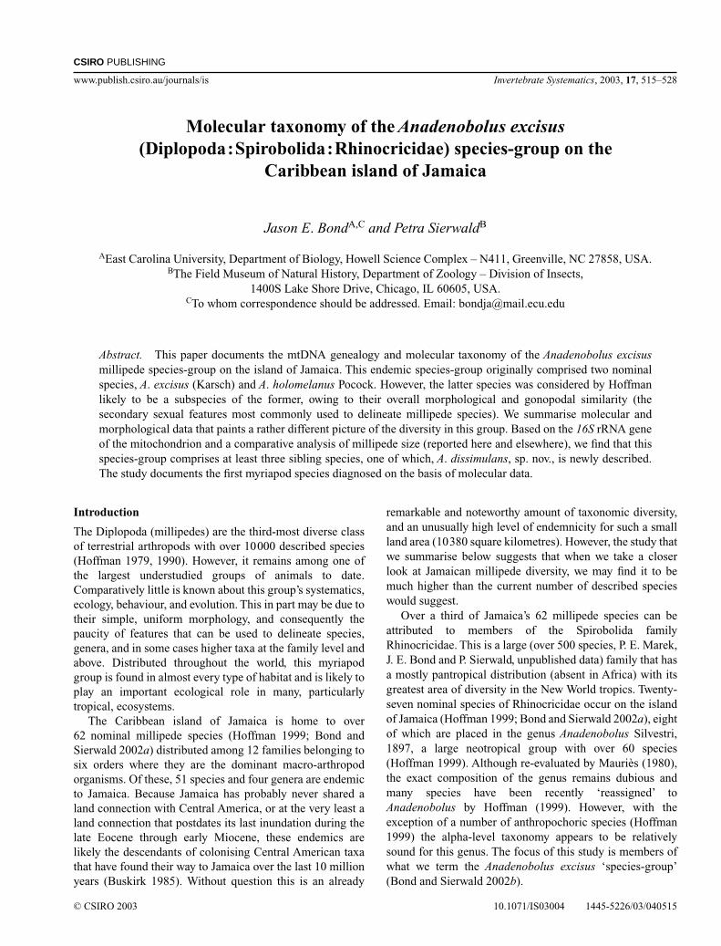
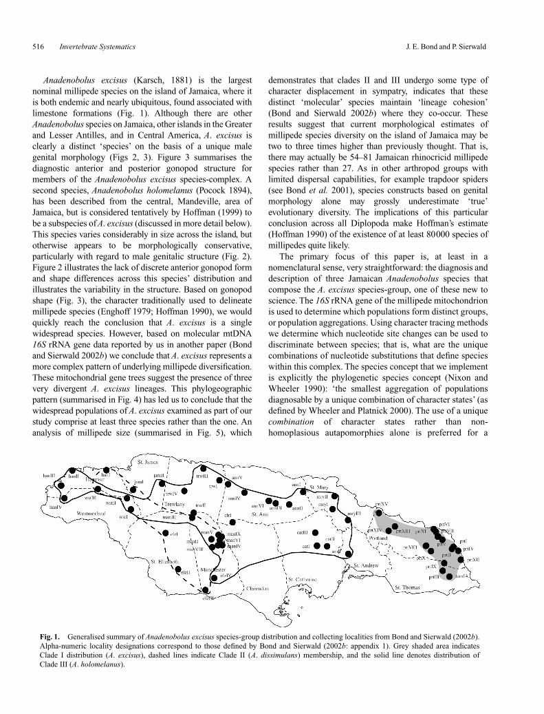
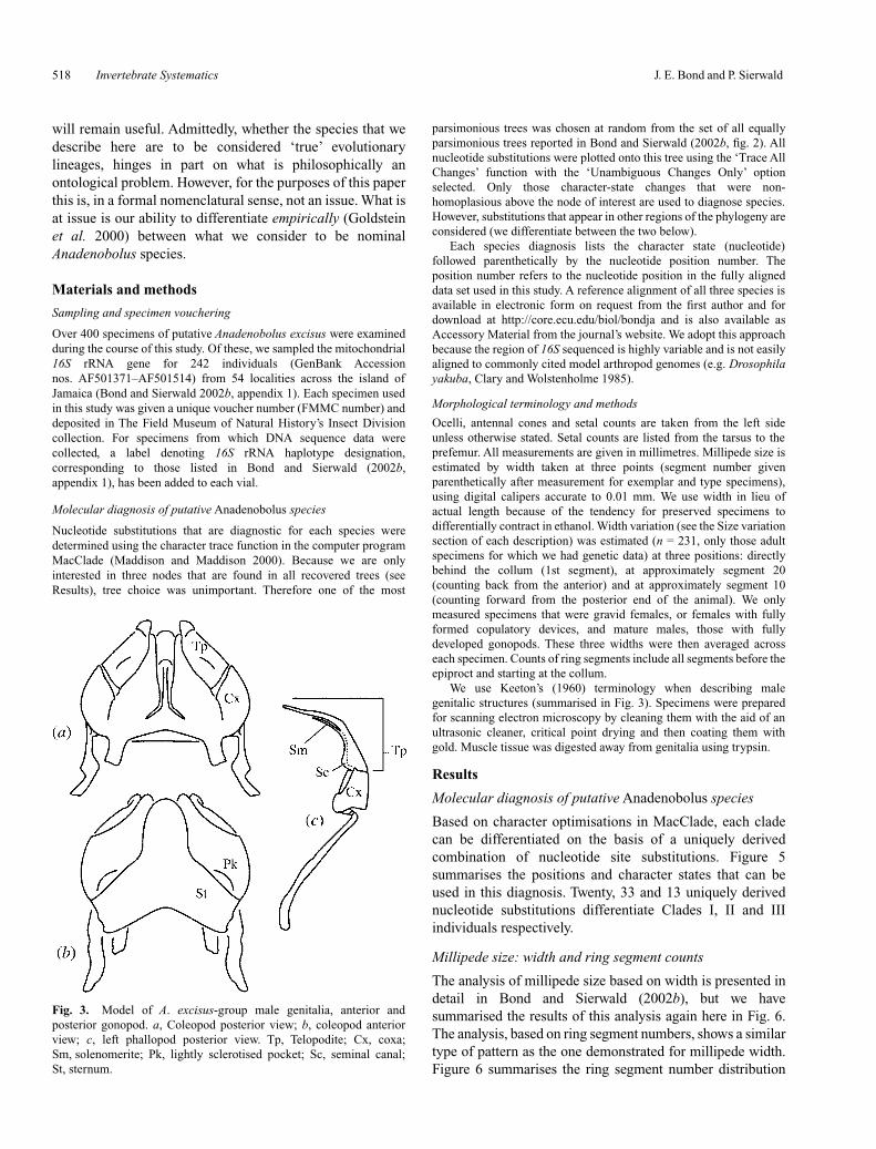
Anadenobolus excisus (Karsch, 1881) is the largestnominal millipede species on the island of Jamaica, where itis both endemic and nearly ubiquitous, found associated withlimestone formations (Fig. 1). Although there are otherAnadenobolus species on Jamaica, other islands in the Greaterand Lesser Antilles, and in Central America, A. excisus isclearly a distinct ‘species’ on the basis of a unique malegenital morphology (Figs 2, 3). Figure 3 summarises thediagnostic anterior and posterior gonopod structure formembers of the Anadenobolus excisus species-complex. Asecond species, Anadenobolus holomelanus (Pocock 1894),has been described from the central, Mandeville, area ofJamaica, but is considered tentatively by Hoffman (1999) tobe a subspecies of A. excisus (discussed in more detail below).This species varies considerably in size across the island, butotherwise appears to be morphologically conservative,particularly with regard to male genitalic structure (Fig. 2).Figure 2 illustrates the lack of discrete anterior gonopod formand shape differences across this species’ distribution andillustrates the variability in the structure. Based on gonopodshape (Fig. 3), the character traditionally used to delineatemillipede species (Enghoff 1979; Hoffman 1990), we wouldquickly reach the conclusion that A. excisus is a singlewidespread species. However, based on molecular mtDNA16S rRNA gene data reported by us in another paper (Bondand Sierwald 2002b) we conclude that A. excisus represents amore complex pattern of underlying millipede diversification.These mitochondrial gene trees suggest the presence of threevery divergent A. excisus lineages. This phylogeographicpattern (summarised in Fig. 4) has led us to conclude that thewidespread populations of A. excisus examined as part of ourstudy comprise at least three species rather than the one. Ananalysis of millipede size (summarised in Fig. 5), which
demonstrates that clades II and III undergo some type ofcharacter displacement in sympatry, indicates that thesedistinct ‘molecular’ species maintain ‘lineage cohesion’(Bond and Sierwald 2002b) where they co-occur. Theseresults suggest that current morphological estimates ofmillipede species diversity on the island of Jamaica may betwo to three times higher than previously thought. That is,there may actually be 54–81 Jamaican rhinocricid millipedespecies rather than 27. As in other arthropod groups withlimited dispersal capabilities, for example trapdoor spiders(see Bond et al. 2001), species constructs based on genitalmorphology alone may grossly underestimate ‘true’evolutionary diversity. The implications of this particularconclusion across all Diplopoda make Hoffman’s estimate(Hoffman 1990) of the existence of at least 80000 species ofmillipedes quite likely.
The primary focus of this paper is, at least in anomenclatural sense, very straightforward: the diagnosis anddescription of three Jamaican Anadenobolus species thatcompose the A. excisus species-group, one of these new toscience. The 16S rRNA gene of the millipede mitochondrionis used to determine which populations form distinct groups,or population aggregations. Using character tracing methodswe determine which nucleotide site changes can be used todiscriminate between species; that is, what are the uniquecombinations of nucleotide substitutions that define specieswithin this complex. The species concept that we implementis explicitly the phylogenetic species concept (Nixon andWheeler 1990): ‘the smallest aggregation of populationsdiagnosable by a unique combination of character states’ (asdefined by Wheeler and Platnick 2000). The use of a uniquecombination of character states rather than non-homoplasious autapomorphies alone is preferred for a
Fig. 1. Generalised summary of Anadenobolus excisus species-group distribution and collecting localities from Bond and Sierwald (2002b).Alpha-numeric locality designations correspond to those defined by Bond and Sierwald (2002b: appendix 1). Grey shaded area indicatesClade I distribution (A. excisus), dashed lines indicate Clade II (A. dissimulans) membership, and the solid line denotes distribution ofClade III (A. holomelanus).
Molecular taxonomy of the Anadenobolus excisus species-group Invertebrate Systematics 517
number of pragmatic reasons. Foremost, as with any speciesdiagnosis, we may never examine all possible sister andoutgroup taxa and can therefore never be confident that any
one diagnostic character is unique to a single taxon. Uniquecombinations of character states potentially ensure that asadditional taxa are discovered and sequenced our diagnoses
Fig. 2. Scanning electron micrographs of anterior male gonopod, coleopod. Left side of figure (a, c, e) showsfull habitus of gonopod (scale bar = 1 mm), right side of figure (b–f) is an enlargement of the gonopod tip (rightside) that shows small distal spines (scale bars = 100 m). a, b, Anadenobolus excisus from prtIII, posterioraspect; c, d, A. dissimulans from wstII, posterior aspect; e, f, A. holomelanus from mryI, posterior aspect;g, A. holomelanus, anterior aspect, small arrow points to lightly sclerotised pocket (Pk in Fig. 3);h, enlargement of sclerotised pocket from adjacent figure.
518 Invertebrate Systematics J. E. Bond and P. Sierwald
will remain useful. Admittedly, whether the species that wedescribe here are to be considered ‘true’ evolutionarylineages, hinges in part on what is philosophically anontological problem. However, for the purposes of this paperthis is, in a formal nomenclatural sense, not an issue. What isat issue is our ability to differentiate empirically (Goldsteinet al. 2000) between what we consider to be nominalAnadenobolus species.
Materials and methods
Sampling and specimen vouchering
Over 400 specimens of putative Anadenobolus excisus were examinedduring the course of this study. Of these, we sampled the mitochondrial16S rRNA gene for 242 individuals (GenBank Accessionnos. AF501371–AF501514) from 54 localities across the island ofJamaica (Bond and Sierwald 2002b, appendix 1). Each specimen usedin this study was given a unique voucher number (FMMC number) anddeposited in The Field Museum of Natural History’s Insect Divisioncollection. For specimens from which DNA sequence data werecollected, a label denoting 16S rRNA haplotype designation,corresponding to those listed in Bond and Sierwald (2002b,appendix 1), has been added to each vial.
Molecular diagnosis of putative Anadenobolus species
Nucleotide substitutions that are diagnostic for each species weredetermined using the character trace function in the computer programMacClade (Maddison and Maddison 2000). Because we are onlyinterested in three nodes that are found in all recovered trees (seeResults), tree choice was unimportant. Therefore one of the most
parsimonious trees was chosen at random from the set of all equallyparsimonious trees reported in Bond and Sierwald (2002b, fig. 2). Allnucleotide substitutions were plotted onto this tree using the ‘Trace AllChanges’ function with the ‘Unambiguous Changes Only’ optionselected. Only those character-state changes that were non-homoplasious above the node of interest are used to diagnose species.However, substitutions that appear in other regions of the phylogeny areconsidered (we differentiate between the two below).
Each species diagnosis lists the character state (nucleotide)followed parenthetically by the nucleotide position number. Theposition number refers to the nucleotide position in the fully aligneddata set used in this study. A reference alignment of all three species isavailable in electronic form on request from the first author and fordownload at http://core.ecu.edu/biol/bondja and is also available asAccessory Material from the journal’s website. We adopt this approachbecause the region of 16S sequenced is highly variable and is not easilyaligned to commonly cited model arthropod genomes (e.g. Drosophilayakuba, Clary and Wolstenholme 1985).
Morphological terminology and methods
Ocelli, antennal cones and setal counts are taken from the left sideunless otherwise stated. Setal counts are listed from the tarsus to theprefemur. All measurements are given in millimetres. Millipede size isestimated by width taken at three points (segment number givenparenthetically after measurement for exemplar and type specimens),using digital calipers accurate to 0.01 mm. We use width in lieu ofactual length because of the tendency for preserved specimens todifferentially contract in ethanol. Width variation (see the Size variationsection of each description) was estimated (n = 231, only those adultspecimens for which we had genetic data) at three positions: directlybehind the collum (1st segment), at approximately segment 20(counting back from the anterior) and at approximately segment 10(counting forward from the posterior end of the animal). We onlymeasured specimens that were gravid females, or females with fullyformed copulatory devices, and mature males, those with fullydeveloped gonopods. These three widths were then averaged acrosseach specimen. Counts of ring segments include all segments before theepiproct and starting at the collum.
We use Keeton’s (1960) terminology when describing malegenitalic structures (summarised in Fig. 3). Specimens were preparedfor scanning electron microscopy by cleaning them with the aid of anultrasonic cleaner, critical point drying and then coating them withgold. Muscle tissue was digested away from genitalia using trypsin.
Results
Molecular diagnosis of putative Anadenobolus species
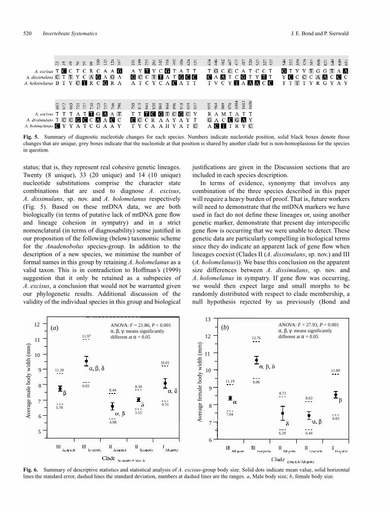
Based on character optimisations in MacClade, each cladecan be differentiated on the basis of a uniquely derivedcombination of nucleotide site substitutions. Figure 5summarises the positions and character states that can beused in this diagnosis. Twenty, 33 and 13 uniquely derivednucleotide substitutions differentiate Clades I, II and IIIindividuals respectively.
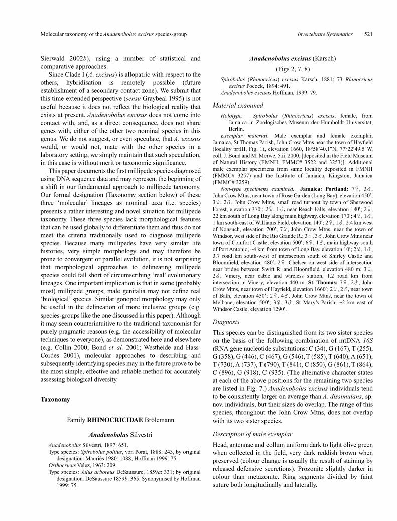
Millipede size: width and ring segment counts
The analysis of millipede size based on width is presented indetail in Bond and Sierwald (2002b), but we havesummarised the results of this analysis again here in Fig. 6.The analysis, based on ring segment numbers, shows a similartype of pattern as the one demonstrated for millipede width.Figure 6 summarises the ring segment number distribution
Fig. 3. Model of A. excisus-group male genitalia, anterior andposterior gonopod. a, Coleopod posterior view; b, coleopod anteriorview; c, left phallopod posterior view. Tp, Telopodite; Cx, coxa;Sm, solenomerite; Pk, lightly sclerotised pocket; Sc, seminal canal;St, sternum.
Molecular taxonomy of the Anadenobolus excisus species-group Invertebrate Systematics 519
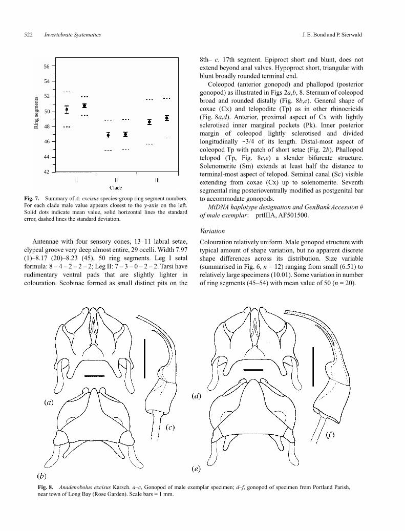
for each species (Clades I–III). Clade II individuals tend tohave fewer ring segments. Furthermore, Clade III has thegreatest range of segment number (e.g. 43–56 for malesexamined). The disparity in ring segment number acrossClade III individuals is also likely correlated with presence,or absence, in the zone of overlap. The number of segmentsfor the three clades significantly differ for males(Kruskal–Wallis test, χ2 = 18.72, P < 0.001) and females
(χ2 = 34.00, P < 0.001); however, these values do not differ ina discrete manner (i.e. the values for each clade overlap).
Discussion
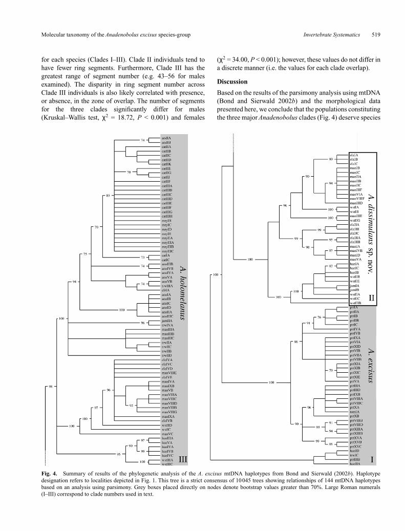
Based on the results of the parsimony analysis using mtDNA(Bond and Sierwald 2002b) and the morphological datapresented here, we conclude that the populations constitutingthe three major Anadenobolus clades (Fig. 4) deserve species
Fig. 4. Summary of results of the phylogenetic analysis of the A. excisus mtDNA haplotypes from Bond and Sierwald (2002b). Haplotypedesignation refers to localities depicted in Fig. 1. This tree is a strict consensus of 10045 trees showing relationships of 144 mtDNA haplotypesbased on an analysis using parsimony. Grey boxes placed directly on nodes denote bootstrap values greater than 70%. Large Roman numerals(I–III) correspond to clade numbers used in text.
520 Invertebrate Systematics J. E. Bond and P. Sierwald
status; that is, they represent real cohesive genetic lineages.Twenty (8 unique), 33 (20 unique) and 14 (10 unique)nucleotide substitutions comprise the character statecombinations that are used to diagnose A. excisus,A. dissimulans, sp. nov. and A. holomelanus respectively(Fig. 5). Based on these mtDNA data, we are bothbiologically (in terms of putative lack of mtDNA gene flowand lineage cohesion in sympatry) and in a strictnomenclatural (in terms of diagnosability) sense justified inour proposition of the following (below) taxonomic schemefor the Anadenobolus species-group. In addition to thedescription of a new species, we minimise the number offormal names in this group by retaining A. holomelanus as avalid taxon. This is in contradiction to Hoffman’s (1999)suggestion that it only be retained as a subspecies ofA. excisus, a conclusion that would not be warranted givenour phylogenetic results. Additional discussion of thevalidity of the individual species in this group and biological
justifications are given in the Discussion sections that areincluded in each species description.
In terms of evidence, synonymy that involves anycombination of the three species described in this paperwill require a heavy burden of proof. That is, future workerswill need to demonstrate that the mtDNA markers we haveused in fact do not define these lineages or, using anothergenetic marker, demonstrate that present day interspecificgene flow is occurring that we were unable to detect. Thesegenetic data are particularly compelling in biological termssince they do indicate an apparent lack of gene flow whenlineages coexist (Clades II (A. dissimulans, sp. nov.) and III(A. holomelanus)). We base this conclusion on the apparentsize differences between A. dissimulans, sp. nov. andA. holomelanus in sympatry. If gene flow was occurring,we would then expect large and small morphs to berandomly distributed with respect to clade membership, anull hypothesis rejected by us previously (Bond and
Fig. 5. Summary of diagnostic nucleotide changes for each species. Numbers indicate nucleotide position, solid black boxes denote thosechanges that are unique, grey boxes indicate that the nucleotide at that position is shared by another clade but is non-homoplasious for the speciesin question.
ANOVA: F = 21.86, P < 0.001α, β, γ: means significantly different at α = 0.05
ANOVA: F = 27.93, P < 0.001α, β, γ: means significantly different at α = 0.05
(a) (b)
Ave
rage
mal
e bo
dy w
idth
(m
m)
Ave
rage
fem
ale
body
wid
th (
mm
)
12
12
10
1110
11
13
9
8
7
6
7
8
9
6
5
11.97
11.20
6.63
5.78
8.448.39
10.01
6.51
5.55
4.986.65
6.446.29
7.04
11.19 8.86
8.728.63
11.89
12.76
Fig. 6. Summary of descriptive statistics and statistical analysis of A. excisus-group body size. Solid dots indicate mean value, solid horizontallines the standard error, dashed lines the standard deviation, numbers at dashed lines are the ranges. a, Male body size; b, female body size.
Molecular taxonomy of the Anadenobolus excisus species-group Invertebrate Systematics 521
Sierwald 2002b), using a number of statistical andcomparative approaches.
Since Clade I (A. excisus) is allopatric with respect to theothers, hybridisation is remotely possible (futureestablishment of a secondary contact zone). We submit thatthis time-extended perspective (sensu Graybeal 1995) is notuseful because it does not reflect the biological reality thatexists at present. Anadenobolus excisus does not come intocontact with, and, as a direct consequence, does not sharegenes with, either of the other two nominal species in thisgenus. We do not suggest, or even speculate, that A. excisuswould, or would not, mate with the other species in alaboratory setting, we simply maintain that such speculation,in this case is without merit or taxonomic significance.
This paper documents the first millipede species diagnosedusing DNA sequence data and may represent the beginning ofa shift in our fundamental approach to millipede taxonomy.Our formal designation (Taxonomy section below) of thesethree ‘molecular’ lineages as nominal taxa (i.e. species)presents a rather interesting and novel situation for millipedetaxonomy. These three species lack morphological featuresthat can be used globally to differentiate them and thus do notmeet the criteria traditionally used to diagnose millipedespecies. Because many millipedes have very similar lifehistories, very simple morphology and may therefore beprone to convergent or parallel evolution, it is not surprisingthat morphological approaches to delineating millipedespecies could fall short of circumscribing ‘real’ evolutionarylineages. One important implication is that in some (probablymost) millipede groups, male genitalia may not define real‘biological’ species. Similar gonopod morphology may onlybe useful in the delineation of more inclusive groups (e.g.species-groups like the one discussed in this paper). Althoughit may seem counterintuitive to the traditional taxonomist forpurely pragmatic reasons (e.g. the accessibility of moleculartechniques to everyone), as demonstrated here and elsewhere(e.g. Collin 2000; Bond et al. 2001; Westheide and Hass-Cordes 2001), molecular approaches to describing andsubsequently identifying species may in the future prove to bethe most simple, effective and reliable method for accuratelyassessing biological diversity.
Taxonomy
Family RHINOCRICIDAE Brölemann
Anadenobolus Silvestri
Anadenobolus Silvestri, 1897: 651.Type species: Spirobolus politus, von Porat, 1888: 243, by original
designation. Mauriès 1980: 1088; Hoffman 1999: 75.Orthocricus Velez, 1963: 209. Type species: Julus arboreus DeSaussure, 1859a: 331; by original
designation. DeSaussure 1859b: 365. Synonymised by Hoffman1999: 75.
Anadenobolus excisus (Karsch)
(Figs 2, 7, 8)
Spirobolus (Rhinocricus) excisus Karsch, 1881: 73 Rhinocricusexcisus Pocock, 1894: 491.
Anadenobolus excisus Hoffman, 1999: 79.
Material examined
Holotype. Spirobolus (Rhinocricus) excisus, female, fromJamaica in Zoologisches Museum der Humboldt Universität,Berlin.
Exemplar material. Male exemplar and female exemplar,Jamaica, St Thomas Parish, John Crow Mtns near the town of Hayfield(locality prtIII, Fig. 1), elevation 1660, 18°58′40.1″N, 77°22′49.5″W,coll. J. Bond and M. Merwe, 5.ii. 2000, [deposited in the Field Museumof Natural History (FMNH; FMMC# 3522 and 3253)]. Additionalmale exemplar specimens from same locality deposited in FMNH(FMMC# 3257) and the Institute of Jamaica, Kingston, Jamaica(FMMC# 3259).
Non-type specimens examined. Jamaica: Portland: 7�, 3�,John Crow Mtns, near town of Rose Garden (Long Bay), elevation 450′;3�, 2�, John Crow Mtns, small road turnout by town of SherwoodForest, elevation 370′; 2�, 1�, near Reach Falls, elevation 180′; 2�,22 km south of Long Bay along main highway, elevation 170′; 4�, 1�,1 km south-east of Williams Field, elevation 140′; 2�, 1�, 2.4 km westof Nonsuch, elevation 700′; 7�, John Crow Mtns, near the town ofWindsor, west side of the Rio Grande R.; 3�, 3�, John Crow Mtns neartown of Comfort Castle, elevation 500′; 6�, 1�, main highway southof Port Antonio, ~4 km from town of Long Bay, elevation 10′; 2�, 1�,3.7 road km south-west of intersection south of Shirley Castle andBloomfield, elevation 480′; 2�, Chelsea on west side of intersectionnear bridge between Swift R. and Bloomfield, elevation 480 m; 3�,2�, Vinery, near cable and wireless station, 1.2 road km fromintersection in Vinery, elevation 440 m. St. Thomas: 7�, 2�, JohnCrow Mtns, near town of Hayfield, elevation 1660′; 2�, 2�, near townof Bath, elevation 450′; 2�, 4�, John Crow Mtns, near the town ofMelbane, elevation 500′; 3�, 3�, St Mary’s Parish, ~2 km east ofWindsor Castle, elevation 1290′.
Diagnosis
This species can be distinguished from its two sister specieson the basis of the following combination of mtDNA 16SrRNA gene nucleotide substitutions: C (34), G (167), T (255),G (358), G (446), C (467), G (546), T (585), T (640), A (651),T (730), A (737), T (790), T (841), C (850), G (861), T (864),C (896), G (918), C (935). (The alternative character statesat each of the above positions for the remaining two speciesare listed in Fig. 7.) Anadenobolus excisus individuals tendto be consistently larger on average than A. dissimulans, sp.nov. individuals, but their sizes do overlap. The range of thisspecies, throughout the John Crow Mtns, does not overlapwith its two sister species.
Description of male exemplar
Head, antennae and collum uniform dark to light olive greenwhen collected in the field, very dark reddish brown whenpreserved (colour change is usually the result of staining byreleased defensive secretions). Prozonite slightly darker incolour than metazonite. Ring segments divided by faintsuture both longitudinally and laterally.
522 Invertebrate Systematics J. E. Bond and P. Sierwald
Antennae with four sensory cones, 13–11 labral setae,clypeal groove very deep almost entire, 29 ocelli. Width 7.97(1)–8.17 (20)–8.23 (45), 50 ring segments. Leg I setalformula: 8 – 4 – 2 – 2 – 2; Leg II: 7 – 3 – 0 – 2 – 2. Tarsi haverudimentary ventral pads that are slightly lighter incolouration. Scobinae formed as small distinct pits on the
8th– c. 17th segment. Epiproct short and blunt, does notextend beyond anal valves. Hypoproct short, triangular withblunt broadly rounded terminal end.
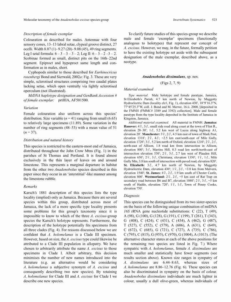
Coleopod (anterior gonopod) and phallopod (posteriorgonopod) as illustrated in Figs 2a,b, 8. Sternum of coleopodbroad and rounded distally (Fig. 8b,e). General shape ofcoxae (Cx) and telopodite (Tp) as in other rhinocricids(Fig. 8a,d). Anterior, proximal aspect of Cx with lightlysclerotised inner marginal pockets (Pk). Inner posteriormargin of coleopod lightly sclerotised and dividedlongitudinally ~3/4 of its length. Distal-most aspect ofcoleopod Tp with patch of short setae (Fig. 2b). Phallopodtelopod (Tp, Fig. 8c,e) a slender bifurcate structure.Solenomerite (Sm) extends at least half the distance toterminal-most aspect of telopod. Seminal canal (Sc) visibleextending from coxae (Cx) up to solenomerite. Seventhsegmental ring posterioventrally modified as postgenital barto accommodate gonopods.
MtDNA haplotype designation and GenBank Accession #of male exemplar: prtIIIA, AF501500.
Variation
Colouration relatively uniform. Male gonopod structure withtypical amount of shape variation, but no apparent discreteshape differences across its distribution. Size variable(summarised in Fig. 6, n = 12) ranging from small (6.51) torelatively large specimens (10.01). Some variation in numberof ring segments (45–54) with mean value of 50 (n = 20).
Rin
g se
gmen
ts
56
54
52
50
48
46
44
42
Fig. 7. Summary of A. excisus species-group ring segment numbers.For each clade male value appears closest to the y-axis on the left.Solid dots indicate mean value, solid horizontal lines the standarderror, dashed lines the standard deviation.
Fig. 8. Anadenobolus excisus Karsch. a–c, Gonopod of male exemplar specimen; d–f, gonopod of specimen from Portland Parish,near town of Long Bay (Rose Garden). Scale bars = 1 mm.
Molecular taxonomy of the Anadenobolus excisus species-group Invertebrate Systematics 523
Description of female exemplar
Colouration as described for males. Antennae with foursensory cones, 13–13 labral setae, clypeal groove distinct, 27ocelli. Width 8.07 (1)–9.27 (20)–9.08 (45), 49 ring segments.Leg I setal formula: 6 – 3 – 3 – 3 – 2; Leg II: 6 – 3 – 2 –3 – 2.Scobinae formed as small, distinct pits on the 16th–22ndsegment. Epiproct and hypoproct same length and con-formation as in males, short.
Cyphopods similar to those described for Eurhinocricusrosenbergi Bond and Sierwald, 2002a: Fig. 3. These are verysimple, sclerotised structures comprising two caudal plateslacking setae, which open ventrally via lightly sclerotisedoperculum (not illustrated).
MtDNA haplotype designation and GenBank Accession #of female exemplar: prtIIIA, AF501500.
Variation
Female colouration also uniform across this species’distribution. Size variable (n = 41) ranging from small (6.65)to relatively large specimens (11.89). Some variation in thenumber of ring segments (48–53) with a mean value of 51(n = 37).
Distribution and natural history
This species is restricted to the eastern-most end of Jamaica,distributed throughout the John Crow Mtns (Fig. 1) in theparishes of St Thomas and Portland. It is found almostexclusively in the thin layer of leaves on and aroundlimestone. This represents a marginal departure in habitatfrom the other two Anadenobolus species described in thispaper since they occur in an ‘interstitial’-like manner amongthe limestone rubble.
Remarks
Karsch’s 1881 description of this species lists the typelocality (implied) only as Jamaica. Because there are severalspecies within this group, distributed across most ofJamaica, the lack of a more specific type locality presentssome problems for this group’s taxonomy since it isimpossible to know to which of the three A. excisus-groupspecies the Karsch’s holotype represents. Furthermore, thedescription of the holotype potentially fits specimens fromall three clades (Fig. 4). For reasons discussed below we areconfident that A. holomelanus is a Clade III specimen.However, based on size, the A. excisus type could likewise beattributed to a Clade III population in allopatry. We havechosen to arbitrarily attribute the name A. excisus to thosespecimens in Clade I. Albeit arbitrary, this decisionminimises the number of new names introduced into theliterature (e.g. an alternative would be consideringA. holomelanus a subjective synonym of A. excisus andconsequently describing two new species). By retainingA. holomelanus for Clade III and A. excisus for Clade I wedescribe one new species.
To clarify future studies of this species-group we describemale and female ‘exemplar’ specimens (functionallyanalogous to holotypes) that represent our concept ofA. excisus. However, we may, in the future, formally petitionto have the existing holotype set aside with the subsequentdesignation of the male exemplar, described above, as aneotype.
Anadenobolus dissimulans, sp. nov.
(Figs 2, 7, 9)
Material examined
Type material. Male holotype and female paratype, Jamaica,St Elizabeth’s Parish, 4.7 km north of Newton, by MaggottyHydroelectric Dam (locality elz1, Fig. 1), elevation 430′, 18°9′16.3″N,77°45′25.3″W, coll. J. Bond and M. Merwe, 16.ii. 2000, [deposited inthe FMNH (FMMC# 3389 and 3392) collection]. Male and femaleparatype from the type locality deposited in the Institute of Jamaica inKingston, Jamaica.
Additional material examined. All material in FMNH. Jamaica:Hanover: 4�, 3�, small side road along coast at the town of Flint R.,elevation 20–30′; 1�, 5.2 km west of Lucea along highway A1,elevation 20′. Manchester: 3�, 2�, 4.5 km east of town of Mark Post,elevation 1318′; 2�, 4�; ~2.5 km east/south-east of Mile Gully,elevation 1720′; 3�, 5.2 km north of Oxford, elevation 1210′; 2�, 2�,north-east of Allison, 1.8 road km from intersection in Allison,elevation 800′; 3�, Martins Hill, 0.3 road km north/north-east ofintersection elevation 530′; 2�, 3�, 2.7 km west of Plauden Hill,elevation 650′; 2�, 3�, Christiana, elevation 1350′; 1�, 1�, MileGully Mtn, 3.8 km south of intersection with paved road, elevation 820′.St. Elizabeth: 3�, 4.7 km north of Newton, by MaggottyHydroelectric Dam, elevation 430′; 1�, 2�, ~3.0 km from Malvern,elevation 1540′; St. James: 4�, 2�, 1.9 km south of Chester Castle,elevation 800′. Westmorland: 2�, 2�, ~5 km east of Rat Trap onsecondary road between B6 and B7, elevation 1080′; 2�, 3�, ~5 kmsouth of Haddo, elevation 720′; 1�, 1�, Town of Penny Cooke,elevation 750′.
Diagnosis
This species can be distinguished from its two sister-specieson the basis of the following unique combination of mtDNA16S rRNA gene nucleotide substitutions: C (22), T (49),A (98), G (100), G (128), G (191), C (199), T (261), T (343),G (408), C (424), C (431), C (434), A (462), G (487),T (527), C (552), C (579), A (606), A (631), C (649),C (672), C (685), G (721), C (727), A (735), C (786),C (795), C (815), G (955), C (970), G (1004), A (1013). (Thealternative character states at each of the above positions forthe remaining two species are listed in Fig. 7.) Wheresympatric with A. holomelanus, female A. dissimulans aremuch smaller and statistically have fewer segments (seeresults section above). Known size ranges in sympatry ofA. dissimulans are 6.44–8.63, whereas sizes ofA. holomelanus are 8.86–12.76 (Fig. 6). These species canalso be discriminated in sympatry on the basis of colour.Anadenobolus dissimulans individuals are much lighter incolour, usually a dull olive-green, whereas individuals of
524 Invertebrate Systematics J. E. Bond and P. Sierwald
A. holomelanus are a shiny black. Scobinae of A. dissimulansare consistently 2–3× larger than those of A. excisus,however, because the scobinae of A. holomelanus arepolymorphic (small and large) this character may not be veryuseful in differentiating these species.
Description of male holotype (FMMC# 3389)
Head, antennae and collum uniform dark olive green whencollected in the field, very dark reddish brown whenpreserved (colour change is usually the result of staining byreleased defensive secretions). Prozonite slightly darker incolour than metazonite. Ring segments divided by faintsuture both longitudinally and laterally.
Antennae with four sensory cones, 14–14 labral setae,clypeal groove very deep almost entire, 28 ocelli. Width 7.79(1)–8.73 (20)–8.50 (45), 47 ring segments. Leg I setalformula: 7 – 2 – 3 – 2 – 2; Leg II: 8 – 2 – 2 – 1 – 2. Tarsi haveprominent ventral pads that are light in colour and dividedlongitudinally. Scobinae formed as large distinct pits on the8th– c. 17th segment. Epiproct short and blunt, does notextend beyond anal valves. Hypoproct short, triangular withblunt broadly rounded terminal end.
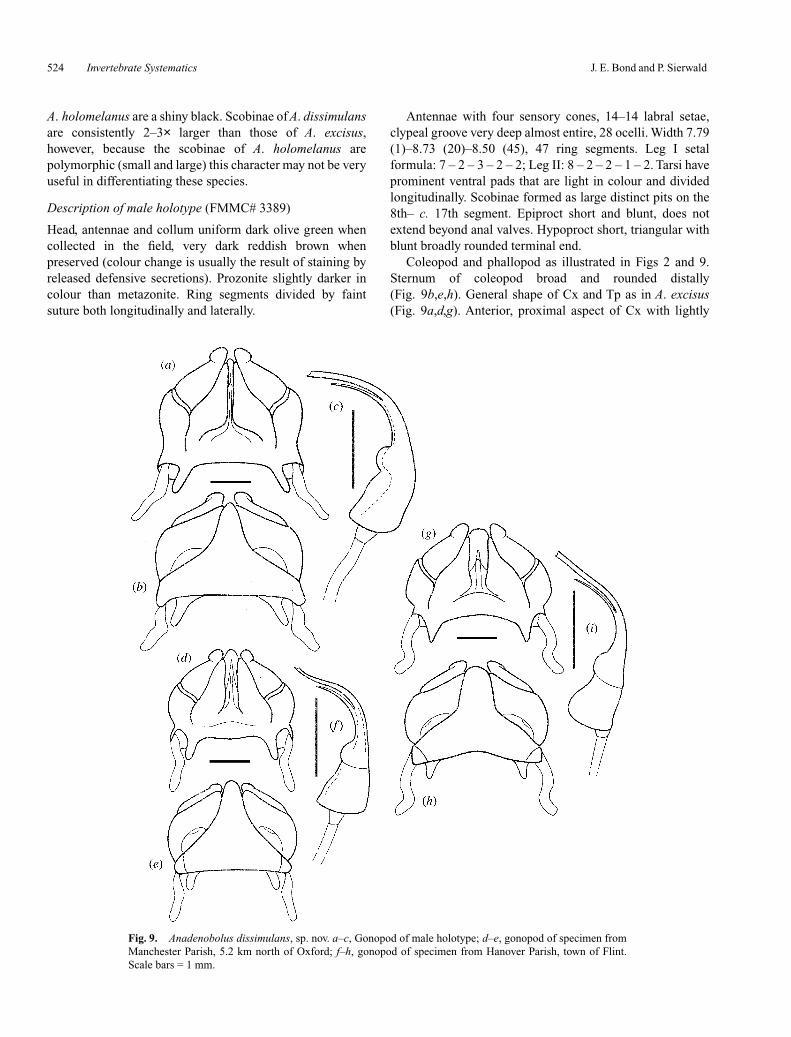
Coleopod and phallopod as illustrated in Figs 2 and 9.Sternum of coleopod broad and rounded distally(Fig. 9b,e,h). General shape of Cx and Tp as in A. excisus(Fig. 9a,d,g). Anterior, proximal aspect of Cx with lightly
Fig. 9. Anadenobolus dissimulans, sp. nov. a–c, Gonopod of male holotype; d–e, gonopod of specimen fromManchester Parish, 5.2 km north of Oxford; f–h, gonopod of specimen from Hanover Parish, town of Flint.Scale bars = 1 mm.
Molecular taxonomy of the Anadenobolus excisus species-group Invertebrate Systematics 525
sclerotised inner marginal Pk. Inner posterior margin ofcoleopod lightly sclerotised and divided longitudinally ~3/4of its length (Fig. 9a,d,g). Distal-most aspect of coleopod Tpwith patch of short setae (Fig. 2d). Phallopod Tp (Fig. 9c,f,i)is a slender bifurcate structure. Solenomerite extends at leasthalf the distance to the terminal-most aspect of the telopod.Sc is visible extending from the Cx up to the solenomerite.Seventh segmental ring posterioventrally modified as apostgenital bar to accommodate gonopods.
MtDNA haplotype designation and GenBank Accession #of male exemplar: elz1B, AF501448.
Variation
Colouration relatively uniform. Male gonopod structure withtypical amount of shape variation, but no apparent discreteshape differences across its distribution. Size variable(summarised in Fig. 6a), specimens relatively small(5.55–8.39, n = 18). Some variation in the number of ringsegments (Fig. 7, 43–52) with a mean value of 47 (n = 31).
Description of female paratype (FMMC# 3392)
Colouration as described for males. Antennae with foursensory cones, 13–14 labral setae, clypeal groove distinct, 33ocelli. Width 7.68 (1)–8.92 (20)–9.12 (45), 45 ring segments.Leg I setal formula: 7 – 4 – 2 – 3 – 1; Leg II: 9 – 3 – 3 –3 – 2.Tarsi lack prominent pads. Scobinae formed as distinct pitson the 7th–20th segment, smaller than in males. Epiproct andhypoproct same length and conformation as in males, short.
Cyphopods similar to those described for Eurhinocricusrosenbergi Bond and Sierwald, 2002a: Fig. 3. These are verysimple, sclerotised structures comprising two caudal plateslacking setae, which open ventrally via lightly sclerotisedoperculum (not illustrated).
MtDNA haplotype designation and GenBank Accession #of female exemplar: elz1A, AF501447.
Variation
Female colouration also uniform across this species’distribution. Size variable (Fig. 6), relatively small(6.29–8.72, n = 36). Some variation in the number of ringsegments (45–50; Fig. 7) with a mean value of 47 (n = 22).
Distribution and natural history
The first author has collected this species throughout thefollowing parishes: St James, Hanover, Westmorland,Manchester and St Elizabeth (Fig. 1). This species occursonly in areas with lots of limestone, where it is found in an‘interstitial’-like manner amongst the limestone rubble.Most of its range overlaps with that of A. holomelanus.
Remarks
See Remarks under A. holomelanus.
Etymology
The specific epithet refers to the cryptic nature of this species.
Anadenobolus holomelanus (Pocock)
(Figs 2, 7, 10)
Rhinocricus holomelanus Pocock 1881: 492. (Female holotypefrom Jamaica most likely in The Natural History Museum(BMNH), not examined, probably lost.)
Anadenobolus holomelanus, Hoffman 1999: 79.
Material examined
Designation of neotype. Male neotype (FMMC# 3421) andfemale neoparatype (FMMC# 3422), Jamaica, Manchester Parish,Christiana, 18°08′34.3″N, 77°29′37.2″W (locality manV, Fig. 1),elevation 2660′, coll. J. Bond and M. Merwe, 18.ii. 2000, (FMNH).
Paratypes. Jamaica, Trewlany Parish, Troy, 18°14′47.9″N,77°36′53.7″W, elevation 1410′, coll. J. Bond and M. Merwe, 16.ii.2000, (Institute of Jamaica in Kingston, Jamaica).
Additional material examined. All material in FMNH. Jamaica:Clarendon: 1�, 2�, town of Burbage, elevation 1750′. Hanover: 1�,small side road between green Island and Santoy, elevation 40′.Manchester: 3�, 2�, Christiana, elevation 1350′; 3�, 8�; 0.5 kmsouth of Newport, elevation 2510′; 12�, 8�, 0.3 km north/north-eastof intersection at Martins Hill, elevation 530 m; 3�, 3�, north-east ofAllison, 1.8 road km from intersection in Allison, elevation 800′; 3�,3�, 5.2 km north of Oxford, elevation 1210′. St. Andrew: 2�, 1�,4.1 km east of Rock Hill, elevation 1440′. St. Ann: 2�, 4�, ~1 kmfrom the south end of Fern Gully, small turnout near the base of largecoral formation, elevation 910′; 4�, 1�; near town of Phoenix Park,elevation 1220′; 2�, 2�, Alderton, elevation 1460′; 3�, ~4.1 km fromtown of Cedar Valley, elevation 2230′; 1�, 2�,~5.6 km north ofAlexandria, elevation 1900′; 1�, 2�, 1.2 road km south of mainintersection in Stewart Town. St. Catherine: 3�, 3�, 3.1 km east ofJackson, elevation 1550′; 7�, 4�; town of Bay Walk, elevation 230′;11�, between Works and Riverdale, elevation 150′. St. James: 2�,2�, ~.07 road km S of Spring Vale, elevation 160′. St. Mary: 3�, 3�,~2 km east of the town of Windsor Castle, elevation 1290′; 7�, 3�,Leinster, 1.8 road km south/south-west of intersection near bridge northof Tranquility, elevation 290 m. Trewlany: just south of Jackson Town;4�, 4�; Cockpit Country, Windsor, Troy Trail, about 100 m south ofDevils Hole, elevation 300 m; 1�; Troy, elevation 1410′.Westmorland: 1�, 2�, ~5 km south of Haddo, elevation 720′; 3�,1�, ~5 km north of Delue Bridge; 2�, Town of Penny Cooke, elevation750′.
Diagnosis
This species can be distinguished from its two sister specieson the basis of the following unique combination of mtDNA16S rRNA gene nucleotide substitutions: C (56), T (59),G (123), C (392), T (473), A (520), A (523), C (537), T (559),C (663), C (937), C (964), T (969), C (1058). (The alternativecharacter states at each of the above positions for theremaining two species are listed in Fig. 7.) Additionalmorphological features that may be helpful in distinguishingA. holomelanus from A. dissimulans are given in theDiagnosis of A. dissimulans. Anadenobolus holomelanus,where sympatric with A. dissimulans, behaves muchdifferently than A. dissimulans (or A. excisus). Whendisturbed, Anadenobolus holomelanus thrashes about andsprays defensive secretions to distances in excess of a metre,whereas the other two species are more likely to curl up intoa tight ball and ooze their defensive secretions.
526 Invertebrate Systematics J. E. Bond and P. Sierwald
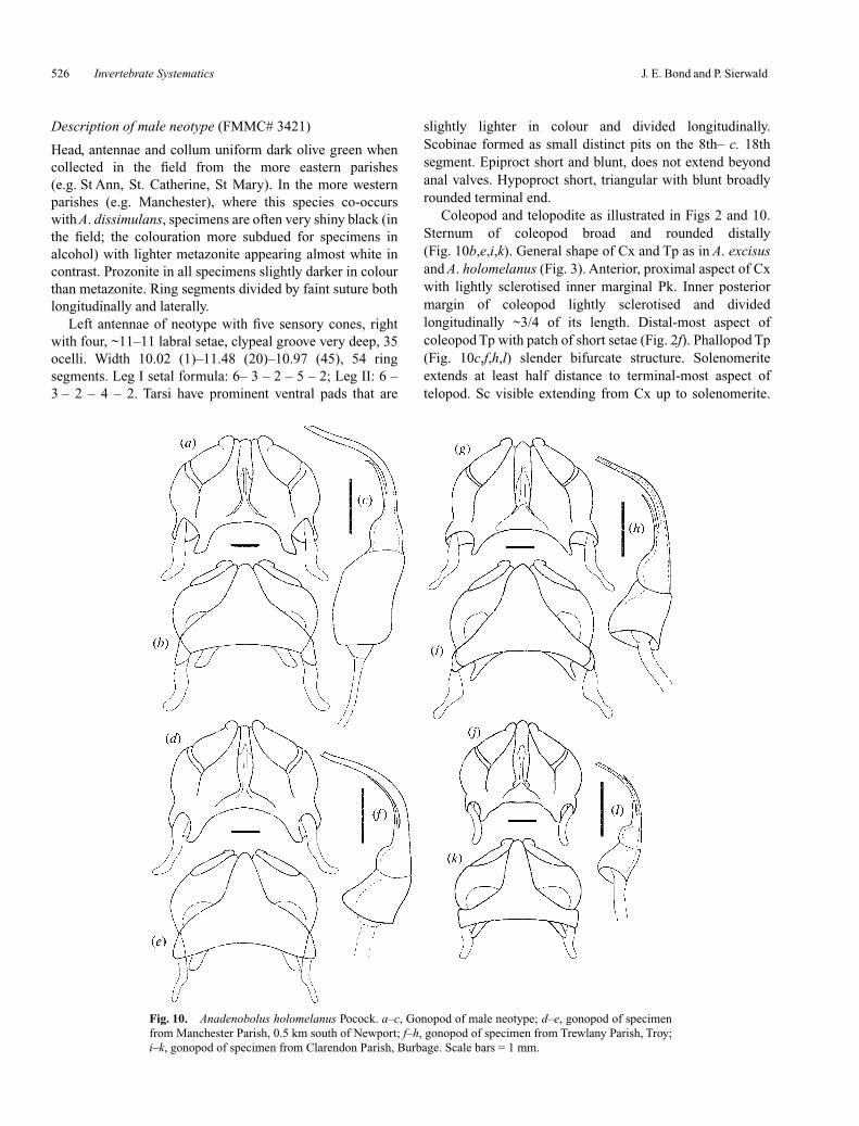
Description of male neotype (FMMC# 3421)
Head, antennae and collum uniform dark olive green whencollected in the field from the more eastern parishes(e.g. St Ann, St. Catherine, St Mary). In the more westernparishes (e.g. Manchester), where this species co-occurswith A. dissimulans, specimens are often very shiny black (inthe field; the colouration more subdued for specimens inalcohol) with lighter metazonite appearing almost white incontrast. Prozonite in all specimens slightly darker in colourthan metazonite. Ring segments divided by faint suture bothlongitudinally and laterally.
Left antennae of neotype with five sensory cones, rightwith four, ~11–11 labral setae, clypeal groove very deep, 35ocelli. Width 10.02 (1)–11.48 (20)–10.97 (45), 54 ringsegments. Leg I setal formula: 6– 3 – 2 – 5 – 2; Leg II: 6 –3 – 2 – 4 – 2. Tarsi have prominent ventral pads that are
slightly lighter in colour and divided longitudinally.Scobinae formed as small distinct pits on the 8th– c. 18thsegment. Epiproct short and blunt, does not extend beyondanal valves. Hypoproct short, triangular with blunt broadlyrounded terminal end.
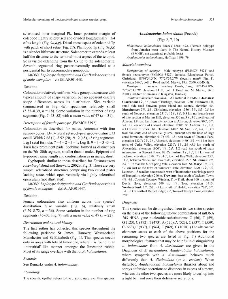
Coleopod and telopodite as illustrated in Figs 2 and 10.Sternum of coleopod broad and rounded distally(Fig. 10b,e,i,k). General shape of Cx and Tp as in A. excisusand A. holomelanus (Fig. 3). Anterior, proximal aspect of Cxwith lightly sclerotised inner marginal Pk. Inner posteriormargin of coleopod lightly sclerotised and dividedlongitudinally ~3/4 of its length. Distal-most aspect ofcoleopod Tp with patch of short setae (Fig. 2f). Phallopod Tp(Fig. 10c,f,h,l) slender bifurcate structure. Solenomeriteextends at least half distance to terminal-most aspect oftelopod. Sc visible extending from Cx up to solenomerite.
Fig. 10. Anadenobolus holomelanus Pocock. a–c, Gonopod of male neotype; d–e, gonopod of specimenfrom Manchester Parish, 0.5 km south of Newport; f–h, gonopod of specimen from Trewlany Parish, Troy;i–k, gonopod of specimen from Clarendon Parish, Burbage. Scale bars = 1 mm.
Molecular taxonomy of the Anadenobolus excisus species-group Invertebrate Systematics 527
Seventh segmental ring posterioventrally modified aspostgenital bar to accommodate gonopods.
MtDNA haplotype designation and GenBank Accession #of male neotype: manVC, AF501439.
Variation
Variation in colouration as described above, variationdependent upon region from which specimen is taken.Scobinae size variable in this species, in some specimensformed only as small pits, whereas in others they are muchlarger. Male gonopod structure with typical amount of shape,however, no apparent discrete shape differences across itsdistribution. Size highly variable (summarised in Fig. 6a),specimens range from relatively small to very large(5.7–11.20, n = 57) and tend to be largest where sympatricwith A. dissimulans. Considerable variation in the number ofring segments (Fig. 7) appears to be concomitant with overallsize variation (43–56) with a mean value of 49 (n = 66).
Description of female paratype (FMMC# 3342)
Colouration as described for males. Antennae with foursensory cones, 11–10 labral setae, clypeal groove distinct, 33ocelli. Width 9.50 (1)–10.24 (20)–10.29 (45), 51 ringsegments. Leg I setal formula: 7 – 3 – 5 – 4 – 2; Leg II: 6 –3 – 2 –4 – 2. Tarsi lack prominent pads. Scobinae formed asdistinct pits on the 8th–17th segment, equal in size to that ofmale neotype. Epiproct and hypoproct same length andconformation as in males, short.
Cyphopods similar to those described for Eurhinocricusrosenbergi Bond and Sierwald, 2002a: fig. 3. These are verysimple, sclerotised structures comprising two caudal plateslacking setae, which open ventrally via lightly sclerotisedoperculum (not illustrated).
MtDNA haplotype designation and GenBank Accession #of female exemplar: manVB, AF501429.
Variation
Female colouration and scobinae size variation similar tothat described for males. Size highly variable (7.04–12.76,n = 67), pattern of size variation as in males. Number of ringsegments likewise variable (43–56) with a mean value of 49.
Distribution and natural history
The first author has collected this species throughout thefollowing parishes: St Catherine, St. Ann, Trewlany,Manchester, Westmorland, St James and Hanover. Thisspecies occurs only in areas with lots of limestone where it isfound in an ‘interstitial’-like manner among the limestonerubble. Much of its range overlaps with that ofA. dissimulans. As mentioned in the Remarks, this speciesbehaves differently when disturbed. Unlike the other twospecies, A. holomelanus is quite active and is able to spray itsdefensive chemicals quite a distance.
Remarks
The original holotype of A. holomelanus was presumed byHoffman (1999) to be deposited in The Natural HistoryMuseum collection in London, but the specimen could not belocated (J. Beccaloni personal communication). Althoughunlikely to be deposited elsewhere, we made additional,unproductive inquiries, to the Museum National d’HistoireNaturelle, Paris; Zoologisk Museum, UniversitetKøbenhavn; and Zoologisches Museum, Hamburg. Theabsence of this type specimen in the BMNH is consistentwith what Hoffman (1999) has observed for many of theBiologi Centrali–Americana and later species that neverwere returned after Pocock’s departure from the BMNH in1903. To ensure the future nomenclatural stability of theAnadenobolus excisus species complex we find it necessaryto designate a neotype for A. holomelanus. Due to the crypticnature and widespread distribution of all three species in thisgroup it is important that each species be represented by atype specimen collected from a specific type locality.
Pocock’s (1894) description contains two clues thatsuggest that the Clade III specimens and the single specimenthat we designate as the A. holomelanus neotype are capturedby his concept of A. holomelanus. First, his generaldescription states that the colour is ‘black and shining’ andthe length is up to 105 mm. These colouration and size arefeatures are consistent with Clade III individuals (Fig. 4) thatare sympatric with A. dissimulans (Clade II). Secondly,Pocock does not give a specific type locality but he doesmention specimens collected from Mandeville andMoneague. Although Clade II individuals have beencollected from Mandeville and surrounding areas (seeabove), only Clade I and III individuals have been collectedin the parishes east of Manchester. The choice of locality forthe neotype specimen reflects our attempt to choose aspecimen from an area close to those mentioned by Pocock(1894) in his description.
Acknowledgments
This project was supported by National Science Foundation(NSF) PEET grant DEB 9712438 to P. Sierwald andW. Shear and NSF grant DEB 9870233 to G. Rosenberg. Thegovernment of Jamaica provided collecting permitsG. Rosenberg. We thank Dr Jason Dunlop, ZoologischesMuseum der Humboldt Universität, Berlin for the loan of theA. excisus holotype. Gonopod illustrations were drawn byLori Grove.
References
Bond, J. E., and Sierwald, P. (2002a). Eurhinocricus rosenbergi, a newspecies of rhinocricid from the Caribbean island of Jamaica(Spirobolida: Rhinocricidae). Proceedings of the Biological Societyof Washington 115, 181–186.
Bond, J. E., and Sierwald, P. (2002b). Cryptic speciation in theAnadenobolus excisus millipede species complex on the island ofJamaica. Evolution 56, 1123–1135.
528 Invertebrate Systematics J. E. Bond and P. Sierwald
http://www.publish.csiro.au/journals/is
Bond, J. E., Hedin, M. C., Ramirez, M. G., and Opell, B. D. (2001).Deep molecular divergence in the absence of morphological andecological change in the Californian coastal dune endemic trapdoorspider Aptostichus simus. Molecular Ecology 10, 899–910.
Brölemann, H. W. (1913). Un noveau systeme de Spirobolides[Myriapoda. Diplopoda]. Bulletin de la Societe Entomologique deFrance 19, 476–478.
Buskirk, R. E. (1985). Zoogeographic patterns and tectonic history ofJamaica and the northern Caribbean. Journal of Biogeography 12,445–461.
Clary, D. O., and Wolstenholme, D. R. (1985). The mitochondrial DNAmolecule of Drosophila yakuba: nucleotide sequence, geneorganization, and genetic code. Journal of Molecular Evolution 2,252–271.
Collin, R. (2000). Phylogeny of the Crepidula plana (Gastropoda:Calyptraeidae) cryptic species complex in North America.Canadian Journal of Zoology 78, 1500–1514.
DeSaussure, M. H. (1859a). Diagnose de divers Myriapodes nouveaux.Linnaea Entomologica 13, 328–336.
DeSaussure, M. H. (1859b). Essai d’une faune des myripodes duMexique: avec la description de quelques espèces des autres partesde l’Amérique. Memoire de la Société de Physique et d’histoirenaturelle de Geneve 2d, 15, 33–393 (331–370 mispaginated as531–570).
Enghoff, H. (1979). The millipede genus Okeanobates (Diplopoda,Julida: Nemasomatidae). Steenstrupia 5, 161–178.
Goldstein, P. Z., DeSalle, R., Amato, G., and Vogler, A. P. (2000).Conservation genetics at the species boundary. ConservationBiology 14, 120–131.
Graybeal, A. (1995). Naming species. Systematic Biology 44, 237–250.Hoffman, R. (1979). ‘Classification of the Diplopoda.’ (Museum de
Geneve: Geneve.)Hoffman, R. (1990). Diplopoda. In ‘Soil Biology Guide’.
(Ed. D. Dindal.) pp. 835–860. (John Wiley & Sons: New York.)Hoffman, R. L. (1999). ‘Checklist of the millipeds of North and Middle
America.’ (Virginia Museum of Natural History: Martinsville,Virginia.)
Karsch, F. (1881). “Neue Juliden des Berliner Museums, als Prodromuseiner Juliden-Monographie.” Zeitschrift fuer die gesamtenNaturwissenschaften. Leipzig, Stuttgart 54, 1–78.
Keeton, W. (1960). A taxonomic study of the milliped familySpirobolidae (Diplopoda; Spirobolida). Memoirs of the AmericanEntomological Society 17, 1–146.
Maddison, W. P., and Maddison, D. R. (2000). ‘MacClade version 4.0.’(Sinauer: Sunderland Massachusetts.)
Mauriès, J. (1980). Diplopodes Chilognathes de la Guadeloupe et sesdependences. Bulletin du Museum National d’Historie Naturelle(Paris) 4th ed. ser. 2, 1059–1111.
Nixon, K. C., and Wheeler, Q. D. (1990). An amplificaton of thephylogenetic species concept. Cladistics 6, 211–223.
Pocock, R. I. (1894). Contributions to our knowledge of the arthropodfauna of the West Indies. -Part III. Diplopoda and Malacopoda, witha supplement on the Arachnida of the Class Pedipalpi. The Journalof the Linnean Society 24157, 473–544.
Silvestri, F. (1897). Systema diplopodum. Annali del Museo Civico diStoria Naturale “Giacomo Doria.” Genova 38, 644–651.
Velez, M. J. (1962). A new genus (Orthocricus) of the familyRhinocricidae (Diploda: Spirobolida). Caribbean Journal ofScience 3, 209–211.
von Porat, C. O. (1888). Uber Einige Exotischen Iuliden des Brusseler-Museums. Annales de la Société Entomologiqué de Belgique 32,15–256.
Westheide, W., and Hass-Cordes, E. (2001). Molecular taxonomy:description of a cryptic Petitia species (Polychaeta: Syllidae) fromthe island of Mahé (Seychelles, Indian Ocean) using RAPD markersand ITS2 sequences. Journal of Zoological Systematics andEvolutionary Research 39, 103–110.
Wheeler, Q. D., and Platnick, N. I.. (2000). The phylogenetic speciesconcept. In ‘Species Concepts and Phylogenetic Theory’.(Eds Q. D. Wheeler and R. Meier.) pp. 55–69. (Columbia UniversityPress: New York.)
Manuscript received 31 January 2003; revised and accepted 16 May2003.


![Molecular Taxonomy of Phytopathogenic Fungi: A Case Study ... · molecular taxonomy has been intensively debated in the literature, particularly regarding barcoding [16–18]. For](https://img.pdfslide.us/doc/110x75/5eddf962ad6a402d66693941/molecular-taxonomy-of-phytopathogenic-fungi-a-case-study-molecular-taxonomy.jpg)