Embed Size (px)
Citation preview
viruses
Article
Molecular Surveillance of EHV-1 Strains Circulatingin France during and after the Major 2009 Outbreak inNormandy Involving Respiratory Infection,Neurological Disorder, and Abortion
Gabrielle Sutton 1,2,*, Marie Garvey 3, Ann Cullinane 3, Marion Jourdan 4, Christine Fortier 1,2,Peggy Moreau 5, Marc Foursin 5, Annick Gryspeerdt 6, Virginie Maisonnier 4,Christel Marcillaud-Pitel 4, Loïc Legrand 1,2, Romain Paillot 1,2,† and Stéphane Pronost 1,2,†
1 LABÉO Frank Duncombe, 14280 Saint-Contest, France; [email protected] (C.F.);[email protected] (L.L.); [email protected] (R.P.);[email protected] (S.P.)
2 NORMANDIE UNIV, UNICAEN, BIOTARGEN, 14000 Caen, France3 Irish Equine Centre, Johnstown, Naas, County Kildare, Eircode: W91 RH93, Ireland;
[email protected] (M.G.); [email protected] (A.C.);4 RESPE, 14280 Saint-Contest, France; [email protected] (M.J.); [email protected] (V.M.);
[email protected] (C.M.-P.);5 Clinique équine de la Boisrie, 61500 Chailloué, France; [email protected] (P.M.);
[email protected] (M.F.)6 Equi Focus Point Belgium, 8900 Ypres, Belgium; [email protected]* Correspondence: [email protected]; Tel.: +33-2-31-41-93-61† The authors have contributed equally to the work.
Received: 14 August 2019; Accepted: 1 October 2019; Published: 4 October 2019�����������������
Abstract: Equine herpesvirus 1 (EHV-1) is an Alphaherpesvirus infecting not only horses but alsoother equid and non-equid mammals. It can cause respiratory distress, stillbirth and neonatal death,abortion, and neurological disease. The different forms of disease induced by EHV-1 infection canhave dramatic consequences on the equine industry, and thus the virus represents a great challengefor the equine and scientific community. This report describes the progress of a major EHV-1 outbreakthat took place in Normandy in 2009, during which the three forms of disease were observed. Acollection of EHV-1 strains isolated in France and Belgium from 2012 to 2018 were subsequentlygenetically analysed in order to characterise EHV-1 strain circulation. The open reading frame30 (ORF30) non-neuropathogenic associated mutation A2254 was the most represented among 148samples analysed in this study. ORF30 was also sequenced for 14 strains and compared to previouslypublished sequences. Finally, a more global phylogenetic approach was performed based on arecently described Multilocus Sequence Typing (MLST) method. French and Belgian strains wereclustered with known strains isolated in United Kingdom and Ireland, with no correlation betweenthe phylogeny and the time of collection or location. This new MLST approach could be a tool to helpunderstand epidemics in stud farms.
Keywords: equine herpesvirus type 1; outbreak; respiratory disease; abortion; neuropathogenicstrain; myeloencephalopathy; phylogeny; ORF30; MLST
1. Introduction
Equine herpesvirus 1 (EHV-1) is a member of the Varicellovirus genus, in the Alphaherpesvirussub-family [1]. EHV-1 is known to infect horses as principal hosts but some cases have been reported
Viruses 2019, 11, 916; doi:10.3390/v11100916 www.mdpi.com/journal/viruses

Viruses 2019, 11, 916 2 of 18
in other equids and non-equid mammals [2–5]. Virus transmission occurs through direct contactbetween horses, infectious aerosols, fomites, and/or indirectly by humans. More recently, transmissionof herpesvirus after survival in water was reported. Experimentally, EHV-1 was shown to be stableand infectious in water for over a week under different conditions of pH, salinity, temperature, andturbidity, and up to three weeks under some of these conditions [6]. EHV-1 infection may induce severalclinical forms of disease including respiratory infection (also called rhinopneumonitis) associated withpyrexia, cough, and respiratory distress, abortion in pregnant mares, stillbirth and neonatal death, andneurological disorders with symptoms ranging from mild ataxia to complete paralysis of the animal(EHV-1 inducing neurological disease is usually referred as Equine Herpesvirus Encephalomyelitis(EHM)) [7]. However, numerous factors may affect the nature and the extent of clinical signs of disease,such as age, sex, physical condition, prior infection history, and/or the nature of the EHV-1 strain [8].After infection, the trigeminal ganglia and leukocytes have been identified as the sites of latency forEHV-1 [9,10]. The virus can be reactivated after environmental stimuli (e.g., stress) or therapeutictreatment (e.g., dexamethasone) and replicate in mucous tissues with subsequent dissemination toother hosts [11]. EHV-1 latency mechanisms are poorly understood, although gene regulation has beenshown to play a major role in the process [12].
The linear double-stranded genome of EHV-1 is 150 kb long and consists of a unique long (UL)and unique short region (US). Each of these regions is surrounded by small inverted sequences(Terminal Repeat Long (TRL)/Internal Repeat Long (IRL) and Terminal Repeat Short (TRS)/Internalrepeat Short (IRS), respectively). The genome contains 80 open reading frames (ORFs), some ofwhich show more genetic variability than others and contain sufficient variation for phylogeneticanalysis [13]. Previous studies performed ORF and/or whole genome sequencing to study geneticpolymorphism [14,15]. Whole genome comparison of two characterised strains, Ab4 [16] and V592 [17],identified a polymorphic region within ORF68 (herpes simplex virus type 1 US2 homologue), whichwas used to examine the genetic heterogeneity of EHV-1 isolates in several different countries [15,18].Furthermore, a single point mutation of adenine to guanine at nucleotide position 2254 within ORF30(DNA polymerase catalytic subunit) is often associated with EHV-1 different forms of disease [19–24].Although it is not exclusive, A2254 (non-neuropathogenic) and G2254 (neuropathogenic) strains weremore frequently associated with abortion and neurological disease, respectively [19,25]. Additionally,genome comparison of Ab4 and V592 identified non-synonymous substitutions that could be used formulti-locus sequence typing (MLST) of EHV-1 strains [19,26].
Due to its impact on animal welfare and performance, EHV-1 represents a major threat to the equineindustry and a great interest and challenge for the equine veterinary and scientific community. Thisreport aims to illustrate how challenging EHV-1 infection could be in the field through the descriptionof a multi-syndromic outbreak that lasted over 9 months in 2009 and 2010. This outbreak involved 28animals and different forms of disease were observed, including two EHM cases and four abortions.Both neuropathogenic and non-neuropathogenic strains were isolated from this major outbreak andwere investigated alongside samples collected from 2012 to 2018. Different molecular analytic methodswere applied to specimens from these outbreaks (2009, and from 2012 to 2018), including the ORF30substitution A2254G typing, complete ORF30 sequencing, and Multi Locus Sequence Typing (MLST).These samples included EHV-1 strains isolated from the 2018 EHV-1 epizootic, which is considered tobe the most important recorded in France for the last 30 years, with at least 56 outbreaks reported andleading to the cancellation of numerous equestrian events.
2. Materials and Methods
2.1. EHV-1 Outbreak Data Collection
Data related to EHV-1 outbreaks were collected from the diagnostic and equine research InstituteLABEO and the French Epidemiological Surveillance Network for Equine Pathologies (Réseaud’Epidémio-Surveillance en Pathologie Equine (RESPE)) and associated equine veterinary practitioners

Viruses 2019, 11, 916 3 of 18
(see Table 1). From August 2009 to June 2010, 242 samples were collected on a stud farm during onesingle EHV-1 infectious episode (four tissue samples, 115 nasal swabs, one cerebrospinal fluid, 114blood samples, and eight vaginal swabs). Concerning this outbreak, which happened on the samepremise, mares and foals from the stud farm were divided in six groups while yearlings were keptapart. Mares with foals were divided in groups 1, 2, 3, and 4, while mares without foals were dividedin groups 5 and 6. From 2012 to 2018, 180 samples were collected from individual EHV-1 outbreaks bythe diagnostic and equine research Institute LABEO (France) (85 tissue samples, 82 nasal swabs, onetracheal liquid, four cerebro-spinal fluids, and 19 blood samples). Detailed information is presented inSupplementary Materials (Table S1).
Table 1. Number of individual outbreaks of equine herpesvirus 1 (EHV-1) reported to Réseaud’Epidémio-Surveillance en Pathologie Equine (RESPE) from 2012 to 2018.
Year Respiratory Abortion Neurological ND 1 Total
2012 MD 2 MD MD MD MD2013 1 1 3 0 52014 14 10 3 0 272015 11 16 1 0 282016 11 8 7 3 322017 5 9 6 3 272018 15 16 5 20 56
1 ND = clinical form of disease not defined or reported. 2 MD = missing data.
2.2. Biological Samples And Nucleic acids
Samples collected included body fluids (nasopharyngeal swabs, cerebrospinal fluid, trachealliquid, vaginal swabs), fetal organs (lungs, liver, and placenta), or whole blood samples (EDTA). Nasalswabs were processed in 4 mL Eagle Minimal Essential medium or phosphate-buffered saline (PBS). Fornucleic acid extraction, 140 µL of biological fluids extracts, 30 mg of organs, or 2–3 mL of blood wereused. DNA was extracted using different extraction kits according to the type of sample: fluid extractsand organs were treated as previously described [27], blood samples were processed using the DNABlood Maxi Kit (Qiagen, Germany) prior to May 2015, and the NucleoSpin Blood L (Macherey-Nagel,Germany) post May 2015 according to manufacturer’s recommendations. Twenty strains amongstthe 180 strains collected were selected for ORF30 sequencing, including three strains collected fromoutbreaks of EHV-1 in Belgium. Belgian strains were available through a diagnostic collaborationwith Equi Focus Point Belgium (equine infectious diseases surveillance network in Belgium) andwere incorporated in this study due to the close epidemiological relationship between these twoneighbouring countries. Sequenced strains were identified according to their location (French Regionor Country) and year of collection, as follows: LOC/ID NUMBER/YEAR.
2.3. EHV-1 Identification And Quantification
Real-time Polymerase Chain Reaction (PCR) was performed on purified nucleic acids using Dialloet al., (2006) [28] primers and probe targeting glycoprotein B of EHV-1 (Forward Primer F1: 5′-CATGTC AAC GCA CTC CCA-3′; Reverse Primer R1: 5′-GGG TCG GGC GTT TCT GT-3′and probeFAM-CCC TAC GCT GCT CC-MGB-NFQ). Viral load quantification was performed as previouslydescribed [27,29]. A standard curve based on a cloned sequence was used. Results are expressedas a copy number per mL of nasopharyngeal swab extract. A volume of 2.5 µL of each sample wasadded to the PCR mixture composed of 12.5 µL TaqMan Universal PCR Master Mix (ThermoFisherScientific, Waltham, MA, USA), 1.25 µL of each primer (at a concentration of 20 µM) and a definedvolume of probe depending on the titration. The mixture was completed with nuclease free water to afinal volume of 25 µL.
Viruses 2019, 11, 916 4 of 18
2.4. EHV-1 Strain Typing (ORF30 DNA Polymerase, Position 2254)
Real-time discriminatory allelic PCR was performed on purified nucleic acids using Allenet al. (2007) [22] primers targeting the EHV-1 ORF30 DNA polymerase (Forward Primer 5′-CCACCC TGG CGC TCG-3′; Reverse Primer 5′-AGC CAG TCG CGC AGC AAG ATG-3′) and probes(“non-neuropathogenic” A2254 Probe VIC-CAT CCG TCA ACT ACT C-MGB; “neuropathogenic”G2254 Probe 6-FAM-TCC GTC GAC TAC TC-MGB). The same mix as described in 2.3. was used forPCR, with 0.6 µL of each primer (at a concentration of 20 µM) and a defined volume of each probe.
2.5. ORF30 Sequencing And Analysis
Twenty EHV-1 samples were chosen for ORF30 sequencing, based on the disease type and the yearof sampling (see Table S2 in Supplementary Materials). Due to a lesser number of neurological outbreaksand low viral load of these samples of neurological origin, only one sample (BRE/13/2018) isolated duringa neurological episode was selected. The EHV-1 strains NORM/1/2009 and NORM/2/2010 correspond tostrains isolated from a foal during the 2009 outbreak (Section 3.1.2.) and the strain isolated from the foetusafter Mare E abortion (Section 3.1.3), respectively. Biofidal (France) performed primer design and ORF30(3.665 kb) sequencing. First, a primer set (EHV-1-ORF30-PCR-F: 5′-GAACGTGCGAGTGCTGTTTT-3′
and EHV-1-ORF30-PCR-RC: 5′TGTGAAGGTCTGTTCGACGG-3′) was designed to amplify a 5kbregion of EHV-1 including ORF30. The amplification mixture was composed of 5 µL of 5× PrimeSTARBuffer (Takara, Japan), 2 µL deoxyribo–nucleotide triphosphates (dNTPs) Mix (2.5 mM each), 1 µLof each primer (10 µM), 0.5 µL PrimeSTAR GXL DNA Polymerase (Takara, Kusatu, Shiga, Japan),13.5 µL RNase Free Water, and 2 µL of sample. Thermocycling conditions were as follows: initialdenaturation at 98 ◦C for 30 s, 35 cycles of denaturation at 96 ◦C for 20 s, annealing at 61 ◦C for20 s, elongation at 68 ◦C for 5 min, and final elongation at 68 ◦C for 5 min. Sequencing PCR wassubsequently performed using the two external (PCR) primers and nine internal (sequencing) primersdetailed in Supplementary Materials (Table S3). Amplicons were purified using ExoSAP-IT TM kit(ThermoFisher Scientific). Sanger sequencing was performed using Big Dye Terminator SequencingMix (ThermoFisher Scientific). Samples were purified before electrophoresis using BigDye XTerminatorkit (ThermoFisher Scientific). Then, 3730XL DNA Analyzer was then used for sequence electrophoresis.Single nucleotide polymorphisms (SNP) and consensus sequences were analysed using BioEditversion 7.0.5.3 (Tom Hall, Carlsbad, CA, US) [30] and CodonCode Aligner version 8.0.2 (Codon CodeCorporation, Centerville, MA, US) [31] software. Sequences were analysed using MEGA 7 softwareversion 7.2.26 (Koichiro Tamura, Glen Stecher, Sudhir Kumar, the Pennsylvania State University,University Park, PA, US) [32] and phylogenetic trees were built using Neighbour-Joining statisticalmethod [33] and Maximum Composite Likelihood model [34]. Finally, all ORF30 sequences used tobuild the tree were converted into a Nexus format using Seqret EMBOSS (The European BioinformaticsInstitute, Hinxton, Cambridgeshire, UK) [35], and a median joining network was built using PopulationAnalysis with Reticulate Trees (PopART) software (Jessica Leigh, David Bryant and Mike Steel, Dunedin,New Zealand) [36].
2.6. Multi-locus Sequence Typing (MLST) Analysis
For Multi Locus Sequence analysis, 37 loci in 26 ORFs were analysed based on non-synonymouschanges identified between different published data including Ab4 and V592 protein coding regionsas reported by Garvey et al. (2019) [14,19,26]. The MSLT was performed according to methodologydescribed by Garvey et al. (2019) [26]. The method and reactional products used were the same asdescribed by the authors. The MLST based on 37 non-synonymous US and UL amino acid changes(located in 26 ORFs; ORF2, 5, 8, 11, 13, 14, 15, 22, 29, 30, 31, 32, 33, 34, 36, 37, 39, 40, 42, 45, 46, 50, 52,57, 73, and 76) was performed on eight EHV-1 strains. A reduced MLST based on 14 of the 37 loci(located in 6 ORFs; ORF11, 13, 30, 37, 52 and 76), suspected to be determinant in clade attribution, wasperformed on seven other EHV-1 strains (see Supplementary Table S2). Amplification products of

Viruses 2019, 11, 916 5 of 18
target ORFs were purified and sequenced using forward primers (except for ORF13, which neededboth forward and reverse sequencing). Sequences were analysed using CodonCode Aligner andBioEdit software. Clade attribution is based on UL sequences distribution and was correlated withMLST segregation by Garvey et al. [14,26]. Sequence analysis was conducted on concatenated aminoacids sequences. Both a phylogenetic tree and a network were built using MEGA7 and Splits Tree4 [37],respectively. The phylogenic tree was built using Maximum Likelihood method and Jones TaylorThornton model, while the network was built using a Neighbour-Net method [38].
2.7. Statistical Analysis
Chi-squared was used to test the null hypothesis that there was no correlation between theA2254/G2254 typing and the form of disease (strains isolated from respiratory cases, abortion cases, orneurological cases).
3. Results
3.1. The 2009 Multi-Syndromic EHV-1 Outbreak
The outbreak occurred on a thoroughbred stud farm in Normandy that consisted of 169 horses intotal (60 broodmares—44 with and 16 without a foal—and 65 yearlings). The first case was reportedat the end of August 2009. Two cases of EHM, 23 cases of respiratory disease, and four abortioncases were reported over a period of nine and a half months (see Figure 1). The mares and their foalswere kept on one site but separated into six groups (see Table 2). Mares were vaccinated twice a yearagainst equine influenza (EI) with the EIV-HA coding recombinant canarypoxvirus–based vaccine(ProteqFlu-Te, Merial SAS, Lyon, France) and against rhinopneumonitis on the fifth, seventh, andninth months of gestation with a whole EHV-1/4 inactivated carbomer-adjuvanted vaccine (DuvaxynEHV1-4, Fort DodgeAnimalHealth, Overland Park, KS, US). Due to reports of EHV-1 outbreaks inother countries in the weeks and months prior to this case (29 August), a booster dose of a wholeinactivated EHV-1 vaccine (Pneumequine, Merial) had been given to mares before their return fromabroad (Ireland, the UK, and the US), approximately two and a half months before the commencementof the outbreak on the French stud farm. None of the foals had received a primary vaccination inAugust. Yearlings were kept on a second site located a few kilometers from the main yard. They hadreceived two immunisations one month apart and a booster immunisation six months later (from April2009 to June 2009, depending on the foals age).
Viruses 2019, 11, x FOR PEER REVIEW 5 of 19
2.7. Statistical Analysis
Chi-squared was used to test the null hypothesis that there was no correlation between the
A2254/G2254 typing and the form of disease (strains isolated from respiratory cases, abortion cases, or
neurological cases).
3. Results
3.1. The 2009 Multi-Syndromic EHV-1 Outbreak
The outbreak occurred on a thoroughbred stud farm in Normandy that consisted of 169 horses
in total (60 broodmares—44 with and 16 without a foal—and 65 yearlings). The first case was reported
at the end of August 2009. Two cases of EHM, 23 cases of respiratory disease, and four abortion cases
were reported over a period of nine and a half months (see Figure 1). The mares and their foals were
kept on one site but separated into six groups (see Table 2). Mares were vaccinated twice a year
against equine influenza (EI) with the EIV-HA coding recombinant canarypoxvirus–based vaccine
(ProteqFlu-Te, Merial SAS, Lyon, France) and against rhinopneumonitis on the fifth, seventh, and
ninth months of gestation with a whole EHV-1/4 inactivated carbomer-adjuvanted vaccine (Duvaxyn
EHV1-4, Fort DodgeAnimalHealth, Overland Park, KS, US). Due to reports of EHV-1 outbreaks in
other countries in the weeks and months prior to this case (29 August), a booster dose of a whole
inactivated EHV-1 vaccine (Pneumequine, Merial) had been given to mares before their return from
abroad (Ireland, the UK, and the US), approximately two and a half months before the
commencement of the outbreak on the French stud farm. None of the foals had received a primary
vaccination in August. Yearlings were kept on a second site located a few kilometers from the main
yard. They had received two immunisations one month apart and a booster immunisation six months
later (from April 2009 to June 2009, depending on the foals age).
Figure 1. The 2009 EHV-1 outbreak chronology from D0 = day of the first case declaration, to 9.5
months after D0. D7 = Day 7 after the first case was declared. The biosecurity measures that were
taken during this outbreak are described in blue boxes.
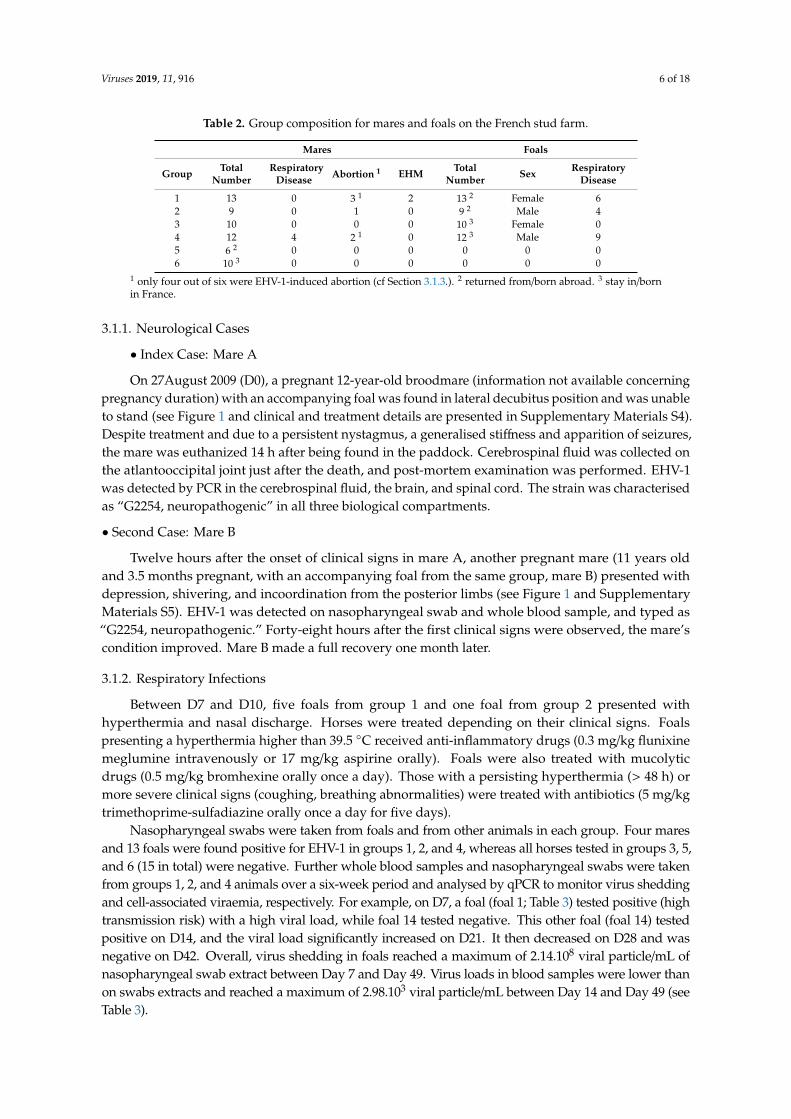
Table 2. Group composition for mares and foals on the French stud farm.
Mares Foals
Group Total
number
Respiratory
Disease Abortion1 EHM
Total
number Sex
Respiratory
Disease
1 13 0 31 2 132 Female 6
2 9 0 1 0 9 2 Male 4
3 10 0 0 0 103 Female 0
4 12 4 21 0 123 Male 9
5 62 0 0 0 0 0 0
Figure 1. The 2009 EHV-1 outbreak chronology from D0 = day of the first case declaration, to 9.5months after D0. D7 = Day 7 after the first case was declared. The biosecurity measures that weretaken during this outbreak are described in blue boxes.
Viruses 2019, 11, 916 6 of 18
Table 2. Group composition for mares and foals on the French stud farm.
Mares Foals
Group TotalNumber
RespiratoryDisease Abortion 1 EHM Total
Number Sex RespiratoryDisease
1 13 0 3 1 2 13 2 Female 62 9 0 1 0 9 2 Male 43 10 0 0 0 10 3 Female 04 12 4 2 1 0 12 3 Male 95 6 2 0 0 0 0 0 06 10 3 0 0 0 0 0 0
1 only four out of six were EHV-1-induced abortion (cf Section 3.1.3.). 2 returned from/born abroad. 3 stay in/bornin France.
3.1.1. Neurological Cases
• Index Case: Mare A
On 27August 2009 (D0), a pregnant 12-year-old broodmare (information not available concerningpregnancy duration) with an accompanying foal was found in lateral decubitus position and was unableto stand (see Figure 1 and clinical and treatment details are presented in Supplementary Materials S4).Despite treatment and due to a persistent nystagmus, a generalised stiffness and apparition of seizures,the mare was euthanized 14 h after being found in the paddock. Cerebrospinal fluid was collected onthe atlantooccipital joint just after the death, and post-mortem examination was performed. EHV-1was detected by PCR in the cerebrospinal fluid, the brain, and spinal cord. The strain was characterisedas “G2254, neuropathogenic” in all three biological compartments.
• Second Case: Mare B
Twelve hours after the onset of clinical signs in mare A, another pregnant mare (11 years oldand 3.5 months pregnant, with an accompanying foal from the same group, mare B) presented withdepression, shivering, and incoordination from the posterior limbs (see Figure 1 and SupplementaryMaterials S5). EHV-1 was detected on nasopharyngeal swab and whole blood sample, and typed as“G2254, neuropathogenic.” Forty-eight hours after the first clinical signs were observed, the mare’scondition improved. Mare B made a full recovery one month later.
3.1.2. Respiratory Infections
Between D7 and D10, five foals from group 1 and one foal from group 2 presented withhyperthermia and nasal discharge. Horses were treated depending on their clinical signs. Foalspresenting a hyperthermia higher than 39.5 ◦C received anti-inflammatory drugs (0.3 mg/kg flunixinemeglumine intravenously or 17 mg/kg aspirine orally). Foals were also treated with mucolyticdrugs (0.5 mg/kg bromhexine orally once a day). Those with a persisting hyperthermia (> 48 h) ormore severe clinical signs (coughing, breathing abnormalities) were treated with antibiotics (5 mg/kgtrimethoprime-sulfadiazine orally once a day for five days).
Nasopharyngeal swabs were taken from foals and from other animals in each group. Four maresand 13 foals were found positive for EHV-1 in groups 1, 2, and 4, whereas all horses tested in groups 3, 5,and 6 (15 in total) were negative. Further whole blood samples and nasopharyngeal swabs were takenfrom groups 1, 2, and 4 animals over a six-week period and analysed by qPCR to monitor virus sheddingand cell-associated viraemia, respectively. For example, on D7, a foal (foal 1; Table 3) tested positive (hightransmission risk) with a high viral load, while foal 14 tested negative. This other foal (foal 14) testedpositive on D14, and the viral load significantly increased on D21. It then decreased on D28 and wasnegative on D42. Overall, virus shedding in foals reached a maximum of 2.14.108 viral particle/mL ofnasopharyngeal swab extract between Day 7 and Day 49. Virus loads in blood samples were lower thanon swabs extracts and reached a maximum of 2.98.103 viral particle/mL between Day 14 and Day 49 (seeTable 3).

Viruses 2019, 11, 916 7 of 18
Table 3. Monitoring of the viral shedding on 14 horses from groups 1, 2, and 3 over a period of seven weeks (copy/mL).
D7 D14 D21 D28 D35 D42 D49
Horse NS Blood NS Blood NS Blood NS Blood NS Blood NS Blood NS Blood
Group 1 andGroup 2
1 2.14E + 08 nd 6.75E + 05 1.05E + 03 7.04E + 02 NEG POS NEG NEG NEG NEG NEG NEG NEG2 3.02E + 08 nd 4.52E + 04 NEG 7.04E + 02 NEG 6.05E + 03 NEG NEG NEG NEG NEG NEG NEG3 9.29E + 07 nd NEG NEG NEG NEG 8.09E + 02 NEG 2.14E + 03 4.58E + 02 1.11E + 05 NEG NEG NEG4 1.91E + 06 nd 1.81E + 05 2.98E + 03 7.34E + 04 NEG NEG POS NEG NEG NEG NEG NEG NEG5 1.66E + 06 nd NEG NEG 3.67E + 04 NEG NEG NEG POS NEG NEG NEG NEG NEG6 1.08E + 07 nd 1.30E + 04 NEG 1.23E + 03 NEG 6.05E + 03 NEG POS NEG NEG NEG NEG NEG7 2.23E + 05 nd NEG POS POS NEG NEG POS NEG POS NEG NEG NEG NEG8 1.28E + 05 nd 3.93E + 04 POS POS NEG NEG NEG NEG NEG NEG NEG NEG NEG9 4.84E + 04 nd NEG NEG NEG POS NEG NEG NEG NEG NEG NEG NEG NEG10 NEG nd NEG POS NEG NEG NEG NEG NEG NEG NEG NEG NEG NEG11 NEG nd NEG NEG NEG NEG NEG NEG NEG NEG NEG NEG NEG NEG12 POS nd NEG NEG NEG NEG NEG NEG NEG NEG NEG NEG NEG NEG13 NEG nd NEG NEG NEG NEG NEG NEG NEG 5.64E + 02 NEG NEG NEG NEG14 NEG nd 6.75E + 05 NEG 1.31E + 08 NEG POS NEG 6.48E + 03 NEG NEG NEG NEG NEG15 nd nd nd nd POS NEG POS NEG NEG NEG POS NEG NEG NEG
Group 416 2.26E + 04 nd NEG POS NEG NEG NEG NEG NEG NEG NEG NEG NEG NEG17 1.18E + 06 nd 4.27E + 03 NEG NEG NEG NEG NEG NEG NEG NEG NEG NEG NEG18 1.97E + 04 nd POS NEG NEG NEG NEG NEG NEG NEG NEG NEG NEG NEG
Viral load in nasal swab (NS) from D0 to D42, and in blood from D7 to D42. POS = positive samples non quantifiable; NEG = negative samples. The boxes in gray indicate the period whenthe horse became negative (when both the NS and blood samples from horses were negative).

Viruses 2019, 11, 916 8 of 18
3.1.3. Abortion Cases
Between four and five months after the first neurological case (see Figure 1), a mare (C) fromgroup 1 and two mares (D and E) from group 4 aborted and were confirmed EHV-1 positive. Testswere carried out on fetal tissues (liver, lung, and kidney) and the mares’ placentas. Mare C foetuskidney tested negative. The EHV-1 strains were identified as “non-neuropathogenic” (A2254) for thethree cases.
Six months after developing neurological signs of disease, mare B (9.5 months pregnant) showedsigns of dystocic abortion (liquid loss and contractions without delivery of the foal). Intra-uterineexamination revealed an anterior dorsal position of the foal and bended legs and head. The foalwas moved in the appropriate position under epidural anaesthesia. It was found dead and icteric.Delivery was immediate and complete (see Supplementary Materials S6). The following day, a uterinewash was performed. The mare was isolated in a stable. and sanitary measures were put in place(Section 3.1.4. Biosafety measures). Post-mortem examination of the foetus revealed a densificationof lung parenchyma with interlobular oedema. Placenta and lung/liver samples tested positive forEHV-1. The strains were identified as “neuropathogenic” (G2254), as previously detected for this mareduring this neurological episode (Section 3.1.1.).
Uterine swabs were performed eight days and 30 days post abortion on mares B, C, D and E. Allsamples were positive for EHV-1 eight days post abortion and negative 30 days post abortion. Mare Bwas moved to Ireland for breeding five weeks post-abortion. Mare B was confirmed pregnant twomonths post-abortion and subsequently delivered a healthy foal after a normal pregnancy.
Overall, six of the 60 mares from this stud farm aborted during the year. EHV-1 infection wasconfirmed for mares B, C, D, and E. One of the two other mares aborted due to Enterobacter amnigenusinfection, while no cause could be confirmed for the last mare (the dead foal could not be recovered).
3.1.4. Biosafety Measures
The first two cases (Mares A and B) were reported to the RESPE. A safety perimeter was putin place around sick horses. Gloves, gowns, and over-boots were used to manipulate the animals.Movement restrictions were put in place, including foot baths with disinfectant installed at the paddockentrance, car wheels cleaning, and movement restriction for horses.
Mares and foals that tested positive for EHV-1 were moved to isolation from the other horses. Allmares were sampled (nasal swabs) before leaving or returning to the stud farm. A negative PCR resultfor EHV-1 was a prerequisite for movement. Booster vaccination against rhinopneumonitis was alsoadministered. The following year, all pregnant mares stayed in France, including mare B.
To conclude, the overall EHV-1-induced morbidity rate reached 16.6% of the herd (28 clinicallyaffected animals out of 169), including 6.7% of the broodmares, 1.2% EHM cases, and 13.6%respiratory cases.
3.2. Surveillance and Phylogeny from 2009 to 2018
3.2.1. Outbreaks, Forms of Disease, and ORF30 A2254G Typing
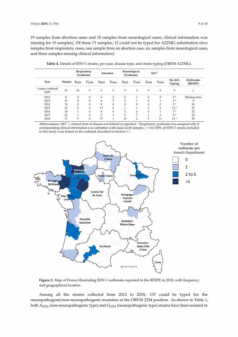
This epidemiological study involved the 2009 outbreak described in Section 3.1 and EHV-1outbreaks from 2012 to 2018. Samples collected from 2012 to 2018 are represented in SupplementaryMaterials (Table S1). From 2012 to 2017, 42 respiratory cases, 44 abortion cases, 20 neurological cases,and six cases with no clinical information were reported. Significantly, in 2018, several outbreaks ofEHV-1 occurred. During this year, an increased number of outbreaks (56 in total) were reported to theRESPE, including 15 respiratory outbreaks, 16 cases of abortion, five neurological outbreaks, and 20outbreaks with no clinical information (see Table 4 and Figure 2). The majority of the outbreaks werereported on the western part of the country, which has the highest concentration of breeding farms.Neurological outbreaks occurred in Normandy and Brittany and lead to the euthanasia of five animals.From these outbreaks, 71 samples were received and analysed (i.e., 15 samples from respiratory cases,
Viruses 2019, 11, 916 9 of 18
19 samples from abortion cases and 18 samples from neurological cases; clinical information wasmissing for 19 samples). Of these 71 samples, 12 could not be typed for A2254G substitution (twosamples from respiratory cases, one sample from an abortion case, six samples from neurological cases,and three samples missing clinical information).
Table 4. Details of EHV-1 strains, per year, disease type, and strain typing (ORF30 A2254G).
RespiratorySyndrome Abortion Neurological
Syndrome ND 1
Year Strains A2254 G2254 A2254 G2254 A2254 G2254 A2254 G2254No A/GTyping
Outbreaks(RESPE)
1 major outbreak2009 30 24 0 2 2 0 2 0 0 0 /
2012 8 0 0 0 2 0 1 0 0 5 * Missing data2013 19 0 0 6 7 2 1 0 1 2 * 42014 10 0 0 8 0 0 0 0 0 2 * 262015 32 0 0 17 0 1 1 1 0 12 * 272016 18 1 0 8 0 2 4 0 1 2 * 322017 22 1 0 2 1 0 7 1 2 8 * 232018 71 9 4 17 1 10 2 5 11 12 * 56
Abbreviations: ND 1 = clinical form of disease not defined or reported. * Respiratory syndrome was assigned only ifcorresponding clinical information was submitted with nasal swab samples. / = for 2009, all EHV-1 strains includedin this study were linked to the outbreak described in Section 3.1.
Viruses 2019, 11, x FOR PEER REVIEW 9 of 19
information was missing for 19 samples). Of these 71 samples, 12 could not be typed for A2254G
substitution (two samples from respiratory cases, one sample from an abortion case, six samples from
neurological cases, and three samples missing clinical information).
Table 4. Details of EHV-1 strains, per year, disease type, and strain typing (ORF30 A2254G).
Respiratory
Syndrome Abortion
Neurological
Syndrome ND1
Year Strains A2254 G2254 A2254 G2254 A2254 G2254 A2254 G2254 No A/G
Typing
Outbreaks
(RESPE)
1 major outbreak
2009 30 24 0 2 2 0 2 0 0 0 /
2012 8 0 0 0 2 0 1 0 0 5* Missing data
2013 19 0 0 6 7 2 1 0 1 2* 4
2014 10 0 0 8 0 0 0 0 0 2* 26
2015 32 0 0 17 0 1 1 1 0 12* 27
2016 18 1 0 8 0 2 4 0 1 2* 32
2017 22 1 0 2 1 0 7 1 2 8* 23
2018 71 9 4 17 1 10 2 5 11 12* 56
Abbreviations: ND1= clinical form of disease not defined or reported. * Respiratory syndrome was
assigned only if corresponding clinical information was submitted with nasal swab samples. / = for
2009, all EHV-1 strains included in this study were linked to the outbreak described in Section 3.1.
Figure 2. Map of France illustrating EHV-1 outbreaks reported to the RESPE in 2018, with frequency
and geographical location.
Among all the strains collected from 2012 to 2018, 137 could be typed for the
neuropathogenic/non-neuropathogenic mutation at the ORF30 2254 position. As shown in Table 4,
both A2254 (non-neuropathogenic type) and G2254 (neuropathogenic type) strains have been isolated in
Figure 2. Map of France illustrating EHV-1 outbreaks reported to the RESPE in 2018, with frequencyand geographical location.
Among all the strains collected from 2012 to 2018, 137 could be typed for theneuropathogenic/non-neuropathogenic mutation at the ORF30 2254 position. As shown in Table 4,both A2254 (non-neuropathogenic type) and G2254 (neuropathogenic type) strains have been isolated in

Viruses 2019, 11, 916 10 of 18
respiratory cases, abortion, and neurological outbreaks. The Chi square test indicates a significantstatistical association between the ORF30 types and both abortion and neurological disorder (p-value =
0.000202, see Table 5). Ninety-one A2254 strains have been isolated over the years (66% of all the strainstyped), when compared with 46 (34% of all the strains typed) G2254 strains.
Table 5. ORF30 2254 mutation according to disease from EHV-1 isolates (2009 to 2018).
Type Respiratory Abortion Neurological Information Missing Total
A2254 Non-neuro. 11 (73%) 58 (84%) 15 (48%) 7 (32%) 91(66%)G2254 Neuro. 4 (27%) 11 (16%) 16 (52%) 15 (68%) 46 (34%)
Chi squarep-value
Resp./Ab. 0.325642Resp./Neu. 0.109608Ab./Neu. 0.000202 *All three 0.000996 *
Non-neuro. = non neuropathogenic; Neuro. = neuropathogenic. (%: percentage of ORF30 2254 type among thedisease category). Chi Square test null hypothesis “There is no correlation between the disease category and theORF30 2254 type. Resp./Ab. = Chi square test for Respiratory and Abortion categories; Resp./Neu.= Chi square testfor Respiratory and Neurological categories; Ab./Neu.= Chi square test for Abortion and Neurological categories; Allthree= Chi square test for Respiratory, Abortion and Neurological categories. * significant result at p-value p < 0.05.
3.2.2. ORF30 Sequence Analysis
Probably because of the DNA quantity and purity, only 14 strains could be completely sequenced.BRE/13/2018, the only strain isolated on a neurological outbreak, could not be sequenced. ORF30sequences from these 14 EHV-1 strains (see Supplementary Table S2) were compared to theneuropathogenic reference strain Ab4 (Genbank accession number AY665713). SNP and aminoacid substitutions are reported in Supplementary Table S7.
Twelve of the 14 strains had an adenine residue at position 2254 of the polymerase gene. Seven ofthese also had a mutation in position 96 (G96A) and five of them in position 2968 (G2968A) comparedto reference AY665713 (Ab4). The mutation (A > G) in position 2254 and 2968 led to an amino acidchange (N752D and K990E, respectively). Other punctual mutations were observed on the other strainssequenced and five of them induced an amino acid change. One nucleotide on NORM/5/2012 sequencecould not be determined during sequencing electrophoresis and was identified as a K (either thymine orguanine). NORM/17/2018 and NORM/18/2018 showed 100% identity at nucleotide and amino acid level.After further investigation about the outbreak location, it appeared that both strains NORM/17/2018and NORM/18/2018 were collected, respectively, on February 2018 and March 2018 on the same premiseafter the abortion of two mares present in the stud farm. Although this comparison only concernsORF30, it is possible that the same strain infected the two pregnant mares and induced their abortion.Both a phylogenetic tree (Neighbour-joining method and Maximum Composite Likelihood model) anda Median Joining Network were constructed (see Figure 3) based on the ORF30 sequences collectedsince 2009 and sequences obtained in a recent study in UK [14]. Two groups could be observed. Thefirst group (Group 1 in Figure 3) formed a cluster of 32 strains including UK strains and referencestrain Ab4. Of these 32, 26 contained the G2254 substitution in ORF30 (neuropathogenic type). BothG2254 strains collected in France (NORM/4/2012 and ILEDEFR/14/2018) belong to this group. Thesecond group (Group 2 in Figure 3) is divided in three clusters. One of these clusters contains similarstrains (NORM/2/2010, NELLEAQU/9/2015, BELG/12/2017, NORM/17/2018, NORM/18/2018) and oneUK G2254 strain. NORM/5/2012 unidentified nucleotide in position 2876 did not discriminate thestrain from ILEDEFR/3/2012 and NORM/8/2015. ORF30 analysis provides a first statement concerningstrain phylogeny and potential neuropathogenicity but it only represents a small part of the genome.Multi-locus typing of EHV-1 as described by Garvey et al. 2019 was used to segregate EHV-1 strainsinto UL clades (Bryant et al. 2018) [14,26].
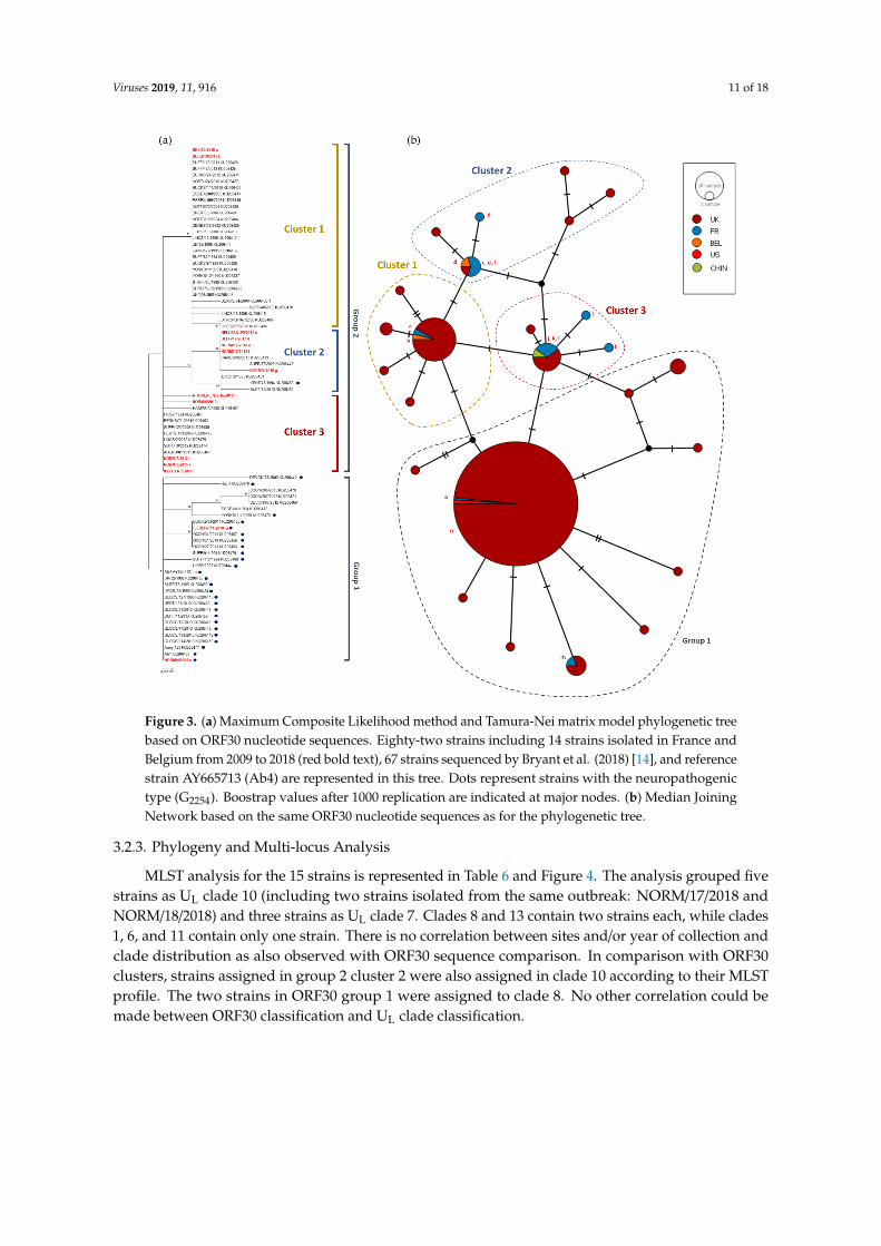
Viruses 2019, 11, 916 11 of 18Viruses 2019, 11, x FOR PEER REVIEW 11 of 19
Figure 3. (a) Maximum Composite Likelihood method and Tamura-Nei matrix model phylogenetic
tree based on ORF30 nucleotide sequences. Eighty-two strains including 14 strains isolated in France
and Belgium from 2009 to 2018 (red bold text), 67 strains sequenced by Bryant et al. (2018) [14], and
reference strain AY665713 (Ab4) are represented in this tree. Dots represent strains with the
neuropathogenic type (G2254). Boostrap values after 1000 replication are indicated at major nodes. (b)
Median Joining Network based on the same ORF30 nucleotide sequences as for the phylogenetic tree.
3.2.3. Phylogeny and Multi-locus Analysis
MLST analysis for the 15 strains is represented in Table 7 and Figure 4. The analysis grouped
five strains as UL clade 10 (including two strains isolated from the same outbreak: NORM/17/2018
and NORM/18/2018) and three strains as UL clade 7. Clades 8 and 13 contain two strains each, while
clades 1, 6, and 11 contain only one strain. There is no correlation between sites and/or year of
Figure 3. (a) Maximum Composite Likelihood method and Tamura-Nei matrix model phylogenetic treebased on ORF30 nucleotide sequences. Eighty-two strains including 14 strains isolated in France andBelgium from 2009 to 2018 (red bold text), 67 strains sequenced by Bryant et al. (2018) [14], and referencestrain AY665713 (Ab4) are represented in this tree. Dots represent strains with the neuropathogenictype (G2254). Boostrap values after 1000 replication are indicated at major nodes. (b) Median JoiningNetwork based on the same ORF30 nucleotide sequences as for the phylogenetic tree.
3.2.3. Phylogeny and Multi-locus Analysis
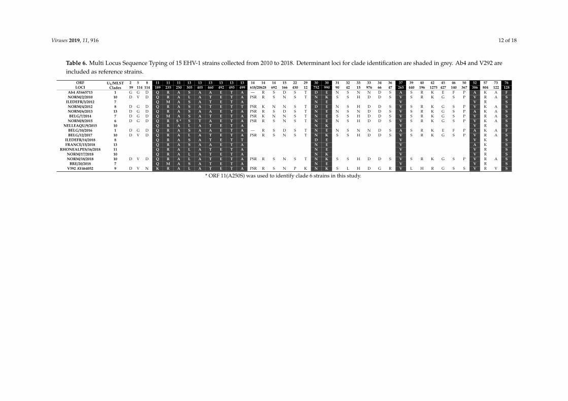
MLST analysis for the 15 strains is represented in Table 6 and Figure 4. The analysis grouped fivestrains as UL clade 10 (including two strains isolated from the same outbreak: NORM/17/2018 andNORM/18/2018) and three strains as UL clade 7. Clades 8 and 13 contain two strains each, while clades1, 6, and 11 contain only one strain. There is no correlation between sites and/or year of collection andclade distribution as also observed with ORF30 sequence comparison. In comparison with ORF30clusters, strains assigned in group 2 cluster 2 were also assigned in clade 10 according to their MLSTprofile. The two strains in ORF30 group 1 were assigned to clade 8. No other correlation could bemade between ORF30 classification and UL clade classification.
Viruses 2019, 11, 916 12 of 18
Table 6. Multi Locus Sequence Typing of 15 EHV-1 strains collected from 2010 to 2018. Determinant loci for clade identification are shaded in grey. Ab4 and V292 areincluded as reference strains.
ORF UL/MLSTClades
2 5 8 11 11 11 13 13 13 13 13 13 14 14 14 15 22 29 30 30 31 32 33 33 34 36 37 39 40 42 45 46 50 52 57 73 76LOCI 59 114 114 189 235 250 305 405 460 492 493 499 618/20628 692 166 430 12 752 990 90 42 15 976 66 47 265 440 196 1275 427 140 367 386 804 122 128
Ab4 AY665713 1 G G D Q R A S A A E T A — R S D S T D E N S N N D S A S R K E F P A K A FNORM/2/2010 10 D V D Q R A L A T E T A PSR R S N S T N K S S H D D S V S R K G S P V R A S
ILEDEFR/3/2012 7 Q M A S A T E T A N E V V R SNORM/4/2012 8 D G D Q R A S A T E T T PSR K N N S T D E N S H D D S V S R K G S P V K A SNORM/6/2013 13 D G D Q R A S A A E T A PSR R S D S T N E N S N D D S V S R K G S P A K A SBELG/7/2014 7 D G D Q M A S A T E T A PSR K N N S T N E S S H D D S V S R K G S P V R A S
NORM/8/2015 6 D G D Q R S * S T A E T A PSR R S N S T N E N S H D D S V S R K G S P V K A SNELLEAQU/9/2015 10 Q R A L A T E T A N K V V R S
BELG/10/2016 1 D G D Q R A S A A E T A — R S D S T N E N S N N D S A S R K E F P A K A FBELG/12/2017 10 D V D Q R A L A T E T A PSR R S N S T N K S S H D D S V S R K G S P V R A S
ILEDEFR/14/2018 8 Q R A S A T E T T D E V V K SFRANCE/15/2018 13 Q R A S A A E T A N E V A K S
RHONEALPES/16/2018 11 Q R A L A T E T A N E V V R SNORM/17/2018 10 Q R A L A T E T A N K V V R SNORM/18/2018 10 D V D Q R A L A T E T A PSR R S N S T N K S S H D D S V S R K G S P V R A S
BRE/20/2018 7 Q M A S A T E T A N E V V R SV592 AY464052 9 D V N K R A L A T E T A PSR R S N P K N K S L H D G R V L H R G S S V R V S
* ORF 11(A250S) was used to identify clade 6 strains in this study.

Viruses 2019, 11, 916 13 of 18
Viruses 2019, 11, x; doi: FOR PEER REVIEW www.mdpi.com/journal/viruses
Figure 4. (a) Maximum Likelihood phylogenic tree based on Jones Taylor Thornton model built with
MLST sequences including 8 French and Belgian strains (in red), 66 EHV-1 UK strains [14], and 22
EHV-1 Irish strains [26]. Dots indicate D752 strains (Neuropathogenic type). Strains with partial
concatenated amino acid sequence were not included in the Maximum Likelihood analysis but their
supposed position in the tree is indicated with red arrows according to their clade identification (see
Table 7). UL Clades [14] are numbered and represented with vertical lines. Boostrap values after 1000
replication are indicated at major nodes. (b) Network built using Neighbour-Net method with the
same sequences as the tree. Only 37 amino acid sequences were included in the network, and French
and Belgian strains are represented in grey boxes.
4. Discussion
The 2009 EHV1 outbreak is regretfully a good example of the impact that an EHV-1 infection
can have in stud farms and illustrates the diversity of diseases that could be observed and faced by
veterinarians. The three clinical forms of disease induced by EHV-1 (respiratory, neurological
Figure 4. (a) Maximum Likelihood phylogenic tree based on Jones Taylor Thornton model built withMLST sequences including 8 French and Belgian strains (in red), 66 EHV-1 UK strains [14], and 22EHV-1 Irish strains [26]. Dots indicate D752 strains (Neuropathogenic type). Strains with partialconcatenated amino acid sequence were not included in the Maximum Likelihood analysis but theirsupposed position in the tree is indicated with red arrows according to their clade identification (seeTable 6). UL Clades [14] are numbered and represented with vertical lines. Boostrap values after 1000replication are indicated at major nodes. (b) Network built using Neighbour-Net method with thesame sequences as the tree. Only 37 amino acid sequences were included in the network, and Frenchand Belgian strains are represented in grey boxes.
MLST sequences from strains collected in Belgium and France were compared to strains sequencedby Bryant et al. [14], and a Maximum Likelihood phylogenic tree was built based on Jones TaylorThornton model (see Figure 4).
4. Discussion
The 2009 EHV1 outbreak is regretfully a good example of the impact that an EHV-1 infectioncan have in stud farms and illustrates the diversity of diseases that could be observed and faced byveterinarians. The three clinical forms of disease induced by EHV-1 (respiratory, neurological disorderand abortion) were observed over a nine-and-a-half-month period on a thoroughbred farm (169 horses),which raises questions about the source of EHV-1 infection and its transmission during this outbreak.The significance of this 2009 EHV-1 outbreak was also the observation of the three clinical forms ofdisease in the same premise over a long period of time. This phenomenon is rarely described in
Viruses 2019, 11, 916 14 of 18
literature [39], and no other outbreak involving all three forms of disease was reported in France since2009. The number of horses on this premise and the breeding activities may be factors that havecontributed to increase the number of cases, which could have been higher in the absence of vaccination.The identification of two different strains, which may be the result from frequent horse movements inFrance and abroad, might also have influenced the diseases observed during this unusual outbreak.EHV-1 outbreaks are frequent but are usually limited to the report of one or two forms of the diseasefor the same outbreak (RESPE, personal communication). However, epidemiological links betweenoutbreaks separated in time are not always available or identified. For example, the 2009 outbreakdescribed here lasted nine and a half months with at least two and a half months between the lastrespiratory infection and the first abortion. Occurrence of multiple forms of disease may be morefrequent than currently imagined.
The three clinical forms of disease were reported on a regular basis from 2009 to 2018 on thedifferent outbreaks of this study. In 2018, a large number of cases were reported during a short period(March 2018 to May 2018), with exceptional sanitary measures needed to control contaminationsbetween horses. This EHV-1 crisis is likely to be associated with the fact that a vaccine shortageoccurred in 2016, implying a lower vaccination rate.
At the time of the 2009 outbreak, only a few tools were available to conduct EHV-1 molecularinvestigation. The PCR designed by Diallo et al. (2006, [28]) was used as an EHV-1 detection andquantification test. Viral loads of samples were quantified, and sanitary measures were lifted whenundetected. Results obtained at the time indicated that virus titers in total blood samples were lowerthan in swab extracts, but these two compartments are not always correlated to each other. Monitoringviral loads provided an overview of the virus excretion and risk of transmission. When none of thegroup 1 foals tested positive after a seven-week isolation period, the day to day management of thestud farm returned to normal. EHV-1 strains were typed (ORF30 SNP A2254G) and both types wereidentified (neuropathogenic and non-neuropathogenic).
Although those tools gave useful indicative information concerning the different strains isolatedfrom this outbreak, they proved to be limited to establish potential relationship between cases andvirus strains. Other tools have been developed (Nugent et al. 2006, Bryant et al. 2018 and Garveyet al. 2019 [14,19,26]) that have motivated our retrospective and molecular analysis of French andBelgian EHV-1 strains isolated from 2009 outbreak to the major 2018 EHV-1 epizootic. Three differentmolecular analysis were performed: the ORF30 A2254G typing, the complete ORF30 sequencing andthe MLST. In 2006, Nugent et al. [19] reported a significant association between the A2254G SNP in theDNA polymerase gene (ORF30), neuropathogenicity. This single point substitution (A2254G) involvesan amino acid change (N752D). N752 (A2254) strains were strongly associated to non-neuropathogenicinfection cases, while D752 (G2254) strains were strongly associated to neuropathogenic cases [19].A2254G typing was subsequently used on a regular basis for strain discrimination [40,41]. In our study,137 strains collected from 2009 to 2018 were typed, and A2254 strains were more significantly associatedwith abortion cases than to neurological cases (p = 0.0002), as recently described by Lechmann et al.(2019) [42]. However, no significant correlation between G2254 strains and the neurological formof disease was measured. This observation is in agreement with some recent studies showing thatA2254G mutation is not exclusively associated to EHM but could be part of a more complex mechanismaffecting strains virulence [41]. Both neuropathogenic and non-neuropathogenic strains could be typedamong 2018 strains, suggesting that more than one strain was circulating during the crisis. StrainA2254G typing is also interesting as it was reported that the point mutation could change sensitivity tosome drugs targeting DNA polymerase activity as it was demonstrated in a study showing that a N752variant was more sensitive to aphidicolin than the D752 variants. Aphidicolin inhibits some dNTPsbinding to a family of DNA polymerase which include herpesvirus DNA polymerase [17,19].
The A2254G typing was completed with ORF30 sequencing on 14 French and Belgian strainsisolated between 2012 and 2018 in order to compare sequences and to perform a phylogenetic analysis.Several synonymous and non-synonymous substitutions were identified. Equine herpesvirus DNA
Viruses 2019, 11, 916 15 of 18
polymerase subunit structures and mechanisms are not well known. Despite a low homology betweenEHV-1 and Human Simplex Virus (HSV) polymerase amino acid sequence (54%), the latter has beendescribed as closest to α polymerase structure [43]. On this basis, SNP found in EHV-1 strains could beattributed to structure domains identified in HSV polymerase [43]. Amino acid substitutions couldbe localised in the pre-NH2 terminal domain (R59G), in the 3′-5′ exonuclease domain (S419L andR429K), in the palm domain (A694V and D752N), in the thumb domain (E990K). Although some of thedomain activities have been studied for human herpesviruses [43,44], it is hard to predict the impactof the substitutions observed in EHV-1 strains on the protein activity. A strong homology (99.86% to100%) was measured among those strains with no obvious evolutionary tendency. Results were inagreement with those published by Bryant et al. in 2018 [14]. According to ORF30 sequencing, EHV-1evolution is not linked to sampling location or year of collection, with the exception of the strainsNORM/17/2018 and NORM/18/2018 that were isolated from the same stud farm, a few weeks apart,and have similar ORF30 sequences. All G2254 strains were grouped in the same cluster, suggesting thatORF30 sequencing does not provide further information when compared with the A2254G typing.However, three clusters were identified for A2254 strains, primarily differentiated by one SNP (e.gA2968G). The significance of these different clusters is unknown. MLST analysis provides a moreglobal view concerning EHV-1 strains evolution as it takes into account 37 loci in 26 different ORFs. Asobserved with ORF30 analysis, there is no obvious correlation between year and location of collection,with the exception of NORM/17/2018 and NORM/18/2018, both located in clade 10, which support aco-circulation of EHV-1 strains from different clades as already described by Bryant et al. and Garveyet al. [14,26]. It is interesting to note that four French strains and one Belgium strain are localisedin clade 10. The UL Clade 12 described by Bryant et al. (2018) [14], which contains a strain fromthe UK, is not shown here. UL Clades 2 and 4 were not represented by any of the European strainsanalysed in this study. It is important to note that the MLST method cannot distinguish UL Clade 2and 12 from MLST Clade 1 and 10, respectively [26]. As all the strains compared in this study are fromEurope, a broader strain selection would be needed to identify a potential geographical effect on cladedifferentiation. Finally, two abortion strains isolated from the same premise in a one-month intervalafter two mare abortions had the exact same MLST profile suggesting that the same strain infected themares. Obviously, EHV-1 strain surveillance is complicated by the fact that EHV-1 can also establishlatency in different sites, implying no viral replication as the viral genome maintains an episomal formblocking transcription and translation of its genes (limited transcription with LATs) [9,11]. This impliesthat strains circulation and outbreaks are potentially dependent on latency and re-activation.
5. Conclusions
To conclude, this study allowed applying and comparing three different typing approaches toconduct a phylogenetic analysis over a six years period. A significant association was measuredbetween EHV-1 induced abortion and the DNA polymerase A2254 genotype of related strains, while nodisease association was observed with the G2254 genotype. This result suggests that the commonlyused “neuropathogenic/non-neuropathogenic” designation is not always appropriate. The ORF30 andMLST analysis highlight the diversity of EHV-1 strains circulating in the French equine population andthe difficulty to link strain evolution, time of collection, and location. However, the MLST offers newpossibilities for EHV-1 epidemiology.
Supplementary Materials: The following are available online at http://www.mdpi.com/1999-4915/11/10/916/s1,Table S1: Samples type collected from EHV-1 outbreaks from 2012 to 2018, Table S2: Strains identification, locationand year of collection. Table S3. ORF30 sequencing Primers. Supplementary Materials S4: Complementaryclinical information concerning Mare A. Supplementary Materials S5: Complementary clinical informationconcerning Mare B. Supplementary Materials S6: Complementary clinical information concerning Mare B abortion.Supplementary Table S7: ORF30 amino acid alignments.
Author Contributions: Here are described the contribution of the authors for this research article:conceptualisation, R.P., S.P., and G.S.; investigation, C.F., M.G., and G.S.; resources, M.J., C.F., P.M., M.F.,A.G., V.M., C.M.-P., and L.L.; writing—original draft preparation, R.P., S.P., and G.S.; writing—review and editing,

Viruses 2019, 11, 916 16 of 18
M.G., A.C., C.F., A.G., and L.L.; supervision, R.P. and S.P.; project administration, R.P. and S.P.; funding acquisition,R.P. and S.P.
Funding: This research was funded by the Fonds Eperon OVERLORD N12-2017, Normandy County Council(17E01598/17EP04324), the IFCE (Institut Français du Cheval et de l’Equitation) grant number 2017-008, CENTAUREEuropean project co-funded by Normandy County Council, European Union in the framework of the ERDF-ESFoperationnal programme 2014-2020.
Acknowledgments: We thank Erika Hue (LABÉO) for her help in solving problems and methodology, FaridaMebarki (BIOFIDAL) for her help in sequencing, and Meriadeg Ar Gouilh (NORMANDIE UNIV) for his helpwith network construction.
Conflicts of Interest: The authors declare no conflict of interest.
References
1. Davison, A.J.; Eberle, R.; Ehlers, B.; Hayward, G.S.; McGeoch, D.J.; Minson, A.C.; Pellett, P.E.; Roizman, B.;Studdert, M.J.; Thiry, E. The Order Herpesvirales. Arch. Virol. 2009, 154, 171–177. [CrossRef] [PubMed]
2. Abdelgawad, A.; Azab, W.; Damiani, A.M.; Baumgartner, K.; Will, H.; Osterrieder, N.; Greenwood, A.D.Zebra-borne equine herpesvirus type 1 (EHV-1) infection in non-African captive mammals. Vet. Microbiol.2014, 169, 102–106. [CrossRef] [PubMed]
3. Wohlsein, P.; Lehmbecker, A.; Spitzbarth, I.; Algermissen, D.; Baumgärtner, W.; Böer, M.; Kummrow, M.;Haas, L.; Grummer, B. Fatal epizootic equine herpesvirus 1 infections in new and unnatural hosts. Vet.Microbiol. 2011, 149, 456–460. [CrossRef] [PubMed]
4. Chowdhury, S.I.; Ludwig, H. Molecular biological characterization of equine herpesvirus type 1 (EHV-1)isolates from ruminant hosts. Virus Res. 1988, 11, 127–139. [CrossRef]
5. Ghanem, Y.M.; Fukushi, H.; Ibrahim, E.S.M.; Ohya, K.; Yamaguchi, T.; Kennedy, M. Molecular phylogeny ofequine herpesvirus 1 isolates from onager, zebra and Thomson’s gazelle. Arch. Virol. 2008, 153, 2297–2302.[CrossRef]
6. Dayaram, A.; Franz, M.; Schattschneider, A.; Damiani, A.M.; Bischofberger, S.; Osterrieder, N.;Greenwood, A.D. Long term stability and infectivity of herpesviruses in water. Sci. Rep. UK 2017, 7.[CrossRef]
7. Paillot, R.; Case, R.; Ross, J.; Newton, R.; Nugent, J. Equine Herpes Virus-1: Virus, Immunity and Vaccines.Open Vet. Sci. J. 2008, 2, 68–91. [CrossRef]
8. Lunn, D.P.; Davis-Poynter, N.; Flaminio, M.J.B.F.; Horohov, D.W.; Osterrieder, K.; Pusterla, N.;Townsend, H.G.G. Equine Herpesvirus-1 Consensus Statement. J. Vet. Intern. Med. 2009, 23, 450–461.[CrossRef]
9. Chesters, P.M.; Allsop, R.; Purewal, A.; Edington, N. Detection of Latency-Associated Transcripts of EquidHerpesvirus 1 in Equine Leukocytes but Not in Trigeminal Ganglia. J. Virol. 1997, 71, 7.
10. Aleman, M.; Pickles, K.J.; Simonek, G.; Madigan, J.E. Latent Equine Herpesvirus-1 in Trigeminal Ganglia andEquine Idiopathic Headshaking. J. Vet. Intern. Med. 2012, 26, 192–194. [CrossRef]
11. Rock, D.L. The molecular basis of latent infections by alphaherpesviruses. Semin. Virol. 1993, 4, 157–165.[CrossRef]
12. Gulati, B.R.; Sharma, H.; Riyesh, T.; Khurana, S.K.; Kapoor, S. Viral and Host Strategies for Regulation ofLatency and Reactivation in Equid Herpesviruses. Asian J. Anim. Vet. Adv. 2015, 10, 669–689. [CrossRef]
13. Telford, E.A.R.; Watson, M.S.; McBride, K.; Davison, A.J. The DNA sequence of equine herpesvirus-1. Virology1992, 189, 304–316. [CrossRef]
14. Bryant, N.A.; Wilkie, G.S.; Russell, C.A.; Compston, L.; Grafham, D.; Clissold, L.; McLay, K.; Medcalf, L.;Newton, R.; Davison, A.J.; et al. Genetic diversity of equine herpesvirus 1 isolated from neurological,abortigenic and respiratory disease outbreaks. Transbound. Emerg. Dis. 2018, 65, 817–832. [CrossRef][PubMed]
15. Cuxson, J.L.; Hartley, C.A.; Ficorilli, N.P.; Symes, S.J.; Devlin, J.M.; Gilkerson, J.R. Comparing the geneticdiversity of ORF30 of Australian isolates of 3 equid alphaherpesviruses. Vet. Microbiol. 2014, 169, 50–57.[CrossRef]
16. Crowhurst, F.A.; Dickinson, G.; Burrows, R. An outbreak of paresis in mares and geldings associated withequid herpesvirus 1. Vet. Rec. 1981, 109, 527–528.

Viruses 2019, 11, 916 17 of 18
17. Mumford, J.A.; Rossdale, P.D.; Jessett, D.M.; Gann, S.J.; Ousey, J.; Cook, R.F. Serological and virologicalinvestigations of an equid herpesvirus 1 (EHV-1) abortion storm on a stud farm in 1985. J. Reprod. Fertil.1987, 35, 509–518.
18. Anagha, G.; Gulati, B.R.; Riyesh, T.; Virmani, N. Genetic characterization of equine herpesvirus 1 isolatesfrom abortion outbreaks in India. Arch. Virol. 2017, 162, 157–163. [CrossRef]
19. Nugent, J.; Birch-Machin, I.; Smith, K.C.; Mumford, J.A.; Swann, Z.; Newton, J.R.; Bowden, R.J.; Allen, G.P.;Davis-Poynter, N. Analysis of Equid Herpesvirus 1 Strain Variation Reveals a Point Mutation of the DNAPolymerase Strongly Associated with Neuropathogenic versus Nonneuropathogenic Disease Outbreaks.J. Virol. 2006, 80, 4047–4060. [CrossRef]
20. Goodman, L.B.; Loregian, A.; Perkins, G.A.; Nugent, J.; Buckles, E.L.; Mercorelli, B.; Kydd, J.H.;Palù, G.; Smith, K.C.; Osterrieder, N.; et al. A Point Mutation in a Herpesvirus Polymerase DeterminesNeuropathogenicity. PLoS Pathog. 2007, 3, e160. [CrossRef]
21. Pronost, S.; Léon, A.; Legrand, L.; Fortier, C.; Miszczak, F.; Freymuth, F.; Fortier, G. Neuropathogenicand non-neuropathogenic variants of equine herpesvirus 1 in France. Vet. Microbiol. 2010, 145, 329–333.[CrossRef] [PubMed]
22. Allen, G.P. Development of a Real-Time Polymerase Chain Reaction Assay for Rapid Diagnosis ofNeuropathogenic Strains of Equine Herpesvirus-1. J. Vet. Diagn. Investig. 2007, 19, 69–72. [CrossRef][PubMed]
23. Van de Walle, G.R.; Goupil, R.; Wishon, C.; Damiani, A.; Perkins, G.A.; Osterrieder, N. A Single-NucleotidePolymorphism in a Herpesvirus DNA Polymerase Is Sufficient to Cause Lethal Neurological Disease. J. Infect.Dis. 2009, 200, 20–25. [CrossRef] [PubMed]
24. Franz, M.; Goodman, L.; Van de Walle, G.; Osterrieder, N.; Greenwood, A. A Point Mutation in a HerpesvirusCo-Determines Neuropathogenicity and Viral Shedding. Viruses 2017, 9, 6. [CrossRef] [PubMed]
25. Gryspeerdt, A.; Vandekerckhove, A.; Van Doorsselaere, J.; Van de Walle, G.; Nauwynck, H. Description of anunusually large outbreak of nervous system disorders caused by equine herpesvirus 1 (EHV1) in 2009 inBelgium. Vlaams Diergeneeskundig Tijdschrif 2011, 80, 147–153.
26. Garvey, M.; Lyons, R.; Hector, R.; Walsh, C.; Arkins, S.; Cullinane, A. Molecular Characterisation of EquineHerpesvirus 1 Isolates from Cases of Abortion, Respiratory and Neurological Disease in Ireland between1990 and 2017. Pathogens 2019, 8, 7. [CrossRef]
27. Pronost, S.; Legrand, L.; Pitel, P.-H.; Wegge, B.; Lissens, J.; Freymuth, F.; Richard, E.; Fortier, G. Outbreak ofEquine Herpesvirus Myeloencephalopathy in France: A Clinical and Molecular Investigation: Outbreak ofEHV-1 myeloencephalopathy. Transbound. Emerg. Dis. 2012, 59, 256–263. [CrossRef]
28. Diallo, I.S.; Hewitson, G.; Wright, L.; Rodwell, B.J.; Corney, B.G. Detection of equine herpesvirus type 1 usinga real-time polymerase chain reaction. J. Virol. Methods 2006, 131, 92–98. [CrossRef]
29. Thieulent, C.J.; Hue, E.S.; Fortier, C.I.; Dallemagne, P.; Zientara, S.; Munier-Lehmann, H.; Hans, A.;Fortier, G.D.; Pitel, P.-H.; Vidalain, P.-O.; et al. Screening and evaluation of antiviral compounds againstEquid alpha-herpesviruses using an impedance-based cellular assay. Virology 2019, 526, 105–116. [CrossRef]
30. Hall, T. BioEdit: A user-friendly biological sequence alignment editor and analysis program for Windows95/98/NT. Nucleic Acids Symp. Ser. 1999, 41, 95–98.
31. CodonCode Aligner. Available online: https://www.codoncode.com (accessed on 27 September 2019).32. Kumar, S.; Stecher, G.; Tamura, K. MEGA7: Molecular Evolutionary Genetics Analysis Version 7.0 for Bigger
Datasets. Mol. Biol. Evol. 2016, 33, 1870–1874. [CrossRef] [PubMed]33. Saitou, N.; Nei, M. The neighbor-joining method: A new method for reconstructing phylogenetic trees. Mol.
Biol. Evol. 1987, 4, 406–425. [PubMed]34. Jones, D.T.; Taylor, W.R.; Thornton, J.M. The rapid generation of mutation data matrices from protein
sequences. Bioinformatics 1992, 8, 275–282. [CrossRef]35. Madeira, F.; Park, Y.M.; Lee, J.; Buso, N.; Gur, T.; Madhusoodanan, N.; Basutkar, P.; Tivey, A.R.N.; Potter, S.C.;
Finn, R.D.; et al. The EMBL-EBI search and sequence analysis tools APIs in 2019. Nucleic Acids Res. 2019, 47,W636–W641. [CrossRef]
36. PopART. Available online: http://popart.otago.ac.nz (accessed on 27 September 2019).37. Huson, D.H.; Bryant, D. Application of Phylogenetic Networks in Evolutionary Studies. Mol. Biol. Evol.
2006, 23, 254–267. [CrossRef]

Viruses 2019, 11, 916 18 of 18
38. Bryant, D. Neighbor-Net: An Agglomerative Method for the Construction of Phylogenetic Networks. Mol.Biol. Evol. 2003, 21, 255–265. [CrossRef]
39. Walter, J.; Seeh, C.; Fey, K.; Bleul, U.; Osterrieder, N. Clinical observations and management of a severe equineherpesvirus type 1 outbreak with abortion and encephalomyelitis. Acta Vet. Scand. 2013, 55, 19. [CrossRef]
40. Vissani, M.A.; Becerra, M.L.; Olguín Perglione, C.; Tordoya, M.S.; Miño, S.; Barrandeguy, M. Neuropathogenicand non-neuropathogenic genotypes of Equid Herpesvirus type 1 in Argentina. Vet. Microbiol. 2009, 139,361–364. [CrossRef]
41. Pronost, S.; Cook, R.F.; Fortier, G.; Timoney, P.J.; Balasuriya, U.B.R. Relationship between equine herpesvirus-1myeloencephalopathy and viral genotype: EHV-1, genotype and EHM. Equine Vet. J. 2010, 42, 672–674.[CrossRef]
42. Lechmann, J.; Schoster, A.; Ernstberger, M.; Fouché, N.; Fraefel, C.; Bachofen, C. A novel PCR protocol fordetection and differentiation of neuropathogenic and non-neuropathogenic equid alphaherpesvirus 1. J. Vet.Diagn. Investig. 2019, 31, 696–703. [CrossRef]
43. Liu, S.; Knafels, J.D.; Chang, J.S.; Waszak, G.A.; Baldwin, E.T.; Deibel, M.R.; Thomsen, D.R.; Homa, F.L.;Wells, P.A.; Tory, M.C.; et al. Crystal Structure of the Herpes Simplex Virus 1 DNA Polymerase. J. Biol. Chem.2006, 281, 18193–18200. [CrossRef] [PubMed]
44. Zarrouk, K.; Piret, J.; Boivin, G. Herpesvirus DNA polymerases: Structures, functions and inhibitors. Vir. Res.2017, 234, 177–192. [CrossRef] [PubMed]
© 2019 by the authors. Licensee MDPI, Basel, Switzerland. This article is an open accessarticle distributed under the terms and conditions of the Creative Commons Attribution(CC BY) license (http://creativecommons.org/licenses/by/4.0/).