Embed Size (px)
Citation preview
The complex body architecture of vertebrates requires efficient and simultaneous transport of gases, liquids, nutrients, signalling molecules and circulating cells between tissues and organs. This task is carried out by two highly branched, tree-like tubular networks: the blood vessels and the lymphatic vessels, which are formed by endothelial cells (ECs) (FIG. 1). These two endothelial structures are interconnected through the largest lymph-atic vessel, the thoracic duct, which drains lymph into the blood circulation. Both endothelial networks are essential for homeostasis in the healthy organism and their malformation or dysfunction contributes to the pathogenesis of many diseases1–4. Insufficient blood-vessel supply causes tissue ischaemia in cardiovascular disease, whereas in cancer, stimulation of angiogenesis can give tumour cells better access to nutrients and oxygen. Cancer cells also hijack the lymphatic vasculature or blood vessels during metastasis to distant tissues2,5,6. Therefore, the ability to control the growth of blood and lymphatic vessels directly should have considerable therapeutic benefits.
The angiogenic growth of the two vascular systems provides an excellent example for the tight coordination of cell proliferation, differentiation, migration, matrix adhesion and cell–cell signalling processes during tissue morphogenesis. In this review, we provide a broad over-view of the known angiogenic and lymphangiogenic regulators, outline their biological roles and highlight open questions that concern the angiogenic growth pro-gramme. Although we mainly focus on the regu lation of angiogenesis during development, some of the findings are relevant to human diseases in which the normal growth and differentiation of blood vessels or lymphatic vessels are compromised.
Angiogenic growth of capillariesBlood circulation proceeds from the heart through arteries into smaller arterioles and, finally, into capillary beds (FIG. 1). Capillaries form extensive networks that facilitate the exchange of gases and metabolic products before blood is returned through venules and veins to the heart and then on to the lungs to be replenished with oxygen. To increase blood transport into growing tissues, arteries and veins expand through circumferential growth and remodel-ling processes, whereas capillaries sprout and branch into larger, more complex networks. This transformation is most obvious early in embryogenesis when the first primitive and uniform vascular structures, known as primary capil-lary plexi, develop into hierarchically organized arteries, capillaries and veins (FIG. 1). Similar remodelling pro cesses are thought to be important for the growth of blood vessels later in development as well as for physio logical and pathological angiogenesis in the adult.
Stimulation of angiogenic growth. The most important mole cule that controls blood-vessel morphogenesis is vas-cular endothelial growth factor A (VEGFA; also known as vascular permeability factor (VPF)), which is part of a large family of potent angiogenic regulators including placental growth factor (PlGF), VEGFB, VEGFC and VEGFD (also known as FIGF)7,8. VEGFA is required for the chemotaxis and differentiation of endothelial precursor cells (EPCs; angio blasts), EC proliferation, the direct assembly of ECs into vascular structures (vasculogenesis) and angiogenic remodel ling (FIG. 1). Some of these roles are equally important for blood-vessel growth in adults and tumours.
The cellular responses to VEGF are tightly regulated by several mechanisms including expression of different family members, expression of alternatively spliced variants
*Vascular Development Laboratory, Cancer Research UK London Research Institute, London WC2A 3PX, UK. ‡Molecular/Cancer Biology Laboratory and Ludwig Institute for Cancer Research, Biomedicum Helsinki and the Haartman Institute, University of Helsinki, Helsinki FI-00014, Finland.e-mails:[email protected]; [email protected] doi:10.1038/nrm2183
AngioblastsMesoderm-derived endothelial precursor cells that are not fully differentiated and retain some stem-cell properties.
Molecular regulation of angiogenesis and lymphangiogenesisRalf H. Adams* and Kari Alitalo‡
Abstract | Blood vessels and lymphatic vessels form extensive networks that are essential for the transport of fluids, gases, macromolecules and cells within the large and complex bodies of vertebrates. Both of these vascular structures are lined with endothelial cells that integrate functionally into different organs, acquire tissue-specific specialization and retain plasticity; thereby, they permit growth during tissue repair or in disease settings. The angiogenic growth of blood vessels and lymphatic vessels coordinates several biological processes such as cell proliferation, guided migration, differentiation and cell–cell communication.
R E V I E W S
464 | JUNE 2007 | VOLUME 8 www.nature.com/reviews/molcellbio
© 2007 Nature Publishing Group

Primary capillaryplexus
Bloodislands
Fusion
Vasculogenesis Angiogenesis Lymphangiogenesis
Assembly into cord-like structures
Formation oflymphaticcapillaries andcollecting ducts
LEC sproutingfrom the vein
Angiogenicremodelling
Branching
Branching
Arteriole
Artery
vSMCsvSMCs
Pericyte
Capillaries
Venule
Vein
Lymphatic sacs
Angioblasts
Vasculogenicincorporationof EPCs
MesodermThe cell layer in the vertebrate embryo that differentiates into mesenchyme, connective tissue, bone, muscle, the cardiovascular system and blood cells.
MesenchymeMesoderm-derived embryonic connective tissue that generates bone, cartilage, fibroblasts, smooth muscle and other cell types.
PericytesMesenchyme-derived cells that cover blood vessels and make direct contact with endothelial cells through numerous long processes. Pericyte–endothelial interactions involve adhesion molecules and ion channels, and stabilize the endothelium.
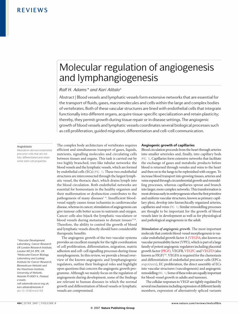
and ligand binding to different receptors. For example, alternative splicing of VEGFA can generate several variants of the growth factor, including the so-called b isoforms that differ in the last six C-terminal amino-acid residues and have anti-angiogenic properties9. The balance between the different VEGFA isoforms can regulate blood-vessel growth and patterning. Binding of VEGFA to the receptor tyrosine kinase VEGFR2 (also known as KDR or FLK1) promotes EC differentiation, proliferation and sprouting. In embryos, this function is counteracted by another receptor, VEGFR1 (also known as FLT1), which has higher affinity for VEGF but weak tyrosine-kinase activity. VEGFR1 also exists in a secreted, catalytically inactive and therefore inhibitory isoform7,8.
Many other factors influence the process of angio-genesis (FIG. 2). The local composition of the extra-cellular matrix (ECM) around the vasculature can affect
angiogenesis positively or negatively10. The proteolytic release of pericellular matrix-bound VEGF isoforms is an important regulator of VEGF bioavailability and blood-vessel patterning11. Furthermore, the physical contact between ECs and pericytes is thought to induce a quiescent, non-sprouting phenotype12,13.
Selection of ECs for sprouting. During angiogenic growth, some ECs within the capillary vessel wall are selected for sprouting (FIG. 2). These ECs are known as the tip cells, which lead the growing sprout. If all cells were to react indiscriminately, the affected section of the vascular network might disintegrate and thereby compromise tissue perfusion. Notch receptors and their Delta-like-4 (DLL4) ligand are essential for sprouting during mouse development. Reduced levels of DLL4 or blocking of Notch signalling enhances the formation of
Figure 1 | Origin of endothelial cells and assembly of the vasculature. Mesodermal cells in the early embryo differentiate into endothelial precursor cells (EPCs, angioblasts) and form aggregates, known as blood islands (left). Fusion of blood islands leads to the vasculogenic formation of honeycomb-shaped primary capillary plexi in the yolk sac and embryo itself. Blood circulation is established and primary plexi are remodelled into a hierarchical network of arterioles and arteries (red), capillaries (grey), and venules and veins (blue). The dorsal aorta and cardinal vein are directly formed through the assembly of angioblasts. The vasculogenic incorporation of circulating EPCs into growing blood vessels may contribute to regenerative or pathological neovascularization in the adult. Vascular smooth-muscle cells (vSMCs) are associated with arteries and veins, whereas capillaries are covered by pericytes (yellow). The first lymphatic endothelial cells (LECs) sprout from the embryonic veins, then migrate and form lymphatic sacs. Further steps of lymphangiogenic growth involve sprouting, branching, proliferation, differentiation and remodelling processes. The recruitment of lymphangioblasts from the adjacent mesenchyme has been speculated to be a further source of LECs. Blind-ending lymphatic capillaries (green) feed into collecting vessels and ducts. These larger lymphatics are sparsely covered by SMCs (purple) and contain valves that prevent backflow.
R E V I E W S
NATURE REVIEWS | MOLECULAR CELL BIOLOGY VOLUME 8 | JUNE 2007 | 465
© 2007 Nature Publishing Group

Growth factorsand inhibitors+ –
b Sprout outgrowth and guidance
Cells
a Selection of sprouting ECs
PDGFB
PDGF
c Sprout fusion and lumen formation d Perfusion and maturation
Bloodflow
Bloodflow
DLL4–NotchVEGF–VEGFRModulation of
EC–EC contacts + –
ECM+ –
Tip cells encounteringrepulsionor adhesion + –
Maintenance of junctions
Stalk-cellproliferation
Vacuole formationand fusion
Stabilization of EC–EC adhesion
Stabilization of PC contacts
Depositionof new ECM
Growth factorsand inhibitors+ –
+ –
Lateralinhibition –
ECproliferation
Pro-quiescent signals (local and systemic) –
↑ Pro-quiescentsignals –
↓ ECproliferation
Changeof polarity
ECMdegradation +
Modulation of PC contacts + –
VEGF–VEGFR2Semaphorin–Neuropilin/PlexinNetrin–UNC5BSLIT–ROBO4
EGFL7ECM
IntegrinsCDC42 and Rac1
Invasivebehaviour+
tip cells, resulting in dramatically increased sprouting, branching and fusion of endothelial tubes14–19. Similarly, endothelial sprouts fail to arrest their angiogenic motile behaviour and branch excessively when Dll4–Notch signalling is compromised in zebrafish embryos20,21. Dll4 expression is induced in the tip cell, whereas the activation of Notch signalling in neighbouring ECs is thought to suppress sprouting of these cells15,19,21 (FIG. 2).
The role of Notch is not confined only to physiological angiogenesis: disruption of DLL4–Notch interactions in tumour models causes extensive sprouting and branch-ing of tumour blood vessels. However, such vessels are poorly functional, which leads to increased hypoxia, poor perfusion and decreased tumour growth. These findings suggest that the Notch pathway can provide a target for anti-angiogenic cancer therapy16,17.
The mechanism by which Notch imposes differen-tial behaviour on ECs that are exposed to similar pro-angiogenic stimuli appears to be directly connected to VEGFA signalling. Dll4 expression is induced in response to VEGFA, whereas the activation of Notch by DLL4 suppresses the expression of Vegfr2 and, in zebrafish, vegfr3 (flt4)14,17–19,21. Therefore, Dll4 expres-sion might correspond to elevated VEGF-mediated signal transduction in selected ECs, whereas Notch activation in their direct neighbouring cells stifles pro-angiogenic responses (FIG. 2). Future work will need to address whether the Notch pathway is pre-dominantly required in cells that are adjacent to the sprout tip or, perhaps in a dynamically regulated and transient fashion, also in the tip itself and other areas of the capillary bed.
Figure 2 | Angiogenic sprouting. a | Sprouting is controlled by the balance between pro-angiogenic signals (+), such as vascular endothelial growth factor (VEGF), and factors that promote quiescence (–), such as tight pericyte (PC; yellow) contact, certain extracellular matrix (ECM) molecules or VEGF inhibitors. In conditions that favour angiogenesis, some endothelial cells (ECs) can sprout (green), whereas others fail to respond (grey). Sprouting requires the flipping of apical–basal polarity, the induction of motile and invasive activity, the modulation of cell–cell contacts and local matrix degradation. b | The growing EC sprout is guided by VEGF gradients. Other signals may include attractive (+) or repulsive (–) matrix cues and guidepost cells in the tissue environment. Release of platelet-derived growth factor B (PDGFB) by the tip cells promotes the recruitment of PCs to new sprouts. EC–EC junctions need to be maintained after lumen formation to prevent excessive leakage. c | Adhesive or repulsive interactions that occur when tip cells encounter each other regulate the fusion of adjacent sprouts and vessels. Lumen formation in stalk ECs involves the fusion of vacuoles but other mechanisms may also contribute. d | Fusion processes at the EC–EC interfaces establish a continuous lumen. Blood flow improves oxygen delivery and thereby reduces pro-angiogenic signals that are hypoxia-induced. Perfusion is also likely to promote maturation processes such as the stabilization of cell junctions, matrix deposition and tight PC attachment. Growth factor withdrawal can trigger sprout retraction and endothelial apoptosis. DLL4, delta-like-4 ligand; EGFL7, epidermal growth factor ligand-7; ROBO4, roundabout homologue-4 (also known as magic roundabout); VEGFR2, VEGF receptor-2.
R E V I E W S
466 | JUNE 2007 | VOLUME 8 www.nature.com/reviews/molcellbio
© 2007 Nature Publishing Group

FilopodiaSlender cellular processes that extend from the front of migrating cells, attach to the surrounding matrix and help to move cells forward.
Mural cellsCells of the outer vessel wall: pericytes and vascular smooth-muscle cells.
Axonal growth conesDynamic guidance structures at the distal end of growing nerve fibres that direct fibres to their appropriate targets and thereby promote the correct ‘wiring’ of the nervous system.
Sprout outgrowth and chemotaxis. Despite insights into the mechanisms that select certain endothelial cells for outgrowth, very little is known about most aspects of the sprouting process. It seems obvious that sprouting ECs need to change their phenotype fundamentally, acquire invasive and motile behaviour, activate secreted or cell-surface-anchored proteases and locally degrade the subendothelial basement membrane. Interactions with surrounding ECs are likely to be modulated, presumably without complete disruption of cell–cell junctions and vessel-wall integrity (FIG. 2). The apical surface of ECs faces the vessel lumen, therefore apical–basal polarity needs to be reversed when new sprouts emerge from the outer (basal) side of the endothelium. Similarly, sprout elongation needs to be polarized and directional.
Consistent with the prominent expression of VEGFR2 in the tips of sprouts, EC guidance is controlled by VEGFA. A spatial concentration gradient of a matrix-anchored isoform of VEGFA (VEGF164 in mice, VEGF165 in humans), which binds to heparan sulphate, functions as a chemoattractive signal that promotes the polarized extension of tip-cell filopodia. A shorter and freely diffusible VEGFA isoform (VEGF120 in mice, VEGF121 in humans) that lacks the heparan sulphate binding motif can support EC proliferation but not tip-cell guidance or proper vascular branching22,23. Neuropilins (NRP1 and NRP2) — transmembrane receptors that can bind to semaphorin family axon-guidance molecules and VEGF — might be involved in this differential response because NRP1 can bind to and enhance signalling through VEGF164 (VEGF165) but not VEGF120 (VEGF121)8,24–26. However, defects in vascular patterning and filopodia extension in Nrp1-knockout mice are relatively mild, which indicates that there are important roles for other VEGFRs in the tip-cell guidance process22,27.
Growing vascular sprouts generate another concentra-tion gradient, namely for platelet-derived growth factor B (PDGFB). In several developing tissues, high levels of PDGFB in tip cells promote the recruitment of pericytes that express the PDGF receptor β (PDGFRβ). This pro-cess ensures that the endothelium of growing vessels is sufficiently stabilized by supporting mural cells23 (FIG. 3).
Common neurovascular guidance cuesEndothelial tip cells function as motile guidance struc-tures that dynamically extend filopodia to explore attractive or repulsive signals that are present in the tissue environment. The dynamic nature, function and morphology of EC tips bear remarkable similarity to axonal growth cones and it has been proposed that nerves and blood vessels have evolved similar processes for the formation of complex networks28,29.
Growing nerve fibres are guided to their targets by a combination of attractive or repulsive cues, such as certain matrix substrates, guidepost cells that provide information in a cell–cell contact-dependent fashion and soluble factors (FIG. 2). Some of these chemotactic signals can also attract endothelial sprouts. For example, the class 3 semaphorins control both axon guidance and vascular patterning (FIG. 3). Many semaphorins mediate
their effects by interacting with receptor complexes that are formed by (semaphorin-binding) neuropilins and (signal-transducing) plexin-family transmembrane proteins30,31 (FIG. 3). At least one class 3 semaphorin, SEMA3E, can signal through plexin D1 independently of neuropilins32. Plexin D1 is expressed in ECs and its role in the pathfinding of blood vessels has been demon-strated by genetic experiments in mice and zebrafish33,34. Loss of plexin D1 causes aberrant sprouting of interseg-mental vessels into SEMA3E-expressing somi tic tissue, which suggests that semaphorins are repulsive cues for ECs33.
Netrins are secreted matrix-binding proteins with homology to the basement-membrane molecule laminin (FIG. 3). In the nervous system, netrins are important axon-guidance cues, which can be either attractive, through interactions with receptors of the deleted in colorectal cancer (DCC) family, or repulsive, through receptors of the uncoordinated-5 (UNC5) family or UNC5–DCC heterodimers28,29. One of these receptors, UNC5B, is strongly expressed in capillaries and endo-thelial tip cells. Loss of Unc5b function in mice or fish leads to increased endothelial sprouting, whereas stimu lation of ECs with the ligand netrin-1 leads to the retraction of tip-cell filopodia35, which is consistent with a negative regulation of blood-vessel growth by netrins and UNC5B. However, some evidence suggests that netrins could promote developmental and thera peutic angio genesis, which may involve as-yet-unknown receptors36.
SLIT proteins form another family of secreted axon-guidance molecules that might have a role in EC sprouting. Interactions of SLIT proteins with round-about (ROBO) receptors (FIG. 3) repel axon growth cones and inhibit the migration of certain cell types. ROBO4 (magic roundabout) is essential for normal vascular patterning, although it is controversial whether ROBO4 is capable of direct binding to any of the three known mammalian SLIT proteins37–39.
These examples highlight that endothelial sprouts use similar attractive and repulsive tissue-derived navigational cues as growing nerve fibres to control the patterning and directional growth of the vascular network.
Transformation of sprouts into vesselsMany further steps are required to convert endo-thelial sprouts into functional, blood-carrying vessels (FIG. 2). For example, sprout extension can involve the migration of the endothelial cells behind the tip. Such chains of migrating ECs are visible during the growth of intersegmental vessels in the trunk of zebrafish embryos20,21,40. Alternatively, local EC proliferation can promote sprout extension. For example, in the developing mouse retina, ECs in the stalk of sprouts proliferate whereas the tip cells do not23. However, only the leading tip cell undergoes a single cell division in zebrafish intersegmental vessels21. These differences in the mitotic pattern together with higher levels of Dll4, Vegfr2 and Pdgfb transcripts and the growth-cone-like morph ology of sprout tips indicate that ECs at the tip and in the stalk of sprouts are different. However, a fraction of the stalk cells
R E V I E W S
NATURE REVIEWS | MOLECULAR CELL BIOLOGY VOLUME 8 | JUNE 2007 | 467
© 2007 Nature Publishing Group
Netrins
LamNT
EGF-like
C-terminaldomain
UNC5B
TM
Ig-like
TSP1
ZO-1-like
DEATH
CUB
Discoidindomain
MAM
TM
Ig
Sema
PSI
Sema
Class 3 semaphorins
Ig-like
TM
PSI
VEGFA
VEGF
TM
ROBO4
Ig-like
Ig-like
SLIT proteins
EGF-like
EGF-like
LamG
Leucine-richrepeats
?
NRP1/2 Plexin D1
N-cadherin
TM
N-cadherin
SAM
Kinase
FN3
Ligandbinding
TM
Ephrin-B2
EphB
EphB4 Ephrin-B2EndothelialEndothelial
Mural
TM TM
Extracellular spaceand matrix
Ephrin-B2
EGF-like
DSL AnkyrinrepeatDSL
Notch1/3/4
EGF-like
TMDLL4
JAG1
TM EGF-like DSLVWC
EC guidance
EC–EC interactions EC–EC interactions
MC motility and adhesion
EC–MC adhesion
EC–MC adhesion
Tip/stalk identityEC proliferationArterial identity
CArepeats
PinocytosisUptake of extracellular liquid into cells in the form of membrane-coated vesicles.
also extend filopodia and show elevated expression of Dll4 or Pdgfb. It remains unclear whether the differences between tip and stalk ECs reflect stable, genetically con-trolled fates or transient adaptations to positional cues within the sprout.
To form new vascular connections, tip cells need to suppress their motile, explorative behaviour upon encountering their targets, the tips of other sprouts or existing capillaries. Strong adhesive interactions and EC–EC junctional contacts need to be established at this joining point. The failure to do so (for example, because of repulsive signalling triggered by cell–cell contact) would presumably preclude the generation of a blood-transporting vessel by these ECs (FIG. 2). Such a process might also help to prevent the fusion between incompatible parts of the vasculature and the formation of arteriovenous shunts.
The establishment of blood flow requires the form-ation of a vascular lumen, which may occur within growing endothelial sprouts before or after they have joined with other vessels (FIG. 2). Cellular structures can be converted into tubes in various ways, as has been shown for epithelial tissues in vertebrates and invertebrates41. Although little is known about the lumenization of blood vessels, there is strong evidence for the involvement of pinocytosis (‘cell drinking’) and vacuole formation. Studies in cultured ECs suggest that these processes require the integrin cell-matrix adhesion machinery and the small GTPases CDC42 and Rac1 (REF. 42). High-resolution time-lapse imaging of growing intersegmental vessels in zebrafish has established that the lumen in these ECs is formed by intracellular and, subsequently, intercellular fusion of large vacuoles40.
Figure 3 | Guidance cues, adhesion molecules and cell-fate regulators that function both in the nervous system and in the vasculature. Black arrows indicate molecular interactions. Navigational cues that direct growing axons such as netrins, semaphorins and SLITs also guide endothelial cells through their corresponding receptors. The specificity of SLIT-ligand binding to ROBO4 is controversial38,39. Notch signalling triggered by DLL4 or JAG1 controls endothelial sprouting, proliferation and arteriovenous identity. Binding of ephrin-B2 — a guidance cue and a modulator of neuronal activity — to its receptor EphB4 modulates endothelial cell (EC)–EC interactions and is essential for angiogenesis. Ephrin-B2 expression in mural cells (MC) controls their motility and adhesion105. This function may be modulated by endothelial ephrin-B2. The homophilic cell-adhesion molecule N-cadherin promotes synapse formation between neurons and stabilizes the EC–pericyte contact119,146,147. CA, cadherin repeats; CUB, cubilin-related; DLL4, Delta-like-4 ligand; DSL, Delta/Serrate/Lag-2; EGF-like, epidermal-growth factor like; FN3, fibronectin type 3-like; Ig, immunoglobulin; JAG1, Jagged1; LamG, laminin globular G domain; LamNT, laminin N-terminal domain; MAM, meprin–A5-protein–PTPmu-related; PSI, plexin–semaphorin–integrin; ROBO4, roundabout homologue-4; SAM, sterile-α motif; Sema, semaphorin; TM, transmembrane; TSP1, thrombospondin-1 like; UNC5B, uncoordinated-5 homologue B; VWC, von Willebrand type C factor-like; ZO-1-like, zona occludens-1-like.
R E V I E W S
468 | JUNE 2007 | VOLUME 8 www.nature.com/reviews/molcellbio
© 2007 Nature Publishing Group

Modulation ofEC–EC junctions
Growth factorsand inhibitors+ –
VEGF–VEGFR2SDF1–CXCR4
VEGF–VEGFR2Invasivebehaviour
Adhesion
a Circulating cells and blood-vessel growth
b Vessel splitting by intussusception
Stimulation of EC proliferation and/orsprouting byperivascular cells
Incorporation into vessel wall and circumferential growth
VEGF–VEGFR1
Circulating precursor
++
Incorporation into vascular sprouts and angiogenic growth
ECproliferation
Insertion oftissue pillar
ECMdeposition
Lumen formation and tubulogenesis are also con-trolled by EGF-like domain-7 (EGFL7; also known as vascular endothelial statin), a small secreted ECM-associated protein that is expressed by the ECs. Although the exact role of EGFL7 requires further investigation, it appears to suppress matrix adhesion without affecting EC proliferation43.
The generation of a lumen and the onset of blood flow may help to stabilize new vascular connections. Improved oxygen delivery lowers local VEGFA expres-sion. The recruitment of pericytes, the deposition of ECM proteins into the subendothelial basement mem-brane and reduced EC proliferation also promote vessel maturation and quiescence (FIG. 2). At this stage, as well as during other steps in the sprouting process, changes in the local balance between pro- and anti-angiogenic factors may lead to the elimination (pruning) of the new connections; for example, by EC retraction or apopto-sis. The pruning process is most extensive around the oxygen-rich arteries in the developing retina, consistent
with the suppression of hypoxia-driven VEGFA expres-sion. Treatment of experimental tumours with VEGFA inhibitors has shown that empty sleeves of basement membrane with associated pericytes remain when immature vessels are eliminated. Because these struc-tures favour the rapid regrowth of blood vessels when the VEGF signal is restored, matrix sleeves might be potential targets in cancer therapy44.
Non-angiogenic growth of blood vesselsAngiogenic sprouting of ECs from existing blood vessels is not the only mechanism that contributes to vascular growth. For example, the dorsal aorta and cardinal vein in the early embryo are formed by vasculo-genesis — the direct assembly of angioblasts and ECs45. Similarly, bone-marrow-derived circulating EPCs are thought to contribute to vasculogenic regener ative or pathological blood-vessel growth in the adult46. Such incorporation of EPCs into the endothelial mono layer could lead to circumferential enlargement of vessels.
Figure 4 | Vasculogenic and intussusceptive growth of blood vessels. a | Recruitment and incorporation of cells from the blood circulation requires positive chemotactic signals and local factors that promote adhesion to endothelial cells (ECs). Transendothelial migration and/or the incorporation of precursor cells into the endothelium require the modulation of existing EC–EC contacts. Circulating precursor cells (angioblasts) have been suggested to differentiate into ECs and lead to an expansion of the vessel diameter (circumferential growth) or, alternatively, they may be recruited to new angiogenic sprouts. Circulation-derived cells do not necessarily acquire an endothelial fate but can instead promote the proliferation and sprouting of local ECs from a perivascular location. b | Intussusception — the splitting of vessels through the insertion of tissue pillars — is a separate mechanism that leads to the expansion of blood vessels. Little is known about the function or regulation of intussusceptive growth but the process is likely to involve EC proliferation, cell movement, the degradation of extracellular matrix (ECM) and the deposition of new ECM molecules. CXCR4, CXC-chemokine receptor-4; SDF1 (also known as CXCL12), stromal cell-derived factor-1; VEGF, vascular endothelial growth factor; VEGFR1, VEGF receptor-1.
R E V I E W S
NATURE REVIEWS | MOLECULAR CELL BIOLOGY VOLUME 8 | JUNE 2007 | 469
© 2007 Nature Publishing Group
Vascular smooth-muscle cellsSpecialized smooth-muscle cells that form the outer layer of arteries, arterioles and larger veins. They provide blood vessels with mechanical stability that is due to their contractile properties and the deposition of matrix and elastic fibres.
Alternatively, recruitment of EPCs to growing endo-thelial sprouts may combine vasculogenic and angio-genic mechanisms (FIG. 4). In most cases, however, the contribution of EPCs to angiogenesis is small.
VEGF and its receptors are crucial for the mobilization of EPCs in the bone marrow and their recruitment to specific sites in the body. In both processes, VEGFR1 expression and signalling in bone-marrow-derived cells have essential roles even though the same receptor inhibits angiogenesis during embryogenesis7,8,46. VEGF–VEGFR1 signalling also mediates the recruitment of bone-marrow-derived monocytic cells, which do not give rise to ECs and are not incorporated into the endothe-lium7,47. For example, recruited bone-marrow-derived circulating cells (RBCCs), which express CXC-chemokine receptor-4 (CXCR4), are retained in the perivascular space. This retention is due to the local expression of its ligand stromal-derived factor-1 (SDF1) in response to VEGFA. The recruitment of RBCCs enhances endothelial proliferation and angiogenesis, whereas interfering with RBCCs attenuates VEGF-mediated pro-angiogenic effects47 (FIG. 4a).
Finally, it should be mentioned that the vascular net-work can be locally extended by intussusception — the splitting of blood vessels through the insertion of tissue pillars48 (FIG. 4b). Despite strong morphological evidence that supports a role for this process in various tissues, little is known about the physiological role and molecular regulation of intussusception.
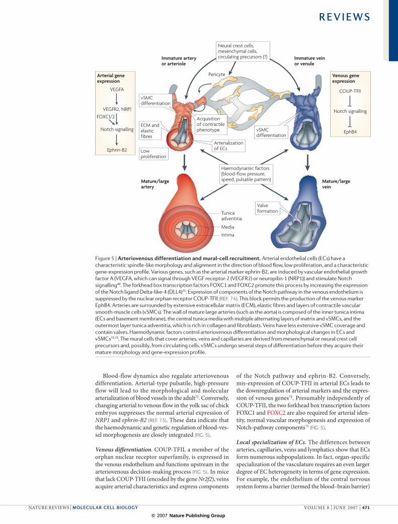
Arteriovenous differentiationDuring the remodelling of the primary capillary plexi in the early embryo, arteries and veins are formed in a process that is known as arteriovenous differentiation3,49. The growth and specialization of arteries and veins con-tinues throughout development and reflects the distinct haemodynamic properties within the vascular network. Concentric layers of contractile vascular smooth-muscle cells (vSMCs), ECM and elastic fibres allow arteries to resist shear stress and pulsatile flow (FIG. 5). Elevated fluid shear stress is also a main factor that mediates the enlargement and remodelling of existing arteries and arterioles (arteriogenesis) in the adult3,50. The venous low-pressure system contains valves, which are flap-like structures that open unidirectionally to prevent backflow (FIG. 5). Veins are enveloped by fewer vSMCs but even this low degree of coverage is physiologically relevant, as shown by the degeneration of the vSMC–matrix layer in varicose veins and some other venous diseases51,52.
Notch controls arterial differentiation. From the earliest stages of cardiovascular development, arterial ECs share the expression of certain transcription factors, signalling molecules, gap-junction proteins, ECM molecules and adhesion proteins that are largely absent from veins. Some of these molecules reflect the specific physiological needs of arteries, whereas others, such as those in the Notch pathway, are directly involved in the differen-tiation of the arterial branch49. Proteolytic processing of activated Notch releases an intracellular fragment, which translocates to the nucleus and associates with
transcriptional regulators of the suppressor of hairless family (CBF1 or RBP-Jκ in vertebrates). This complex allows the expression of basic helix–loop–helix (bHLH) proteins of the hairy/enhancer-of-split-related transcrip-tion factor family — Hey2 and Her in zebrafish, and Hey and Hes in mammals — which, in turn, control the expression of other downstream target genes53,54.
Knockout mice that lack components of the Notch pathway — NOTCH1 and/or NOTCH4, the ligands Jagged1 (JAG1) or DLL4, presenilin proteins that are required for Notch proteolytic processing, the ubiquitin-ligase mind bomb (a regulator of ligand endocytosis and Notch signalling), the transcription factor RBP-Jκ or the Hey family members HEY1 and HEY2 — have severe vascular defects and are embryonic lethal55–62. Although it is likely that some or all of these molecules are also required for endothelial sprouting and capillary growth, the knockout phenotypes and their defective arterio venous-marker expression support important roles for these molecules in arterial differentiation. Similarly, manipulation of Notch signalling or the hey2 gene in zebrafish embryos shows that Notch signalling is necessary and sufficient for the expression of arterial markers49.
Arterial markers and arteriovenous identity. Probably the best-known arterial EC marker is the transmembrane protein ephrin-B2 (encoded by EFNB2) which is a highly promiscuous ligand that can bind to at least six different receptors within the large Eph tyrosine kinase family63,64. Expression of the cognate receptor (EphB4) is largely con-fined to the venous endothelium so that ephrin-B2 and EphB4 are frequently used as molecular markers for the identification of arteries and veins. Although ephrin-B2 and EphB4 are essential for angiogenesis in the early mouse embryo, their precise function in the vasculature and their potential involvement in the arteriovenous-fate decision are unclear28,29.
VEGF is also an important regulator of arterial diff erentiation. Exposure of cultured ECs to VEGFA leads to increased Dll4 expression, Notch signalling and ephrin-B2 upregulation65,66 (FIG. 5). In mouse embryonic skin, VEGFA secreted by peripheral nerves controls arterial differentiation67. Similarly, the expression of Notch pathway genes and ephrin-B2 is regulated by VEGFA in zebrafish49. How can VEGFA simult aneously control EC proliferation, vasculogenesis, angiogenesis and arterial differentiation? Local differences in VEGFR expression and signalling are possible explanations. In the zebrafish, Vegfr2 is abundant in the dorsal aorta, whereas Vegfr3 (Flt4) is predominantly found in veins49.
Neuropilins also show distinct arteriovenous-specific patterns. NRP1 is found in arteries, whereas NRP2 is restricted to veins and lymphatic vessels68–70. The two neuropilins can bind to different VEGF isoforms, which may lead to distinct signalling responses in arteries and veins8,29. Local regulation might also involve Vegf splic-ing, the availability of heparan sulphate proteoglycans, proteolytic processing of VEGF and the modulation of its matrix-binding properties11,71.
R E V I E W S
470 | JUNE 2007 | VOLUME 8 www.nature.com/reviews/molcellbio
© 2007 Nature Publishing Group
Arterial geneexpression
VEGFA
FOXC1/2VEGFR2, NRP1
Notch signalling
Ephrin-B2
Immature arteryor arteriole
Mature/largeartery
Mature/largevein
Immature veinor venule
Venous geneexpression
COUP-TFII
Notch signalling
EphB4
Neural crest cells,mesenchymal cells,circulating precursors (?)
Haemodynamic factors(blood-flow pressure,speed, pulsatile pattern)
Pericyte
Media
Intima
Tunicaadventitia
Acquisition of contractilephenotype
vSMCdifferentiation
vSMCdifferentiation
Lowproliferation
ECM andelastic fibres
Arterializationof ECs
Valveformation
Blood-flow dynamics also regulate arteriovenous differentiation. Arterial-type pulsatile, high-pressure flow will lead to the morphological and molecular arterial ization of blood vessels in the adult72. Conversely, changing arterial to venous flow in the yolk sac of chick embryos suppresses the normal arterial expression of NRP1 and ephrin-B2 (REF. 73). These data indicate that the haemodynamic and genetic regulation of blood-ves-sel morphogenesis are closely integrated (FIG. 5).
Venous differentiation. COUP-TFII, a member of the orphan nuclear receptor superfamily, is expressed in the venous endothelium and functions upstream in the arteriovenous decision-making process (FIG. 5). In mice that lack COUP-TFII (encoded by the gene Nr2f2), veins acquire arterial characteristics and express components
of the Notch pathway and ephrin-B2. Conversely, mis-expression of COUP-TFII in arterial ECs leads to the downregulation of arterial markers and the expres-sion of venous genes74. Presumably independently of COUP-TFII, the two forkhead box transcription factors FOXC1 and FOXC2 are also required for arterial iden-tity, normal vascular morphogenesis and expression of Notch-pathway components75 (FIG. 5).
Local specialization of ECs. The differences between arteries, capillaries, veins and lymphatics show that ECs form numerous subpopulations. In fact, organ-specific specialization of the vasculature requires an even larger degree of EC heterogeneity in terms of gene expression. For example, the endothelium of the central nervous system forms a barrier (termed the blood–brain barrier)
Figure 5 | Arteriovenous differentiation and mural-cell recruitment. Arterial endothelial cells (ECs) have a characteristic spindle-like morphology and alignment in the direction of blood flow, low proliferation, and a characteristic gene-expression profile. Various genes, such as the arterial marker ephrin-B2, are induced by vascular endothelial growth factor A (VEGFA, which can signal through VEGF receptor-2 (VEGFR2) or neuropilin-1 (NRP1)) and stimulate Notch signalling49. The forkhead box transcription factors FOXC1 and FOXC2 promote this process by increasing the expression of the Notch ligand Delta-like-4 (DLL4)75. Expression of components of the Notch pathway in the venous endothelium is suppressed by the nuclear orphan receptor COUP-TFII (REF. 74). This block permits the production of the venous marker EphB4. Arteries are surrounded by extensive extracellular matrix (ECM), elastic fibres and layers of contractile vascular smooth-muscle cells (vSMCs). The wall of mature large arteries (such as the aorta) is composed of the inner tunica intima (ECs and basement membrane), the central tunica media with multiple alternating layers of matrix and vSMCs, and the outermost layer tunica adventitia, which is rich in collagen and fibroblasts. Veins have less extensive vSMC coverage and contain valves. Haemodynamic factors control arteriovenous differentiation and morphological changes in ECs and vSMCs72,73. The mural cells that cover arteries, veins and capillaries are derived from mesenchymal or neural crest cell precursors and, possibly, from circulating cells. vSMCs undergo several steps of differentiation before they acquire their mature morphology and gene-expression profile.
R E V I E W S
NATURE REVIEWS | MOLECULAR CELL BIOLOGY VOLUME 8 | JUNE 2007 | 471
© 2007 Nature Publishing Group
PodocyteSpecialized, highly branched epithelial cell in the filtering units (glomeruli) of the kidney. Numerous podocyte foot processes cover the glomerular capillary basement membrane and thereby form a size-selective filtration barrier that is permeable to water, salts and glucose but retains macromolecules in the bloodstream.
LymphoedemaHarmful interstitial liquid accumulation that is due to insufficient lymphatic drainage.
that tightly controls transendothelial transport and cell migration. By contrast, vessels in endocrine glands and the pancreas, intestine and kidney are highly permeable owing to pore-like fenestrations, which can be induced by VEGF or endocrine gland–VEGF (EG–VEGF), a tissue-specific angiogenic regulator76. Also, the form-ation of valves in lymphatic vessels and veins most probably involves as-yet-unidentified instructive signals recognized by a few ECs but not their neighbouring cells. The identification and characterization of such local signals are important areas for future research.
Growth and identity of lymphatic vesselsThe lymphatic vasculature forms a unidirectional net-work of blind-ended capillaries (terminal lymphatics) that collect the protein-rich fluid that exudes from blood vessels and then drain through a conduit system of collecting vessels, lymph nodes, lymphatic trunks and ducts into the venous circulation2,4,77,78. Loss of lymphatic function in humans as a result of hereditary disease, lymphatic-vessel damage or surgical removal of lymph nodes causes lymphoedema4. The lymphatic
network is also involved in the immune surveillance of the body. Lymphocytes and antigen-presenting dendritic cells travel from peripheral tissues through lymphatic vessels to the lymphoid organs, in which immune defences against pathogens are mounted.
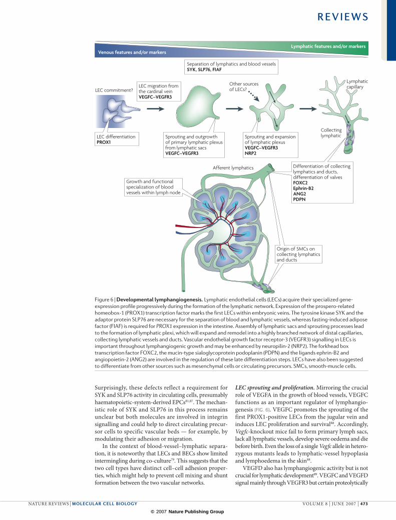
Although the ECs of blood vessels (BECs) and lym-phatics (LECs) share many features, such as their flat shape, strong apical–basal polarity and the expression of certain endothelial markers, the distinct functional roles of the two vascular networks require extensive specializ ation. The endothelium of terminal lym-phatics lacks a continuous basement membrane and LEC–LEC contacts are not tightly sealed by junctional complexes. Owing to anchoring filaments that link LECs to the interstitial ECM, fluid accumulation in the tissue opens inter cellular gaps and enhances the uptake of interstitial fluid. The distinct architecture and func-tion of lymphatic vessels requires some specific gene products that are not shared with the BECs2,79 (BOX 1). Because the mammalian lymphatic system originates from the embryonic veins, its morphogenesis involves the acquisition of LEC identity and separation from the blood-vessel network (FIG. 6). A small part of the LEC population may be generated from mesenchymal or circulating precursor cells80,81, but little is yet known about these processes (FIG. 6).
LEC identity and separation from blood vessels. In mouse mid-gestation embryos, a distinct cluster of ECs on the dorsal side of the jugular vein express the prospero-related homeobox-1 (PROX1) transcription factor and differentiate into the first LECs (FIG. 6). The activity that induces the local expression of this tran-scription factor is unknown but the role of PROX1 in the upregulation of many lymphatic-specific genes and suppression of certain blood-vessel markers has been firmly established by gene-expression profiling studies in cultured cells79,82.
PROX1 function is also essential for lymphatic development in vivo (FIG. 6). Inactivation of Prox1 in mice results in defective extension of ECs from the cardinal vein, loss of lymphatic-marker expression and lack of the lymphatic vasculature83,84. PROX1 function is not confined to the onset of lymphatic development. Heterozygous Prox1-mutant mice are viable but have mispatterned, dysfunctional and ruptured lymphatic vessels. This defect is most obvious in the gastrointes-tinal system and leads to obesity because leaked lymph promotes the differentiation of adipocytes85. The expression of Prox1 in the LECs of the small intestine is severely reduced in mice that lack fasting-induced adipose factor (FIAF; also known as ANGPTL4). Fiaf mutants die within a few weeks of birth with dilated and blood-filled lymphatics that are aberrantly connected to blood vessels86.
The critical importance of the separation of blood vessels and lymphatic vessels is also demonstrated by studies in mice that lack the cytoplasmic tyrosine kinase SYK or the adaptor protein SLP76 (REFS 81,87). Both mutants develop abnormal connections between BECs and LECs during embryogenesis.
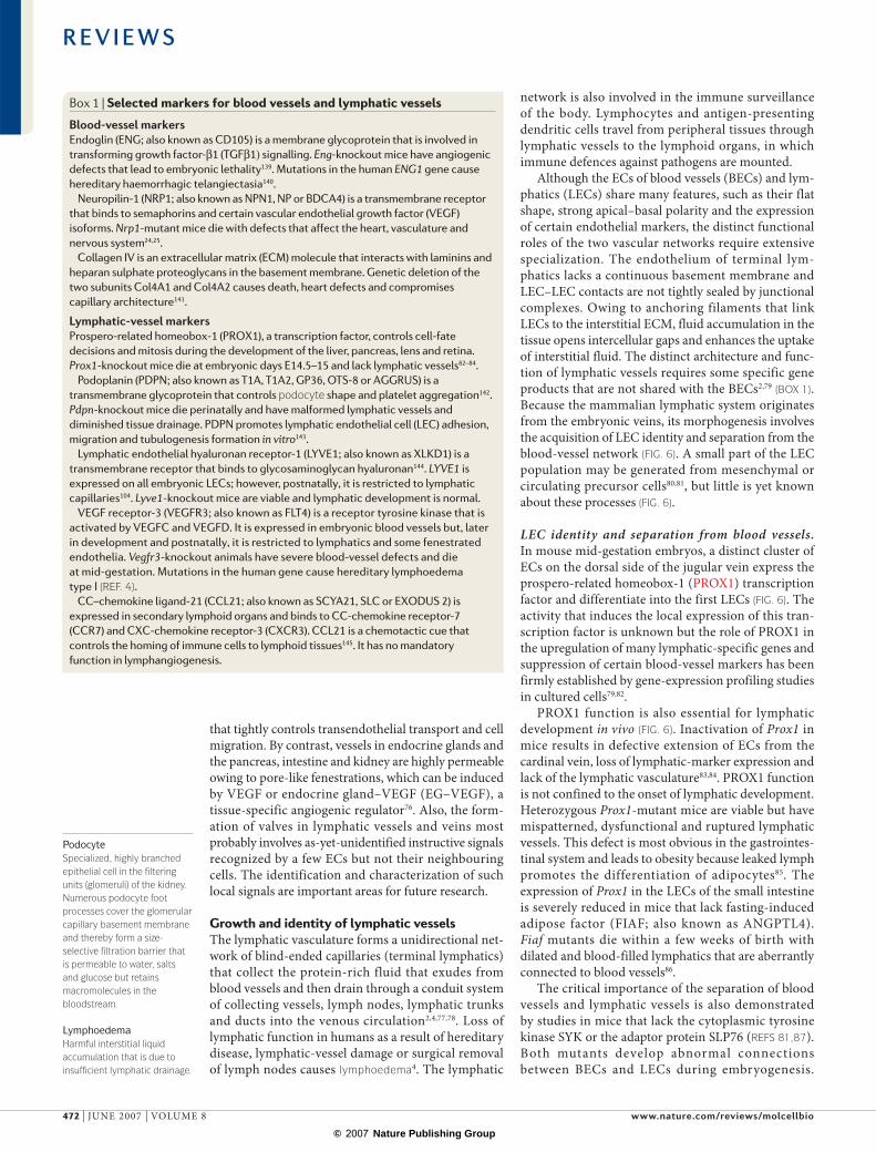
Box 1 | Selected markers for blood vessels and lymphatic vessels
Blood-vessel markersEndoglin (ENG; also known as CD105) is a membrane glycoprotein that is involved in transforming growth factor-β1 (TGFβ1) signalling. Eng-knockout mice have angiogenic defects that lead to embryonic lethality139. Mutations in the human ENG1 gene cause hereditary haemorrhagic telangiectasia140.
Neuropilin-1 (NRP1; also known as NPN1, NP or BDCA4) is a transmembrane receptor that binds to semaphorins and certain vascular endothelial growth factor (VEGF) isoforms. Nrp1-mutant mice die with defects that affect the heart, vasculature and nervous system24,25.
Collagen IV is an extracellular matrix (ECM) molecule that interacts with laminins and heparan sulphate proteoglycans in the basement membrane. Genetic deletion of the two subunits Col4A1 and Col4A2 causes death, heart defects and compromises capillary architecture141.
Lymphatic-vessel markersProspero-related homeobox-1 (PROX1), a transcription factor, controls cell-fate decisions and mitosis during the development of the liver, pancreas, lens and retina. Prox1-knockout mice die at embryonic days E14.5–15 and lack lymphatic vessels82–84.
Podoplanin (PDPN; also known as T1A, T1A2, GP36, OTS-8 or AGGRUS) is a transmembrane glycoprotein that controls podocyte shape and platelet aggregation142. Pdpn-knockout mice die perinatally and have malformed lymphatic vessels and diminished tissue drainage. PDPN promotes lymphatic endothelial cell (LEC) adhesion, migration and tubulogenesis formation in vitro143.
Lymphatic endothelial hyaluronan receptor-1 (LYVE1; also known as XLKD1) is a transmembrane receptor that binds to glycosaminoglycan hyaluronan144. LYVE1 is expressed on all embryonic LECs; however, postnatally, it is restricted to lymphatic capillaries104. Lyve1-knockout mice are viable and lymphatic development is normal.
VEGF receptor-3 (VEGFR3; also known as FLT4) is a receptor tyrosine kinase that is activated by VEGFC and VEGFD. It is expressed in embryonic blood vessels but, later in development and postnatally, it is restricted to lymphatics and some fenestrated endothelia. Vegfr3-knockout animals have severe blood-vessel defects and die at mid-gestation. Mutations in the human gene cause hereditary lymphoedema type I (REF. 4).
CC–chemokine ligand-21 (CCL21; also known as SCYA21, SLC or EXODUS 2) is expressed in secondary lymphoid organs and binds to CC-chemokine receptor-7 (CCR7) and CXC-chemokine receptor-3 (CXCR3). CCL21 is a chemotactic cue that controls the homing of immune cells to lymphoid tissues145. It has no mandatory function in lymphangiogenesis.
R E V I E W S
472 | JUNE 2007 | VOLUME 8 www.nature.com/reviews/molcellbio
© 2007 Nature Publishing Group
Lymphatic features and/or markersVenous features and/or markers
Collectinglymphatic
LEC commitment?
LymphaticcapillaryOther sources
of LECs?
Afferent lymphatics
LEC differentiationPROX1
LEC migration fromthe cardinal veinVEGFC–VEGFR3
Separation of lymphatics and blood vesselsSYK, SLP76, FIAF
Sprouting and expansionof lymphatic plexusVEGFC–VEGFR3NRP2
Sprouting and outgrowthof primary lymphatic plexus from lymphatic sacsVEGFC–VEGFR3
Differentiation of collectinglymphatics and ducts,differentiation of valvesFOXC2Ephrin-B2ANG2PDPN
Growth and functionalspecialization of bloodvessels within lymph node
Origin of SMCs on collecting lymphaticsand ducts
Surprisingly, these defects reflect a requirement for SYK and SLP76 activity in circulating cells, presumably haematopoietic-system-derived EPCs81,87. The mechan-istic role of SYK and SLP76 in this process remains unclear but both molecules are involved in integrin signalling and could help to direct circulating precur-sor cells to specific vascular beds — for example, by modulating their adhesion or migration.
In the context of blood-vessel–lymphatic separa-tion, it is noteworthy that LECs and BECs show limited intermingling during co-culture79. This suggests that the two cell types have distinct cell–cell adhesion proper-ties, which might help to prevent cell mixing and shunt formation between the two vascular networks.
LEC sprouting and proliferation. Mirroring the crucial role of VEGFA in the growth of blood vessels, VEGFC functions as an important regulator of lymphangio-genesis (FIG. 6). VEGFC promotes the sprouting of the first PROX1-positive LECs from the jugular vein and induces LEC proliferation and survival88. Accordingly, Vegfc-knockout mice fail to form primary lymph sacs, lack all lymphatic vessels, develop severe oedema and die before birth. Even the loss of a single Vegfc allele in hetero-zygous mutants leads to lymphatic-vessel hypoplasia and lymphoedema in the skin88.
VEGFD also has lymphangiogenic activity but is not crucial for lymphatic development89. VEGFC and VEGFD signal mainly through VEGFR3 but certain proteolytically
Figure 6 | Developmental lymphangiogenesis. Lymphatic endothelial cells (LECs) acquire their specialized gene-expression profile progressively during the formation of the lymphatic network. Expression of the prospero-related homeobox-1 (PROX1) transcription factor marks the first LECs within embryonic veins. The tyrosine kinase SYK and the adaptor protein SLP76 are necessary for the separation of blood and lymphatic vessels, whereas fasting-induced adipose factor (FIAF) is required for PROX1 expression in the intestine. Assembly of lymphatic sacs and sprouting processes lead to the formation of lymphatic plexi, which will expand and remodel into a highly branched network of distal capillaries, collecting lymphatic vessels and ducts. Vascular endothelial growth factor receptor-3 (VEGFR3) signalling in LECs is important throughout lymphangiogenic growth and may be enhanced by neuropilin-2 (NRP2). The forkhead box transcription factor FOXC2, the mucin-type sialoglycoprotein podoplanin (PDPN) and the ligands ephrin-B2 and angiopoietin-2 (ANG2) are involved in the regulation of these late differentiation steps. LECs have also been suggested to differentiate from other sources such as mesenchymal cells or circulating precursors. SMCs, smooth-muscle cells.
R E V I E W S
NATURE REVIEWS | MOLECULAR CELL BIOLOGY VOLUME 8 | JUNE 2007 | 473
© 2007 Nature Publishing Group
Neural crest cellsEctodermal cells that delaminate from the neural tube in vertebrate embryos, migrate to various locations and contribute to different body structures such as the peripheral nervous system, bone and cartilage, skeletal and smooth muscle, or pigment cells in the skin (melanocytes).
Vascular stem cellsStem cells that can differentiate into endothelial or mural cells in the blood vessel wall.
HaemangioblastsPrecursor cells that can differentiate into endothelial and haematopoietic cells.
processed forms can also interact with VEGFR2 (REFS 4,90). These binding properties and expression of Vegfr3 in BECs explain the severe vasculogenic and angio genic defects during early embryogenesis in VEGFR3 loss-of-function models91,92. Perinatally, Vegfr3 expression becomes increasingly confined to the lymphatic vas culature so that disruption of VEGFC–VEGFR3 sig-nalling selectively compromises lymphangiogenesis93,94. However, some inhibition of angiogenesis in tumours and wounds has also been observed, and correlates with the re-expression of Vegfr3 in blood vessels95.
NRP2, which is expressed in lymphatic vessels, can interact with VEGFR3 and bind to VEGFC and VEGFD, and is essential for lymphangiogenesis68,96. Nrp2-knockout mice show reduced LEC proliferation and fail to develop small-diameter lymphatic vessels and capillaries68.
VEGFA also stimulates lymphatic growth in several experimental systems97–99 but this activity might be indirect (for example, through the recruitment of inflam-matory cells and increased VEGFC expression100,101). Other growth factors and chemokines that have been suggested to have lymphangiogenic activity include fibroblast growth factor (FGF), hepatocyte growth factor (HGF), PDGF, insulin-like growth factor-1 (IGF1) and IGF2 (REF. 2). However, some of these molecules may stimulate lymphatic growth indirectly or only in pathological settings.
Differentiation of lymphatic vesselsLittle is known about the lymphatic maturation pro-gramme but known processes include the gradual sup-pression of LEC proliferation and sprouting, protection against VEGFC withdrawal, valve formation and the differentiation of the network into lymphatic capillaries and smooth-muscle-covered collecting lymphatics, lymphatic trunks and ducts (FIG. 6).
One important regulator of these processes is VEGFC, which remains essential until developmental lymphangio-genesis is completed postnatally93. The patterning of the lymphatic network and SMC recruitment to the collecting lymphatics are defective in angiopoietin-2 (Ang2)-deficient mice. ANG1 can also promote lymphangio genesis, trig-ger LEC proliferation and rescue the lymphatic defects of Ang2-knockout mice102,103.
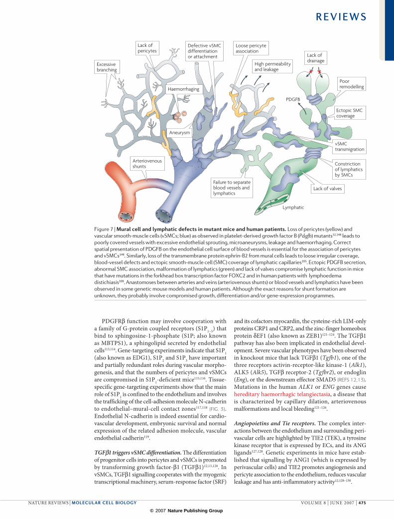
Similar to the angiopoietins, ephrin-B2 is involved in the angiogenic growth of both blood vessels and lym-phatic vessels, as shown by LEC sprouting, lymphatic patterning and valve-formation defects in mutant mice104. A further interesting feature of the ephrin-B2 mutant is the appearance of ectopic SMC coverage on cutaneous lymphatic capillaries (FIG. 7). Although this is caused by a cell-autonomous differentiation defect of the lymphatic endothelium, SMCs are also found on terminal lymphatics in mice that lack ephrin-B2 in mural cells. In these mutants, pericytes and vSMCs fail to associate stably with blood vessels and some migrate to lymph atics105 (FIG. 7). Lymphatics also acquire ectopic vSMCs in Pdgfrb-knockout mice, which suggests that intact pericyte and vSMC chemotaxis helps to ensure that only the correct vessels acquire mural cells105.
Abnormal association of smooth-muscle actin-positive cells with lymphatic vessels is associated with compro-mised tissue drainage in lymphoedema distichiasis (LD), a congenital disease caused by inactivating, autosomal dominant mutations in the human FOXC2 gene106–108. Similarly, Foxc2-deficient mice have dysfunctional lymphatics that express several BEC markers including PDGFB, are covered by smooth-muscle cells, and lack valves (FIG. 7). Normal early lymphatic devel opment in these mutants indicates that FOXC2 is mainly required for the late maturation steps108.
Maturation of blood vesselsThe term maturation describes the stepwise transition from an actively growing vascular bed to a quiescent, fully formed and functional network. This involves the suppres-sion of endothelial proliferation and sprouting, protection against growth-factor (VEGF) withdrawal, stabilization of existing vascular tubes, cellular-differentiation processes such as the formation of valves, fenestrations or tight-junction barriers, and the incorporation of mural cells into the vessel wall (see below)3,45. Maturation is frequently compromised in pathological settings, particularly in the vasculature of tumours109,110.
A prominent and widely recognized feature of the maturation programme is the recruitment of mural cells — pericytes and vSMCs (FIG. 5). Pericytes establish direct cell–cell contact with ECs and cover capillaries and immature blood vessels, whereas vSMCs cover mature and larger diameter vessels, such as arteries and veins, and are separated from the endothelium by a basement-membrane layer. Both mural cell types share a mesen-chymal, fibroblast-like morphology and their phenotypes are possibly interconvertible, although this has not been formally demonstrated. Understanding the relationships between mural cells is complicated by the difficulty in identifying pericytes. Molecular markers such as the pro-teoglycan NG2, the intermediate filament protein desmin, α-smooth-muscle actin, the regulator of G-protein signalling RGS5 or the reporter transgene X-LacZ4 label only subsets of the highly heterogeneous pericyte popu-lation and are also expressed by vSMCs and other cell types12,13. Furthermore, it remains unclear whether there are other, as-yet-unidentified and perhaps tissue-specific subsets of these cells.
PDGFRβ cooperates with G-protein coupled receptors. The ontogeny of pericytes and vSMCs is similarly puzzling and complex (FIG. 5). Some mural cells are derived from neural crest cells that delaminate from the neural tube early in embryogenesis, whereas others descend from undifferentiated mesenchymal cells or, possibly, vascular stem cells12,13. Expression of the receptor tyrosine kinase PDGFRβ in mesenchymal precursor cells, pericytes and vSMCs is required for mural-cell proliferation, guided migration and incorporation into the vessel wall. The expression, matrix-binding and correct spatial presenta-tion of PDGFB are indispensable in mice12,111, although PDGFD can also bind and activate PDGFRβ (FIG. 7). It has recently been suggested that PDGFB and PDGFRβ also have a role in the differentiation of haemangioblasts112.
R E V I E W S
474 | JUNE 2007 | VOLUME 8 www.nature.com/reviews/molcellbio
© 2007 Nature Publishing Group
Haemorrhaging
Lack of valves
Aneurysm
Loose pericyteassociation
Poorremodelling
Ectopic SMCcoverage
vSMCtransmigration
Constriction of lymphaticsby SMCs
Defective vSMCdifferentiationor attachment
Failure to separateblood vessels andlymphatics
High permeabilityand leakage
Arteriovenousshunts
Lack ofpericytes
Lack ofdrainage
Excessivebranching
Lymphatic
PDGFB
PDGFRβ function may involve cooperation with a family of G-protein coupled receptors (S1P1–5) that bind to sphingosine-1-phosphate (S1P; also known as MBTPS1), a sphingolipid secreted by endothelial cells113,114. Gene-targeting experiments indicate that S1P1 (also known as EDG1), S1P2 and S1P3 have important and partially redundant roles during vascular morpho-genesis, and that the numbers of pericytes and vSMCs are compromised in S1P1-deficient mice115,116. Tissue-specific gene-targeting experiments show that the main role of S1P1 is confined to the endothelium and involves the trafficking of the cell-adhesion molecule N-cadherin to endothelial–mural-cell contact zones117,118 (FIG. 3). Endothelial N-cadherin is indeed essential for cardio-vascular development, embryonic survival and normal expression of the related adhesion molecule, vascular endothelial cadherin119.
TGFβ1 triggers vSMC differentiation. The differentiation of progenitor cells into pericytes and vSMCs is promoted by transforming growth factor-β1 (TGFβ1)12,13,120. In vSMCs, TGFβ1 signalling cooperates with the myogenic transcriptional machinery, serum-response factor (SRF)
and its cofactors myocardin, the cysteine-rich LIM-only proteins CRP1 and CRP2, and the zinc-finger homeobox protein δEF1 (also known as ZEB1)121–124. The TGFβ1 pathway has also been implicated in endothelial devel-opment. Severe vascular phenotypes have been observed in knockout mice that lack TGFβ1 (Tgfb1), one of the three receptors activin-receptor-like kinase-1 (Alk1), ALK5 (Alk5), TGFβ receptor-2 (Tgfbr2), or endoglin (Eng), or the downstream effector SMAD5 (REFS 12,13). Mutations in the human ALK1 or ENG genes cause hereditary haemorrhagic telangiectasia, a disease that is characterized by capillary dilation, arteriovenous malformations and local bleeding121–126.
Angiopoietins and Tie receptors. The complex inter-actions between the endothelium and surrounding peri-vascular cells are highlighted by TIE2 (TEK), a tyrosine kinase receptor that is expressed by ECs, and its ANG ligands127,128. Genetic experiments in mice have estab-lished that signalling by ANG1 (which is expressed by perivascular cells) and TIE2 promotes angiogenesis and pericyte association to the endothelium, reduces vascular leakage and has anti-inflammatory activity12,128–130.
Figure 7 | Mural cell and lymphatic defects in mutant mice and human patients. Loss of pericytes (yellow) and vascular smooth-muscle cells (vSMCs; blue) as observed in platelet-derived growth factor B (Pdgfb) mutants12,148 leads to poorly covered vessels with excessive endothelial sprouting, microaneurysms, leakage and haemorrhaging. Correct spatial presentation of PDGFB on the endothelial cell surface of blood vessels is essential for the association of pericytes and vSMCs149. Similarly, loss of the transmembrane protein ephrin-B2 from mural cells leads to loose irregular coverage, blood-vessel defects and ectopic smooth-muscle cell (SMC) coverage of lymphatic capillaries105. Ectopic PDGFB secretion, abnormal SMC association, malformation of lymphatics (green) and lack of valves compromise lymphatic function in mice that have mutations in the forkhead box transcription factor FOXC2 and in human patients with lymphoedema distichiasis108. Anastomoses between arteries and veins (arteriovenous shunts) or blood vessels and lymphatics have been observed in some genetic mouse models and human patients. Although the exact reasons for shunt formation are unknown, they probably involve compromised growth, differentiation and/or gene-expression programmes.
R E V I E W S
NATURE REVIEWS | MOLECULAR CELL BIOLOGY VOLUME 8 | JUNE 2007 | 475
© 2007 Nature Publishing Group
ANG2, which is produced and stored by ECs for rapid release, functions as an antagonist of ANG1 and pro-motes inflammatory responses130,131. However, ANG2 can also promote angiogenesis depending on the tissue and context130. Further biological roles of the angiopoietins include the regulation of EC migration, endothelial tube formation, tumour angiogenesis, haematopoiesis130,132–135
and lymphangiogenesis (see above).Tie1-knockout mice die during late embryogen-
esis and perinatally, displaying haemorrhaging and oedema128,130. Although it remains unclear which ligands can bind and activate TIE1, it has been recently shown that the TIE1 receptor can interact with TIE2 and signal in a heterodimeric complex136.
ConclusionsResearch in recent years has dramatically improved our understanding of the angiogenic and lymphangiogenic gene-expression and cell-differentiation programmes. New molecular regulators have been identified and are studied in increasingly sophisticated animal models such as transgenic fish or tissue-specific gene-knockout mice. Better imaging techniques allow the characteriza-tion of phenotypes at cellular or subcellular resolution as well as dynamic studies in live animals. These and other advances have made vascular morphogenesis much more accessible to research. Although we still know little about some fundamental processes, such
as the formation of vascular lumens, the differentia-tion of venous and lymphatic valves, the interactions between endothelial sprouts, or the dynamic regulation of pericyte–endothelial contacts, progress in other areas (such as endothelial sprouting or lymphangiogenesis) is breathtaking.
One surprising discovery has been that many signal-ling pathways are shared between the nervous system and the vasculature. For example, VEGFA and VEGFC are not only important growth factors for the ECs but also function on neural cells137,138. Moreover, many molecular players, such as VEGFs, angiopoietins, ephrins and neuropilins, are involved in the morphogenesis of blood vessels and lymphatic vessels. Given that the inhibition of the angiogenic growth of blood vessels is already used in cancer therapy, simultaneous interference with the growth of both endothelial networks may prove to be increasingly powerful by inhibiting metastatic tumour-cell entry into blood and lymphatic vessels. Similarly, pro-angiogenic therapy (for example, in the treatment of chronic leg ulcers) may benefit from the simultaneous growth of new blood and lymphatic vessels.
In any case, thorough knowledge of the relevant molecular players and their functional roles in the growth, patterning and differentiation of endothelial networks will be of great importance. Fortunately, we can expect that our knowledge in these areas will continue to grow with the rapid pace seen during the past few years.
1. Carmeliet, P. Angiogenesis in health and disease. Nature Med. 9, 653–660 (2003).
2. Cueni, L. N. & Detmar, M. New insights into the molecular control of the lymphatic vascular system and its role in disease. J. Invest. Dermatol. 126, 2167–2177 (2006).
3. Jain, R. K. Molecular regulation of vessel maturation. Nature Med. 9, 685–693 (2003).
4. Alitalo, K., Tammela, T. & Petrova, T. V. Lymphangiogenesis in development and human disease. Nature 438, 946–953 (2005).
5. He, Y. et al. Vascular endothelial cell growth factor receptor 3-mediated activation of lymphatic endothelium is crucial for tumor cell entry and spread via lymphatic vessels. Cancer Res. 65, 4739–4746 (2005).
6. Achen, M. G. & Stacker, S. A. Tumor lymphangiogenesis and metastatic spread — new players begin to emerge. Int. J. Cancer 119, 1755–1760 (2006).
7. Shibuya, M. Differential roles of vascular endothelial growth factor receptor-1 and receptor-2 in angiogenesis. J. Biochem. Mol. Biol. 39, 469–478 (2006).
8. Ferrara, N., Gerber, H. P. & LeCouter, J. The biology of VEGF and its receptors. Nature Med. 9, 669–676 (2003).
9. Ladomery, M. R., Harper, S. J. & Bates, D. O. Alternative splicing in angiogenesis: the vascular endothelial growth factor paradigm. Cancer Lett. 249, 133–142 (2006).
10. Nyberg, P., Xie, L. & Kalluri, R. Endogenous inhibitors of angiogenesis. Cancer Res. 65, 3967–3979 (2005).
11. Lee, S., Jilani, S. M., Nikolova, G. V., Carpizo, D. & Iruela-Arispe, M. L. Processing of VEGF-A by matrix metalloproteinases regulates bioavailability and vascular patterning in tumors. J. Cell Biol. 169, 681–691 (2005).
12. Armulik, A., Abramsson, A. & Betsholtz, C. Endothelial/pericyte interactions. Circ. Res. 97, 512–523 (2005).
13. Bergers, G. & Song, S. The role of pericytes in blood-vessel formation and maintenance. Neuro-oncology 7, 452–464 (2005).
14. Sainson, R. C. et al. Cell-autonomous notch signaling regulates endothelial cell branching and proliferation
during vascular tubulogenesis. FASEB J. 19, 1027–1029 (2005).
15. Hellstrom, M. et al. Dll4 signalling through Notch1 regulates formation of tip cells during angiogenesis. Nature 445, 776–780 (2007).One of several papers, which shows that endothelial sprouting and the selection of tip cells in the developing mouse retina are controlled by DLL4–Notch signalling.
16. Ridgway, J. et al. Inhibition of Dll4 signalling inhibits tumour growth by deregulating angiogenesis. Nature 444, 1083–1087 (2006).
17. Noguera-Troise, I. et al. Blockade of Dll4 inhibits tumour growth by promoting non-productive angiogenesis. Nature 444, 1032–1037 (2006).References 16 and 17 demonstrate that blocking of DLL4-mediated signalling dramatically enhances angiogenic sprouting of tumour blood vessels. This process leads to compromised vessel formation, increased hypoxia and reduced tumour growth.
18. Lobov, I. B. et al. Delta-like ligand 4 (Dll4) is induced by VEGF as a negative regulator of angiogenic sprouting. Proc. Natl Acad. Sci. USA 104, 3219–3224 (2007).
19. Suchting, S. et al. The Notch ligand Delta-like 4 negatively regulates endothelial tip cell formation and vessel branching. Proc. Natl Acad. Sci. USA 104, 3225–3230 (2007).
20. Leslie, J. D. et al. Endothelial signalling by the Notch ligand Delta-like 4 restricts angiogenesis. Development 134, 839–844 (2007).
21. Siekmann, A. F. & Lawson, N. D. Notch signalling limits angiogenic cell behaviour in developing zebrafish arteries. Nature 445, 781–784 (2007).References 20 and 21 show that Notch signalling by Dll4 controls the angiogenic behaviour of endothelial cells in zebrafish intersegmental vessels.
22. Ruhrberg, C. et al. Spatially restricted patterning cues provided by heparin-binding VEGF-A control blood vessel branching morphogenesis. Genes Dev. 16, 2684–2698 (2002).
23. Gerhardt, H. et al. VEGF guides angiogenic sprouting utilizing endothelial tip cell filopodia. J. Cell Biol. 161, 1163–1177 (2003).
Characterization of the endothelial tip cell in the retina and the role of matrix-bound VEGF gradients in the guidance of vascular sprouts.
24. Klagsbrun, M., Takashima, S. & Mamluk, R. The role of neuropilin in vascular and tumor biology. Adv. Exp. Med. Biol. 515, 33–48 (2002).
25. Neufeld, G. et al. The neuropilins: multifunctional semaphorin and VEGF receptors that modulate axon guidance and angiogenesis. Trends Cardiovasc. Med. 12, 13–19 (2002).
26. Pan, Q. et al. Blocking neuropilin-1 function has an additive effect with anti-VEGF to inhibit tumor growth. Cancer Cell 11, 53–67 (2007).
27. Gerhardt, H. et al. Neuropilin-1 is required for endothelial tip cell guidance in the developing central nervous system. Dev. Dyn. 231, 503–509 (2004).
28. Carmeliet, P. & Tessier-Lavigne, M. Common mechanisms of nerve and blood vessel wiring. Nature 436, 193–200 (2005).
29. Eichmann, A., Makinen, T. & Alitalo, K. Neural guidance molecules regulate vascular remodeling and vessel navigation. Genes Dev. 19, 1013–1021 (2005).
30. Kruger, R. P., Aurandt, J. & Guan, K. L. Semaphorins command cells to move. Nature Rev. Mol. Cell Biol. 6, 789–800 (2005).
31. Neufeld, G. et al. Semaphorins in cancer. Front. Biosci. 10, 751–760 (2005).
32. Gu, C. et al. Semaphorin 3E and plexin-D1 control vascular pattern independently of neuropilins. Science 307, 265–268 (2005).
33. Gitler, A. D., Lu, M. M. & Epstein, J. A. PlexinD1 and semaphorin signaling are required in endothelial cells for cardiovascular development. Dev. Cell 7, 107–116 (2004).
34. Torres-Vazquez, J. et al. Semaphorin–plexin signaling guides patterning of the developing vasculature. Dev. Cell 7, 117–123 (2004).
35. Lu, X. et al. The netrin receptor UNC5B mediates guidance events controlling morphogenesis of the vascular system. Nature 432, 179–186 (2004).Identification of UNC5B as a guidance receptor that controls vascular sprouting, which is reminiscent of the role of UNC5 molecules in the pathfinding of axonal growth cones.
R E V I E W S
476 | JUNE 2007 | VOLUME 8 www.nature.com/reviews/molcellbio
© 2007 Nature Publishing Group

36. Wilson, B. D. et al. Netrins promote developmental and therapeutic angiogenesis. Science 313, 640–644 (2006).
37. Bedell, V. M. et al. roundabout4 is essential for angiogenesis in vivo. Proc. Natl Acad. Sci. USA 102, 6373–6378 (2005).
38. Park, K. W. et al. Robo4 is a vascular-specific receptor that inhibits endothelial migration. Dev. Biol. 261, 251–267 (2003).
39. Suchting, S., Heal, P., Tahtis, K., Stewart, L. M. & Bicknell, R. Soluble Robo4 receptor inhibits in vivo angiogenesis and endothelial cell migration. FASEB J. 19, 121–123 (2005).
40. Kamei, M. et al. Endothelial tubes assemble from intracellular vacuoles in vivo. Nature 442, 453–456 (2006).Beautiful demonstration that the lumen of endothelial cells in zebrafish intersegmental vessels is formed through the fusion of intracellular vacuoles. This is followed by intercellular fusion processes.
41. Lubarsky, B. & Krasnow, M. A. Tube morphogenesis: making and shaping biological tubes. Cell 112, 19–28 (2003).
42. Davis, G. E. & Bayless, K. J. An integrin and Rho GTPase-dependent pinocytic vacuole mechanism controls capillary lumen formation in collagen and fibrin matrices. Microcirculation 10, 27–44 (2003).
43. Parker, L. H. et al. The endothelial-cell-derived secreted factor Egfl7 regulates vascular tube formation. Nature 428, 754–758 (2004).
44. Mancuso, M. R. et al. Rapid vascular regrowth in tumors after reversal of VEGF inhibition. J. Clin. Invest. 116, 2610–2621 (2006).
45. Cleaver, O. & Melton, D. A. Endothelial signaling during development. Nature Med. 9, 661–668 (2003).
46. Rafii, S., Lyden, D., Benezra, R., Hattori, K. & Heissig, B. Vascular and haematopoietic stem cells: novel targets for anti-angiogenesis therapy? Nature Rev. Cancer 2, 826–835 (2002).
47. Grunewald, M. et al. VEGF-induced adult neovascularization: recruitment, retention, and role of accessory cells. Cell 124, 175–189 (2006).Demonstration that the recruitment of perivascular bone-marrow-derived circulating cells has an important role in adult angiogenesis.
48. Djonov, V. & Makanya, A. N. New insights into intussusceptive angiogenesis. EXS 17–33 (2005).
49. Torres-Vazquez, J., Kamei, M. & Weinstein, B. M. Molecular distinction between arteries and veins. Cell Tissue Res. 314, 43–59 (2003).
50. Heil, M., Eitenmuller, I., Schmitz-Rixen, T. & Schaper, W. Arteriogenesis versus angiogenesis: similarities and differences. J. Cell. Mol. Med. 10, 45–55 (2006).
51. Brouillard, P. & Vikkula, M. Vascular malformations: localized defects in vascular morphogenesis. Clin. Genet. 63, 340–351 (2003).
52. Bergan, J. J. et al. Chronic venous disease. N. Engl. J. Med. 355, 488–498 (2006).
53. Le Borgne, R., Bardin, A. & Schweisguth, F. The roles of receptor and ligand endocytosis in regulating Notch signaling. Development 132, 1751–1762 (2005).
54. Bray, S. J. Notch signalling: a simple pathway becomes complex. Nature Rev. Mol. Cell Biol. 7, 678–689 (2006).
55. Limbourg, F. P. et al. Essential role of endothelial Notch1 in angiogenesis. Circulation 111, 1826–1832 (2005).
56. Krebs, L. T. et al. Notch signaling is essential for vascular morphogenesis in mice. Genes Dev. 14, 1343–1352 (2000).
57. Koo, B. K. et al. Mind bomb 1 is essential for generating functional Notch ligands to activate Notch. Development 132, 3459–3470 (2005).
58. Fischer, A., Schumacher, N., Maier, M., Sendtner, M. & Gessler, M. The Notch target genes Hey1 and Hey2 are required for embryonic vascular development. Genes Dev. 18, 901–911 (2004).
59. Krebs, L. T. et al. Haploinsufficient lethality and formation of arteriovenous malformations in Notch pathway mutants. Genes Dev. 18, 2469–2473 (2004).
60. Gale, N. W. et al. Haploinsufficiency of delta-like 4 ligand results in embryonic lethality due to major defects in arterial and vascular development. Proc. Natl Acad. Sci. USA 101, 15949–15954 (2004).
61. Duarte, A. et al. Dosage-sensitive requirement for mouse Dll4 in artery development. Genes Dev. 18, 2474–2478 (2004).
62. Nakajima, M. et al. Abnormal blood vessel development in mice lacking presenilin-1. Mech. Dev. 120, 657–667 (2003).
63. Himanen, J. P. & Nikolov, D. B. Eph receptors and ephrins. Int. J. Biochem. Cell Biol. 35, 130–134 (2003).
64. Murai, K. K. & Pasquale, E. B. ‘Eph’ective signaling: forward, reverse and crosstalk. J. Cell Sci. 116, 2823–2832 (2003).
65. Williams, C. K., Li, J. L., Murga, M., Harris, A. L. & Tosato, G. Up-regulation of the Notch ligand Delta-like 4 inhibits VEGF-induced endothelial cell function. Blood 107, 931–939 (2006).
66. Hainaud, P. et al. The role of the vascular endothelial growth factor-Delta-like 4 ligand/Notch4-Ephrin b2 cascade in tumor vessel remodeling and endothelial cell functions. Cancer Res. 66, 8501–8510 (2006).
67. Mukouyama, Y. S., Gerber, H. P., Ferrara, N., Gu, C. & Anderson, D. J. Peripheral nerve-derived VEGF promotes arterial differentiation via neuropilin1-mediated positive feedback. Development 132, 941–52 (2005).
68. Yuan, L. et al. Abnormal lymphatic vessel development in neuropilin 2 mutant mice. Development 129, 4797–4806 (2002).
69. Stalmans, I. et al. Arteriolar and venular patterning in retinas of mice selectively expressing VEGF isoforms. J. Clin. Invest. 109, 327–336 (2002).
70. Gu, C. et al. Neuropilin-1 conveys semaphorin and VEGF signaling during neural and cardiovascular development. Dev. Cell 5, 45–57 (2003).
71. Jakobsson, L. et al. Heparan sulfate in trans potentiates VEGFR-mediated angiogenesis. Dev. Cell 10, 625–634 (2006).
72. Kwei, S. et al. Early adaptive responses of the vascular wall during venous arterialization in mice. Am. J. Pathol. 164, 81–89 (2004).
73. le Noble, F. et al. Flow regulates arterial-venous differentiation in the chick embryo yolk sac. Development 131, 361–375 (2004).
74. You, L. R. et al. Suppression of Notch signalling by the COUP-TFII transcription factor regulates vein identity. Nature 435, 98–104 (2005).Shows that the nuclear orphan receptor COUP-TFII suppresses the expression of components of the Notch pathway in venous endothelial cells. Because Notch signalling controls arterial differentiation, COUP-TFII is crucial for the specification of arteriovenous identity.
75. Seo, S. et al. The forkhead transcription factors, Foxc1 and Foxc2, are required for arterial specification and lymphatic sprouting during vascular development. Dev. Biol. 294, 458–470 (2006).
76. LeCouter, J. et al. Identification of an angiogenic mitogen selective for endocrine gland endothelium. Nature 412, 877–884 (2001).
77. Oliver, G. Lymphatic vasculature development. Nature Rev. Immunol. 4, 35–45 (2004).
78. Oliver, G. & Alitalo, K. The lymphatic vasculature: recent progress and paradigms. Annu. Rev. Cell Dev. Biol. 21, 457–483 (2005).
79. Petrova, T. V. et al. Lymphatic endothelial reprogramming of vascular endothelial cells by the Prox-1 homeobox transcription factor. EMBO J. 21, 4593–4599 (2002).
80. Wilting, J. et al. Dual origin of avian lymphatics. Dev. Biol. 292, 165–173 (2006).
81. Sebzda, E. et al. Syk and Slp-76 mutant mice reveal a cell-autonomous hematopoietic cell contribution to vascular development. Dev. Cell 11, 349–361 (2006).
82. Hong, Y. K. et al. Prox1 is a master control gene in the program specifying lymphatic endothelial cell fate. Dev. Dyn. 225, 351–357 (2002).
83. Wigle, J. T. et al. An essential role for Prox1 in the induction of the lymphatic endothelial cell phenotype. EMBO J. 21, 1505–1513 (2002).
84. Wigle, J. T. & Oliver, G. Prox1 function is required for the development of the murine lymphatic system. Cell 98, 769–778 (1999).Identification of PROX1 as the regulator of the first steps of lymphangiogenic growth in the mouse embryo.
85. Harvey, N. L. et al. Lymphatic vascular defects promoted by Prox1 haploinsufficiency cause adult-onset obesity. Nature Genet. 37, 1072–1081 (2005).
86. Backhed, F., Crawford, P. A., O’Donnell, D. & Gordon, J. I. Postnatal lymphatic partitioning from the blood vasculature in the small intestine requires fasting-induced adipose factor. Proc. Natl Acad. Sci. USA 104, 606–611 (2007).
87. Abtahian, F. et al. Regulation of blood and lymphatic vascular separation by signaling proteins SLP-76 and Syk. Science 299, 247–251 (2003).
88. Karkkainen, M. J. et al. Vascular endothelial growth factor C is required for sprouting of the first lymphatic vessels from embryonic veins. Nature Immunol. 5, 74–80 (2004).Demonstration that the sprouting of PROX1-expressing lymphatic endothelial cells from embryonic veins is controlled by VEGFC.
89. Baldwin, M. E. et al. Vascular endothelial growth factor D is dispensable for development of the lymphatic system. Mol. Cell. Biol. 25, 2441–2449 (2005).
90. Tammela, T., Enholm, B., Alitalo, K. & Paavonen, K. The biology of vascular endothelial growth factors. Cardiovasc. Res. 65, 550–563 (2005).
91. Ober, E. A. et al. Vegfc is required for vascular development and endoderm morphogenesis in zebrafish. EMBO Rep. 5, 78–84 (2004).
92. Dumont, D. J. et al. Cardiovascular failure in mouse embryos deficient in VEGF receptor-3. Science 282, 946–949 (1998).
93. Karpanen, T. et al. Lymphangiogenic growth factor responsiveness is modulated by postnatal lymphatic vessel maturation. Am. J. Pathol. 169, 708–718 (2006).
94. Makinen, T. et al. Inhibition of lymphangiogenesis with resulting lymphedema in transgenic mice expressing soluble VEGF receptor-3. Nature Med. 7, 199–205 (2001).
95. Laakkonen, P. et al. Vascular endothelial growth factor receptor 3 is involved in tumor angiogenesis and growth. Cancer Res. 67, 593–599 (2007).
96. Karpanen, T. et al. Functional interaction of VEGF-C and VEGF-D with neuropilin receptors. FASEB J. 20, 1462–72 (2006).
97. Nagy, J. A. et al. Vascular permeability factor/vascular endothelial growth factor induces lymphangiogenesis as well as angiogenesis. J. Exp. Med. 196, 1497–1506 (2002).
98. Hong, Y. K. et al. VEGF-A promotes tissue repair-associated lymphatic vessel formation via VEGFR-2 and the α1β1 and α2β1 integrins. FASEB J. 18, 1111–1113 (2004).
99. Hirakawa, S. et al. VEGF-A induces tumor and sentinel lymph node lymphangiogenesis and promotes lymphatic metastasis. J. Exp. Med. 201, 1089–1099 (2005).
100. Baluk, P. et al. Pathogenesis of persistent lymphatic vessel hyperplasia in chronic airway inflammation. J. Clin. Invest. 115, 247–257 (2005).
101. Cursiefen, C. et al. VEGF-A stimulates lymphangiogenesis and hemangiogenesis in inflammatory neovascularization via macrophage recruitment. J. Clin. Invest. 113, 1040–1050 (2004).
102. Gale, N. W. et al. Angiopoietin-2 is required for postnatal angiogenesis and lymphatic patterning, and only the latter role is rescued by Angiopoietin-1. Dev. Cell 3, 411–423 (2002).
103. Tammela, T. et al. Angiopoietin-1 promotes lymphatic sprouting and hyperplasia. Blood 105, 4642–4648 (2005).
104. Makinen, T. et al. PDZ interaction site in ephrinB2 is required for the remodeling of lymphatic vasculature. Genes Dev. 19, 397–410 (2005).
105. Foo, S. S. et al. Ephrin-B2 controls cell motility and adhesion during blood-vessel-wall assembly. Cell 124, 161–173 (2006).
106. Fang, J. et al. Mutations in FOXC2 (MFH-1), a forkhead family transcription factor, are responsible for the hereditary lymphedema-distichiasis syndrome. Am. J. Hum. Genet. 67, 1382–1388 (2000).
107. Kriederman, B. M. et al. FOXC2 haploinsufficient mice are a model for human autosomal dominant lymphedema-distichiasis syndrome. Hum. Mol. Genet. 12, 1179–1185 (2003).
108. Petrova, T. V. et al. Defective valves and abnormal mural cell recruitment underlie lymphatic vascular failure in lymphedema distichiasis. Nature Med. 10, 974–981 (2004).
109. Baluk, P., Hashizume, H. & McDonald, D. M. Cellular abnormalities of blood vessels as targets in cancer. Curr. Opin. Genet. Dev. 15, 102–111 (2005).
110. Jain, R. K. Normalization of tumor vasculature: an emerging concept in antiangiogenic therapy. Science 307, 58–62 (2005).
111. Betsholtz, C., Lindblom, P. & Gerhardt, H. Role of pericytes in vascular morphogenesis. EXS 115–125 (2005).
R E V I E W S
NATURE REVIEWS | MOLECULAR CELL BIOLOGY VOLUME 8 | JUNE 2007 | 477
© 2007 Nature Publishing Group

112. Rolny, C. et al. Platelet-derived growth factor receptor-β promotes early endothelial cell differentiation. Blood 108, 1877–1886 (2006).
113. Allende, M. L. & Proia, R. L. Sphingosine-1-phosphate receptors and the development of the vascular system. Biochim. Biophys. Acta 1582, 222–227 (2002).
114. Spiegel, S. & Milstien, S. Sphingosine-1-phosphate: an enigmatic signalling lipid. Nature Rev. Mol. Cell Biol. 4, 397–407 (2003).
115. Kono, M. et al. The sphingosine-1-phosphate receptors S1P1, S1P2, and S1P3 function coordinately during embryonic angiogenesis. J. Biol. Chem. 279, 29367–29373 (2004).
116. Liu, Y. et al. Edg-1, the G protein-coupled receptor for sphingosine-1-phosphate, is essential for vascular maturation. J. Clin. Invest. 106, 951–961 (2000).
117. Allende, M. L., Yamashita, T. & Proia, R. L. G-protein-coupled receptor S1P1 acts within endothelial cells to regulate vascular maturation. Blood 102, 3665–3667 (2003).
118. Paik, J. H. et al. Sphingosine 1-phosphate receptor regulation of N-cadherin mediates vascular stabilization. Genes Dev. 18, 2392–2403 (2004).
119. Luo, Y. & Radice, G. L. N-cadherin acts upstream of VE-cadherin in controlling vascular morphogenesis. J. Cell Biol. 169, 29–34 (2005).
120. Chen, S. & Lechleider, R. J. Transforming growth factor-β-induced differentiation of smooth muscle from a neural crest stem cell line. Circ. Res. 94, 1195–1202 (2004).
121. Pipes, G. C., Creemers, E. E. & Olson, E. N. The myocardin family of transcriptional coactivators: versatile regulators of cell growth, migration, and myogenesis. Genes Dev. 20, 1545–1556 (2006).
122. Miano, J. M. et al. Restricted inactivation of serum response factor to the cardiovascular system. Proc. Natl Acad. Sci. USA 101, 17132–17137 (2004).
123. Nishimura, G. et al. δEF1 mediates TGF-β signaling in vascular smooth muscle cell differentiation. Dev. Cell 11, 93–104 (2006).
124. Chang, D. F. et al. Cysteine-rich LIM-only proteins CRP1 and CRP2 are potent smooth muscle differentiation cofactors. Dev. Cell 4, 107–118 (2003).
125. Bertolino, P., Deckers, M., Lebrin, F. & ten Dijke, P. Transforming growth factor-β signal transduction in angiogenesis and vascular disorders. Chest 128, 585S–590S (2005).
126. Goumans, M. J., Lebrin, F. & Valdimarsdottir, G. Controlling the angiogenic switch: a balance between two distinct TGF-b receptor signaling pathways. Trends Cardiovasc. Med. 13, 301–307 (2003).
127. Ward, N. L. & Dumont, D. J. The angiopoietins and Tie2/Tek: adding to the complexity of cardiovascular development. Semin. Cell Dev. Biol. 13, 19–27 (2002).
128. Thurston, G. Role of angiopoietins and Tie receptor tyrosine kinases in angiogenesis and lymphangiogenesis. Cell Tissue Res. 314, 61–68 (2003).
129. Thurston, G. et al. The anti-inflammatory actions of angiopoietin-1. EXS 233–245 (2005).
130. Eklund, L. & Olsen, B. R. Tie receptors and their angiopoietin ligands are context-dependent regulators of vascular remodeling. Exp. Cell Res. 312, 630–641 (2006).
131. Fiedler, U. et al. Angiopoietin-2 sensitizes endothelial cells to TNF-α and has a crucial role in the induction of inflammation. Nature Med. 12, 235–239 (2006).
132. Arai, F. et al. Tie2/angiopoietin-1 signaling regulates hematopoietic stem cell quiescence in the bone marrow niche. Cell 118, 149–161 (2004).
133. Tait, C. R. & Jones, P. F. Angiopoietins in tumours: the angiogenic switch. J. Pathol. 204, 1–10 (2004).
134. Kobayashi, H. & Lin, P. C. Angiopoietin/Tie2 signaling, tumor angiogenesis and inflammatory diseases. Front. Biosci. 10, 666–674 (2005).
135. Jones, N., Iljin, K., Dumont, D. J. & Alitalo, K. Tie receptors: new modulators of angiogenic and lymphangiogenic responses. Nature Rev. Mol. Cell Biol. 2, 257–267 (2001).
136. Saharinen, P. et al. Multiple angiopoietin recombinant proteins activate the Tie1 receptor tyrosine kinase and promote its interaction with Tie2. J. Cell Biol. 169, 239–243 (2005).
137. Le Bras, B. et al. VEGF-C is a trophic factor for neural progenitors in the vertebrate embryonic brain. Nature Neurosci. 9, 340–348 (2006).
138. Storkebaum, E. et al. Treatment of motoneuron degeneration by intracerebroventricular delivery of VEGF in a rat model of ALS. Nature Neurosci. 8, 85–92 (2005).References 137 and 138 show that VEGF signalling is not confined to endothelial cells. VEGFC stimulates the proliferation of glial-cell precursors and VEGFA promotes the survival of motoneurons in an animal model of amyotrophic lateral sclerosis (ALS).
139. Li, D. Y. et al. Defective angiogenesis in mice lacking endoglin. Science 284, 1534–1537 (1999).
140. McAllister, K. A. et al. Endoglin, a TGF-β binding protein of endothelial cells, is the gene for hereditary haemorrhagic telangiectasia type 1. Nature Genet. 8, 345–351 (1994).
141. Poschl, E. et al. Collagen IV is essential for basement membrane stability but dispensable for initiation of its assembly during early development. Development 131, 1619–1628 (2004).
142. Matsui, K., Breitender-Geleff, S., Soleiman, A., Kowalski, H. & Kerjaschki, D. Podoplanin, a novel 43-kDa membrane protein, controls the shape of podocytes. Nephrol. Dial. Transplant. 14 (Suppl.1), 9–11 (1999).
143. Schacht, V. et al. T1α/podoplanin deficiency disrupts normal lymphatic vasculature formation and causes lymphedema. EMBO J. 22, 3546–3556 (2003).
144. Jackson, D. G. Biology of the lymphatic marker LYVE-1 and applications in research into lymphatic trafficking and lymphangiogenesis. APMIS 112, 526–538 (2004).
145. Hirakawa, S. et al. Identification of vascular lineage-specific genes by transcriptional profiling of isolated blood vascular and lymphatic endothelial cells. Am. J. Pathol. 162, 575–586 (2003).
146. Bamji, S. X. Cadherins: actin with the cytoskeleton to form synapses. Neuron 47, 175–178 (2005).
147. Tillet, E. et al. N-cadherin deficiency impairs pericyte recruitment, and not endothelial differentiation or sprouting, in embryonic stem cell-derived angiogenesis. Exp. Cell Res. 310, 392–400 (2005).
148. Tallquist, M. D., French, W. J. & Soriano, P. Additive effects of PDGF receptor β signaling pathways in vascular smooth muscle cell development. PLoS Biol. 1, e52 (2003).
149. Lindblom, P. et al. Endothelial PDGF-B retention is required for proper investment of pericytes in the microvessel wall. Genes Dev. 17, 1835–1840 (2003).
AcknowledgementsThe authors thank several colleagues for collaboration and apologise to those whose work could not be cited owing to lack of space. Work in the authors’ laboratories is supported by grants from Finnish Cancer Organizations, Sigrid Juselius Foundation, Academy of Finland, Novo Nordisk Foundation, European Union, National Institutes of Health, Louis Jeantet Foundation and Helsinki University Central Hospital.
Competing interests statementThe authors declare no competing financial interests.
DATABASESThe following terms in this article are linked online to:Entrez Gene: http://www.ncbi.nlm.nih.gov/entrez/query.fcgi?db=genevegfr3 OMIM: http://www.ncbi.nlm.nih.gov/entrez/query.fcgi?db=OMIMhereditary haemorrhagic telangiectasia | lymphoedema distichiasis UniProtKB: http://ca.expasy.org/sprot/DLL4 | EphB4 | ephrin-B2 | NRP1 | NRP2 | PDGFB | PDGFRβ | PROX1 | VEGFA | VEGFC | VEGFD | VEGFR2
FURTHER INFORMATIONRalf Adams’s homepage: http://science.cancerresearchuk.org/research/loc/london/lifch/adamsr/ Kari Alitalo’s homepage: http://www.ltdk.helsinki.fi/cancerbio/alitaloAccess to this links box is available online.
R E V I E W S
478 | JUNE 2007 | VOLUME 8 www.nature.com/reviews/molcellbio
© 2007 Nature Publishing Group